Embed Size (px)
Citation preview

The effect of prolonged cold storage of eland (Taurotragus oryx) cauda epididymides on the spermatozoa: possible implications for the conservation of biodiversity C. Bissett and R.T.F. Bernard Department of Zoology and Entomology, Rhodes University, Grahamstown 6139, South Africa
Abstract
The objectives of this study were to investigate the effects of prolonged storage of cauda epididymides at 4 °C on
spermatozoa, and to determine the practicality of utilising epididymal sperm, harvested from testes collected during
routine culling of game animals, in assisted reproductive technologies. Testes from eland (Taurotragus oryx) were
collected and epididymides removed and maintained at 4 °C. Sperm motility, viability, morphology and membrane
integrity were examined at 12 h intervals for 108 h. Sperm motility and viability were significantly lower at the end
of the experiment than at the start (P < 0.05) and there was individual variation in the rate at which motility and
viability declined. The total number of normal sperm decreased significantly with prolonged storage at 4 °C.
Midpiece defects were the most common and head and tail abnormalities were rare. A significant decrease in
acrosomal and nuclear membrane integrity was observed with prolonged cold storage but there was no significant
change in cell membrane integrity. However, about 30% of epididymal sperm survived for 3 days at 4 °C and more
than 10% survived for 4 days, and it should be possible to use sperm from culled animals in some assisted
reproductive technologies.
1. Introduction
One goal of conservation is to maintain healthy, genetically diverse, animal and plant populations. Genome Resource
Banks can contribute to this, by providing a source of genes that can be infused into small or fragmented populations
and thus counter the effects of unnatural selection pressures, genetic drift and inbreeding depression [1], [2], [3] and
[4]. Rather than transporting stress-sensitive wild animals from one site to another, genetic heterogeneity could be
maintained by shipping gametes or embryos. It may also be feasible to artificially inseminate free-living females with
sperm from males of other wild, captive or hunted populations [1]. Much of the wildlife in Africa is now found in
protected areas where population size is managed by culling or hunting. This presents an opportunity to develop
gamete recovery procedures that will ensure that the genetic diversity of the culled or hunted animals is not lost.
Previous preliminary studies on some African ungulates (African buffalo (Syncerus caffer) [5] and [6], Burchell's
zebra (Equus burchellii), white rhino (Ceratotherium simum) [7]; roan (Hippotragus equines), gemsbuck (Oryx
gazella) [8] and eland [5], and full studies on the African buffalo [9] and boar [10] have shown that sperm motility
and vitality gradually decease when sperm are stored at 4 °C and that some sperm survive for up to 4 days.
After an animal dies its germ cells remain alive for a certain period of time and if these cells are collected and
fertilised in vitro, it may be possible to produce progeny of an animal after its death [11]. However, the viability of

germ cells can be affected by the duration and the temperature at which the dead animal is held before the germ cells
are collected [12]. Indeed, mouse sperm have a normal fertilising ability after cooling to 4 °C for 4 h [13] and live
mice have been produced by in vitro fertilisation with sperm retrieved from male carcasses that had been held at
22 °C for 24 h after death [11]. However, if longer preservation of sperm was possible, it would provide greater
flexibility and more options for producing embryos from the sperm of dead animals. Thus the aim of this study is to
examine the effect of prolonged cold storage of the cauda epididymis on the stored sperm.
Sperm motility is an inadequate single measure of functional competence [14] and gaur sperm cryopreserved in two
different diluents can have similar post thaw motility, yet differ in acrosome integrity [15]. Therefore, in this study,
we assessed motility, viability, morphology and membrane integrity of sperm that had been stored in the cauda
epididymis at 4 °C.
In this study the eland (Taurotragus oryx) was used as a model species for testing the viability of epididymal sperm
after death. The eland is the largest African antelope. Its range is in the Somali-Masai Arid Zone from southern Sudan
to Tanzanzia, the southern Savanna and most of the South-West Arid zones [16]. It was eliminated from most of
southern Africa because of its high susceptibility to rinderpest and its intolerance to human settlement. Its range was
also greatly reduced in other parts of Africa [16]. Today eland have been re-introduced to a number of protected
areas, including national parks and game reserves in southern Africa [16] and [17]. The eland is a sort after trophy
animal and it was possible to obtain material from hunted animals. Although the eland is not endangered, the giant
eland (Taurotragus derbianus) is endangered in central and western Africa and we hope that results from this study
may assist in the development of a similar storage protocol for the giant eland.
2. Materials and methods
2.1. Source of gametes
Testes were collected from eland bulls (n = 9) within 1 h of death and stored in zip lock plastic bags at 4 °C in a
portable refrigerator (Minus 40, Cape Town) while transported to the laboratory. Eland bulls were shot at the Rietvlei
Nature Reserve (25°53′S, 28°17′E) in Gauteng Province, South Africa, as part of a regular population control
exercise. Transport to the laboratory took 4 h after which the experiment was immediately started. Thus t0 (see
further) was typically 4–5 h after death. In the laboratory, the cauda epididymides were removed with a sterile scalpel
blade and excess blood and connective tissue were removed. The exposed seminiferous tubules were punctured using
a 20-gauge needle and the first sample (t0) of spermatozoa collected using a micropipette [18] and [19]. The cauda
epididymides were kept at 4 °C in a closed plastic bag and sperm samples collected as above, every 12 h, for 108 h.
Spermatozoa were evaluated as given further.
2.2. Sperm quality analysis
2.2.1. Motility
A 10 µl sample of sperm from the epididymides was diluted in 1 ml of Biladyl A (Minitube, Germany) and mixed
thoroughly. Sperm motility was evaluated by placing a 10 µl sample of the diluted sperm on a pre-warmed (35 °C)
microscope slide. Ten fields were examined using 100× light microscopy [20] and sperm were assessed as showing
movement that was either linear and progressive or non-progressive and those that were non-motile.
2.2.2. Viability
A 10 µl droplet of the diluted epididymal sperm, as used for the motility study, was mixed with a 10 µl droplet of
eosine/nigrosine stain (Onderstepoort, South Africa) on a clean microscope slide. Using a 100× light microscope, two
hundred spermatozoa were counted in each smear. Sperm that did not absorb the stain were considered live and
sperm that absorbed the stain were considered dead [21].
2.2.3. Morphology
A 10 µl sample of epididymal sperm was fixed in 2.5% glutaraldehyde in 4% Millonig's buffer (Taurus Stock
Improvement Co-op., South Africa) and assessment of morphological defects was done using 100× objective phase
contrast microscopy. Two hundred spermatozoa were assessed on each smear and the following defects were
counted. Tail defects included broken flagellum, abaxial tail, coiled endpiece and dag defect; midpiece defects
included bent midpieces and distal midpiece reflex and head defects included knobbed acrosomes, pyriform,
microcephalic, rolled head and narrow head [22] and [23]. Normal spermatozoa, both with and without cytoplasmic
droplets were also counted.
2.2.4. Membrane integrity
A sub sample of the sperm fixed in 2.5% glutaraldehyde was prepared for transmission electron microscopy using
standard techniques. The samples were centrifuged in separate eppendorf tubes to form a pellet, the fixative decanted
off and the pellet washed in Millonig's buffer. The pellet was secondarily fixed in 1% osmium tetroxide (90 min),
washed in the buffer and then dehydrated through a graded series of alcohols. The pellets were embedded in resin and
sectioned on an LKB ultramicrotome. Twenty sperm were examined at 0 and 108 h for each animal. The cell
membrane, outer acrosomal membrane, inner acrosomal membrane and nuclear membrane were examined and sperm
with intact or damaged membranes were counted. Membranes were scored as either intact or damaged and no attempt
was made to distinguish between levels of damage.
2.3. Statistical analysis
All counts of sperm have been converted to percentages of the total number of sperm observed and hence we report
on percentage occurrence of, for example, motile sperm. Using Sigma Stat a Friedman RM ANOVA was used to test
the effect of time on sperm motility and viability. A one-way ANOVA tested the effect of time on the occurrence of
morphological defects. A paired t-test (Sigma Stat) was used to compare the extent of the damage to the sperm
membranes at 0 and 108 h. Data are presented as mean ± 1S.D. and α = 0.05.
3. Results
3.1. Motility
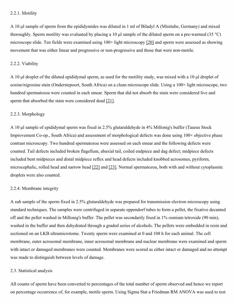
Cold storage of the eland epididymides at 4 °C resulted in a statistically significant decrease in sperm motility with time (RM ANOVA; P < 0.05; Fig. 1A). The reduction in motility was not statistically significant from t0 to t12 but there was a significant reduction after 24 h. Between t24 and t60 there was no significant reduction in motility but after 60 h, the rate of reduction in motility increased and differences between t24 and t72 were statistically significant. Sperm from seven of the nine eland showed a gradual decrease in sperm motility over time, but in two animals (specimens 1 and 8), there was a sharp decrease in the number of motile sperm between 48 and 60 h that is not reflected in the mean values (Fig. 1A).
Fig. 1. Changes in the motility (A) and viability (B) of eland sperm, from cauda epididymides stored at 4 °C (data are mean ± 1S.D.).

3.2. Viability
The percentage of live spermatozoa in the epididymis stored at 4 °C decreased significantly with time from ≈70 to
≈40% (RM ANOVA; P < 0.05; Fig. 1B). There was a significant reduction in viability from t0 to t12 but from t12 to t48
the rate of reduction in viability was slower and only at t72 was the viability significantly less than at t12. In specimens
1 and 8, there was a sharp decrease in percentage live sperm between 48 and 60 h.
3.3. Morphology
The mean percentage occurrence of normal sperm (including sperm with cytoplasmic droplets) decreased
significantly from 38 to ≈20% through the study (P < 0.05) and there was a concomitant increase in the total number
of sperm defects (Table 1). The percentage occurrence of sperm with distal cytoplasmic droplets decreased
significantly over time (P < 0.05; Table 1; Fig. 2) but there was no significant change in the occurrence of sperm with
proximal droplets or of normal sperm with no cytoplasmic droplet (Fig. 2).
Table 1. Changes in the occurrence of normal and defective eland sperm over time
Time (h) Total Normal sperm Sperm with defects
No droplet
Distal droplet
Proximal droplet Total Head
defects
Midpiece defects (%)
Tail defects
0 38 ± 25 5 27 6 62 ± 24 3 57 (92) 2
12 31 ± 22 3 23 5 69 ± 23 1 67 (97) 1
24 30 ± 17 6 19 5 70 ± 13 4 64 (91) 2
36 30 ± 19 7 18 5 70 ± 15 2 66 (94) 2
48 28 ± 13 6 17 5 72 ± 11 3 67 (93) 2
60 27 ± 19 10 13 4 73 ± 20 6 63 (86) 4
72 27 ± 19 8 14 5 73 ± 17 5 64 (88) 4
84 23 ± 15 10 10 3 77 ± 16 6 65 (84) 6
96 17 ± 10 7 8 2 83 ± 9 8 70 (84) 5
Time (h) Total Normal sperm Sperm with defects
No droplet
Distal droplet
Proximal droplet Total Head
defects
Midpiece defects (%)
Tail defects
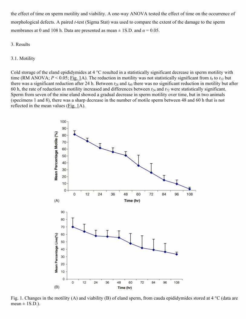
108 20 ± 10 10 6 4 80 ± 4 9 62 (78) 9
All figures are mean percentages of 200 sperm from each of nine animals per time, except for midpiece defects where
the numbers in parenthesis are midpiece defects as a percentage of all defects. Standard deviations are given for the
totals only.
Fig. 2. Changes in the occurrence of normal sperm and spermatozoa with proximal or distal cytoplasmic droplets over
time (data are mean ± 1S.D.).
The majority (78–97%) of defects were midpiece defects and these included bent midpiece and distal midpiece reflex
(Table 1). There were very few head or tail defects (Table 1). The percentage occurrence of all sperm with head
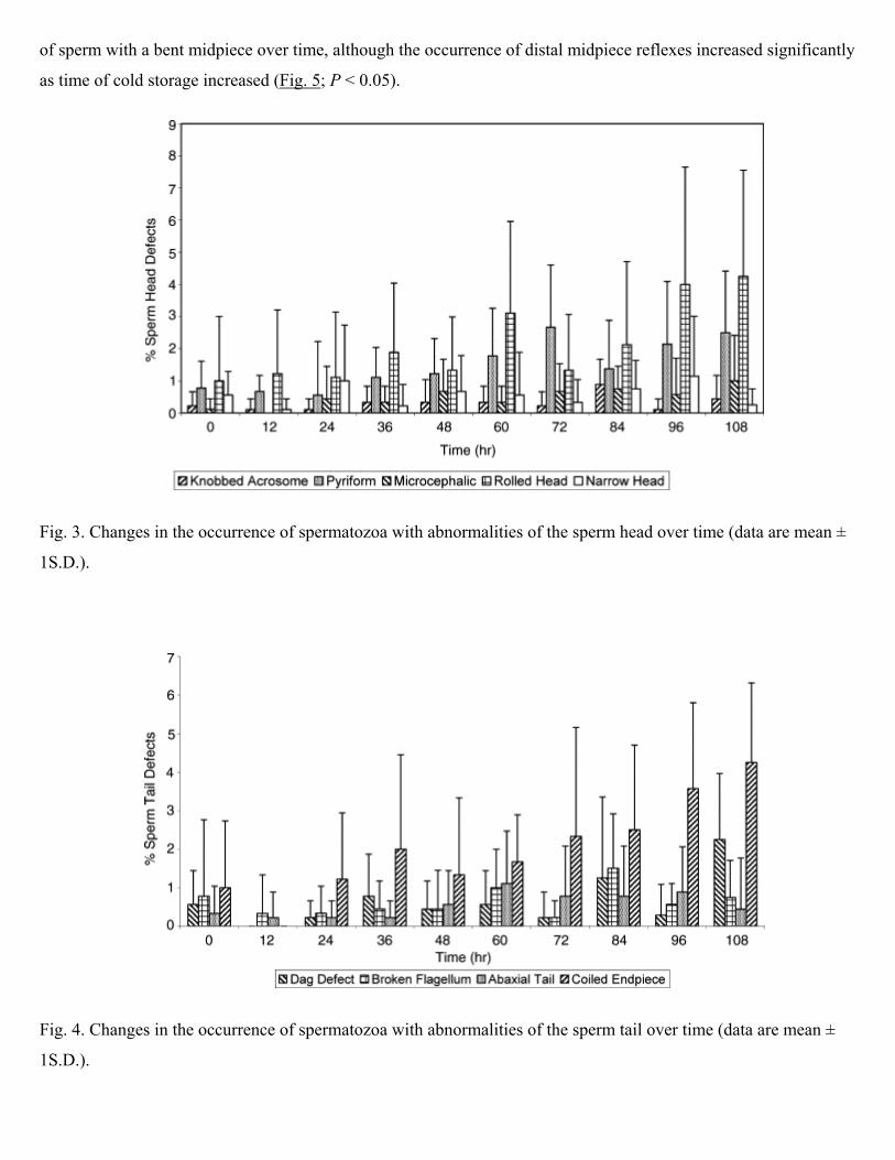
defects increased with time (Table 1) although this change was not statistically significant. However, the percentage
occurrence of sperm with knobbed acrosomes, pyriform and microcephalic defects increased significantly over the
course of the experiment (P < 0.05; Fig. 3). There was no significant change in the occurrence of narrow and rolled
heads (P > 0.05). The percentage occurrence of sperm with the dag defect and coiled endpiece increased significantly
with time (P < 0.05; Fig. 4). However, the occurrence of abaxial tail and broken flagellum did not change
significantly (P > 0.05) (Fig. 4). Since the occurrence of head and tail defects increased with time, it is not surprising
that the percentage occurrence of midpiece defects (as a percentage of all defects) decreased slightly through the
experiment (Table 1). Two midpiece defects were recognised, and there was no significant change in the occurrence
of sperm with a bent midpiece over time, although the occurrence of distal midpiece reflexes increased significantly
as time of cold storage increased (Fig. 5; P < 0.05).
Fig. 3. Changes in the occurrence of spermatozoa with abnormalities of the sperm head over time (data are mean ±
1S.D.).
Fig. 4. Changes in the occurrence of spermatozoa with abnormalities of the sperm tail over time (data are mean ±
1S.D.).
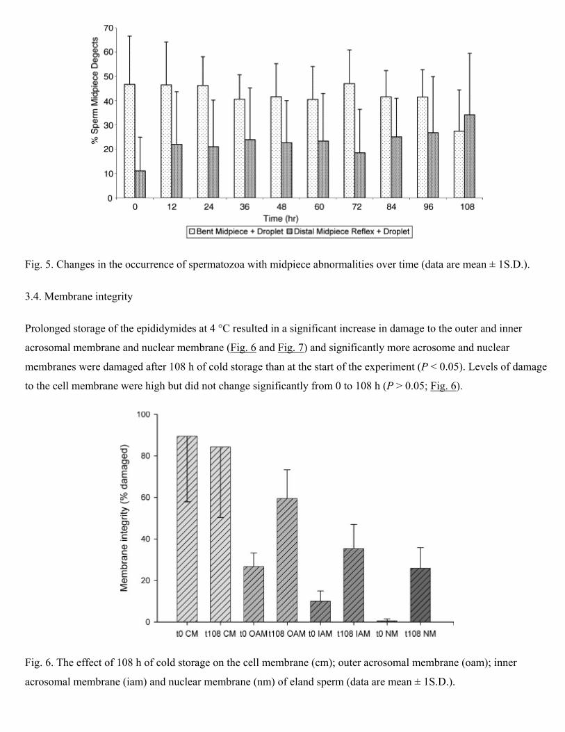
Fig. 5. Changes in the occurrence of spermatozoa with midpiece abnormalities over time (data are mean ± 1S.D.).
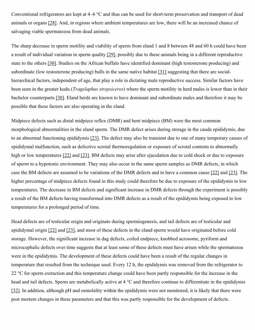
3.4. Membrane integrity
Prolonged storage of the epididymides at 4 °C resulted in a significant increase in damage to the outer and inner
acrosomal membrane and nuclear membrane (Fig. 6 and Fig. 7) and significantly more acrosome and nuclear
membranes were damaged after 108 h of cold storage than at the start of the experiment (P < 0.05). Levels of damage
to the cell membrane were high but did not change significantly from 0 to 108 h (P > 0.05; Fig. 6).
Fig. 6. The effect of 108 h of cold storage on the cell membrane (cm); outer acrosomal membrane (oam); inner
acrosomal membrane (iam) and nuclear membrane (nm) of eland sperm (data are mean ± 1S.D.).
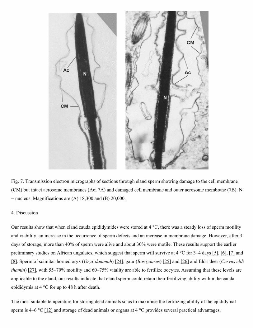
Fig. 7. Transmission electron micrographs of sections through eland sperm showing damage to the cell membrane
(CM) but intact acrosome membranes (Ac; 7A) and damaged cell membrane and outer acrosome membrane (7B). N
= nucleus. Magnifications are (A) 18,300 and (B) 20,000.
4. Discussion
Our results show that when eland cauda epididymides were stored at 4 °C, there was a steady loss of sperm motility
and viability, an increase in the occurrence of sperm defects and an increase in membrane damage. However, after 3
days of storage, more than 40% of sperm were alive and about 30% were motile. These results support the earlier
preliminary studies on African ungulates, which suggest that sperm will survive at 4 °C for 3–4 days [5], [6], [7] and
[8]. Sperm of scimitar-horned oryx (Oryx dammah) [24], gaur (Bos gaurus) [25] and [26] and Eld's deer (Cervus eldi
thamin) [27], with 55–70% motility and 60–75% vitality are able to fertilize oocytes. Assuming that these levels are
applicable to the eland, our results indicate that eland sperm could retain their fertilizing ability within the cauda
epididymis at 4 °C for up to 48 h after death.
The most suitable temperature for storing dead animals so as to maximise the fertilizing ability of the epididymal
sperm is 4–6 °C [12] and storage of dead animals or organs at 4 °C provides several practical advantages.
Conventional refrigerators are kept at 4–6 °C and thus can be used for short-term preservation and transport of dead
animals or organs [28]. And, in regions where ambient temperatures are low, there will be an increased chance of
salvaging viable spermatozoa from dead animals.
The sharp decrease in sperm motility and viability of sperm from eland 1 and 8 between 48 and 60 h could have been
a result of individual variation in sperm quality [29], possibly due to these animals being in a different reproductive
state to the others [30]. Studies on the African buffalo have identified dominant (high testosterone producing) and
subordinate (low testosterone producing) bulls in the same native habitat [31] suggesting that there are social-
hierarchical factors, independent of age, that play a role in dictating male reproductive success. Similar factors have
been seen in the greater kudu (Tragelaphus strepsiceros) where the sperm motility in herd males is lower than in their
bachelor counterparts [30]. Eland herds are known to have dominant and subordinate males and therefore it may be
possible that these factors are also operating in the eland.
Midpiece defects such as distal midpiece reflex (DMR) and bent midpiece (BM) were the most common
morphological abnormalities in the eland sperm. The DMR defect arises during storage in the cauda epididymis, due
to an abnormal functioning epididymis [23]. The defect may also be transient due to one of many temporary causes of
epididymal malfunction, such as defective scrotal thermoregulation or exposure of scrotal contents to abnormally
high or low temperatures [22] and [23]. BM defects may arise after ejaculation due to cold shock or due to exposure
of sperm to a hypotonic environment. They may also occur in the same sperm samples as DMR defects, in which
case the BM defects are assumed to be variations of the DMR defects and to have a common cause [22] and [23]. The
higher percentage of midpiece defects found in this study could therefore be due to exposure of the epididymis to low
temperatures. The decrease in BM defects and significant increase in DMR defects through the experiment is possibly
a result of the BM defects having transformed into DMR defects as a result of the epididymis being exposed to low
temperatures for a prolonged period of time.
Head defects are of testicular origin and originate during spermiogenesis, and tail defects are of testicular and
epididymal origin [22] and [23], and most of these defects in the eland sperm would have originated before cold
storage. However, the significant increase in dag defects, coiled endpiece, knobbed acrosome, pyriform and
microcephalic defects over time suggests that at least some of these defects must have arisen while the spermatozoa
were in the epididymis. The development of these defects could have been a result of the regular changes in
temperature that resulted from the technique used. Every 12 h, the epididymis was removed from the refrigerator to
22 °C for sperm extraction and this temperature change could have been partly responsible for the increase in the
head and tail defects. Sperm are metabolically active at 4 °C and therefore continue to differentiate in the epididymis
[32]. In addition, although pH and osmolality within the epididymis were not monitored, it is likely that there were
post mortem changes in these parameters and that this was partly responsible for the development of defects.

Final maturation of sperm takes place during passage through the epididymis, where they undergo morphological and
functional changes including displacement of the cytoplasmic droplet from a proximal to a distal position [33]. This
explains why there were more sperm with distal droplets than proximal droplets at the start of the experiment. The
significant decline in the number of sperm with distal droplets through the experiment is probably a result of the
continued migration of the droplet while the sperm were in the epididymis [32].
Cold shock can induce changes that are similar to those of capacitation and the acrosome reaction [28], [34] and [35]
and prolonged storage of the spermatozoa at 4 °C could therefore have caused the damage to the cell membrane and
induced the acrosome-like reaction in the eland sperm. This would explain the increase in acrosomal membrane
damage over time.
The physiological processes that ensure the survival of spermatozoa in the epididymis at 4 °C are unclear although
epididymal fluid may contain an unknown cold shock protective factor(s) such as lecithin [36] that is effective for
approximately 3 days [10].
In conclusion, this study has shown that prolonged storage of the cauda epididymis at 4 °C, interrupted every 12 h by
removal of the epididymis to 22 °C, caused structural changes and a reduction in viability and motility to a
considerable proportion of eland spermatozoa. It is likely that the regular changes of temperature would have
increased the rate of sperm degradation and that storage of epididymides at a constant 4 °C would result in a higher
percentage of viable sperm. Although some sperm remained viable for up to 4 days post mortem, functional tests
should be done to ensure that prolonged cold storage is practical. Low temperature transportation within and between
many countries is available and it is possible to deliver epididymides to a laboratory within 1 or 2 days of their
collection. The method of storing epididymides described here can be used to conserve male genetic resources in
eland when epididymal spermatozoa cannot be collected and cryopreserved. Further study combined with artificial
insemination or embryo transfer of in vitro produced oocytes is needed to confirm the full potential of this method.
Acknowledgements
We thank the management of the Rietvlei Nature Reserve for providing samples for this research; the Wildlife
Biological Resource Centre, Rhodes University and the NRF for financial support and facilities; the staff of the
Electron Microscopy Unit at Rhodes University for their expertise and assistance; Martin Villet for statistical advice
and Daniel Parker and Claire Jackson for comments on this manuscript.
References
[1] Wildt DE, Rall WF, Critser JK, Monfort SL, Seal US. Genome Resource Banks: living collections for biodiversity conservation. Bioscience 1997; 47(10):689–701. [2] Wildt DE, Schiewe MC, Schmidt PM, Goodrowe KL, Howard JG, Phillips LG, et al. Developing animal model systems for embryo technologies in rare and endangered wildlife. Theriogenology 1986; 25(1):33–51.

[3] O’Brien SJ, Wildt DE, Goldman D, Merril CR, Bush M. The cheetah is depauperate in genetic variation. Science 1983; 221:459–62. [4] O’Brien SJ. Genetic phylogenetic analysis of endangered species. Ann Rev Genet 1994; 28:467–89. [5] Bezuidenhout C, Fourie le RF, Meintjies M, Bornman MS, Bartels P, Godke RA. Comparative epididymal sperm cell motility of African ungulate and equid game species stored at 4 8C. Theriogenology 1995; 43(1):167 [abstract]. [6] Friedmann Y, Lubbe K, Kilian I, Grobler DG, Denniston RS. Changes in motility and morphological characteristics of African buffalo (Syncerus caffer) sperm during storage of the epididymis. Theriogenology 2000; 53(1):332 [abstract]. [7] Lubbe K, Bartels P, Kilian I, FriedmannY. Godke RA. Comparing motility and morphology of horse, zebra and rhinoceros epididymal spermatozoa when cryopreserved with two different cryodiluents or stored at 4 8C. Theriogenology 2000; 53(1):338 [abstract]. [8] Kilian I, Lubbe K, Bartels P, Friedmann Y, Denniston RS. Evaluating epididymal sperm of African wild ruminants: longevity when stored at 4 8C and viability following cryopreservation. Theriogenology 2000; 53(1):330 [abstract]. [9] Lambrecht H, van Niekerk FE, CoetzerWA, Cloete SWP, van der Horst G. The effects of cryopreservation on the survivability, viability and motility of epididymal African buffalo (Syncerus caffer) spermatozoa. Theriogenology 1999; 52:1241–9. [10] Kikuchi K, Nagai T, KashiwazakiW, Ideki H, Noguchi J, Shimada A, et al. Cryopreservation and ensuing in vitro fertilisation ability of boar spermatozoa from epididymides stored at 4 8C. Theriogenology 1998; 50:615–23. [11] Songsasen N, Tang J, Leibo SP. Birth of live mice derived by in vitro fertilisation with spermatozoa retrieved up to 24 h after death. J Exp Zool 1998; 280:189–96. [12] An TZ, Wada S, Edashige K, Sakurai T, Kasai M. Viable spermatozoa can be recovered from refrigerated mice up to 7 days after death. Cryobiology 1999; 38:27–34. [13] Fuller SJ, Whittingham DG. Effect of cooling mouse sperm to 4 8C on fertilisation and embryonic development. J Reprod Fertil 1996; 108:139–45. [14] Amann RP. Can the fertility potential of a seminal sample be predicted accurately? J Androl 1989; 10:89–98. [15] Schiewe MC. Species variations in semen cryopreservation of non-domestic ungulates. Proceedings of the Wild Cattle Symposium; 1991. p. 56–64. [16] Estes RD. The behaviour guide to African mammals. Halfway House, South Africa: Russel Friedman Books; 1997. [17] Skinner JD, Smithers RH. The mammals of the Southern African subregion. Pretoria, South Africa: University of Pretoria; 1990. [18] Hinton BT, Dott HM, Setchell BP. Measurement of the motility of rat spermatozoa collected by micropuncture from the testis and from different regions along the epididymis. J Reprod Fertil 1979; 55:167–72. [19] Mahony MC, Oehinger S, Doncel G, Marshedi M, Acosta A, Hodger GD. Functional and morphological features of spermatozoa microaspirated from the epididymal region of the Cynomolgus Monkey (Macaca fasicularis). Biol Reprod 1993; 48:613–20.
[20] World Health Organisation. WHO Laboratory Manual for the Examination of Human Semen and Sperm-Cervical Mucus Interaction. (3e). Cambridge: The Press Syndicate of the University of Cambridge; 1997. [21] Dott HM, Foster GC. A technique for studying the morphology of mammalian spermatozoa which are eosinophilic in a differential ‘live/dead’ stain. J Reprod Fertil 1972;29:443–5. [22] Nöthling JO. Sperm morphology in the bull. Pretoria, South Africa: Department of Production Animal Science, Faculty of Veterinary Science, University of Pretoria; 1984. [23] Barth AD, Oko RJ. Abnormal morphology of bovine spermatozoa. Ames, IA: Iowa State University Press; 1989. [24] Roth TL, Bush LM, Wildt DE, Weiss RB. Scimitar-Horned Oryx (Oryx dammah) spermatozoa are functionally competent in a heterologous bovine in vitro fertilisation system after cryopreservation on dry ice, in a dry shipper, or over liquid nitrogen vapour. Biol Reprod 1999;60:493–8. [25] Johnston LA, Parrish JJ, Monson R, Leifried-Rutledge L, Susko-Parrish JL, Northey DL, et al. Oocyte maturation, fertilisation and embryo development in vitro and in vivo in the gaur (Bos gaurus). J Reprod Fertil 1994; 100:131–6. [26] Hopkins SM, Armstrong DL, Hummel KC, Junoir SM. Successful cryopreservation of gaur (Bos gaurus) epididymal spermatozoa. J Zoo Anim Med 1988; 19:195–201. [27] Monfort SL, Asher GW, Wildt DE, Wood TC, Schiewe MC, Williamson LR, et al. Successful intrauterine insemination of Eld’s deer (Cervus eldi thamin) with frozen-thawed spermatozoa. J Reprod Fertil 1993; 99:459–65. [28] Bartels P, Friedmann Y, Lubbe K, Mortimer D, Rasmussen LA, Godke RA. The live birth of an eland (Taurotragus oryx) calf following estrous synchronization and artificial insemination using frozen thawed epididymal sperm. Theriogenology 2001; 53:381 [abstract]. [29] Loskutoff NM, Simmons HA, Goulding M, Thompson G, de Jongh T, Simmons LG. Species and individual variations in cryoprotectant toxicities and freezing resistance of epididymal sperm from African antelope. Anim Reprod Sci 1996; 42:527–35. [30] Schiewe MC, Bush M, de Vos V, Brown JL,Wildt DE. Semen characteristics, sperm freezing and endocrine profiles in free-living wildebeest (Connochaetes taurinus) and greater kudu (Tragelaphus strepticeros). J Zoo Wildlife Med 1991; 22(1):58–72. [31] Brown JL, Wildt DE, Raathe JR, de Vos V, Howard JG, Janssen DL, et al. Impact of season on seminal characteristics and endocrinetatus of adult free-ranging African buffalo (Syncerus caffer). J Reprod Fertil 1991; 92(1):47–57. [32] Fawcett DW, Phillips DM. Observations on the release of spermatozoa and on changes in the head during passage through the epididymis. J Reprod Fertil Suppl 1969; 6:405–18. [33] Axnér E, Linde-Forsberg C, Einarsson S. Morphology and motility of spermatozoa from different regions of the epididymal duct in the domestic cat. Theriogenology 1999; 52:767–78. [34] Jones RC, Stewart DL. The effects of cooling to 5 8C and freezing and thawing on the ultrastructure of bull spermatozoa. J Reprod Fertil 1979; 56:233–8. [35] Aalseth EP, Saacke RG. Morphological change of the acrosome on motile bovine spermatozoa due to storage at 48C. J Reprod Fertil 1985; 74:473–8.
[36] Robaire B, Hermo L. Efferent ducts, epididymis and vas deferens: structure, function and regulation. In: Knobil E, Neill JD, editors. The physiology of reproduction. New York: Raven Press; 1994. p. 999–1080.