Embed Size (px)
Citation preview
13C NMR
Any nucleus with an odd atomic number or odd mass will have a nuclear magnetic resonance
A proton is the most common example in organic chemistry so by default when someone talks about NMR it usually means a 1H NMR
Carbon-12, the most abundant source of carbon, has an even atomic number and even mass so therefore this nuclei is NMR inactive
The isotope C-13, though, has an odd mass and is therefore NMR active
Another very useful nucleus to determine organic structures
Carbon-13
The natural abundance of C-13 isotope is ~1%
Therefore only 1% probability that a given carbon atom is NMR active -therefore the signal-to-noise ratio for carbon NMR is less
Put another way – if studying 1-chloroethane only 1 in 100 molecules will be active at C1
In addition the gyromagnetic ratio, γ, for carbon is ~1/4 that of hydrogen (remember that each nucleus has its own unique gyromagnetic ratio)
-this also causes a lower signal-to-noise for carbon relative to hydrogen NMR
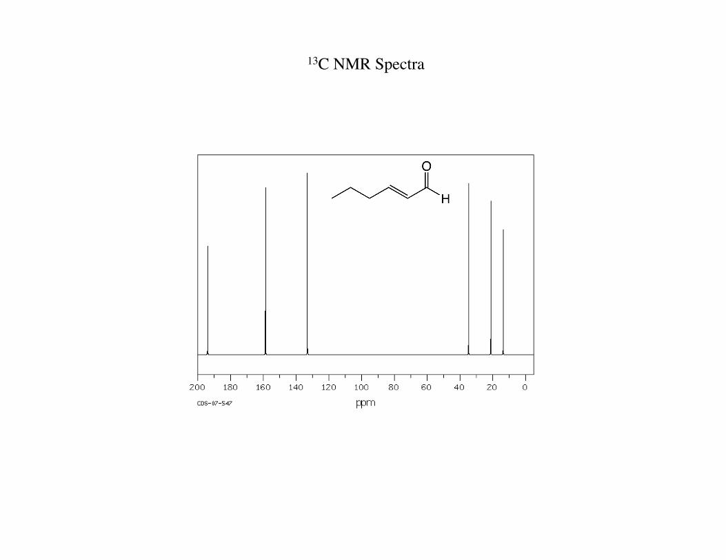
13C NMR Spectra
Unique Features of 13C NMR Relative to 1H NMR
1) The scale is much larger for 13C than 1H NMR Notice spectrum is 0-200 ppm instead of 0-11 ppm, due to carbon being more shielded than hydrogen
[8 electrons around carbon rather than 2 electrons around hydrogen]
2) Relative placement of functional groups though is the same in the above spectrum Which functional group was more deshielded in 1H NMR is still more deshielded in 13C NMR
-aldehyde carbon 194 ppm, sp2 carbons 158 and 133 ppm, sp3 carbons below 40 ppm
3) Each carbon has its own unique chemical shift big advantage with 13C NMR, can instantly tell number of
distinct symmetrically different carbons [the spectrum shown is a proton-decoupled 13C NMR, most common example]
4) Do not observe 13C-13C splitting Due to low probability of finding two adjacent carbons both 13C isotope with only 1%
probability for each
Splitting
Notice that carbon-carbon splitting is not detected in NMR
Can still detect 13C-1H splitting from hydrogens attached to carbon of interest This is called off-resonance decoupled
Have same N+1 rule
Normally, though, a 13C NMR is proton spin decoupled In this mode a sharp singlet is observed for each carbon
-far easier to interpret spectra
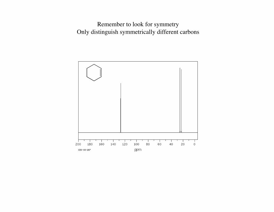
Remember to look for symmetry Only distinguish symmetrically different carbons
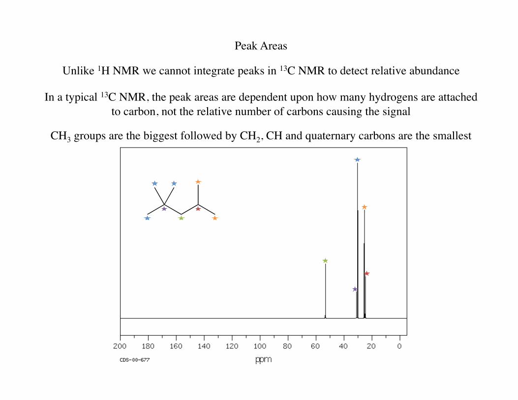
Peak Areas
Unlike 1H NMR we cannot integrate peaks in 13C NMR to detect relative abundance
In a typical 13C NMR, the peak areas are dependent upon how many hydrogens are attached to carbon, not the relative number of carbons causing the signal
CH3 groups are the biggest followed by CH2, CH and quaternary carbons are the smallest
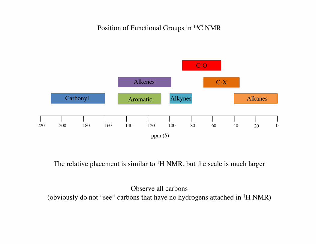
Position of Functional Groups in 13C NMR
The relative placement is similar to 1H NMR, but the scale is much larger
Observe all carbons (obviously do not “see” carbons that have no hydrogens attached in 1H NMR)
020406080100120140160180200220
ppm (!)
Alkanes
C-X
C-O
Alkynes
Alkenes
Aromatic Carbonyl
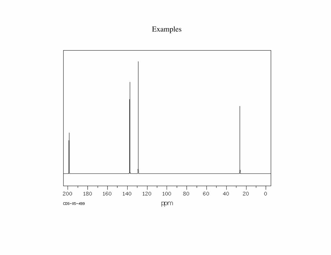
Examples
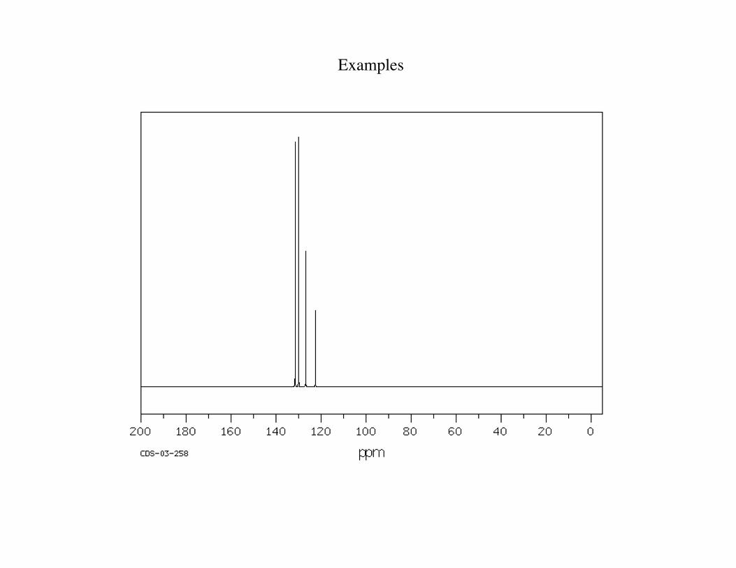
Examples
MRI Imaging
MRI (magnetic resonance imaging) is in theory identical to our discussions MRI = NMR
Consumers are hesitant with anything that has “nuclear” in its descriptor so the name was changed
One main difference is that instead of spinning the sample to make the magnetic field homogeneous throughout the sample in a NMR experiment (thus the chemical shift will be identical for all hydrogens
with the same amount of shielding), in MRI the field is inhomogeneous throughout the sample
This inhomegeneity allows one to measure either placement of protons in a body (where is the water)
or also changes in movement of the protons (i.e. how mobile is water inside a tumor relative to a normal tissue)
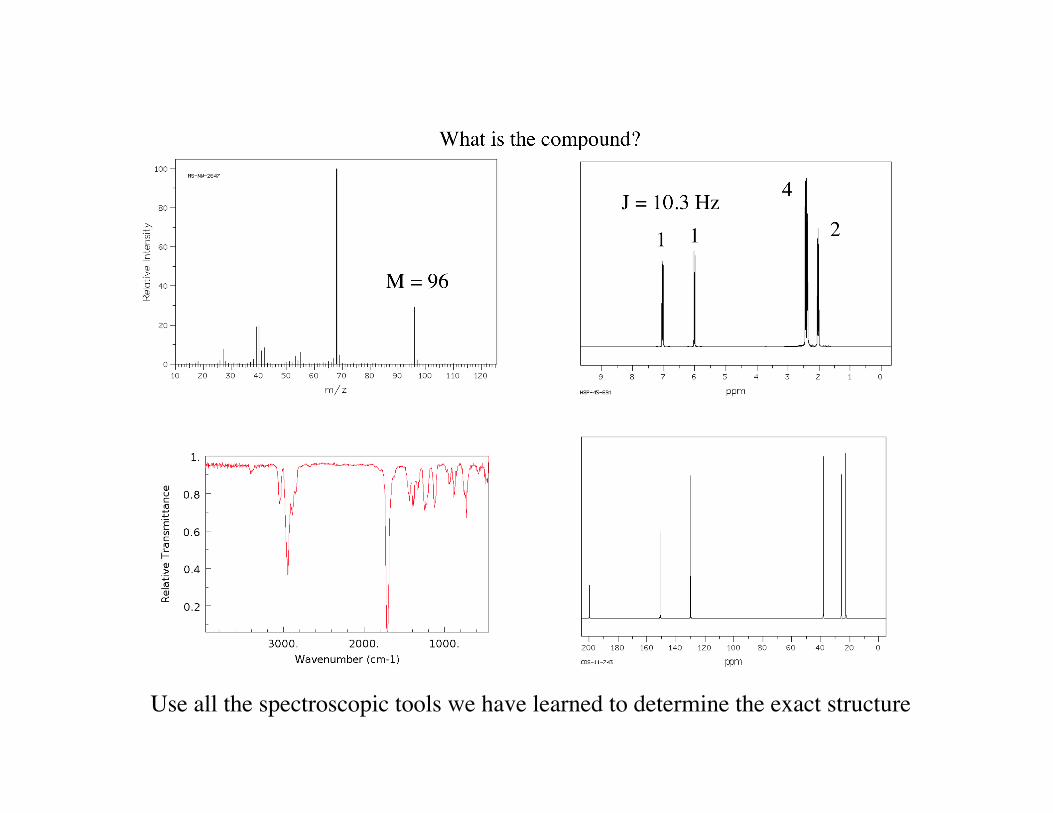
Use all the spectroscopic tools we have learned to determine the exact structure


![High-spin states in the odd–odd nucleus Y · Preliminary results concerningseveral rotational bands identified in this nucleus have been published in Ref. [4]. With new measurements,](https://img.pdfslide.net/doc/110x75/607f2cb77061d245d1250f1f/high-spin-states-in-the-oddaodd-nucleus-y-preliminary-results-concerningseveral.jpg)