Embed Size (px)
Citation preview

rapamycin, mTOR (14), but more importantly throughphosphorylation and inactivation of tuberin (TSC2), anmTOR inhibitor (15). Inhibition of mTOR stops the pro-tein synthesis machinery due to inactivation of its effec-tor, p70 S6 kinase, and activation of the eukaryotic initi-ation factor, 4E binding protein 1 (4E-EP1), an inhibitorof translation (16,17).
Specificity/Sensitivity: Phospho-Akt (Ser473)Antibody (IHC Specific) detects Akt1 only when phos-phorylated at serine 473, and Akt2 and Akt3 only whenphosphorylated at equivalent sites. This antibody wasdeveloped for and is recommended for immunohisto-chemical application only. It does not detect Akt phos-phorylated at other sites or related kinases such as PKCand p70 S6 kinase.
Source/Purification: Polyclonal antibodies are pro-duced by immunizing rabbits with a synthetic phospho-peptide (KLH-coupled) corresponding to residuesaround Ser473 of mouse Akt. Antibodies are purified byprotein A and peptide affinity chromatography.
Tyr315
P
GSK-3β
TSC2 BAD
MDM2
eNOSNO synthesis
FKHR
Ser166/186
AFX
Receptor
Growth factors, insulin, etc.
p21CIP1
PTEN
survival
Akt/PKB
growth
Ser241
Thr308
Ser473
Akt/PKB
Thr308
Ser473
Ser1177
Ser9 Ser136
Thr145/Ser146
Ser256 Thr24/32
glycogensynthesis
proliferationsurvival
proliferationsurvival
Thr1462Ser939
Ser193
PI3KPIP3
PIP3
Tyr376Tyr373
SHIP
Tyr326
Src
YAP
Mem
bran
ePDK1
P P P P PP PP P P
P
P
P
PP
P
P
PP
P
PPPP
PP
P
PP
PPP
PP
P
Akt/PKB Signaling Pathway page
1 o
f 6
Storage: Supplied in 10 mM sodium HEPES (pH 7.5),150 mM NaCl and 50% glycerol. Store at –20°C. Donot aliquot the antibody.
Recommended Antibody Dilutions:Immunohistochemistry (Floating) 1:100Immunohistochemistry (Paraffin) 1:50IHC Protocol: Citrate/TBSTFlow cytometry 1:200
Companion Products:Phospho-Akt (Thr308) (244F9) Rabbit mAb #4056
Akt2 (5B5) Rabbit mAb #2964
Akt2 (54G8) Rabbit mAb (IHC Specific)#4057
Phospho-Akt (Ser473) (193H12) Rabbit mAb #4058
Phospho-Akt (Ser473) (736E11) Rabbit mAb (IHCSpecific) #3787
SignalSilence™ Akt siRNA Kit #6210
SignalSilence™ Akt2 siRNA Kit #6395
PathScan™ Phospho-Akt1 (Ser473) Sandwich ELISAKit #7160
PathScan™ Akt1 Sandwich ELISA Kit #7170
Akt Antibody #9272
Phospho-Akt Pathway Sampler Kit #9916
LY294002 (PI3 Kinase Inhibitor) #9901
Phospho-Akt (Ser473) Blocking Peptide (IHC Specific)#1140
Survival Marker: Signal Stain™ Phospho-Akt (Ser473)IHC Detection Kit #8100
Stor
e at
–20
°C#9
277
Phospho-Akt (Ser473)Antibody (IHC Specific)
nn Small 160 µl (40 sections)
nn Large 480 µl (120 sections)
Background: Akt, also referred to as PKB or Rac, playsa critical role in controlling the balance between survivaland apoptosis (1–3). This protein kinase is activated byinsulin and various growth and survival factors, andfunctions in a wortmannin-sensitive pathway involvingPI3 kinase (2,3). Akt is activated by phospholipid bind-ing and activation loop phosphorylation at Thr308 byPDK1 (4), and by phosphorylation within the carboxy-terminus at Ser473. Akt promotes cell survival byinhibiting apoptosis through its ability to phosphorylateand inactivate several targets, including Bad (5),Forkhead transcription factors (6) and caspase-9. PTENphosphatase is a major negative regulator of the PI3kinase/Akt signaling pathway (7). LY294002 is a specif-ic PI3 kinase inhibitor (8).
One of the essential functions of Akt is the regulation ofglycogen synthesis through phosphorylation and inacti-vation of glycogen synthase kinase-3α and β (9,10).Akt may also play a role in insulin stimulation of glu-cose transport (9).
In addition to its role in survival and glycogen synthe-sis, Akt is involved in cell cycle regulation by preventingGSK3β-mediated phosphorylation and degradation ofcyclin D1 (11), and by negatively regulating the cyclin-dependent kinase inhibitors p27 KIP (12) and p21WAF1 (13). Akt also plays a critical role in cell growthby directly phosphorylating the mammalian target of
Orders n 877-616-CELL (2355)[email protected]
Support n 877-678-TECH (8324)[email protected]
Web n www.cellsignal.comrev. 07/08/05
Applications Species Cross-Reactivity Source
IHC, F (H, M, R) Rabbit
Immunohistochemical analysis of paraffin-embedded human breast carcinoma, showing cytoplamic, nuclear and membranelocalization, using Phopsho-Akt (Ser473) Antibody (IHC Specific) (left) or the same antibody preincubated with Phospho-Akt(Ser473) Blocking Peptide (IHC Specific) #1140 (right).
© 2
005
Cell
Sign
alin
g Te
chno
logy
, Inc
.
This product is for in vitro research use only and is not intended for use in humans or animals.

Orders n 877-616-CELL (2355) [email protected] Support n 877-678-TECH (8324) [email protected] Web n www.cellsignal.com#927
7
page
2of
6
© 2
005
Cell
Sign
alin
g Te
chno
logy
, Inc
.
Selected Application References:Choe, G. et al. (2003) Analysis of the phosphatidylinosi-tol 3'-kinase signaling pathway in glioblastoma patientsin vivo. Cancer Res. 63, 2742–2746. Applications: IHC(paraffin).
Dinulescu, D.M. et al. (2005) Role of K-ras and Pten inthe development of mouse models of endometriosis andendometrioid ovarian cancer. Nature Med. 11 (1),63–70. Applications: IHC (paraffin).
Gupta, A.K. et al. (2002) Local recurrence in head andneck cancer: relationship to radiation resistance and sig-nal transduction. Clin. Cancer Res. 8, 885–892.Applications: IHC (paraffin).
Kim, J.M. et al. (2002) Cooperativity of Nkx3.1 and Ptenloss of function in a mouse model of prostate carcino-genesis. Proc. Natl. Acad. Sci. USA 99 (5), 2884–2889.Applications: IHC (floating/frozen).
Lee, S.H. et al. (2002) Non-small cell lung cancers fre-quently express phosphorylated Akt; an immunohisto-chemical study. APMIS 110, 587–592. Applications:IHC (paraffin).
Malik, S.N. et al. (2002) ImmunohistochemicalDemonstration of Phospho-Akt in High Gleason GradeProstate Cancer. Clin. Cancer Res. 8, 1168–1171.Applications: IHC (paraffin).
Noshita, N. et al. (2002) Akt phosphorylation and neu-ronal survival after traumatic brain injury in mice.Neurobiol. Dis. 9, 294–304. Applications: IHC (paraffin).
Okudela, K. et al. (2004) K-ras gene mutation enhancesmotility of immortalized airway cells and lung adenocar-cinoma cells via Akt activation: possible contribution tonon-invasive expansion of lung adenocarcinoma. Am. J.Pathol. 164, 91–100. Applications: IHC (paraffin).
Akt (5G3) Monoclonal Antibody #2966 and Phospho-Akt (Ser473) Antibody staining of untreated (left) or LY294002 treated(right) Jurkat cells.
Control
Confocal image of immunohistochemical staining withPhospho-Akt (Ser473) Antibody (IHC Specific) (green) andpropidium iodide (red) in rat dentate gyrus regions, usingsections from 15 minute transient cerebral ischemia followedby 30 minutes of reperfusion. (Provided by Dr. Bingren Hu,University of Miami School of Medicine, Florida.)
Roy, H.K. et al. (2002) AKT proto-oncogene overexpres-sion is an early event during sporadic colon carcinogen-esis. Carcinogenesis 23 (1), 201–205. Applications:IHC (paraffin).
Schmitz, K.J. et al. (2004) Prognostic relevance of acti-vated Akt kinase in node-negative breast cancer: a clini-copathological study of 99 cases. Mod. Pathol. 17,15–21.
Background References:(1) Franke, T.F. (1997) Cell 88, 435–437.
(2) Burgering, B.T. and Coffer, P.J. (1995) Nature 376,599–602.
(3) Franke, T.F. et al. (1995) Cell 81, 727–736.
(4) Alessi, D.R. et al. (1996) EMBO J. 15, 6541–6551.
(5) Cardone, M.H. et al. (1998) Science 282,1318–1321.
(6) Brunet, A. et al. (1999) Cell 96, 857–868.
(7) Cantley, L.C. et al. (1999) Proc. Natl. Acad. Sci.USA 96, 4240–4245.
(8) Vlahos, C. et al. (1994) J. Biol. Chem. 269,5241–5248.
(9) Hajduch, E. et al. (2000) FEBS Lett. 492, 199–203.
(10) Cross, D.A. et al. (1995) Nature 373, 785–789.
(11) Diehl, et al. (1998) Genes Dev. 12, 3499–3511.
(12) Gesbert, F. et al. (2000) J. Biol. Chem. 273,39223–39230.
(13) Zhou, B.P. et al. (2001) Nat. Cell Biol. 3, 245–252.
(14) Nave, B.T. et al. (1999) Biochem. J. 344, 427–431.
(15) Manning, B.D. et al. (2000) Mol. Cell 4, 648–657.
(16) Manning, B.D. et al. (2002) Mol. Cell 10, 151–162.
(17) Inoki, K. et al. (2002) Nat. Cell Biol. 4, 648–657
Immunohistochemical analysis of paraffin-embeddedhuman breast carcinoma, using Phospho-Akt (Ser473)Antibody (IHC Specific) preincubated with irrelevant controlpeptide (left) or Phospho-Akt (Ser473) Blocking Peptide(IHC Specific) (right).
Applications Key: W—Western IP—Immunoprecipitation IHC—Immunohistochemistry IC—Immunocytochemistry IF—Immunofluoresence F—Flow cytometry E—ELISA D—DELFIA®
Species Cross-Reactivity Key: H—human M—mouse R—rat Hm—hamster Mk—monkey Mi—mink C—chicken X—Xenopus Z—zebra fish B—bovine All—all species expectedSpecies enclosed in parentheses are predicted to react based on 100% sequence homology.
Orders n 877-616-CELL (2355) [email protected] Support n 877-678-TECH (8324) [email protected] Web n www.cellsignal.com#927
7
page
3of
6
© 2
005
Cell
Sign
alin
g Te
chno
logy
, Inc
.
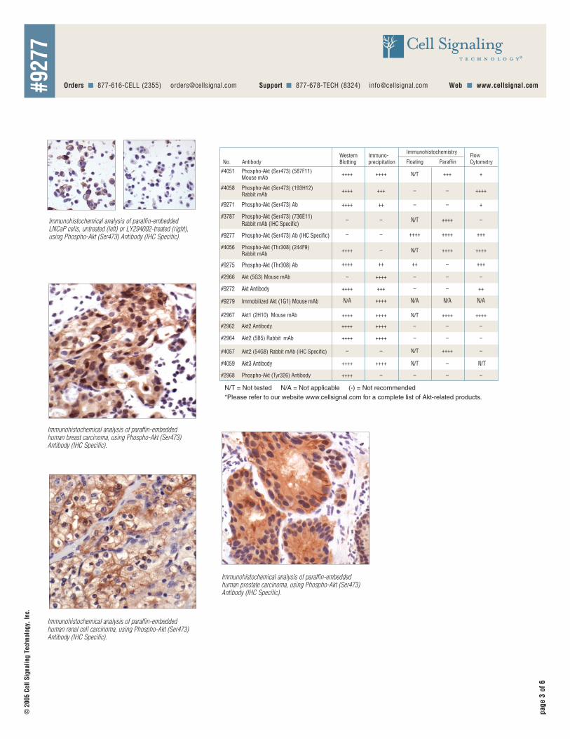
Immunohistochemical analysis of paraffin-embeddedLNCaP cells, untreated (left) or LY294002-treated (right),using Phospho-Akt (Ser473) Antibody (IHC Specific).
Immunohistochemical analysis of paraffin-embeddedhuman breast carcinoma, using Phospho-Akt (Ser473)Antibody (IHC Specific).
Immunohistochemical analysis of paraffin-embeddedhuman renal cell carcinoma, using Phospho-Akt (Ser473)Antibody (IHC Specific).
N/T = Not tested N/A = Not applicable (-) = Not recommended
#2962 Akt2 Antibody ++++ ++++ – – –
#2964 Akt2 (5B5) Rabbit mAb ++++ ++++ – – –
#2966 Akt (5G3) Mouse mAb ++++ –– – –
#9272 Akt Antibody ++++ +++ ++– –
#9275 Phospho-Akt (Thr308) Ab ++++ ++ ++ +++–
#9271 Phospho-Akt (Ser473) Ab ++++ ++ +– –
#2968 Phospho-Akt (Tyr326) Antibody ++++ –– – –
Phospho-Akt (Ser473) (193H12) Rabbit mAb
++++ +++ ++++– –#4058
#2967 Akt1 (2H10) Mouse mAb ++++ ++++ ++++ ++++N/T
#4051 Phospho-Akt (Ser473) (587F11) Mouse mAb
++++ ++++ +++ +N/T
#4056 Phospho-Akt (Thr308) (244F9) Rabbit mAb
++++ ++++ ++++– N/T
Akt2 (54G8) Rabbit mAb (IHC Specific) – ––#4057 ++++N/T
#9277 Phospho-Akt (Ser473) Ab (IHC Specific) +++–– ++++++++
Phospho-Akt (Ser473) (736E11) Rabbit mAb (IHC Specific)
#3787 – –– ++++N/T
#9279 Immobilized Akt (1G1) Mouse mAb ++++N/A N/A N/A N/A
Akt3 Antibody ++++ ++++ –N/T N/T#4059
Western Blotting
Immuno-precipitation Floating
Immunohistochemistry
ParaffinNo. AntibodyFlowCytometry
*Please refer to our website www.cellsignal.com for a complete list of Akt-related products.
Immunohistochemical analysis of paraffin-embeddedhuman prostate carcinoma, using Phospho-Akt (Ser473)Antibody (IHC Specific).
page
4of
6
© 2
005
Cell
Sign
alin
g Te
chno
logy
, Inc
.
#927
7
Immunohistochemistry Protocol for Floating SectionsPhosphoproteins are very sensitive to intracellular ATP levels in tis-sues. When intracellular ATP levels decrease, phosphoproteins aremore easily dephosphorylated. Intracellular ATP levels are depletedduring perfusion fixation of animal tissues. Thus, it is highly rec-ommended that animals be perfused as quickly as possible usingice-cold solutions.
Fix tissues by intracardiac perfusion with ice-cold PBS for 1 minutefollowed by ice-cold 4% paraformaldehyde in phosphate bufferdelivered with a peristaltic pump at 50 ml/min for 10 minutes.Remove tissues and keep in the same fixative solution at 4°C for24 hours, then section with a Vibratome at a thickness of 50 µm.Vibratome sections can be stored in an antifreeze solution at –20°Cfor at least several months.
Note: If more than one animal is used, keep fixation time constant.
Note: Although this protocol is by a floating method, many of ourantibodies have been demonstrated successfully with paraffinembedding or cryosectioning methods.
Solutions and Reagentsn Phosphate Buffer:
0.1 M Na2HP04/NaH2P04 (pH 7.5)
n Antifreeze Solution:320 ml 1X PBS (pH 7.4), 240 ml ethylene glycol (30%),240 ml glycerol (30%)
n Tris Buffered Saline (TBS):0.1 M Tris-HCl (pH 7.4), 0.15 M NaCl
n Wash Buffer:1X TBS, 0.1% Triton X-100 (TBS/Triton)
n Bovine Serum Albumin (BSA)
n ABC Reagent:(Vectastain ABC Kit, Vector Laboratories, Inc., Burlingame,CA) 1 drop reagent A and 1 drop reagent B in 5 ml TBS with1% BSA, 0.1% Triton X-100
n DAB Reagent (0.5 mg/ml):To prepare: Use 10 ml TBS, 500 µl of 10 mg/ml DAB stock solu-tion, 50 µl of glucose oxidase (30 mg/10 ml TBS), 20 µl NH4Cl(2.0 g/10 ml TBS) and 50 µl D (+) glucose (2.5 g/10 ml TBS).
n Gelvatol Preparation:a. Add 2.4 g of polyvinyl alcohol (Mol. Wt. 30,000–70,000) to
6 ml of glycerol. Stir well to mix. Add 6 ml of dH2O andleave for at least 2 hours at room temperature.
b. Add 12 ml of 0.2 M Tris (pH 8.5). Heat to 50°C for 10 min-utes with occasional mixing. After polyvinyl alcohol is dis-solved, clarify by centrifugation (5000 x g) for 15 minutes.Collect supernatant liquid.
c. Add DABCO (1,4-diazabicyclo [2.2.2] octane; Sigma#D2522) to 2.5% as antifade medium. Aliquot in micro-tubes and store at –20°C. Stocks of Gelvatol are stable atroom temperature for several weeks after thawing.
ABC-DAB Method1. Wash tissue sections in TBS/Triton.
2. Treat sections with freshly made 1% H2O2 (0.1 ml of 30%H2O2 in 3 ml TBS) for 30 minutes.
3. Wash the sections with TBS/Triton three times for 30 minuteseach at room temperature.
4. Block nonspecific binding sites with 3% BSA in TBS/Triton for30 minutes to 1 hour.
5. Incubate the sections with primary antibody diluted in3% BSA in TBS/Triton overnight at 4°C.
6. Wash the sections in TBS/Triton three times for 10 minuteseach at room temperature.
7. Incubate the sections in biotinylated anti-rabbit secondaryantibody (for polyclonal primaries) or biotinylated anti-mousesecondary antibody (for monoclonal primaries) diluted in 1%BSA in TBS/Triton for 1 hour at room temperature.
8. Prepare avidin-biotin-peroxidase complex (ABC Reagent)solution and leave the solution at room temperature for atleast 15 minutes.
9. Wash the sections in TBS/Triton three times for 10 minuteseach at room temperature.
10. Incubate the sections in the ABC reagent for 1 hour at roomtemperature.
11. Wash the sections in TBS/Triton three times for 10 minuteseach at room temperature.
12. Incubate the sections in DAB reagent until staining is optimalas determined by light microscopic examination. Note: HandleDAB reagent with gloves.
13. Wash the sections in TBS three times for 5 minutes each.
14. Mount the sections on gelatin coated slides and dry them atroom temperature.
15. Dehydrate the sections sequentially in 50%, 70%, 95% and100% ethanol for 2 minutes each, 50%:50% ethanol/xylenesfor 2 minutes and 100% xylenes for 5 minutes.
16. Mount the coverslides using Permount.
Double Fluorescent Labeling Protocol1. Wash tissue sections in TBS/Triton.
2. Block nonspecific binding sites with 3% BSA in TBS/Triton for30 minutes to 1 hour.
3. Incubate the sections with a first primary antibody (e.g., phos-pho-specific rabbit antibody) diluted in 3% BSA in TBS/Tritonovernight at 4°C.
4. Wash the sections in TBS/Triton three times for 10 minuteseach at room temperature.
5. Incubate the sections with a second primary antibody from adifferent species than the first primary antibody (e.g., mouse)diluted as above for 2 hours at 4°C.
6. Wash the sections in TBS/Triton three times for 30 minuteseach at room temperature.
7. Incubate the sections in a fluorescent secondary antibodymixture containing fluorescence-labeled secondary antibodyagainst the first primary (i.e., fluorescence-labeled anti-rabbit)and fluorescence-labeled secondary antibody against the sec-ond primary antibody (i.e., fluorescence-labeled anti-mouse)each at a dilution of 1:200 in 1% BSA in TBS/Triton for 1 hourat room temperature. Incubation chambers should be coveredwith foil paper to avoid exposure to light.
8. Wash the sections in TBS three times for 30 minutes each atroom temperature.
9. Mount and coverslip the sections using Gelvatol. Add a smalldrop of Gelvatol to sections. Carefully place coverslips on thedrops, avoiding air bubbles.
10. The mounting media will set overnight (at 4°C) or within 2–3hours at room temperature.

A Solutions and ReagentsA1 Xylene
A2 Ethanol, anhydrous denatured, histological grade (100% and 95%)
A3 Deionized water (dH2O)
A4 Hematoxylin (optional)
A5 *Wash Buffer:A5a For Citrate/PBST OR EDTA/PBST: 1X PBS/0.1% Tween-20 (wash buffer): To prepare 1 L add 100 ml 10X PBS to 900 ml dH2O. Add 1ml Tween-20 and mix.
10X Phosphate Buffered Saline (PBS): To prepare 1 L add 80 g sodium chloride (NaCl), 2 g potassium chloride (KCl), 14.4 g sodium phophate, dibasic (Na2HPO4) and 2.4 g potassium phosphate, monobasic (KH2PO4) to 1 L dH2O. Adjust pH to 7.4.
A5b For Citrate/TBST OR EDTA/TBST: 1X TBS/0.1% Tween-20 (wash buffer): To prepare 1 L add 100 ml 10X TBS to 900 ml dH2O. Add 1 ml Tween-20and mix.
10X Tris Buffered Saline (TBS): To prepare 1 L add 24.2 g Trizma® base (C4H11NO3) and 80 g sodium chloride (NaCl) to 1 L dH2O. Adjust pH to 7.6 with concentrated HCl.
A6 *Antigen Unmasking Solution:A6a For Citrate/PBST OR Citrate/TBST: 10 mM Sodium Citrate Buffer: To prepare 1 L, add 2.94 g sodium citrate trisodium salt dihydrate (C6H5Na3O7•2H2O) to 1 L dH2O. Adjust pH to 6.0.
A6b For EDTA/PBST OR EDTA/TBST: 1 mM EDTA: To prepare 1 L add 0.372g EDTA (C10H14N2O8Na2•2H2O) to 1 L dH2O. Adjust pH to 8.0.
A6c Alternative Unmasking: 10 mM Tris: To prepare 1 L add 1.21 gTrizma® Base (C4H11NO3) to 1 L dH2O. Adjust pH to 10.0.
A7 3% Hydrogen Peroxide: To prepare, add 10 ml 30% H2O2 to 90 ml dH2O.
A8 Blocking Solution: 5% horse serum or goat serum diluted in recommended wash buffer.
A9 Biotinylated secondary antibody.
A10 ABC Reagent: (Vectastain ABC Kit, Vector Laboratories, Inc., Burlingame, CA) Prepare according to manufacturer’s instructions 30 minutes before use.
A11 DAB Reagent or suitable substrate: Prepare according to manufacturer’s recommendations.
B Deparaffinization/Rehydration
Note: Do not allow slides to dry at any time during this procedure.
Note: Consult product data sheet for recommended wash buffer.
B1 Deparaffinize/hydrate sections:B1a Incubate sections in three washes of xylene for 5 minutes each.
B1b Incubate sections in two washes of 100% ethanol for 10 minutes each.
B1c Incubate sections in two washes of 95% ethanol for 10 minutes each.
B2 Wash sections twice in dH2O for 5 minutes each.
C *Antigen Unmasking
Note: Consult product data sheet for specific recommendation for the unmasking solution.C1 For Citrate/PBST OR Citrate/TBST: Bring slides to a boil in 10 mM sodium
citrate buffer pH 6.0 then maintain at a sub-boiling temperature for 10 minutes. Cool slides on bench top for 30 minutes.
C2 For EDTA/PBST OR EDTA/TBST: Bring slides to a boil in 1 mM EDTA pH 8.0followed by 15 minutes at a sub-boiling temperature. No cooling is necessary.
C3 Alternate: Bring slides to a boil in 10 mM Tris pH 10.0 followed by 10 minutes at a sub boiling temperature. Cool slides on bench top for 30 minutes.
D Staining
D1 Wash sections in dH2O three times for 5 minutes each.
D2 Incubate sections in 3% hydrogen peroxide for 10 minutes.
D3 Wash sections in dH2O twice for 5 minutes each.
Note: Consult product data sheet for recommended wash buffer.
D4 Wash section in wash buffer for 5 minutes.
D5 Block each section with 100-400 µl blocking solution for 1 hour at room temperature.
D6 Remove blocking solution and add 100-400 µl diluted primary antibody to each section. (Dilute antibody in blocking solution.) Incubate overnight at 4°C.
D7 Remove antibody solution and wash sections in wash buffer three times for 5 minutes each.
D8 Add 100-400 µl secondary antibody, diluted in blocking solution per manufactur-er’s recommendation, to each section. Incubate 30 minutes at room temperature.
D9 If using ABC avidin/biotin method, make ABC reagent according to the manufac-turer’s instructions and incubate solution for 30 minutes at room temperature.
D10 Remove secondary antibody solution and wash sections three times with wash buffer for 5 minutes each.
D11 Add 100-400 µl ABC reagent to each section and incubate for 30 minutes at room temperature.
D12 Remove ABC reagent and wash sections three times in wash buffer for 5 minutes each.
D13 Add 100-400 µl DAB or suitable substrate to each section and monitor stainingclosely.
D14 As soon as the sections develop, immerse slides in dH2O.
D15 If desired, counterstain sections in hematoxylin per manufacturer’s instructions.
D16 Wash sections in dH2O two times for 5 minutes each.
D17 Dehydrate sections:D17a Incubate sections in 95% ethanol two times for 10 seconds each.
D17b Repeat in 100% ethanol, incubating sections two times for 10 seconds each.
D17c Repeat in xylene, incubating sections two times for 10 seconds each.
D18 Mount coverslips.
Immunohistochemistry Protocol (Paraffin)*IMPORTANT: See product data sheet for the appropriate wash buffer and antigen unmasking procedure.
• For Citrate/PBST, use steps A5a, A6a and C1.
• For Citrate/TBST, use steps A5b, A6a and C1.• For EDTA/PBST, use steps A5a, A6b and C2.• For EDTA/TBST, use steps A5b, A6b and C2.
page
5of
6
© 2
005
Cell
Sign
alin
g Te
chno
logy
, Inc
.
#927
7

page
6of
6
© 2
005C
ell S
igna
ling
Tech
nolo
gy, I
nc.
#927
7
A. Solutions and ReagentsA1. 1X Phosphate Buffered Saline (PBS): Dissolve 8g NaCl,
0.2g KCl, 1.44g Na2HPO4 and 0.24g KH2PO4 in 800mL dis-tilled water (dH2O). Adjust the pH to 7.4 with HCl and the vol-ume to 1 liter. Store at room temperature.
A2. Formaldehyde (methanol free)
A3. 90% Methanol: For 100ml, add 90ml methanol to 10ml water.Store at –20°C.
A4. 0.25% Triton X-100: Prepare stock of 20% Triton in PBS;rotate tube overnight to dissolve. Dilute to 0.25% in PBS.
A5. Wash Buffer: Dissolve 0.5g bovine serum albumin (BSA) in100mL 1X PBS. Store at 4°C.
B. FixationB1. Collect cells by spinning in 15mL centrifuge tube and aspirate
supernatant.
B2. Fix cells by resuspending with 0.25–0.5% formaldehyde inPBS for 10 minutes at 37°C.
B3. Spin cells to pellet and remove fixative.
B4. Wash cells once with PBS, spin and aspirate.
C. Permeabilization C1. Permeabilize cells by adding ice-cold 90% methanol slowly to
cells while vortexing.
C2. Incubate 30 minutes on ice or at 4°C.
C3. Note: Cells can be stored at –20°C in 90% methanol orprocessed for staining.
C4. Alternatively, permeabilize cells by adding 5mL 0.25% TritonX-100, vortex and incubate on ice or 4°C for 5 minutes.
D. StainingD1. Note: Allow for isotype matched controls for monoclonal anti-
bodies or rabbit IgG for polyclonal antibodies.
D2. Wash cells once with wash buffer.
D3. Resuspend cells in wash buffer to 5x10⁶/ml and aliquot100µl/assay tube.
D4. Add the primary antibody as appropriate to the assay tubes.
D5. Incubate for 30 minutes at room temperature.
D6. Wash cells twice with 500ul wash buffer.
D7. Dilute the fluorochrome-conjugated secondary antibody permanufacturer’s recommendations in Wash Buffer and add toassay tubes.
D8. Incubate for 30 minutes at room temperature.
D9. Wash cells twice with wash buffer.
E. AnalysisE1. Resuspend cells in wash buffer and analyze on flow
cytometer.
Flow Cytometry for Intracellular Staining