Embed Size (px)
Citation preview
Chapter 23
The Nephron(functional unit of the kidney
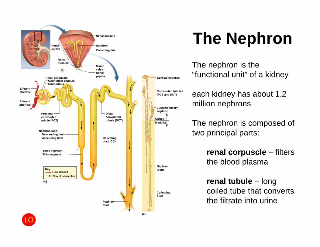
Renal capsule
Collecting ductNephron
(a)
(c)
Cortical nephron
CortexMedulla
GlomerulusGlomerular capsule
Renal corpuscle:
Nephron loop:Descending limbAscending limb
Thick segmentThin segment
Flow of tubular fluidFlow of blood
Key
(b)
Renalcortex
Renalmedulla
Renalpapilla
Minorcalyx
Efferentarteriole
Afferentarteriole
Proximalconvolutedtubule (PCT)
Distalconvolutedtubule (DCT)
Collectingduct (CD)
Papillaryduct
Collectingduct
Nephronloops
Juxtamedullarynephron
Convoluted tubules(PCT and DCT)
The NephronThe nephron is the “functional unit” of a kidney
each kidney has about 1.2 million nephrons
The nephron is composed of two principal parts:
renal corpuscle – filters the blood plasma
renal tubule – long coiled tube that converts the filtrate into urine
The Renal Corpuscle
• renal corpuscle – Glomerulus
• Unique capillary bed• Designed to selectively filter solutes
– glomerular capsule (Bowman capsule) that encloses glomerulus
• parietal (outer) layer of Bowman capsule is simple squamousepithelium
• visceral (inner) layer of Bowman capsule consists of elaborate cells called podocytes that wrap around the capillaries of the glomerulus
• capsular space separates the two layers of Bowman capsule– Extension of this space at the “urinary pole” creates the nephron’s tubules
• vascular pole – the side of the corpuscle where the afferent arterial enter the corpuscle and the efferent arteriole leaves
• urinary pole – the opposite side of the corpuscle where the renal tubule begins
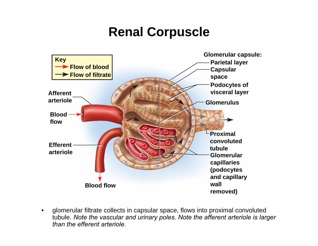
Renal Corpuscle
• glomerular filtrate collects in capsular space, flows into proximal convoluted tubule. Note the vascular and urinary poles. Note the afferent arteriole is larger than the efferent arteriole.
Flow of filtrateFlow of blood
Key
Afferentarteriole
Bloodflow
Efferentarteriole
Blood flow
Glomerularcapillaries(podocytesand capillarywallremoved)
Proximalconvolutedtubule
Glomerulus
Podocytes ofvisceral layer
Capsularspace
Parietal layerGlomerular capsule:
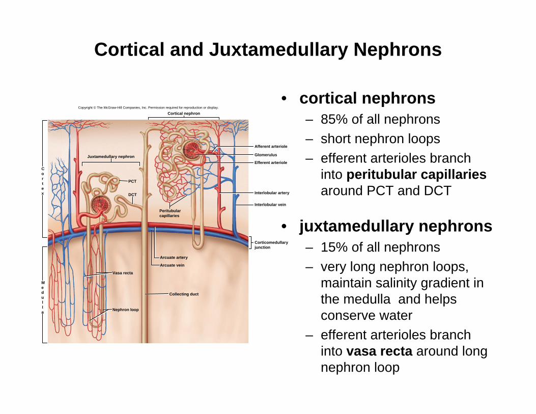
Cortical and Juxtamedullary Nephrons
• cortical nephrons– 85% of all nephrons– short nephron loops– efferent arterioles branch
into peritubular capillaries around PCT and DCT
• juxtamedullary nephrons– 15% of all nephrons– very long nephron loops,
maintain salinity gradient in the medulla and helps conserve water
– efferent arterioles branch into vasa recta around long nephron loop
Copyright © The McGraw-Hill Companies, Inc. Permission required for reproduction or display.
Arcuate vein
Arcuate artery
Vasa recta
Nephron loop
Collecting duct
Cortical nephron
Juxtamedullary nephron Glomerulus
Efferent arteriole
Afferent arteriole
Interlobular artery
Interlobular veinPeritubularcapillaries
Corticomedullaryjunction
PCT
DCT
Medulla
Cortex
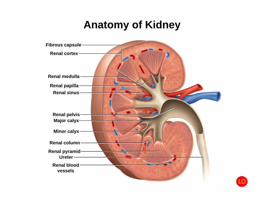
Anatomy of KidneyFibrous capsule
Renal cortex
Renal medulla
Renal pelvisMajor calyx
Minor calyx
Ureter
Renal papillaRenal sinus
Renal column
Renal pyramid
Renal bloodvessels

Blood Supply Diagram
kidneys receive 21% of cardiac output
Inferior vena cava
Arcuate v.
Peritubular capillaries Vasa recta
Efferent arterioleGlomerulus
Afferent arteriole
Interlobular a.
Arcuate a.
Interlobar a.
Segmental a.
Renal a.
(b)
Aorta
Renalmedulla
Renalcortex
Interlobularartery and vein
Interlobarartery and vein
Segmentalartery
Renalarteryandvein
Arcuatearteryand vein
Interlobular v.
Interlobar v.
Renal v.
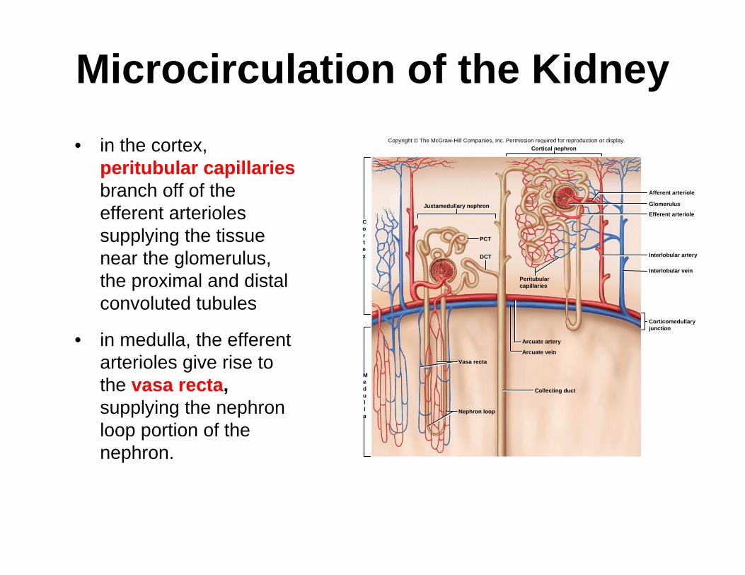
Microcirculation of the Kidney• in the cortex,
peritubular capillariesbranch off of the efferent arterioles supplying the tissue near the glomerulus, the proximal and distal convoluted tubules
• in medulla, the efferent arterioles give rise to the vasa recta, supplying the nephronloop portion of the nephron.
Copyright © The McGraw-Hill Companies, Inc. Permission required for reproduction or display.
Arcuate vein
Arcuate artery
Vasa recta
Nephron loop
Collecting duct
Cortical nephron
Juxtamedullary nephron Glomerulus
Efferent arteriole
Afferent arteriole
Interlobular artery
Interlobular veinPeritubularcapillaries
Corticomedullaryjunction
PCT
DCT
Medulla
Cortex
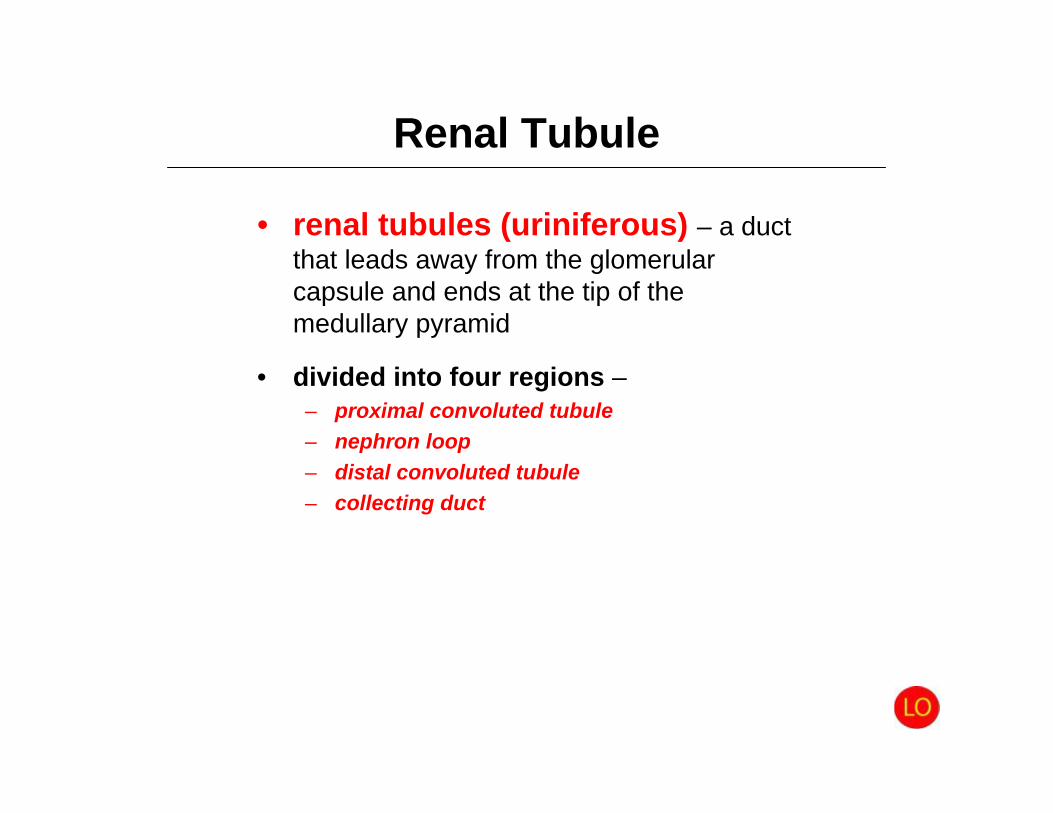
Renal Tubule
• renal tubules (uriniferous) – a duct that leads away from the glomerularcapsule and ends at the tip of the medullary pyramid
• divided into four regions –– proximal convoluted tubule– nephron loop– distal convoluted tubule – collecting duct
Renal Tubule
• proximal convoluted tubule (PCT) – arises from glomerular capsule
– longest and most coiled region– simple cuboidal epithelium with prominent microvilli for
majority of absorption
• nephron loop (loop of Henle) – long U-shaped portion of renal tubule
– descending limb and ascending limb– thick segments have simple cuboidal epithelium
• initial part of descending limb and part or all of the ascendinglimb
• heavily engaged in the active transport of salts and have many mitochondria
– thin segment has simple squamous epithelium• forms lower part of descending limb• cells very permeable to water
Renal Tubule• distal convoluted tubule (DCT) – begins shortly
after the ascending limb reenters the cortex– shorter and less coiled that PCT– cuboidal epithelium without microvilli– DCT is the end of the nephron
• collecting duct – receives fluid from the DCTs of several nephrons as it passes back into the medulla– numerous collecting ducts converge toward the tip of
the medullary pyramid– papillary duct – formed by merger of several collecting
ducts• 30 papillary ducts end in the tip of each papilla• collecting and papillary ducts lined with simple cuboidal
epithelium
Renal TubuleThis is the path for the flow of fluid from the point where the glomerular filtrate is formed to the point where urine leaves the body:
glomerular capsule →proximal convoluted tubule →nephron loop →distal convoluted tubule →collecting duct →papillary duct →minor calyx →major calyx →renal pelvis →ureter →urinary bladder →urethra → (use urethra to pass urine from body)
Renal Innervation• renal plexus – nerves and ganglia wrapped
around each renal artery– follows branches of the renal artery into the
parenchyma of the kidney– issues nerve fibers to the blood vessels and
convoluted tubules of the nephron– carries sympathetic innervation from the abdominal
aortic plexus• stimulation reduces glomerular blood flow and rate of
urine production• respond to falling blood pressure by stimulating the
kidneys to secrete renin, an enzyme that activates hormonal mechanisms to restore blood pressure
– carries parasympathetic innervation from the vagusnerve – increases rate of urine production

Overview of Urine Formation
• kidneys convert blood plasma to urine in four stages
– glomerular filtration– tubular reabsorption– tubular secretion– water conservation
• glomerular filtrate– fluid in capsular space– blood plasma without protein
• tubular fluid– fluid in renal tubule– similar to above except
tubular cells have removed and added substances
• urine– once it enters the collecting
duct– only remaining change is
water content
Glomerular filtrationCreates a plasmalikefiltrate of the blood
Tubular reabsorption Removes useful solutes from the filtrate, returns them to the blood
Tubular secretionRemoves additionalwastes from the blood,adds them to the filtrate
Water conservation Removes water from theurine and returns it toblood; concentrates wastes
Renal corpuscle
Flow of filtrate
Peritubularcapillaries
Renal tubule
H2O
H2O
H2O
Urine
Blood flow
1
2
3
and