Embed Size (px)
Citation preview

BiomaterialsScience
PAPER
Cite this: Biomater. Sci., 2013, 1, 52
Received 20th April 2012,Accepted 16th May 2012
DOI: 10.1039/c2bm00026a
www.rsc.org/biomaterialsscience
Citrate-based biodegradable injectable hydrogelcomposites for orthopedic applications
Dipendra Gyawali,†a Parvathi Nair,†a Harry K. W. Kimb and Jian Yang*a,c
Previous studies have confirmed that natural bone apatite crystals are bound with citrate-rich molecules.
Citrates on apatite crystals impact bone development and its load-bearing function. However, such
understanding has never been translated into bone biomaterials design. Herein, the first citrate-based
injectable composite material for orthopedic applications is developed based on our recently developed
biodegradable poly(ethylene glycol) maleate citrate (PEGMC) and hydroxyapatite (HA). PEGMC contains
rich carboxylic groups that could chelate with calcium-containing HA, thus facilitating polymer/HA inter-
actions, similar to natural citrate-bound apatite crystal. The crosslinking of poly(ethylene glycol) diacrylate
(PEGDA) with PEGMC/HA composites allows additional control over the degradation and mechanical
properties of the crosslinked PEGMC/HA (CPEGMC/HA) composites. CPEGMC/HA composite can serve as
an ideal injectable cell carrier as confirmed by the enhanced DNA content, ALP activity, and calcium pro-
duction through a human fetal osteoblast encapsulation study. An ex vivo study on a porcine femoral
head demonstrated that PEGMC/HA is a potentially promising injectable biodegradable bone material
for the treatment of osteonecrosis of the femoral head. Development of biodegradable citrate-based
injectable PEGMC/HA composite is an initial step for the development of the next generation of bone
tissue engineering and orthopedic biomaterials.
Introduction
Mimicking nature with respect to the tissue molecular, biologi-cal, and dynamic environment, and the tissue’s compositionaland morphological structure has been a widely adopted strat-egy and the ultimate goal in tissue engineering scaffoldingdesign. Significant progress in bioconjugation,1–3 growthfactor delivery,4 nanostructure fabrication,5 recombinant pro-teins/matrices,6–8 self-assembly peptides,9,10 and stem cellniches11,12 has been made and are all within the scope ofmimicking nature in tissue engineering. Bone scaffoldingmaterials have been heavily studied in the past two decades,probably the most studied in tissue engineering. Examplesinclude natural polymers such as polysaccharides (starch, algi-nate, chitosan, hyaluronic acid) and synthetic biodegradablepolymers such as poly(lactic acid) (PLA), poly(glycolic acid)(PGA), and polycaprolacton (PCL). It has long been recognized
that hydroxyapatite (HA, Ca10(PO4)6(OH)2) consists of about60 wt% of bone13 in composite with collagen.14 Thus, mixingpolymers with HA or other related calcium phosphate (e.g.α-TCP and β-TCP) to form micro/nano-composites has been aconventional strategy to improve the mechanical propertiesand bioactivity of scaffolds with considerable success bothin vitro and in vivo for bone tissue engineering. Examples ofdegradable polymers that are composited with HA or relatedcalcium phosphate include poly(L-lactic acid), poly(D,L-lacticacid), polycaprolactone, and poly(propylene fumarate) (PPF).15
However, seeking bioactive bone tissue engineering materialsand scaffolds seems still to be an endless on-going effort and achallenge due to the unsatisfactory osteoinductivity andosteointegration and significant inflammatory responses ofthe existing bone biomaterials in vivo.
By carefully reviewing the literature on natural bone compo-sitions, it is clear that one important fact has been inadver-tently overlooked in the design of bone biomaterials andscaffolds in the past. Natural bone is an organic–inorganicnanocomposite where thin nanocrystals of apatite areembedded in collagen. The small nanocrystals (3 nm thick)control the mechanical properties of bone and prevent crackpropagation. Early in the 1960s, it was found that citrate mole-cules consist of about 5 wt% of the organic components inbone, which was thought to regulate the stabilization of thinnanocrystal apatite.16 More recently, a careful solid-state†Equal Contribution.
aDepartment of Bioengineering, The University of Texas at Arlington, Arlington, TX,
USAbCenter for Excellence in Hip Disorders and Sarah M. and Charles E. Seay Center for
Musculoskeletal Research, Texas Scottish Rite Hospital for Children, Dallas, TX
75219, USAcDepartment of Bioengineering, The Pennsylvania State University, University Park,
PA 16802, USA. E-mail: [email protected]
52 | Biomater. Sci., 2013, 1, 52–64 This journal is © The Royal Society of Chemistry 2013

nuclear magnetic resonance (NMR) study revealed that thesurface of apatite nanocrystals is studded with strongly boundcitrate molecules.17 Citrate is not a dissolved calcium-solubil-izing agent but a strongly bound, integral part of the bonenanocomposite. Surprisingly, citrate was not even mentionedin most of the literature in the past 30 years, not even those onbone biomaterials and scaffolds.
The natural existence of citrate in bone hints that citrateshould be considered in bone biomaterial and scaffold designalthough its function on bone development is still largelyunknown. A recent study on poly(diol citrate)/HA compositesfurther support this strategy. Poly(diol citrate) is the firstcitrate-based biodegradable elastomer that holds promise fortissue engineering and other biomedical applications.18,19 Useof poly (diol citrates) allows the formation of a degradablecomposite containing up to 60% of HA within it, while theconventional polylactide can only form a composite with up to35% of HA. Poly(diol citrate)/HA demonstrated excellent osteo-conductivity and induced rapid mineralization. Surprisingly, itelicited no chronic inflammatory response at the tissue-com-posite interface in vivo.19 It was inferred to that the rich–COOH from citrate units of poly(diol citrate) prompt calciumchelation thus facilitating poly(diol citrate)/HA interaction,enhancing mechanical properties, and promoting mineraliz-ation. Clear understanding on why poly(diol citrate)/HA exhib-ited excellent in vivo tissue/bone-compatibility is stillunknown. Although poly(diol citrate)/HA was not developedunder an hypothesis that the existence of citrate in bone mayimplicate the bone development, its excellent in vivo perform-ance prompts further systematic study on the development ofcitrate-based orthopedic biomaterials.
With the goal of mimicking nature, it is our belief thatcitrate-based polymer/HA (or other related calcium phosphate)composite materials could constitute the next generation ofbone tissue engineering and orthopedic biomaterials. Our labhas long been developing citrate-based biodegradable elasto-mers for versatile biomedical applications, including therecently developed crosslinked urethane-doped polyester(CUPE),20–22 poly(alkylene maleate citrate) (PAMC),23–25 andbiodegradable photoluminescent polymer (BPLP).26,27 Withcareful molecular design, these citrate-based polymers offerversatile processability and tunable mechanical, degradable,and optical properties. These versatile citrate-based biodegrad-able polymers form a platform for us to understand andcustom-design biomaterials toward bone tissue engineeringand orthopedic applications.
Recently, significant attention has been placed on deliver-ing various types of cells (autologous chondrocytes, mesenchy-mal stem cells, and osteoblasts) to treat irregularly shapedbone defects for bone regeneration. It has been recognizedthat in situ crosslinkable injectable hydrogels would be morebeneficial for cell/drug delivery in filling irregular defects asthey can homogeneously fit into the void in situ prior tosetting via a minimally invasive injection. However, most ofthe synthetic hydrogels lack of osteoconductivity. Therefore,injectable composites composed of injectable hydrogels with
osteoconductive HA microparticles can be an alternative solu-tion. Very recently, a few groups have started working on inject-able hydrogel/HA composites for cell/drug delivery fororthopaedic applications using injectable polymers such asPPF,15 Poly(ethylene glycol)-diacrylate,28 Poly(2-hydroxyethylmethacrylate),29 poly(ethylene glycol)-poly(ε-caprolactone)-poly(ethylene glycol),30 and acrylamide-based polymers.31 Ithas been confirmed that HA microparticles within the hydro-gels not only enhance the mechanical strength but also serveas bioactive molecules to enhance cellular responses. However,none of these injectable composites considered the benefits ofthe aforementioned citrate effects in natural bone.
In the present study, a new bioactive citrate-based injectablebiodegradable composite is developed for delivering cells toirregular bony defects. Given the aforementioned benefits ofPOC/HA used for orthopaedic applications, we have recentlyinvestigated and reported the synthesis and characterization ofthe first citrate-based water-soluble, in situ crosslinkable, andbiodegradable polymers, poly (ethylene glycol maleate citrate)(PEGMC) for cell delivery.24 These hydrogels were found to beelastic when compressed and demonstrated minimal cytotoxi-city in vitro comparable to the FDA approved Poly (ethyleneglycol) diacrylate (PEGDA) and completely degrade in vivo withminimal inflammatory responses.23,32 Thus, an injectablecomposite derived by combining PEGMC and HA particleswould have characteristics potentially ideal for injectableorthopaedic applications. The objective of this study is toevaluate the physicochemical properties, polymer and HAinteraction, cytotoxicity, and injectability of CPEGMC/HA com-posites potentially serving as injectable materials for osteo-blast delivery in orthopaedic applications.
Experimental sectionMaterials
HA [Mw: 502.32, assay >90% (as Ca3(PO4)2); particle size:>75 μm (0.5%), 45–75 μm (1.4%), <45 μm (98.1%)] was pur-chased from Fluka (St Louis, MO, USA). All other chemicalswere purchased from Sigma Aldrich (St Louis, MO), exceptwhere mentioned otherwise. All chemicals were used asreceived.
Synthesis and preparation of CPEGMC/HA composite
PEGMC polymer was synthesized by a simple and controlledpolycondensation reaction of poly(ethylene glycol) (PEG) (Mw =200), citric acid (CA) and maleic anhydride (MA), as reportedpreviously.23,24 In a typical procedure, PEG, CA and MA wereadded to a 250 ml round-bottom flask fitted with an inlet andan outlet adapter. The flask was immersed in a silicon oil bathat 160 °C at constant stirring under continuous flow of nitro-gen. Following the melting of the monomers, the temperatureof the oil bath was reduced to 140 °C until the desired PEGMCpolymer viscosity was achieved. To remove any unreactedmonomers and small molecular weight oligomers, PEGMCwas dissolved in de-ionized (DI) water and dialyzed (Spectrum,
Biomaterials Science Paper
This journal is © The Royal Society of Chemistry 2013 Biomater. Sci., 2013, 1, 52–64 | 53

500 Dalton cut-off ) for 2 days with frequent changes of DIwater, followed by freeze-drying.
The HA content of the PEGMC/HA composite was definedby the weight percentage of HA over the entire composite. Puri-fied PEGMC polymer was dissolved in DI water to make a 30%(w/v) solution in a container (centrifuge tube or culture dish).The pH of the mixture was adjusted to 5.5 by adding sodiumbicarbonate under continuous monitoring of pH. HA powderwas then added to the polymer solution under thoroughlymixing. In the next step, 15% (w/v) of poly(ethylene glycol dia-crylate) (PEGDA) (Mw = 3.4 K) was added as a crosslinker fol-lowed by the addition of an aqueous radical initiatorammonium persulfate (APS) and N,N,N′,N′-tetramethylenedi-amine (TEMED) at 25 mM concentration. The entire containerwas tightly sealed and then placed in an incubator at 37 °C for∼15 minutes to obtain crosslinked poly(ethylene glycolmaleate citrate)/HA (CPEGMC/HA) composites. The schematicof PEGMC synthesis and CPEGMC/HA crosslinking is shownin Scheme 1. Three different compositions of CPEGMC/HAwere fabricated where the weight ratios of PEGMC : HA wasvaried from 70 : 30, 55 : 45 and 40 : 60 to yield CPEGMC/HA(30), CPEGMC/HA (45) and CPEGMC/HA (60) respectively.
Characterization of CPEGMC/HA composites
The sol content, swelling ratio, and degradation of theCPEGMC/HA composites were investigated by a mass differen-tial method. CPEGMC/HA composites with different HA com-position were made as previously described. The CPEGMC/HAsamples were cut into circular discs of dimensions 10 mmdiameter by 2 mm thickness (n = 8). Samples were lyophilizedfor 48 hours to remove all water and weighed (Mi).
For the sol–gel fraction study, samples were immersed in10 ml of 1,4-dioxane for 24 hours, freeze-dried and weighed(Md). Sol fraction (SF) of the hydrogel composites was deter-mined by using following eqn (1):
%SF ¼ Mi �Md
Mi� 100 ð1Þ
For the swelling study, the crosslinked composites wereswollen in DI water to equilibrium swelling, blotted withKimwipe paper, and weighed (Mw) and then lyophilized andweighed again (Md). The swelling ratio (SR) was calculatedusing the formula in eqn (2)
%SR ¼ Mw �Md
Md� 100 ð2Þ
For the degradation study, CPEGMC/HA disks were incu-bated in phosphate buffer saline (PBS) (pH = 7.4) at 37 °C. Thebuffer was changed regularly to ensure that the pH of thebuffer was maintained at 7.4 during degradation. The initialweight of CPEGMC/HA discs was recorded as Mi. At pre-deter-mined time points, the samples were removed from PBS,washed in DI water thrice to remove any residual salts and lyo-philized and weighed (Mt). Degradation was determined byweight loss ratio as shown in eqn (3).
%ML ¼ Mi �Mt
Mi� 100 ð3Þ
Microstructural characterization of CPEGMC/HA composites
The microstructures of the CPEGMC/HA composites wereexamined using a Leica DMLP microscope (Leica MicrosystemsInc., Bannockburn, IL) fitted with a Nikon E500 camera (NikonCorp., Japan). Hydrated CPEGMC/HA samples were firstembedded in OCT freezing media (Polysciences Inc., Warring-ton, PA), cryosectioned into 10 μm thick sections, and thenphotomicrographed to characterize the porous morphology ofthe composites and the distribution of HA in the composites.
Mechanical properties of CPEGMC/HA composites
Un-confined compressive mechanical tests of CPEGMC/HAcomposites were studied under a MTS Insight 2 mechanicaltester equipped with a 10 N load cell (MTS, Eden Prairie, MN).Compressive tests of CPEGMC/HA at two different conditionswere conducted, where CPEGMC/HA was compressed immedi-ately after crosslinking (as prepared) and after equilibriumswelling (fully hydrated). Briefly, the cylindrical shaped speci-mens of dimensions 10 mm by 10 mm (diameter × height)
Scheme 1 (A) Schematic representation of PEGMC synthesis and crosslinking;(B) schematic representation of CPEGMC/HA composites. PEGMC is crosslinkedusing redox initiators APS/TEMED in the presence of HA and PEGDA to producecrosslinked PEGMC/HA network.
Paper Biomaterials Science
54 | Biomater. Sci., 2013, 1, 52–64 This journal is © The Royal Society of Chemistry 2013

were compressed at a rate of 1 mm min−1 to 50% strain. Theinitial modulus was calculated by measuring the gradient at10% of compression of the stress–strain curve. Fully hydratedsamples were subjected to cyclic compression to 20% strainfor ten cycles and the hysteresis curves of load vs. strain wereplotted. The results were presented as mean ± standard devi-ation (n = 5). Cancellous bone cylinders removed from afemoral head of a domestic pig were decellularized and sub-jected to compression tests as a control.
In vitro mineralization on CPEGMC/HA composites
Simulated body fluids (SBF) were prepared as describedpreviously33–35 to study in vitro mineralization on theCPEGMC/HA composites. The SBF with an inorganic ion con-centration five times human blood plasma was denoted as SBF(5×). Disk shaped polymer scaffolds were immersed in 6-wellplates containing SBF (5×) for up to 7 days. The SBF wasreplaced every other day. After incubation for various periodsof times, the specimens were washed carefully with DI water toremove any soluble inorganic ions, and dried in air and coatedwith silver prior to analysis under a Hitachi 3000N scanningelectron microscope (SEM) (Hitachi, Pleasanton, CA).
In vitro cell culture on CPEGMC/HA composites
CPEGMC and CPEGMC/HA films were cut into cylindricaldiscs (10 mm in diameter and 1 mm in thickness). Thesamples were sterilized by soaking in 70% ethanol for30 minutes, then exposing to UV light for 1 hour, and thenwashed with PBS for 3 × 5 minutes. Human fetal osteoblasts(hFOBs) (ATCC) were cultured according to ATCC protocol inphenol free Dulbecco’s modified Eagle’s medium (DMEM) –
Ham’s F12 1 : 1 medium supplemented with 10% fetal bovineserum (FBS) (HyClone) and geneticin (300 μg ml−1; Sigma-Aldrich, St. Louise) . The culture flasks were kept in an incuba-tor maintained at 37 °C, 5% CO2, and 95% relative humidity.The cells were trypsinized, centrifuged, and suspended inmedia to obtain a seeding density of 2 × 10⁵ cells per ml onCPEGMC/HA films. After 48 hours of culture, the cells werefixed in 3% (v/v) of gluteraldehyde in PBS and sequentiallydehydrated with a graded series of ethanol, lyophilized, andsputter coated with silver. The samples were then observedunder SEM to view the morphology of the attached cells. Inaddition to SEM images, films seeded with hFOBs werestained with carboxyfluorescein diacetate, succinimidyl ester(CFDA-SE) (Invitrogen, Carlsbad, CA) green fluorescent celltracer according to the manufacturer’s protocol and viewedunder a Zeiss Auxiovert inverted microscope (Carl Zeiss Micro-Imaging, Thornwood, NY).
The cytotoxicity of the composites was further evaluated byencapsulating hFOB cells within the networks of the CPEGMC/HA composites. Briefly, PEGMC solution was formulated asdescribed in Section 2.2. The solution was then sterilized viafiltering through 0.22 μm filter. hFOBs were added to polymersolution to achieve a final cell density of 15 × 106 cells per ml.After being held at 37 °C for approximately 15 minutes in anincubator, the cells/composite constructs were transferred to a
6 well plate and incubated in osteoblast media. During the 21-day culture period, the culture media was changed every48 hours and the cell-encapsulated composites were taken out atweek 1, 2 and 3 and stained using LIVE/DEAD assay (LIVE/DEADViability/Cytotoxicity Kit, Invitrogen, Carlsbad, CA). Cell viabilityand distribution were observed under a Zeiss Auxiovert invertedmicroscope (Carl Zeiss MicroImaging, Thornwood, NY).
For biochemical analysis, at various time points, the cell-cultured CPEGMC/HA constructs were removed from mediaand homogenized in PBS. 500 μl of 0.2% Triton X-100 solutionwas added to the constructs. The samples were subjected totwo freeze-thaw cycles. The homogenates were then sonicatedfor 30 s. The homogenates were centrifuged for 10 minutes at4000 rpm. The supernatant after centrifuging was used tomeasure the DNA content of each construct using PicoGreenassay according to the manufacturer protocol. Measurementswere performed in triplicate.36,37
To characterize the alkaline phosphatase (ALP) activitywithin the composite, alkaline buffer solution was added tothe constructs and sonicated to ensure that the constructswere completely homogenized. The lysate was centrifuged at4000 rpm for 10 minutes at 4 °C. 100 μl of supernatant wasincubated with 100 μl of p-nitrophenyl phosphate solution in a96 well plate at 37 °C for 1 hour. The reaction was stopped byadding 0.2 M NaOH solution to each well. The production ofp-nitrophenyl in the presence of ALP was measured by moni-toring the light absorbance at 405 nm using a microplatereader (Infinite 200, Tecon Group Ltd, Switzerland).38
To quantify the calcium content within the composite,supernatant was collected and 50 μl of the supernatant wasadded to a 96 well plate. The calcium concentration of the celllysate was analyzed using cresolphthalein complexone (Sigma).After 10 minutes, the absorbance was read at 575 nm usingthe Tecan microplate reader (Infinite 200).38
Ex vivo injectability of the CPEGMC/HA composite
The ex vivo study was conducted on a porcine femoral head.Femoral head was cored to create a cylindrical cavity. PEGMC/HA solution was injected into the cavity and incubated at37 °C for in situ crosslinking. To visualize the in situ formedcomposites, the femur head was further sectioned and photo-graphed using a digital camera.
Statistical analysis
Data was expressed as mean ± standard deviation. The statisti-cal significance between two sets of data was calculated usingone-way ANOVA. Data were taken to be significant, whenp < 0.05 was obtained.
ResultsPolymer synthesis, cross linking and characterization ofPEGMC/HA composite
PEGMC polymer was synthesized by a simple, one pot, andcontrolled polycondensation reaction from maleic anhydride,
Biomaterials Science Paper
This journal is © The Royal Society of Chemistry 2013 Biomater. Sci., 2013, 1, 52–64 | 55

PEG, and citric acid. The polymer chains consist of degradableester bonds, crosslinkable carbon–carbon double bonds, andpendant carboxyl and hydroxyl functional group as confirmedby previous FTIR and 1H-NMR.23 Due to these pendent car-boxylic groups present throughout the polymeric network,there are strong interactions between the calcium ions of HAmolecules. This was confirmed in Fig. 1, as the peak maximadue to bond vibration of hydroxyl groups at 3570 cm−1 of thepolymer shift to 3590 cm−1 as in the case of PEGMC/HA com-posite where the characteristic HA peaks (1049 cm−1 for PO−
4)and degradable ester bonds (CvO at 1690–1750 cm−1 and C–Oat 1000–1260 cm−1) are still evident at their respective wavenumbers. PEGMC is completely soluble in water and can behomogeneously mixed with inorganic minerals such as hydro-xyapatite without observable sedimentation before, during,and after the crosslinking process. In our previous study, adetailed rheology study of the PEGMC/HA composite demon-strated that the hydrogel composite precursor is highly inject-able (via a 27-gauge needle) with low viscosity even in higher(50%) HA content and can be crosslinked through radicalpolymerization aided by PEGDA crosslinkers within 3 to15 minutes at 37 °C to create mechanically stable hydrogelcomposites.32
Fig. 2A showed the overall percentage of leachable (sol)content of the crosslinked composites. The sol content did notvary significantly with increasing HA concentration. Forexample, The sol content for CPEGMC/HA(30), CPEGMC/HA(45), and CPEGMC/HA(60) were 6.90 ± 0.74%, 7.06 ± .28%, and5.66 ± 1.13%, respectively. Fig. 2B showed the swelling kineticsof CPEGMC/HA containing various concentration of HA par-ticles. These hydrogel composites started uptaking water andreached equilibrium swelling as early as 10 minutes and didnot significantly increase swelling till 12 hours. Increasing theamount of HA in composites decreased swelling ratios. Forinstance, the swelling ratio reduced from 9.56 ± 0.56(CPEGMC/HA(30)) to 7.89 ± 0.96 (CPEGMC/HA(45)) and 3.42 ±0.66 (CPEGMC/HA(60)).
The degradation profile of CPEGMC/HA in PBS showedincreasing mass losses with lowering concentrations of HA. Asshown in Fig. 2C, CPEGMC/HA (30) showed a mass loss of58.18 ± 6.60% at the end of 22 weeks. The high HA concen-tration CPEGMC/HA(60) showed a mass loss of 32.24 ± 1.79%at the end of 22 weeks. This result indicated that the mass lossof the composites was primarily due to the degradation of thepolymers and that HA particles did not leach out from thenetwork in noticeable amounts during degradation. The com-posites maintained their integrity throughout the 22 weeks ofdegradation study.
Microstructure characterization of CPEGMC/HA composites
The pore morphology of the fully hydrated CPEGMC/HA com-posite sections (5 μm) was observed under microscope. It wasobserved that the pure PEGMC gel network was highly porouswith a pore size of 200–400 μm (Fig. 3A). Multiple sections ofthe fully hydrated cylindrical hydrogels cut at various locationsand angles were analyzed and observed with similar mor-phologies. This technique allowed us to visualize the inter-action of the polymer and HA particles under fully hydratedconditions which is otherwise impossible to observe by SEMor cryo-SEM.28 The pore structure of the composites was notaltered with the addition of HA particles. In fact while under afully hydrated state, these mineral particles were distributedwithin the polymer matrix rather than simply encapsulated in
Fig. 1 FTIR spectra of HA powder, PEGMC hydrogel and CPEGMC/HAcomposite.
Fig. 2 Physiochemical characterization of CPEGMC/HA. (A) Degree of sol content as a function of HA concentration; (B) degree of swelling as a function of HAconcentrations; (C) in vitro mass loss of CPEGMC/HA in PBS (pH 7.4; 37 °C) over time up to 22 weeks.
Paper Biomaterials Science
56 | Biomater. Sci., 2013, 1, 52–64 This journal is © The Royal Society of Chemistry 2013

the pores. As shown in Fig. 3B–D, the more HA was incorpor-ated in the composite, the more HA could be observed withinthe polymer matrix.
Mechanical properties of CPEGMC/HA composite
CPEGMC/HA composites with varying HA content (30, 45,60)% were prepared and subjected to unconfined compressivetests as soon as they were freshly prepared (as-prepared) andafter equilibrium swelling (fully hydrated). As shown inFig. 4A–B, the modulus of the as-prepared CPEGMC/HA(30)-(10 ± 5 kPa) did not change significantly compared to CPEGMC/HA(45)(26 ± 12 kPa). The modulus of CPEGMC/HA(30)(12.5 ± 5kPa) and CPEGMC/HA(45)(30 ± 7 kPa) also showed no signifi-cant difference when tested after full hydration. However, themodulus of CPEGMC/HA(60) (205 ± 26 kPa) was significantlyincreased compared to CPEGMC/HA(30) and CPEGMC/HA(45)and it also significantly decreased in the fully hydrated state
(100 ± 20 kPa). Good overlaps of the stress–strain curves wereobserved when 10 consecutive compressive loading andunloading cycles with 20% strain on fully hydrated CPEGMC/HA samples (Fig. 4C). These mechanical results demonstratedthat the CPEGMC/HA confers the constructs with excellentelastic properties and the HA contributed to the increasedstrength. As shown in Fig. 4D, increasing the crosslinkingtimes also increases the composite mechanical strength. Thedried PEGMC/HA (60) demonstrated a close ultimate compres-sive strength to that of decellularized cancellous bone offemur head of porcine model.
In vitro mineralization
When incubated in simulated body fluid, by day 1, homo-geneous nucleation and 2-dimension growth of calcium phos-phate was evident as mineral nodules appeared on the surfaceof CPEGMC/HA composite (Fig. 5A). By allowing
Fig. 3 Representative light microphotographs of macroporous CPEGMC hydrogel and CPEGMC/HA composite thin sections (10 μm). (A) CPEGMC; (B) CPEGMC/HA(30) composite; (C) CPEGMC/HA(45) composite; and (D) CPEGMC/HA(60) composite. The black dots denote the HA microparticles within the polymer matrix. Thewhite spaces represent void pore spaces.
Biomaterials Science Paper
This journal is © The Royal Society of Chemistry 2013 Biomater. Sci., 2013, 1, 52–64 | 57

mineralization to proceed for a longer period of time (day 7),more distinct mineral nodules continuously covered the entiresurface (several micron) of the CPEGMC/HA composite(Fig. 5B). Upon evaluation by the calibrated energy dispersivespectroscopy (EDS), the apatite nodules on the surface of thecomposite revealed a Ca/P ratio (1.6 ± 0.1) similar to that ofsynthetic HA.
In vitro cell culture
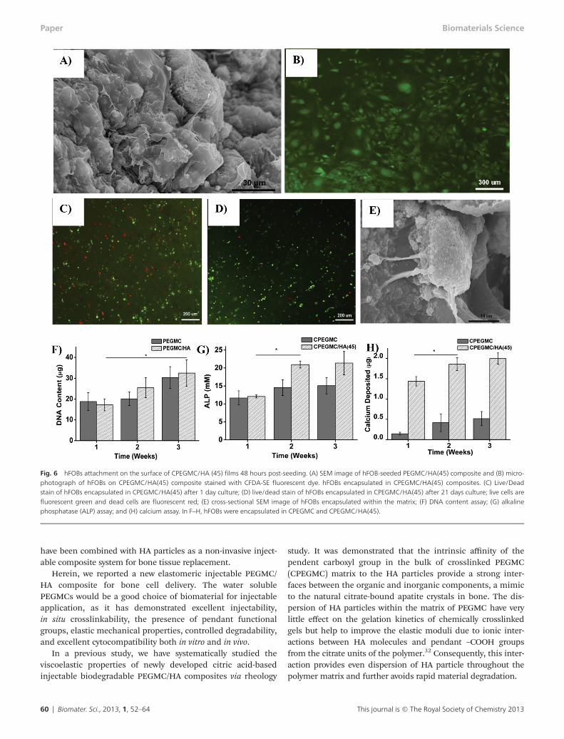
hFOB seeded on the surface of the PEGMC/HA composites sup-ports cell adhesion and promotes cell spreading throughout thesurface over the experimental period of 48 hours when observedunder fluorescent microscope and SEM (Fig. 6A and B).
hFOB cells were further encapsulated within the CPEGMC/HA hydrogel composites to test their potential for cell encapsu-lation/delivery. It was found that the cells survived the encap-sulation procedure and proliferated during the 3 weeks of
subculture. Fluorescent images of the encapsulated osteoblastsstained by LIVE/DEAD assay at day 1 and day 21 of encapsula-tion are shown in Fig. 6C and D, where red fluorescencedenotes dead cells and green fluorescence indicates live cells.In Fig. 6D, the majority of hFOB cells were green, indicatingthat the CPEGMC/HA polymer network provided a cytocompa-tible environment for hFOB delivery. Only a few cells diedunder the crosslinking conditions. Furthermore, the imagesalso indicated a uniform cell distribution in the composites.hFOBs could adhere and spread on CPEGMC/HA composites,which could be clearly seen in Fig. 6E.
The deoxyribonucleic acid (DNA) content of hydrogels con-taining osteoblasts was assessed using the PicoGreen assayand the results were shown in Fig. 6F. DNA content increasedfrom week 1 to week 3, demonstrating that cells were prolifer-ating. No significant difference was found from both the purePEGMC group and the CPEGMC/HA group at each time point.
Fig. 4 Mechanical tests of as-prepared and fully hydrated CPEGMC/HA composites. (A) Compressive modulus of as-prepared CPEGMC/HA composites; (B) compres-sive modulus of fully hydrated CPEGMC/HA composites; (C) cyclic compression tests of fully hydrated CPEGMC/HA composites. Ten consecutive cycles of controlledloading and unloading was applied to fully hydrated CPEGMC/HA specimens using MTS Insight II mechanical tester using a 10 N load cell. (D) Compression test ofdecellularized cancellous bone and synthetic hydrogel composites at different conditions.
Paper Biomaterials Science
58 | Biomater. Sci., 2013, 1, 52–64 This journal is © The Royal Society of Chemistry 2013

As shown in Fig. 6G, the ALP production of the encapsulatedhFOBs in CPEGMC/HA showed a significant increased aftertwo weeks. In contrast, the ALP production on pure CPEGMCdid not show any significant change within 3 week of culture.The calcium content deposited by the hFOBs in CPEGMC/HAwas significantly higher compared to that of the pure CPEGMCand CPEGMC/HA composites without cells. The significantincrease was observed at the 2nd week (Fig. 6H).
Ex vivo study
An ex vivo in situ delivery study was conducted on a porcinefemoral head (Fig. 7A–D). PEGMC/HA could be easily injectedin the defect in the central region of the femur head using asyringe fitted with a biopsy cannula. No leakage was observedprior to crosslinking. Visual examination of the polymerizationsite after sectioning the femoral head suggested that the in situcrosslinked CPEGMC/HA completely filled up the irregularimplantation site and reinforced the femoral head.
Discussion
Hydroxyapatite (HA, Ca10(PO4)6(OH)2) constitutes around 60 wt% of bone and has long been recognized as a crucial
biomaterial to design tissue-engineered bone substitutes.13 Ithas been confirmed that HA and related calcium phosphate(e.g. α-TCP and β-TCP) not only enhances the mechanical prop-erties but also plays a critical role in the osteoconduction andosseointegration of the implanted bone graft.39,40 Thus, devel-oping composite materials based on biodegradable polymersand HA became an intense focus in bone tissue engineering.Such composites take advantage of the formability of polymersand the bioactivity of HA to enhance the mechanical proper-ties of the fabricated scaffolds. In the meantime, the poorbioactivity of most of synthetic polymers can be improved.41
Examples of degradable polymers compositing with HA orrelated calcium phosphate include poly(L-lactic acid), poly(D,L-lactic acid), polycaprolactone, and poly(propylene fumarate).15
These prefabricated polymer composites may be excellentchoice for critical bone defects, however, may not be appli-cable in the case of irregular bone defects, as in the case offemoral head osteonecrosis where complete evacuation of thenecrotic tissue is performed by core decompression. It is desir-able to use an injectable scaffolding system that can perfectlyfill the void, solidifies within the site of injection, and induce/promote bone tissue regeneration. In fact, hydrogels basedupon materials such as gelatin,42 poly(propylene fumarate),15
polymethylmethacrylate,43 and hydroxypropylmethyl cellulose44
Fig. 5 Scanning electron microscope images of in vitro mineralization of CPEGMC/HA composite. (A) CPEGMC/HA (45) composite when incubated in 5× SBF for 1day; (B) CPEGMC/HA(45), (C) CPEGMC/HA(30), and (D) CPEGMC/HA(60) composites when incubated in 5× SBF for 7 day.
Biomaterials Science Paper
This journal is © The Royal Society of Chemistry 2013 Biomater. Sci., 2013, 1, 52–64 | 59
have been combined with HA particles as a non-invasive inject-able composite system for bone tissue replacement.
Herein, we reported a new elastomeric injectable PEGMC/HA composite for bone cell delivery. The water solublePEGMCs would be a good choice of biomaterial for injectableapplication, as it has demonstrated excellent injectability,in situ crosslinkability, the presence of pendant functionalgroups, elastic mechanical properties, controlled degradability,and excellent cytocompatibility both in vitro and in vivo.
In a previous study, we have systematically studied theviscoelastic properties of newly developed citric acid-basedinjectable biodegradable PEGMC/HA composites via rheology
study. It was demonstrated that the intrinsic affinity of thependent carboxyl group in the bulk of crosslinked PEGMC(CPEGMC) matrix to the HA particles provide a strong inter-faces between the organic and inorganic components, a mimicto the natural citrate-bound apatite crystals in bone. The dis-persion of HA particles within the matrix of PEGMC have verylittle effect on the gelation kinetics of chemically crosslinkedgels but help to improve the elastic moduli due to ionic inter-actions between HA molecules and pendant –COOH groupsfrom the citrate units of the polymer.32 Consequently, this inter-action provides even dispersion of HA particle throughout thepolymer matrix and further avoids rapid material degradation.
Fig. 6 hFOBs attachment on the surface of CPEGMC/HA (45) films 48 hours post-seeding. (A) SEM image of hFOB-seeded PEGMC/HA(45) composite and (B) micro-photograph of hFOBs on CPEGMC/HA(45) composite stained with CFDA-SE fluorescent dye. hFOBs encapsulated in CPEGMC/HA(45) composites. (C) Live/Deadstain of hFOBs encapsulated in CPEGMC/HA(45) after 1 day culture; (D) live/dead stain of hFOBs encapsulated in CPEGMC/HA(45) after 21 days culture; live cells arefluorescent green and dead cells are fluorescent red; (E) cross-sectional SEM image of hFOBs encapsulated within the matrix; (F) DNA content assay; (G) alkalinephosphatase (ALP) assay; and (H) calcium assay. In F–H, hFOBs were encapsulated in CPEGMC and CPEGMC/HA(45).
Paper Biomaterials Science
60 | Biomater. Sci., 2013, 1, 52–64 This journal is © The Royal Society of Chemistry 2013

One of the prime concern regarding injectables is the leach-able (sol content) component following the solidification.Especially for hydrogel/particle composites, the likelihood ofparticle leaching in a fully hydrated condition is high. Asdemonstrated earlier, PEGMCs were found to have a lower solcontent after crosslinking (<15%, w/w).23 Similar results werefound with our CPEGMC/HA composites. In fact, an increasein the HA component did not show any significant increase inleachable components from the system. HA particles were alsofound to be homogeneously dispersed within the matrixwithout any settling by gravity during solidification. Excellentstructural integration of the organic and inorganic
components were evident through FTIR spectroscopy (Fig. 1)and morphology analysis (Fig. 2). Furthermore, no evidentamount of HA particles leached out from the composite for 22week of incubation in PBS during the degradation study.Another crucial parameter to be addressed while designingbone scaffolds is its porosity.45 Cancellous bone, where osteo-necrosis occurs, is a highly porous environment with 50–90%porosity with average pore size of 10–400 um to facilitate nutri-ent exchange and osteoblast cellular infusion.46 Interestingly,CPEGMC/HA composites were found to be highly porous withopen and interconnected pores within the range of 200 to 400um as similar to other hydrogel systems. An increase in the HA
Fig. 7 Ex vivo study demonstrates that biodegradable injectable PEGMC/HA(45) can be injected into collapsed femoral head for reinforcement. (A) PEGMC/HA(45)solution loaded in cannula injection tool; (B) The cored femoral head with a cylindrical cavity; (C) PEGMC/HA(45) being injected into collapsed femoral head; and(D) the sectional view of cemented femoral head with CPEGMC/HA(45) composite. Crosslinking was achieved within 5 minutes of injection.
Biomaterials Science Paper
This journal is © The Royal Society of Chemistry 2013 Biomater. Sci., 2013, 1, 52–64 | 61

content did not influence pore formation within the formu-lations that were investigated. Rather, the particles distributedevenly within the polymer matrices.
In our previous study, redox crosslinked PEGMC hydrogeldegraded almost 70% of its mass by the end of one month inPBS at 37 °C.23 Interestingly, even at the end of 22 weeks of anin vitro degradation study, the CPEGMC/HA composites werefound to maintain their structural integrity without any notice-able amount of HA release from the system. It was inferredthat the residual polymer network could still well chelate withHA particles to maintain the integrity of the composites. Themorphology change of the composites at longer degradationtimes needs to be studied in the future. Nonetheless,CPEGMC/HA maintained excellent integrity during degra-dation, a favourable factor when used for tissue engineeringapplications. The degradation of CPEGMC/HA networks in vivowill be reported in our future studies as others have suggestedthat local enzymes and biological environments may impactmaterial degradation in vivo.47–52
In general, polymers themselves have poor osteoconductiv-ity. Therefore, the incorporation of HA into polymers to inducemineralization has been a common way in designing bonetissue engineering composite biomaterials.53,54 It was reportedthat the apatite formed as a result of biomimetic process couldhave biological properties that are more similar to naturalbone mineral than that of synthetic calcium phosphate bio-ceramics and provide a more conducive environment for boneformation38,55,56 and differentiation of mesenchymal cells toosteoblasts. Studies have shown that osteoblasts seeded onPLGA/HA scaffolds covered with a bone like apatite coatingboost cell growth, ALP activity, and mineralization. Thus, bonelike apatite may provide a favourable environment for osteo-blast attachment and growth.57 Inducing mineralization is oneimportant property that should be inherited by bone scaffolds.Various strategies have been employed in biomaterials todemonstrate template-driven biomineralization of the system.For example, urea mediated surface hydrolysis was performedon poly(2-hydroxyethyl methacrylate) (pHEMA) to create apendent carboxylic group with an aim to create high mineralcontent hydrogel.14 Our CPEGMC/HA is saturated with car-boxylic acid groups from citrate molecules of CEPGMC thatnot only provide strong HA chelation within the network butalso promote high affinity nucleation and growth of calciumphosphate in simulated body fluid. After 7 days of incubation,SEM micrographs indicated a robust surface mineral layer dueto apatite deposition covering entire surface of the composite.The induced apatite crystals on the composite hydrogel demon-strated typical ‘cauliflower’. This data clearly demonstrates thatthe PEGMC/HA composite is a promising candidate for bonetissue engineering with high affinity for calcium ions.
Material stiffness has been reported to play an importantrole in adhesion, proliferation, and differentiation of cellsseeded/encapsulated on/in biomaterials. Studies showed that amatrix with a stiffness of 20–110 kPa could promote differen-tiation of mesenchymal stem cells (MSCs) into osteoblasts.58
Thus, we formulated hydrogel composites mimicking similar
stiffness. The as-prepared CPEGMC/HA composites exhibitedstiffness within 110 kPa. Given the results, our compositesinjected into bone defects should exhibit favourable mechan-ical stiffness conducive for bone differentiation. It is also note-worthy that complete hydration did not sacrifice the ability ofCPEGMC/HA composites to withstand cyclic compression with20% compressive strain. Furthermore, the mechanical proper-ties of CPEGMC/HA composites can be further tuned byadjusting the polymer concentrations and crosslinking times.
In addition to surface mineralization, PEGMC/HA couldalso serve as a template for surface cell adhesion and prolifer-ation. Upon implantation, it is extremely important forscaffolds to encourage cell adhesion and tissue infiltrationwithin the scaffold as it helps to improve tissue-material inte-gration. Furthermore, we encapsulated osteoblasts within thematrix of PEGMC/HA composite and evaluated their function-alities for a 3 week period. Although minimal cytotoxicity wasexpected during the process of encapsulation, viable cellswithin CEPGMC/HA proliferated and displayed characteristicosteoblastic functionality in bone formation. ALP, an ectoen-zyme usually produced in the early stages of osteoblastic differ-entiation,59 was found to be significantly increased by week 2,in agreement with the increase of calcium deposition at week2 and week 3 of the subculture. In a review of the literature, itwas rarely reported that cells could be encapsulated in inject-able polymer/HA composites. Most of the cell encapsulationcarriers are pure hydrogels with or without bioconjugation ofbiological molecules.60,61 The presence of HA within injectablePEGMC/HA composites favoured ALP production and calciumdeposition as compared to CPEGMC gels alone.
To demonstrate the feasibility of injectable PEGMC/HA, anex-vivo study was conducted on a porcine femoral head afterthe removal of the cancellous bone from its central region.The experiment clearly showed that the composite precursorcan be easily injected and crosslinked in situ after completelyfiling the entire void space of the collapsed femoral head priorto crosslinking. PEGMC/HA with a HA concentration up to60% was still injectable and crosslinkable in situ. Once fullyfilled with PEGMC/HA, the pocketed femoral head could bereinforced without collapse.
Conclusion
In summary, an injectable citrate-based PEGMC/HA hydrogelcomposite was developed. PEGMC/HA demonstrated highlyporous micro-architecture, tunable mechanical and degradableproperties, and exhibited excellent osteoconductivity sup-ported by increased ALP production and calcium depositionby the cells seeded/encapsulated on/into the composites.PEGMC/HA with high concentrations of HA (up to 60%) couldbe injected into porcine femoral head bone defects andreinforce it within a short crosslinking time. By mimicking thecitrate-bound apatite crystals, PEGMC/HA was the first citrate-based injectable composite scaffold with great potentialserving as a cell delivery carrier for bone tissue engineering.
Paper Biomaterials Science
62 | Biomater. Sci., 2013, 1, 52–64 This journal is © The Royal Society of Chemistry 2013
The discovery of citrate-bound apatite crystal in naturalbone left many unanswered questions.17 For example, where isthe citrate formed in bones? When does the citrate come intoplay for bone development? How does the abundance ofcitrate on the crystal surface influence on bone formation?These questions can be translated into questions in bone bio-material design. How much citrate should be placed in bonebiomaterials? How long and when should the citrate exist inbiomaterials? Although our current study does not answer theabove questions, it constitutes an initial step in the develop-ment of a citrate-based injectable biomaterial platform tostudy the above questions. The development of injectablePEGMC/HA also expands the repertoire of existing orthopaedicbiomaterials.
Acknowledgements
This work was supported in part by a R01 award EB012575from the National Institute of Biomedical Imaging and Bio-engineering (NIBIB), a National Science Foundation (NSF)CAREER award 0954109, and small research funding by theTexas Scottish Rite Hospital for Children.
Notes and references
1 M. P. Lutolf and J. A. Hubbell, Nat. Biotechnol., 2005, 23,47–55.
2 J. Elisseeff, F. Yang, C. G. Williams, D. A. Wang, H. Lee andP. N. Manson, Biomaterials, 2005, 26, 5991–5998.
3 A. G. Mikos, H. Shin and S. Jo, Biomaterials, 2003, 24,4353–4364.
4 A. G. Mikos, J. E. Babensee and L. V. McIntire, Pharm. Res.,2000, 17, 497–504.
5 G. B. Wei and P. X. Ma, Adv. Funct. Mater., 2008, 18, 3568–3582.
6 A. H. Reddi and M. Nakashima, Nat. Biotechnol., 2003, 21,1025–1032.
7 D. L. Kaplan, C. M. Li, C. Vepari, H. J. Jin and H. J. Kim,Biomaterials, 2006, 27, 3115–3124.
8 D. A. Tirrell and S. A. Maskarinec, Curr. Opin. Biotechnol.,2005, 16, 422–426.
9 S. Zhang, J. Kisiday, M. Jin, B. Kurz, H. Hung, C. Seminoand A. J. Grodzinsky, Proc. Natl. Acad. Sci. U. S. A., 2002, 99,9996–10001.
10 S. I. Stupp, M. O. Guler, L. Hsu, S. Soukasene,D. A. Harrington and J. F. Hulvat, Biomacromolecules, 2006,7, 1855–1863.
11 D. E. Discher, D. J. Mooney and P. W. Zandstra, Science,2009, 324, 1673–1677.
12 J. P. Vacanti, A. Khademhosseini, R. Langer andJ. Borenstein, Proc. Natl. Acad. Sci. U. S. A., 2006, 103, 2480–2487.
13 J. R. Porter, T. T. Ruckh and K. C. Popat, Biotechnol. Prog.,2009, 25, 1539–1560.
14 J. Song, E. Saiz and C. R. Bertozzi, J. Am. Chem. Soc., 2003,125, 1236–1243.
15 K. W. Lee, S. Wang, M. J. Yaszemski and L. Lu, Biomaterials,2008, 29, 2839–2848.
16 R. L. Hartles, Adv. Oral. Biol., 1964, 1, 225–253.17 K. Schmidt-Rohr, Y. Y. Hu and A. Rawal, Proc. Natl. Acad.
Sci. U. S. A., 2010, 107, 22425–22429.18 J. Yang, A. R. Webb, S. J. Pickerill, G. Hageman and
G. A. Ameer, Biomaterials, 2006, 27, 1889–1898.19 H. Qiu, J. Yang, P. Kodali, J. Koh and G. A. Ameer, Biomater-
ials, 2006, 27, 5845–5854.20 J. Dey, H. Xu, J. Shen, P. Thevenot, S. R. Gondi,
K. T. Nguyen, B. S. Sumerlin, L. Tang and J. Yang, Biomater-ials, 2008, 29, 4637–4649.
21 J. Dey, H. Xu, K. T. Nguyen and J. Yang, J. Biomed. Mater.Res., Part A, 2010, 95, 361–370.
22 J. Dey, R. T. Tran, J. Shen, L. Tang and J. Yang, Macromol.Mater. Eng., 2011, 296, 1149–1157.
23 D. Gyawali, P. Nair, Y. Zhang, R. T. Tran, C. Zhang,M. Samchukov, M. Makarov, H. K. W. Kim and J. Yang, Bio-materials, 2010, 31, 9092–9105.
24 D. Gyawali, R. T. Tran, K. J. Guleserian, L. Tang andJ. Yang, J. Biomater. Sci., Polym. Ed., 2010, 21, 1761–1782.
25 R. T. Tran, P. thevenot, D. Gyawali, Y. Zhang and J. Yang,Soft Matter, 2010, 6, 2449–2461.
26 J. Yang, Y. Zhang, S. Gautam, L. Liu, J. Dey, W. Chen,R. P. Mason, C. A. Serrano, K. A. Schug and L. Tang, Proc.Natl. Acad. Sci. U. S. A., 2009, 106, 10086–10091.
27 C. A. Serrano, Y. Zhang, J. Yang and K. A. Schug, RapidCommun. Mass Spectrom., 2011, 25, 1152–1158.
28 A. K. Gaharwar, S. A. Dammu, J. M. Canter, C. J. Wu andG. Schmidt, Biomacromolecules, 2011, 12, 1641–1650.
29 J. Song, J. W. Xu, T. Filion, E. Saiz, A. P. Tomsia, J. B. Lian,G. S. Stein, D. C. Ayers and C. R. Bertozzi, J. Biomed. Mater.Res., Part A, 2009, 89A, 1098–1107.
30 S. Z. Fu, G. Gun, C. Y. Gong, S. Zeng, H. Liang, F. Luo,X. N. Zhang, X. Zhao, Y. Q. Wei and Z. Y. Qian, J. Phys.Chem. B, 2009, 113, 16518–16525.
31 T. T. Demirtas, A. G. Karakecili and M. Gumusderelioglu,J. Mater. Sci.: Mater. Med., 2008, 19, 729–735.
32 Y. Jiao, D. Gyawali, J. M. Stark, P. Akcora, P. Nair, R. T. Tranand J. Yang, Soft Matter, 2012, 8, 1499–1507.
33 C. Tas, Biomaterials, 2000, 21, 1429–1438.34 A. Oyane, H. Kim, T. Furuya, T. Kokubo, T. Miyazaki
and T. Nakamura, J. Biomed. Mater. Res., 2003, 65,188–195.
35 S. Kim, M. Sun Park, O. Jeon, C. Yong Choi and B. Kim,Biomaterials, 2006, 27, 1399–1409.
36 D. Wang, C. Williams, F. Yang, N. Cher, H. Lee andJ. Elisseeff, Tissue Eng., 2005, 11, 201–213.
37 J. Burdick and K. Anseth, Biomaterials, 2002, 23, 4315–4323.
38 S. Kim, M. Park, S. Gwak, C. Choi and B. Kim, Tissue Eng.,2006, 12, 2997–3006.
39 R. Khanna, K. S. Katti and D. R. Katti, Acta Biomater., 2011,7, 1173–1183.
Biomaterials Science Paper
This journal is © The Royal Society of Chemistry 2013 Biomater. Sci., 2013, 1, 52–64 | 63
40 T. Yoshikawa, H. Ohgushi and S. Tamai, J. Biomed. Mater.Res., 1996, 32, 481–492.
41 S. Weiner and H. Wagner, Annu. Rev. Mater. Sci., 1998, 28,271–298.
42 W. B. Hillig, Y. Choi, S. Murthy, N. Natravali and P. Ajayan,J. Mater. Sci.: Mater. Med., 2008, 19, 11–17.
43 A. M. Moursi, A. V. Winnard, P. L. Winnard, J. J. Lannuttiand R. R. Seghi, Biomaterials, 2002, 23, 133–144.
44 P. Weiss, S. Bohic, M. Lapkowski and G. Daculsi, J. Biomed.Mater. Res., 1998, 41, 167–170.
45 V. Karageorgiou and D. Kaplan, Biomaterials, 2005, 26,5474–5491.
46 M. Dadsetan, T. Hefferan, J. Szatkowski, P. Mishra, S. Macura,L. Lu and M. Yaszemski, Biomaterials, 2008, 29, 2193–2202.
47 S. J. Peter, S. T. Miller, G. Zhu, A. W. Yasko andA. G. Mikos, J. Biomed. Mater. Res., 1998, 41, 1–7.
48 M. Tracy, K. Ward, L. Firouzabadian, Y. Wang, N. Dong,R. Qian and Y. Zhang, Biomaterials, 1999, 20, 1057–1062.
49 R. Suuronen, T. Pohjonen, J. Hietanen and C. Lindqvist,J. Oral Maxillofacial Surg., 1998, 56, 604–614.
50 R. Smith, C. Oliver and D. Williams, J. Biomed. Mater. Res.,1987, 21, 991–1003.
51 D. Williams, Clin. Mater., 1992, 10, 9–12.
52 M. Timmer, H. Shin, R. Horch, C. Ambrose and A. Mikos,Biomacromolecules, 2003, 4, 1026–1033.
53 M. Kikuchi, S. Itoh, S. Ichinose, K. Shinomiya andJ. Tanaka, Biomaterials, 2001, 22, 1705–1711.
54 Y. F. Chou, W. A. Chiou, Y. Xu, J. C. Y. Dunn and B. M. Wu,Biomaterials, 2004, 25, 5323–5331.
55 S. Kalita, D. Rokusek, S. Bose, H. Hosick andA. Bandyopadhyay, J. Biomed. Mater. Res., 2004, 71, 35–44.
56 D. Lickorish, J. A. M. Ramshaw, J. A. Werkmeister,V. Glattauer and C. R. Howlett, J. Biomed. Mater. Res., 2004,68, 19–27.
57 C. Loty, J. M. Sautier, H. Boulekbache, T. Kokubo,H. M. Kim and N. Forest, J. Biomed. Mater. Res., 2000, 49,423–434.
58 H. J. Kong, T. R. Polte, E. Alsberg and D. J. Mooney, Proc.Natl. Acad. Sci. U. S. A., 2005, 102, 4300–4305.
59 J. Temenoff, H. Park, E. Jabbari, T. Sheffield, R. LeBaron,C. Ambrose and A. Mikos, J. Biomed. Mater. Res., 2004, 70,235–244.
60 G. Nicodemus and S. Bryant, Tissue Eng., Part B: Rev., 2008,14, 149–165.
61 D. Benoit, A. Durney and K. Anseth, Tissue Eng., 2006, 12,1663–1673.
Paper Biomaterials Science
64 | Biomater. Sci., 2013, 1, 52–64 This journal is © The Royal Society of Chemistry 2013


![Journal of Biomaterials Applications ‘Green’ biocompatible ... Biomater Appl-20… · PVA/ chitosan/nano-ZnO composite nanofibrous membranes Antibacterial and antifungal [16]](https://img.pdfslide.net/doc/110x75/605be37fd9239d416832e8c2/journal-of-biomaterials-applications-agreena-biocompatible-biomater-appl-20.jpg)