Embed Size (px)
Citation preview
C3 Glomerulopathy: role of complement for pathogenesis and treatment
Marina Vivarelli
Division of Nephrology and DialysisBambino Gesù Children’s Hospital, IRCCS
Rome, Italy
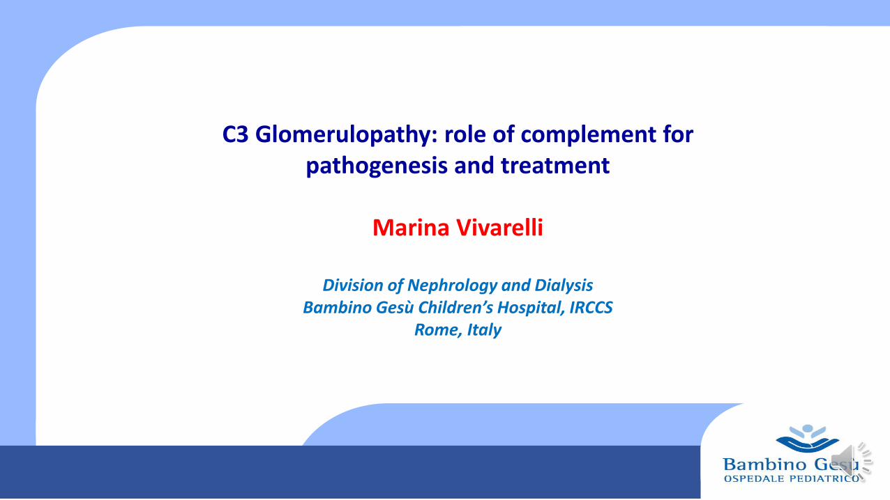
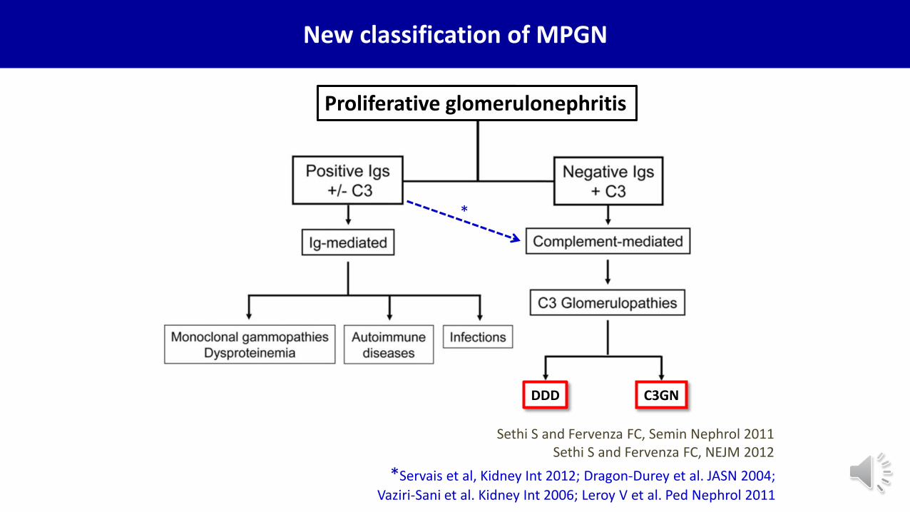
MPGN: the old classification…
MPGN Type ISubendothelial depositsWest et al, J Pediatr 1965
MPGN Type II / DDDIntramembranous depositsGalle, Thesis 1962; Habib et al, Kidney Int 1975
MPGN Type IIISubendothelial andsubepithelial depositsBurkholder et al, Am J Pathol 1969Anders et al, Virchows Arch A Pathol Anat Histol 1997Strife et al, Clin Nephrol 1984
C
C
*C
C
C
C
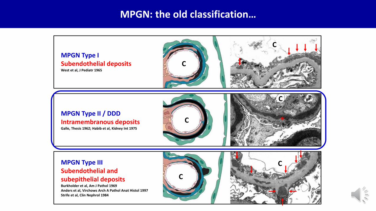
19 patients with unusual glomerulonephritis and:
- C3NeF positivity (7), CFH (3), CFI (2) or MCP (1) mutations
- overt mesangial and epimembranous C3 deposits
- absence of dense intramembranous deposits (no DDD)
- no Ig deposition
C3GN
A new disease entity: C3GN
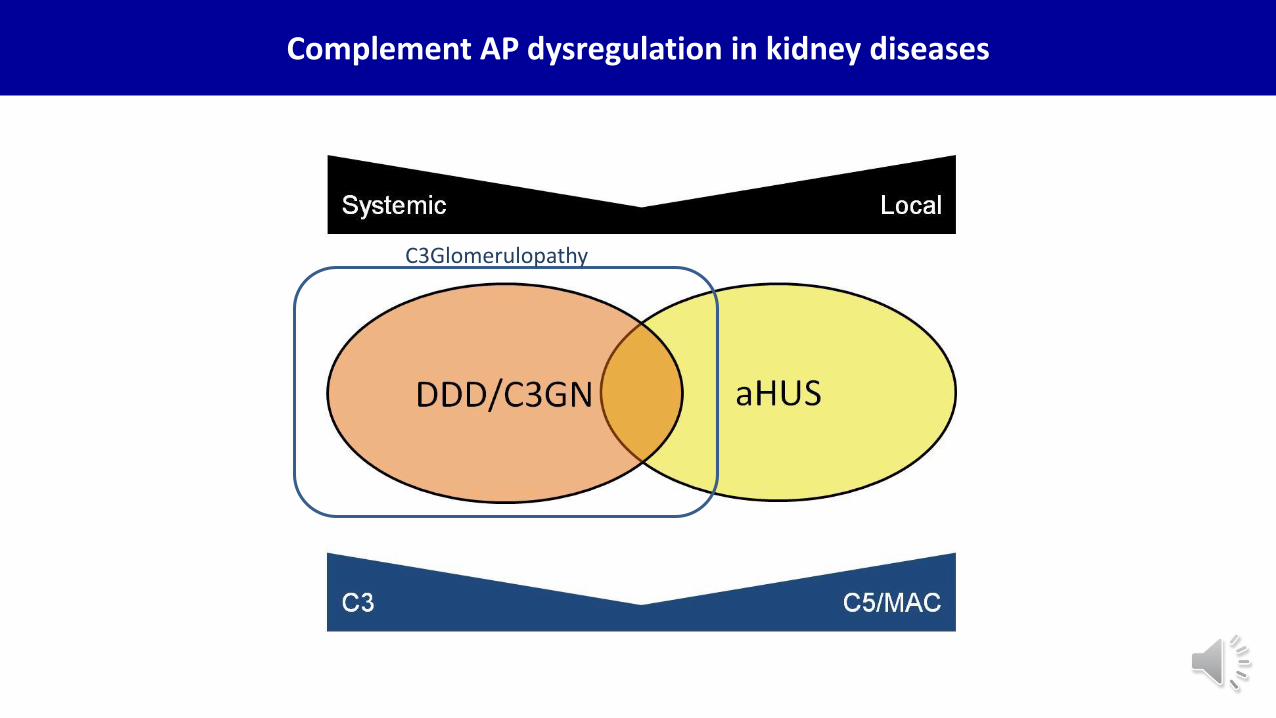
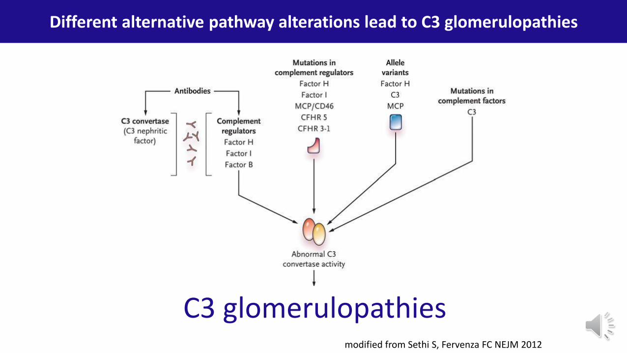
Complement AP dysregulation in kidney diseases
C3Glomerulopathy
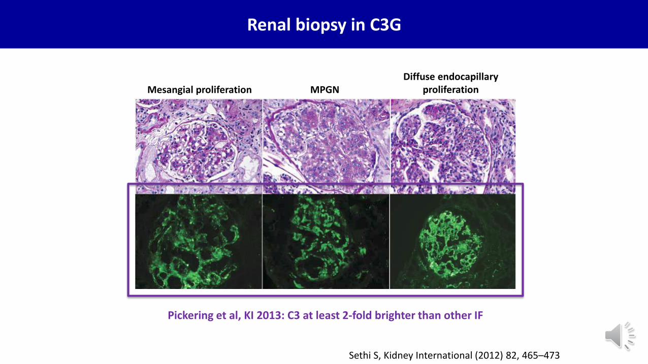
Sethi S, Kidney International (2012) 82, 465–473
MPGNMesangial proliferationDiffuse endocapillary
proliferation
Renal biopsy in C3G
Pickering et al, KI 2013: C3 at least 2-fold brighter than other IF
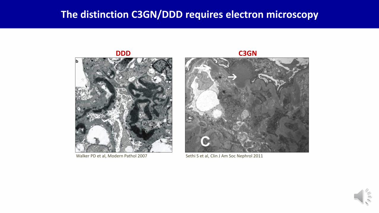
Walker PD et al, Modern Pathol 2007 Sethi S et al, Clin J Am Soc Nephrol 2011
DDD C3GN
The distinction C3GN/DDD requires electron microscopy
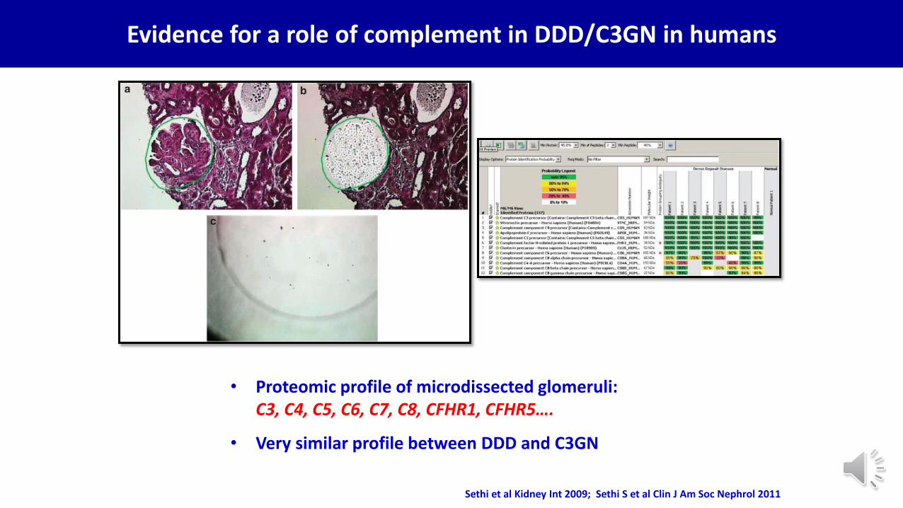
• Proteomic profile of microdissected glomeruli:C3, C4, C5, C6, C7, C8, CFHR1, CFHR5….
• Very similar profile between DDD and C3GN
Sethi et al Kidney Int 2009; Sethi S et al Clin J Am Soc Nephrol 2011
Evidence for a role of complement in DDD/C3GN in humans
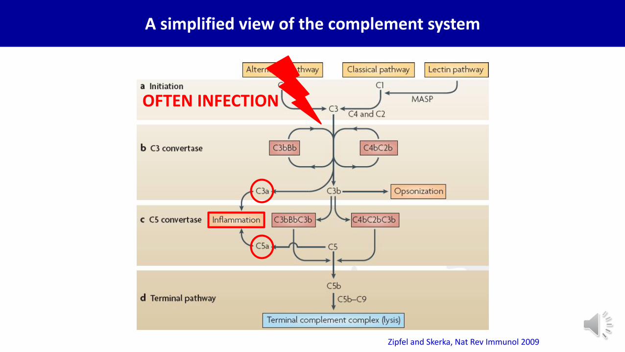
Zipfel and Skerka, Nat Rev Immunol 2009
A simplified view of the complement system
OFTEN INFECTION
modified from Sethi S, Fervenza FC NEJM 2012
Different alternative pathway alterations lead to C3 glomerulopathies
C3 glomerulopathies
Sethi S and Fervenza FC, Semin Nephrol 2011Sethi S and Fervenza FC, NEJM 2012
*Servais et al, Kidney Int 2012; Dragon-Durey et al. JASN 2004;
Vaziri-Sani et al. Kidney Int 2006; Leroy V et al. Ped Nephrol 2011
*
DDD C3GN
New classification of MPGN
Proliferative glomerulonephritis
Servais et al, Kidney Int 2012
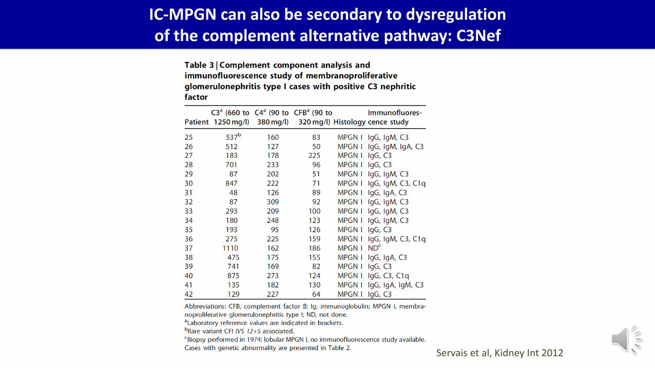
IC-MPGN can also be secondary to dysregulation of the complement alternative pathway: C3Nef
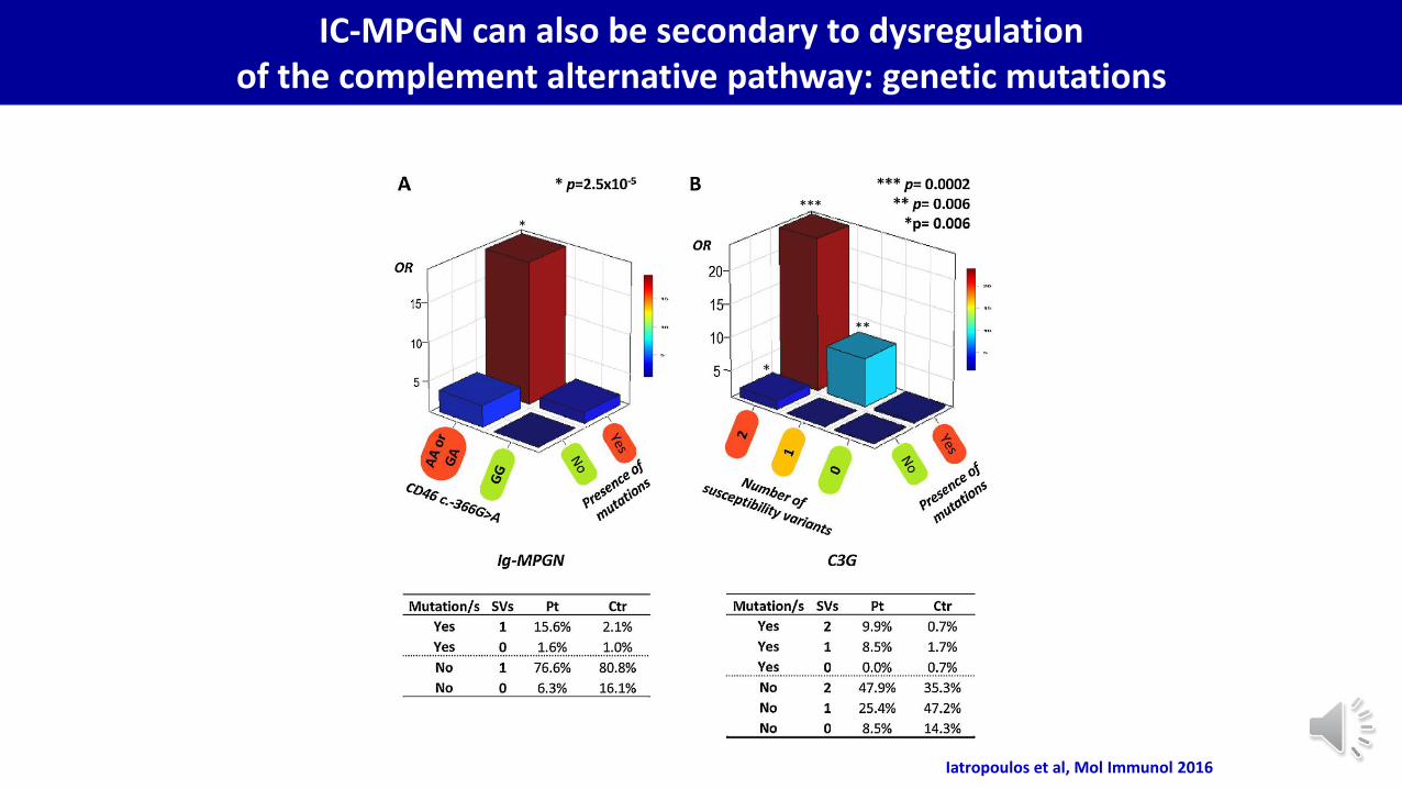
IC-MPGN can also be secondary to dysregulation of the complement alternative pathway: genetic mutations
Iatropoulos et al, Mol Immunol 2016
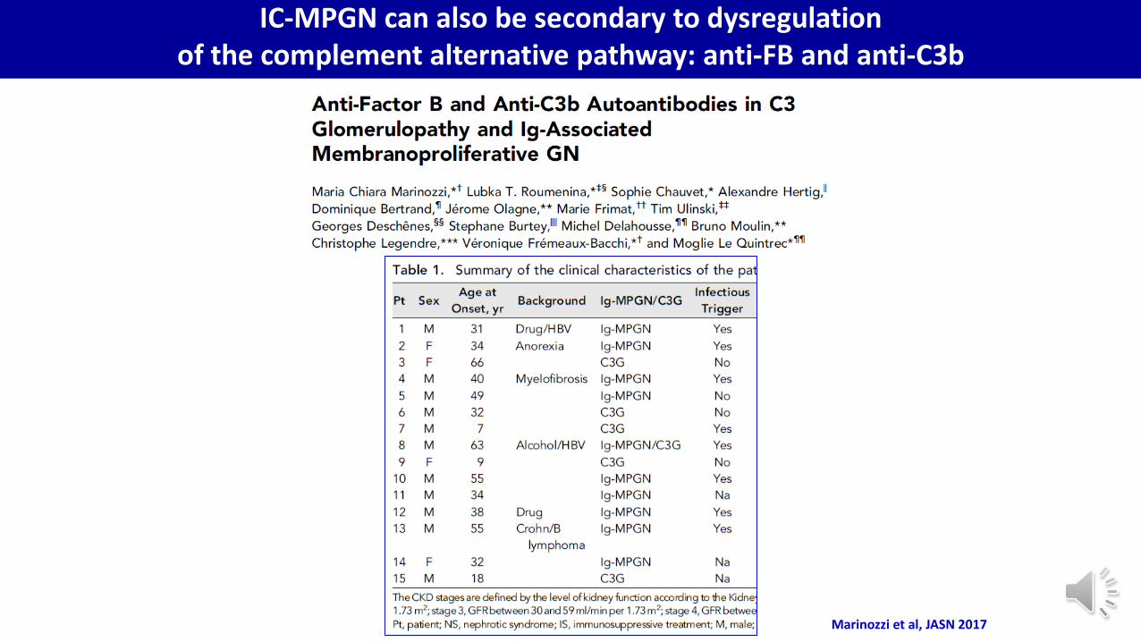
IC-MPGN can also be secondary to dysregulation of the complement alternative pathway: anti-FB and anti-C3b
Marinozzi et al, JASN 2017
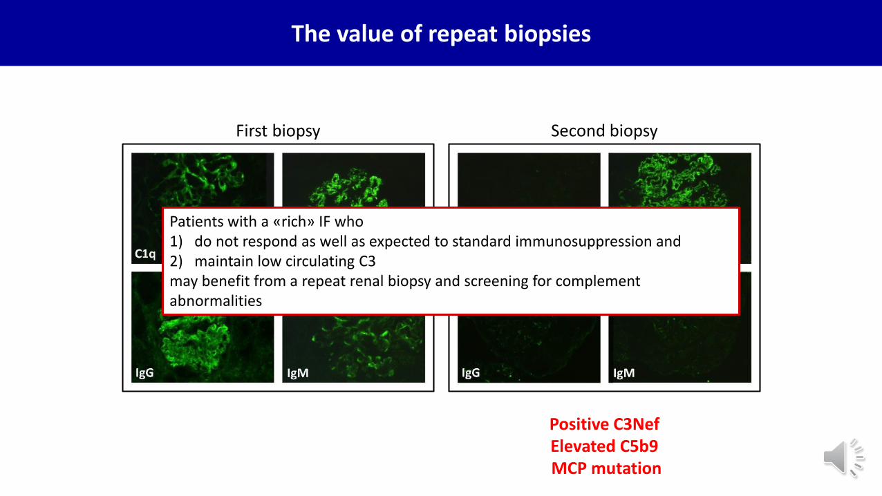
The value of repeat biopsies
First biopsy Second biopsy
Positive C3NefElevated C5b9MCP mutation
Patients with a «rich» IF who1) do not respond as well as expected to standard immunosuppression and 2) maintain low circulating C3may benefit from a repeat renal biopsy and screening for complementabnormalities
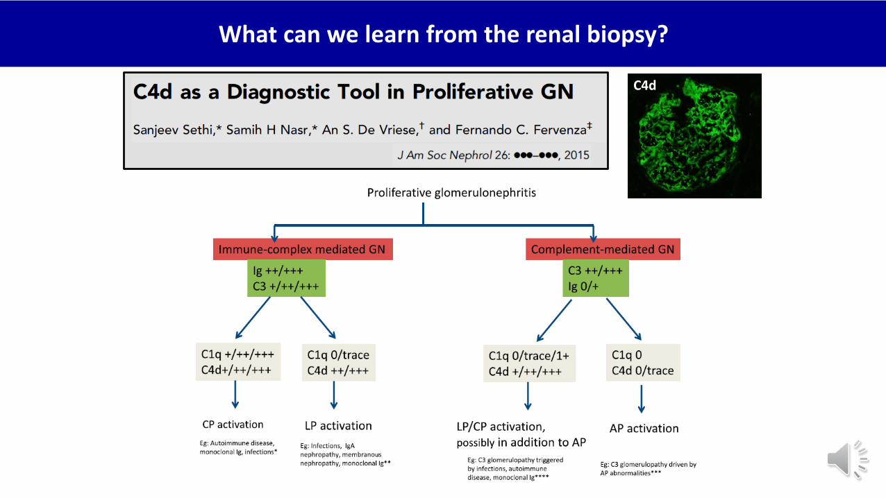
What can we learn from the renal biopsy?
C4d
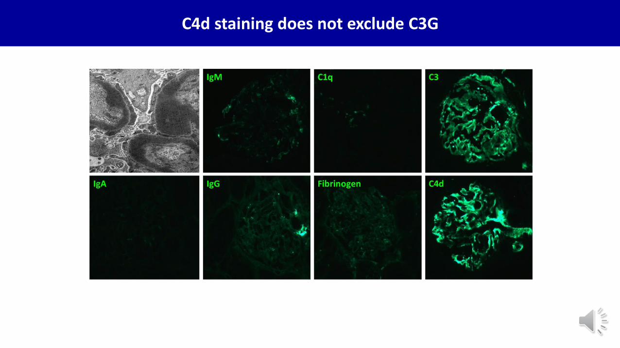
C4d staining does not exclude C3G
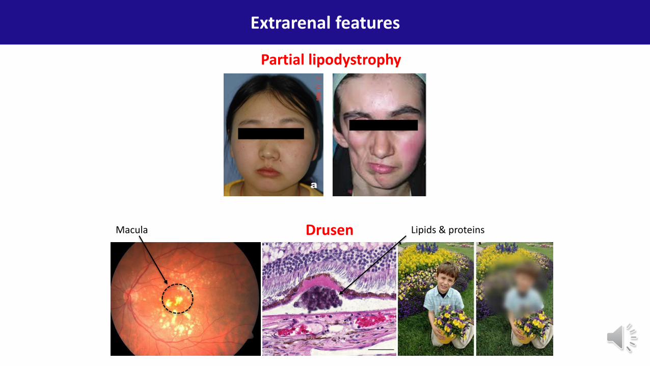
Extrarenal features
Partial lipodystrophy
DrusenMacula Lipids & proteins
C3G: clinical presentation is heterogenous
• Post-infectious glomerulonephritis with low C3
• Infection triggering macrohematuria, as in IgA nephropathy
• Nephrotic syndrome
• Accidental finding of non-nephrotic proteinuria, microhematuria
• Atypical

C3G presenting as acute PIGN
• Post-infectious glomerulonephritis with
1) low C3 that persists > 12 weeks or with2) recurrent macrohematuria
Sethi, Kidney International 2012
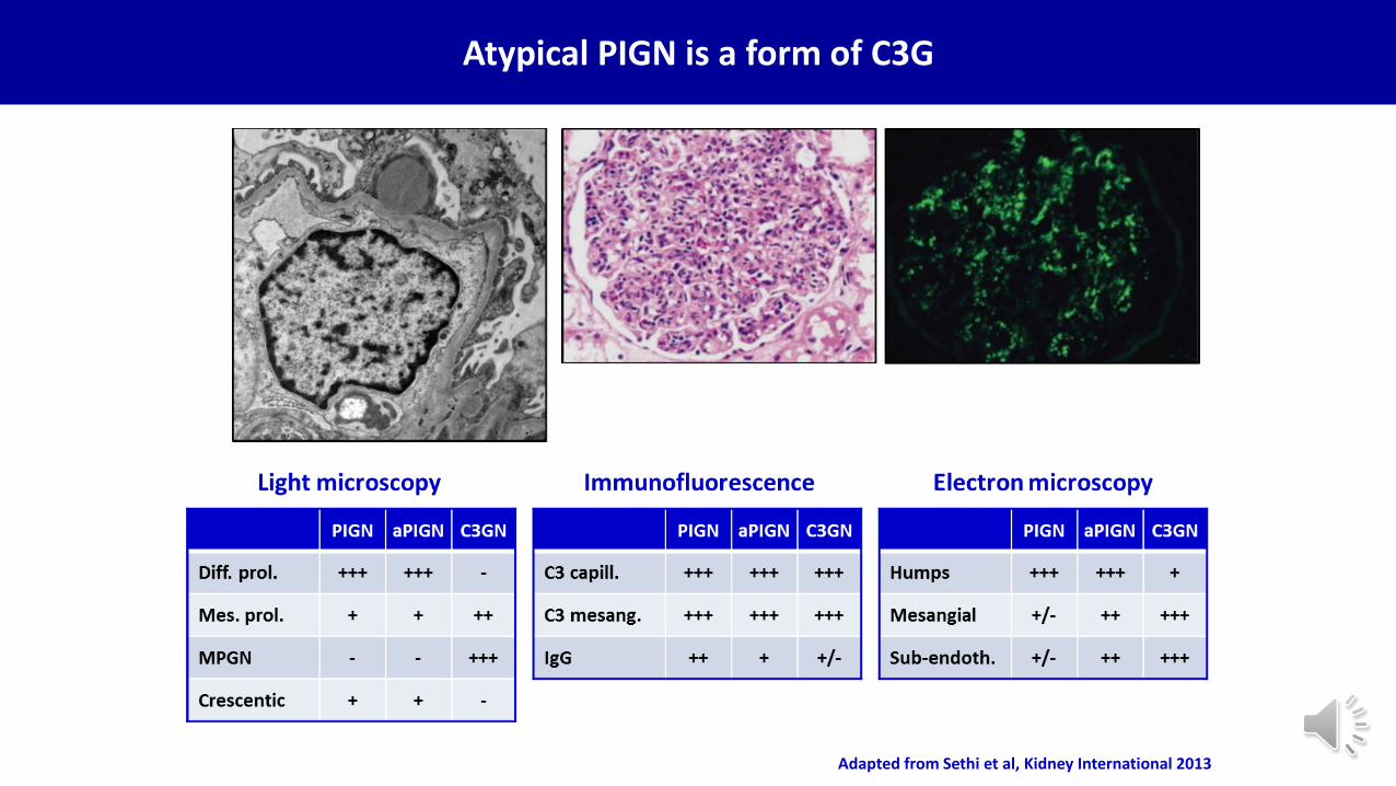
Adapted from Sethi et al, Kidney International 2013
Atypical PIGN is a form of C3G
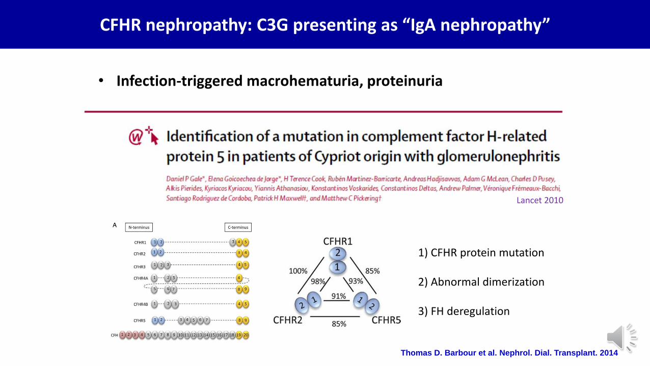
CFHR nephropathy: C3G presenting as “IgA nephropathy”
• Infection-triggered macrohematuria, proteinuria
Thomas D. Barbour et al. Nephrol. Dial. Transplant. 2014
1) CFHR protein mutation
2) Abnormal dimerization
3) FH deregulation
Lancet 2010
C3G can present as nephrotic syndrome
• Post-infectious glomerulonephritis with low C3
• Infection triggering macrohematuria, as in IgA nephropathy
• Nephrotic syndrome
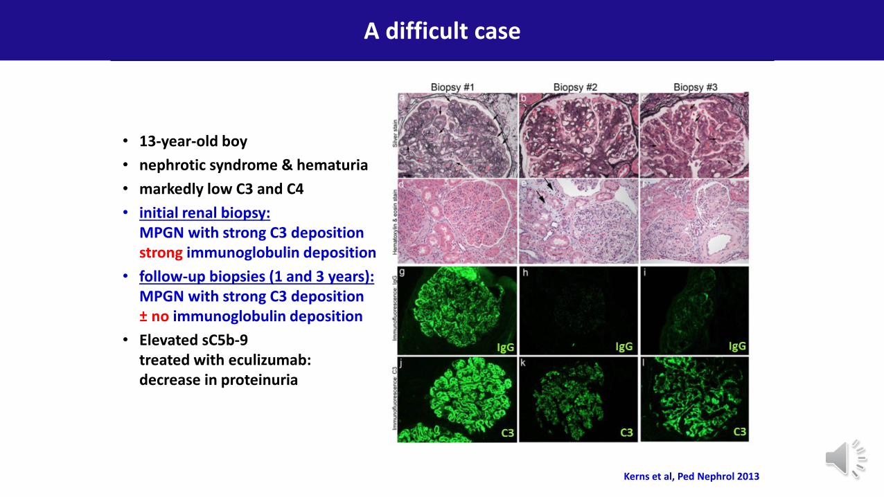
• 13-year-old boy
• nephrotic syndrome & hematuria
• markedly low C3 and C4
• initial renal biopsy: MPGN with strong C3 depositionstrong immunoglobulin deposition
• follow-up biopsies (1 and 3 years):MPGN with strong C3 deposition± no immunoglobulin deposition
• Elevated sC5b-9treated with eculizumab: decrease in proteinuria
A difficult case
Kerns et al, Ped Nephrol 2013
C3G can be found on routine urinalysis
• Post-infectious glomerulonephritis with low C3
• Infection triggering macrohematuria, as in IgA nephropathy
• Nephrotic syndrome
• Accidental finding of non-nephrotic proteinuria, microhematuria
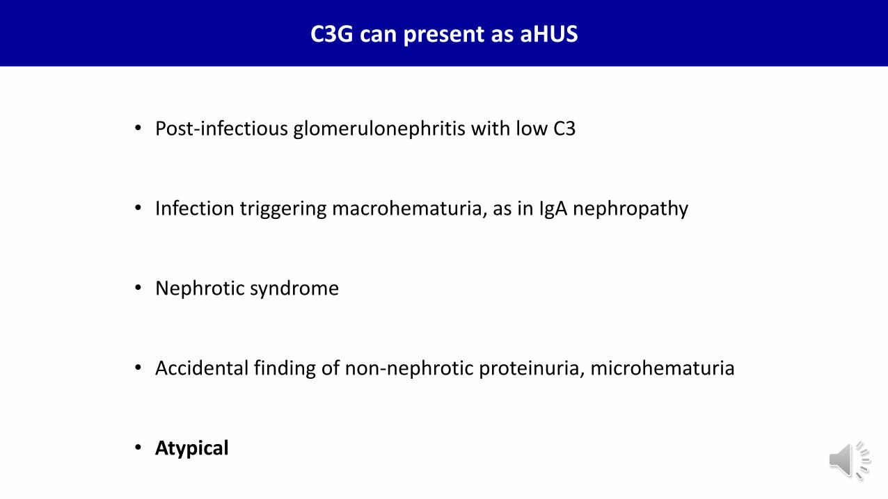
C3G can present as aHUS
• Post-infectious glomerulonephritis with low C3
• Infection triggering macrohematuria, as in IgA nephropathy
• Nephrotic syndrome
• Accidental finding of non-nephrotic proteinuria, microhematuria
• Atypical
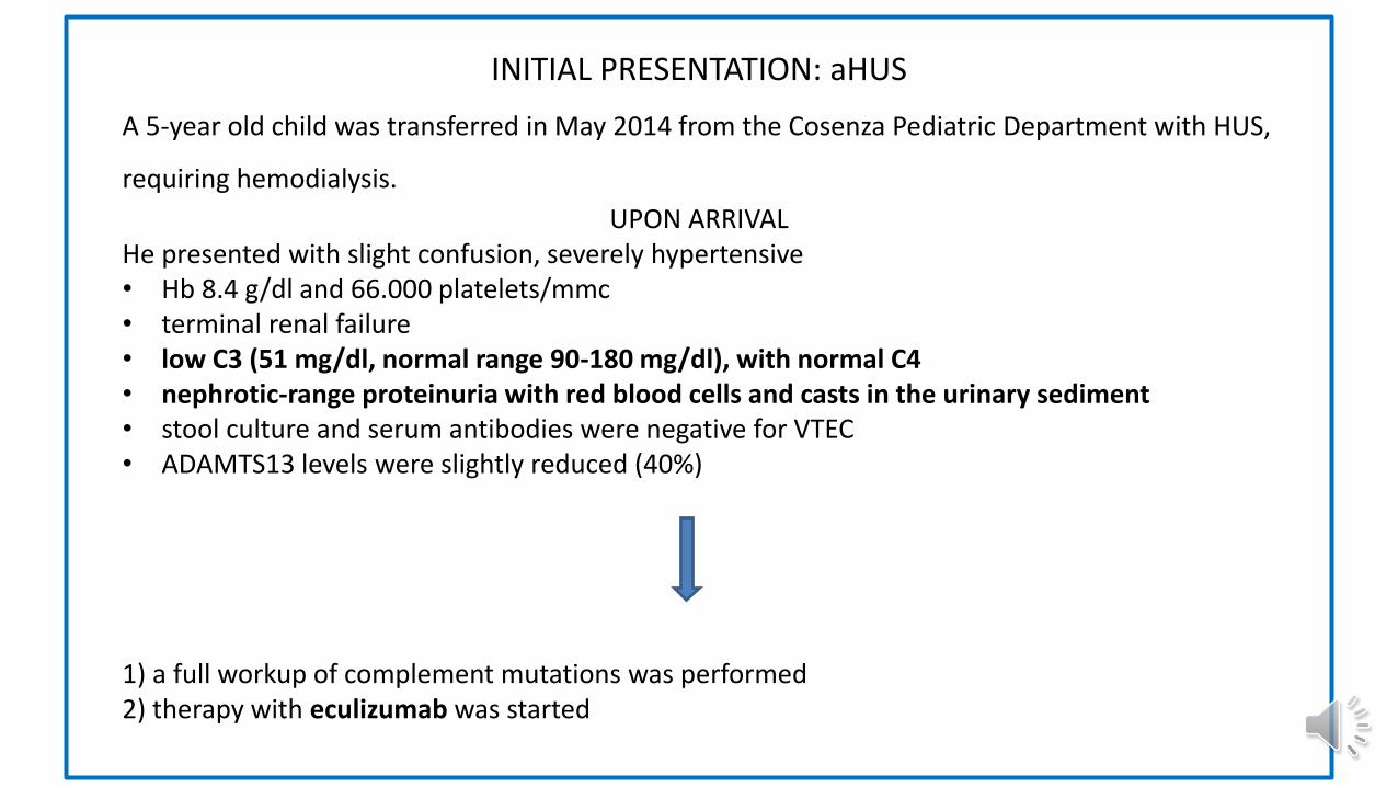
INITIAL PRESENTATION: aHUS
A 5-year old child was transferred in May 2014 from the Cosenza Pediatric Department with HUS,
requiring hemodialysis.
UPON ARRIVALHe presented with slight confusion, severely hypertensive • Hb 8.4 g/dl and 66.000 platelets/mmc • terminal renal failure• low C3 (51 mg/dl, normal range 90-180 mg/dl), with normal C4• nephrotic-range proteinuria with red blood cells and casts in the urinary sediment• stool culture and serum antibodies were negative for VTEC • ADAMTS13 levels were slightly reduced (40%)
1) a full workup of complement mutations was performed 2) therapy with eculizumab was started
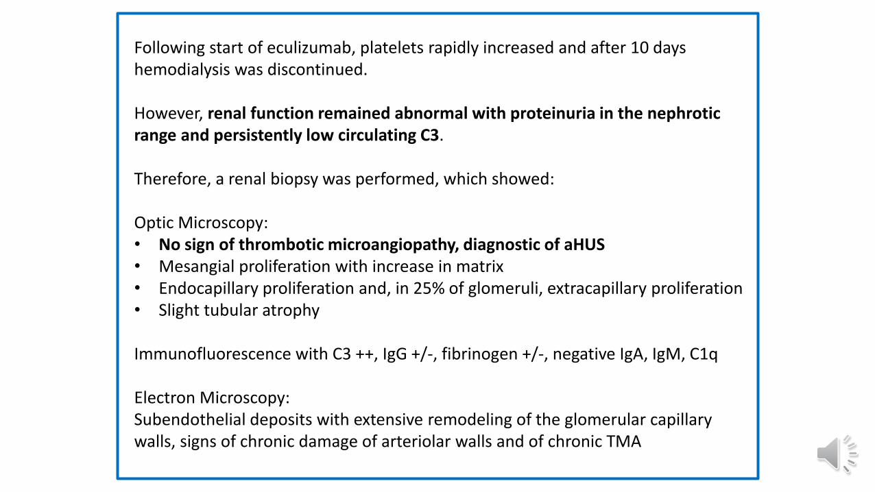
Following start of eculizumab, platelets rapidly increased and after 10 dayshemodialysis was discontinued.
However, renal function remained abnormal with proteinuria in the nephroticrange and persistently low circulating C3.
Therefore, a renal biopsy was performed, which showed:
Optic Microscopy: • No sign of thrombotic microangiopathy, diagnostic of aHUS• Mesangial proliferation with increase in matrix• Endocapillary proliferation and, in 25% of glomeruli, extracapillary proliferation• Slight tubular atrophy
Immunofluorescence with C3 ++, IgG +/-, fibrinogen +/-, negative IgA, IgM, C1q
Electron Microscopy: Subendothelial deposits with extensive remodeling of the glomerular capillary walls, signs of chronic damage of arteriolar walls and of chronic TMA
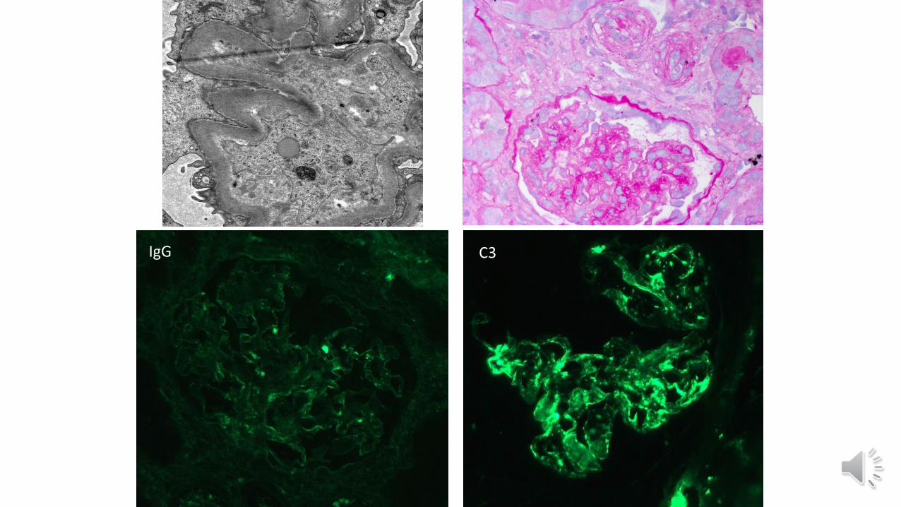
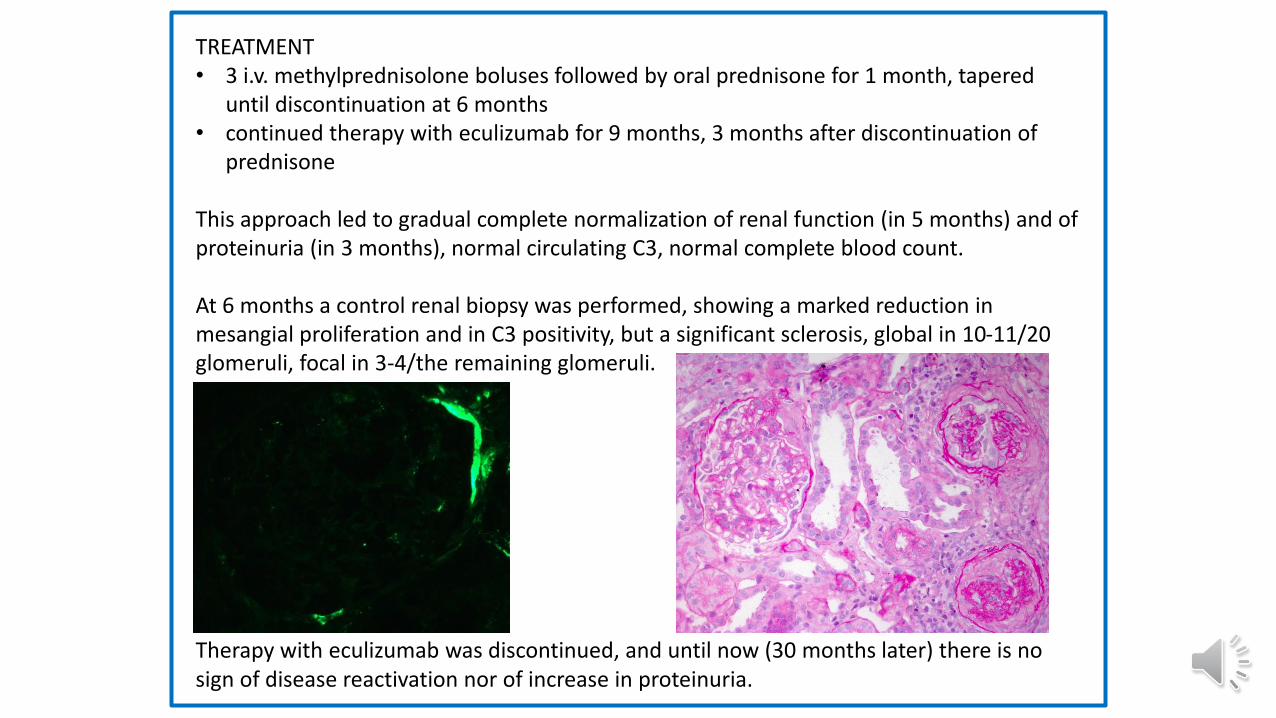
IgG C3
TREATMENT• 3 i.v. methylprednisolone boluses followed by oral prednisone for 1 month, tapered
until discontinuation at 6 months• continued therapy with eculizumab for 9 months, 3 months after discontinuation of
prednisone
This approach led to gradual complete normalization of renal function (in 5 months) and of proteinuria (in 3 months), normal circulating C3, normal complete blood count.
At 6 months a control renal biopsy was performed, showing a marked reduction in mesangial proliferation and in C3 positivity, but a significant sclerosis, global in 10-11/20 glomeruli, focal in 3-4/the remaining glomeruli.
Therapy with eculizumab was discontinued, and until now (30 months later) there is no sign of disease reactivation nor of increase in proteinuria.
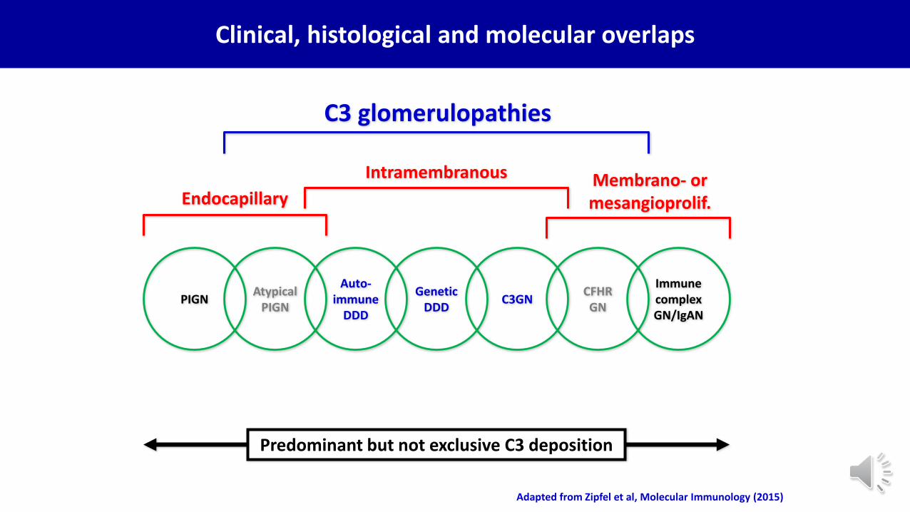
Adapted from Zipfel et al, Molecular Immunology (2015)
Clinical, histological and molecular overlaps
PIGNAtypical
PIGN
Auto-immune
DDD
Genetic DDD
C3GNCFHRGN
ImmunecomplexGN/IgAN
C3 glomerulopathies
Endocapillary
Intramembranous Membrano- ormesangioprolif.
Predominant but not exclusive C3 deposition
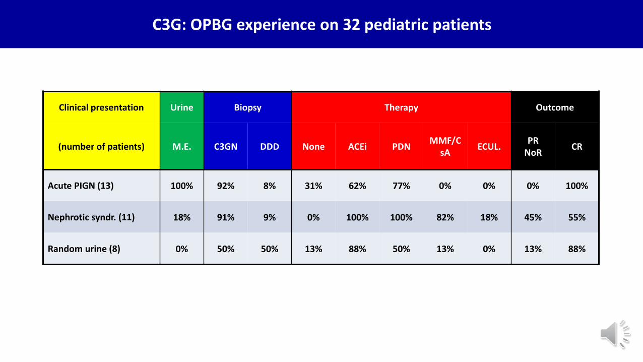
C3G: OPBG experience on 32 pediatric patients
Clinical presentation Urine Biopsy Therapy Outcome
(number of patients) M.E. C3GN DDD None ACEi PDNMMF/C
sAECUL.
PRNoR
CR
Acute PIGN (13) 100% 92% 8% 31% 62% 77% 0% 0% 0% 100%
Nephrotic syndr. (11) 18% 91% 9% 0% 100% 100% 82% 18% 45% 55%
Random urine (8) 0% 50% 50% 13% 88% 50% 13% 0% 13% 88%
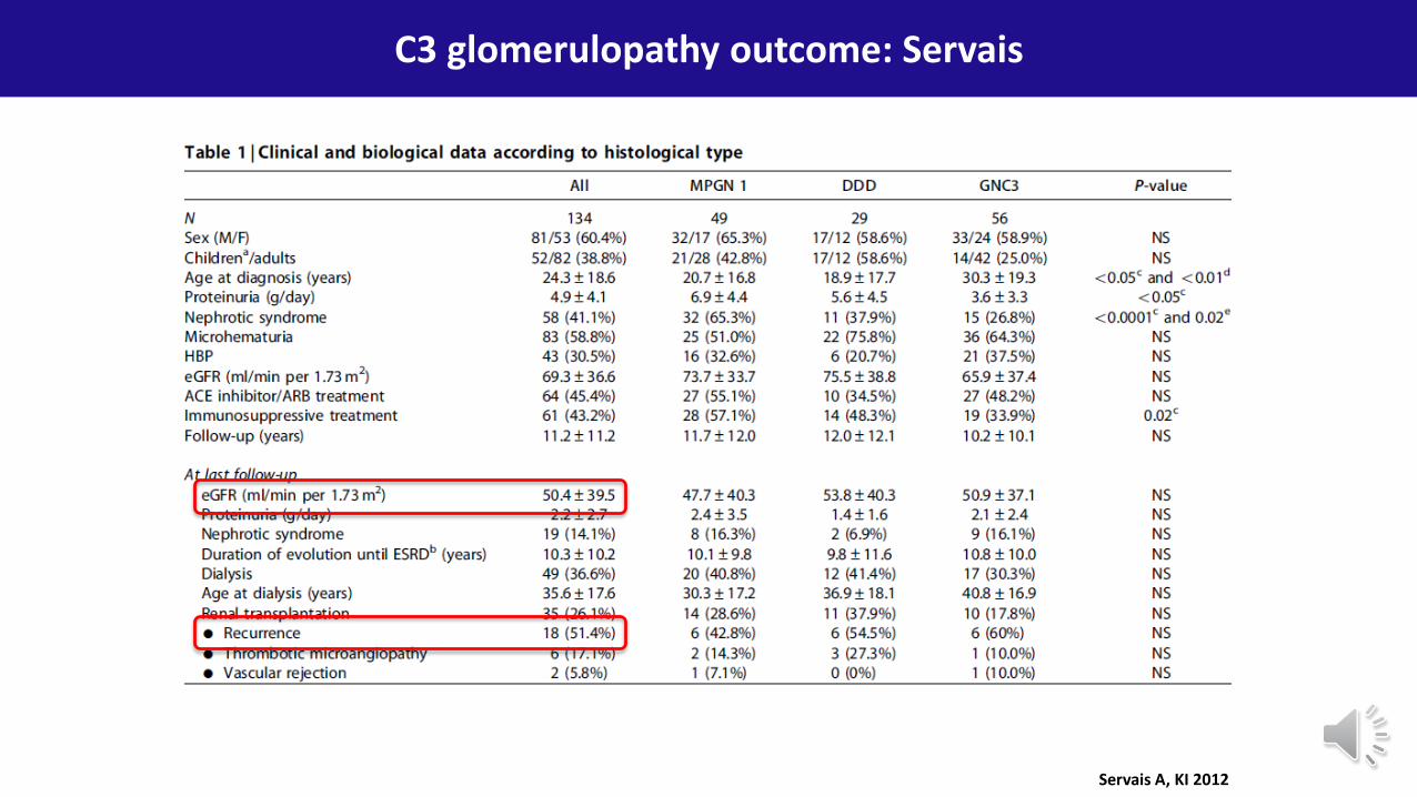
C3 glomerulopathy outcome: Servais
Servais A, KI 2012
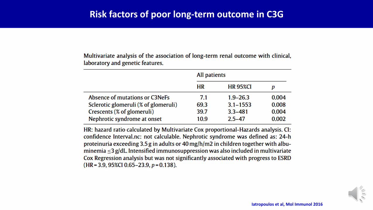
Risk factors of poor long-term outcome in C3G
Iatropoulos et al, Mol Immunol 2016
Sethi S, Fervenza FC NEJM 2012
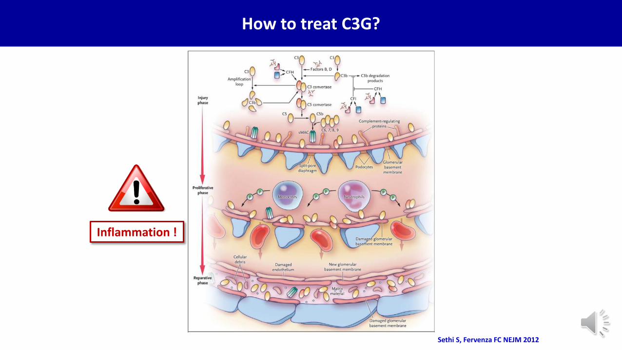
How to treat C3G?
Inflammation !
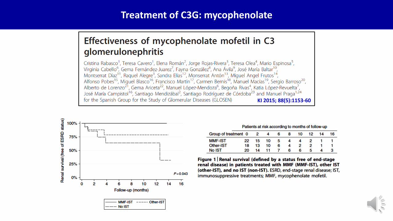
Treatment of C3G: mycophenolate
KI 2015; 88(5):1153-60
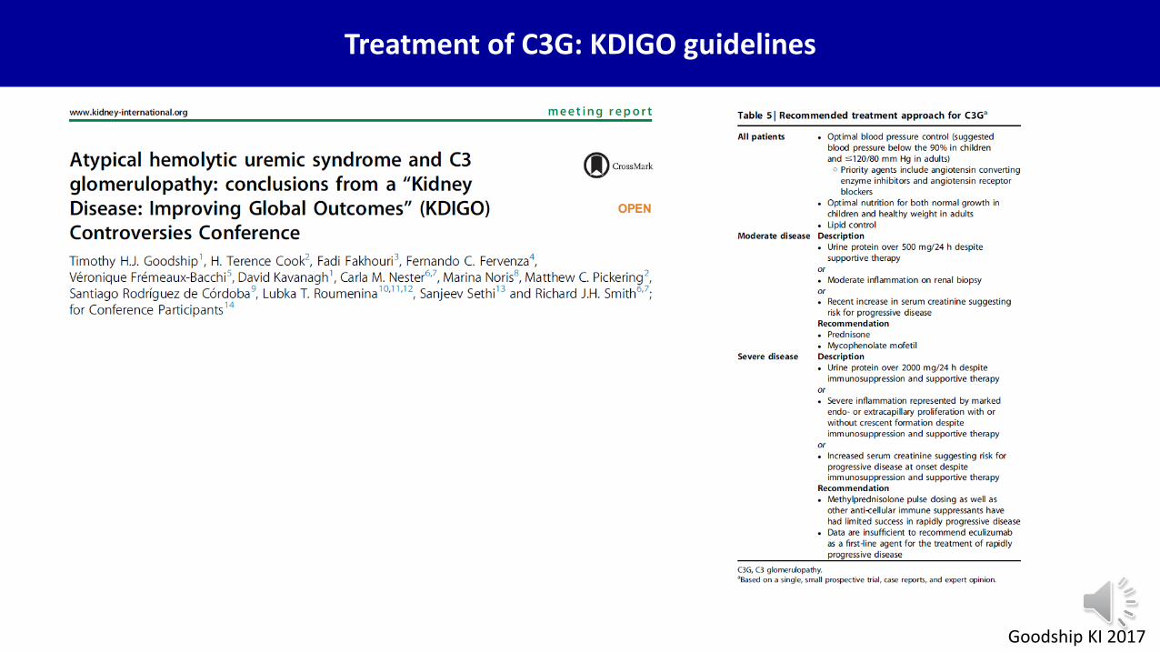
Goodship KI 2017
Treatment of C3G: KDIGO guidelines
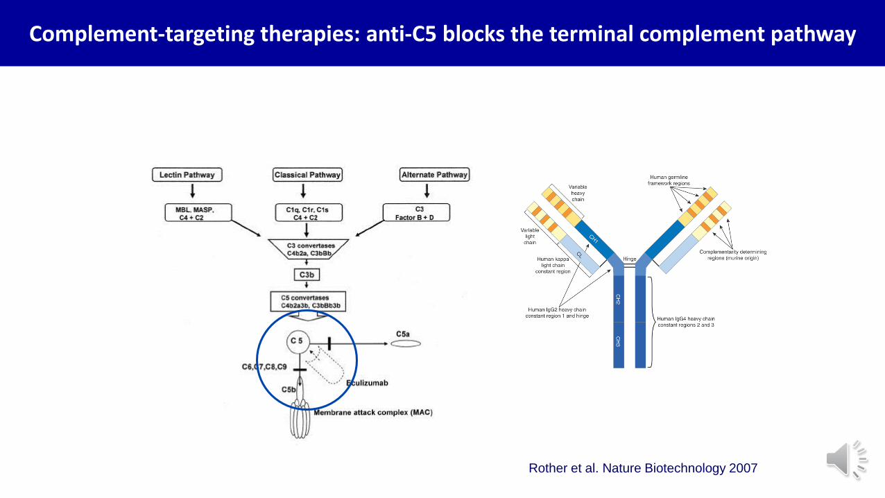
Rother et al. Nature Biotechnology 2007
Complement-targeting therapies: anti-C5 blocks the terminal complement pathway
Vivarelli et al, New England J Med 2012
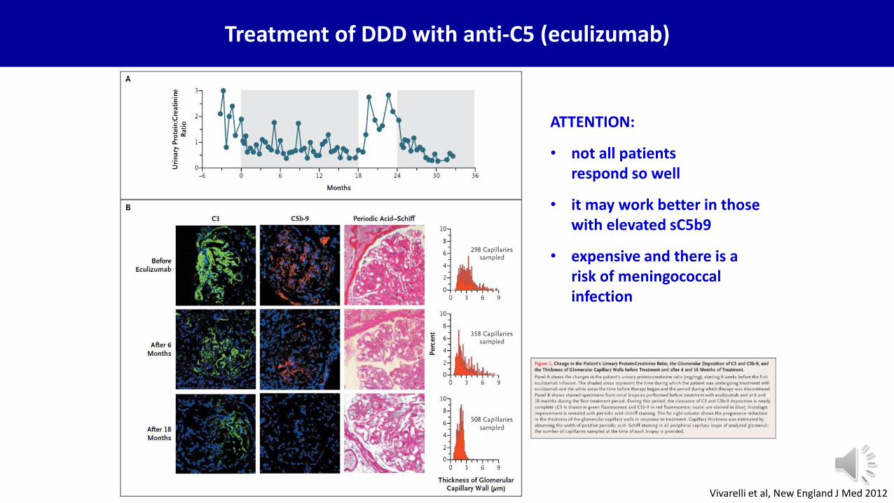
Treatment of DDD with anti-C5 (eculizumab)
ATTENTION:
• not all patientsrespond so well
• it may work better in those with elevated sC5b9
• expensive and there is a risk of meningococcal infection
Yuzhou Zhang et al. CJASN 2014
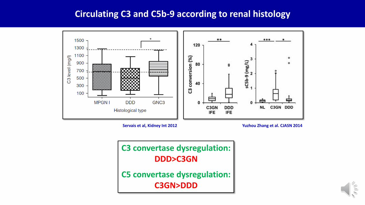
Circulating C3 and C5b-9 according to renal histology
Servais et al, Kidney Int 2012
C3 convertase dysregulation:DDD>C3GN
C5 convertase dysregulation:C3GN>DDD
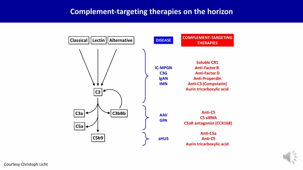
Complement-targeting therapies on the horizon
Courtesy Christoph Licht
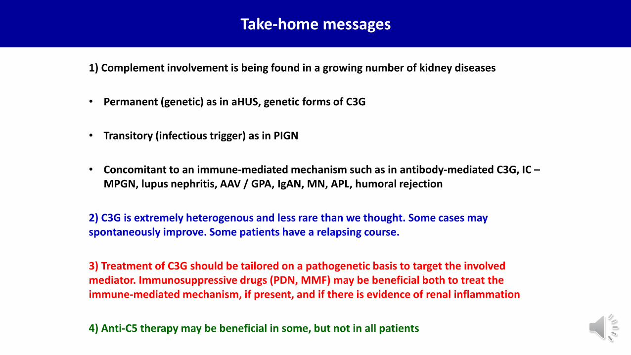
1) Complement involvement is being found in a growing number of kidney diseases
• Permanent (genetic) as in aHUS, genetic forms of C3G
• Transitory (infectious trigger) as in PIGN
• Concomitant to an immune-mediated mechanism such as in antibody-mediated C3G, IC –MPGN, lupus nephritis, AAV / GPA, IgAN, MN, APL, humoral rejection
2) C3G is extremely heterogenous and less rare than we thought. Some cases may spontaneously improve. Some patients have a relapsing course.
3) Treatment of C3G should be tailored on a pathogenetic basis to target the involved mediator. Immunosuppressive drugs (PDN, MMF) may be beneficial both to treat the immune-mediated mechanism, if present, and if there is evidence of renal inflammation
4) Anti-C5 therapy may be beneficial in some, but not in all patients
Take-home messages
THANK YOU
• Prof Joshua Thurman• Prof Carla Nester• Prof Christoph Licht
• Dr Marina Noris• Dr Elena Bresin• Dr Veronique Fremeaux-Bacchi
• Prof Francesco Emma
The patients and their families