Embed Size (px)
Citation preview
Br. J. Pharmac. (1986), 88, 857-866
Novel interactions ofcations with dihydropyridinecalcium antagonist binding sites in brainGordon T. Bolger & Phil Skolnick
Laboratory of Bioorganic Chemistry, National Institute of Arthritis, Digestive Diseases and Kidney Diseases,Building 4, Room 212, National Institutes of Health, Bethesda, MD 20205, U.S.A.
1 The effects of monovalent (Na', Li', K+, Rb+) and divalent (Ca2', Mg2', Mn2+) cations on
dihydropyridine calcium antagonist binding sites in brain and cardiac membranes were investigatedusing a low ionic strength buffer (5 mM Tris-HCI, pH 7.4), and the dihydropyridine, [3H]-nitrendipine.2 At 25'C, the monovalent cations Na', Li+, and K+ (100 mM) but not Rb' significantly decreasedthe apparent dissociation constant (KD) but had no effect on the maximum binding site capacity (Bmax)of [3H]-nitrendipine in brain. The divalent cations Ca2", Mg2+, and Mn2+ (2 mM) significantlyincreased the B but did not affect the KD of [3H1-nitrendipine. The effects of cations were
concentration-dependent (EC50 monovalent cations 10-25 mM; EC50 divalent cations 50-200 ZM) anddemonstrated brain region selectivity. The effect of Ca2+, but not Mg2+ or Mn2+ on [3H]-nitrendipinebinding was described by a two-site model.3 At 25°C, neither mono- nor divalent cations altered the characteristics of [3H]-nitrendipine bindingto rat cardiac membranes.4 At 37C, Na+ (100mM) but not K+ (100 mM) significantly increased the B,,.U of[3H]-nitrendipine inrat brain membranes. Ca2+ (2 mM) significantly increased the B,,.,, of [3HJ-nitrendipine binding to ratbrain membranes to a greater extent than at 25°C. Both Na+ and K+ had no effect on [3H]-nitrendipinebinding to cardiac membranes, while Ca2+ (2 mM) significantly decreased the KD of [3H]-nitrendipine.5 It is suggested that the selective effects ofmono- and divalent cations on [3H]-nitrendipine bindingto rat brain and cardiac membranes may be associated with differences in the calcium current blockingactivity of dihydropyridine calcium antagonists in brain and cardiac tissues.
lntroduction
High affinity binding sites for dihydropyridine calcium channel antagonists (DHPCAs), have been de-tected in cardiac, smooth and skeletal muscles andbrain (Belleman et al., 1981; Janis et al., 1982; Bolger etal., 1982; 1983; Ferry & Glossmann, 1982; Murphy &Snyder, 1982; Ehlert et al., 1982; Fosset et al., 1983).Biochemical and electrophysiological evidence sug-gests that the ability of DHPCAs to inhibit calciumcurrents in cardiac and smooth muscles (Fleckenstein,1977; Rosenberger & Triggle, 1978; Triggle & Swamy,1980; Triggle, 1981) is due to an interaction of theDHPCA with a binding site that constitutes a 're-gulatory' component of calcium channels (Venter etal., 1983; Goll etal., 1983; Horneetal., 1984;Triggle&Janis, 1984b; Rengasamy et al., 1985). In contrast totheir effects in cardiac and smooth muscles, DHPCAspossess very low potencies or are ineffective at inhibit-ing calcium-dependent processes (e.g. 45Ca2"-uptake,neurotransmitter release) in brain (Toll, 1982; Daniell
et al., 1983; Freedman et al., 1984; Starke et al., 1984;Ogura & Takahasi, 1984; Rampe et al., 1984) despitethe presence of high affinity brain DHPCA bindingsites (Murphy et al., 1982; Cortes et al., 1983; 1984).Several hypotheses have been advanced to provide anexplanation for discrepancies in the calcium currentblocking activity ofDHPCAs between tissues (Triggle& Janis, 1984a, b; Bean, 1984). There is, however, littledirect biochemical evidence to support such largedifferential tissue activities for DHPCAs. Nonethe-less, a marked similarity in the properties ofDHPCAbinding sites in brain and cardiac tissues (e.g. bindingsite affinity, structure-activity relationships, ion-de-pendence, interactions of other non-DHP calciumantagonists) (Triggle & Janis, 1984a) implies that thebrain DHPCA binding site may serve an analogousfunction in regulating calcium-dependent processes asin pheripheral tissues. Both behavioural (Hoffmeisteret al., 1982; Mendelson et al., 1984; Bolger et al.,
Q The Macmillan Press Ltd 1986
858 G.T. BOLGER & P. SKOLNICK
1985a) and biochemical (Turner & Goldin, 1985;Middlemiss & Spedding, 1985; Kendall & Nahorski,1985) observations indicate that DHPCAs can re-gulate neuronal processes in the central nervoussystem (CNS).
[3H]-nitrendipine binding to a partially purifiedmembrane perparation from guinea-pig cardiac tissuewas shown to be dependent on the ionic strength oftheassay buffer, being increased by mono- and divalentcations and anions (Glossmann & Ferry, 1983). Re-cently, it was found that Na' could increase the KDand Bmax of [3H]-nitrendipine binding to rat cardiactissue but not brain (Schwartz & Velly, 1985), provid-ing evidence for differences in the biochemical charac-teristics of DHPCA binding sites in these tissues. Werecently observed that phencyclidine could increasethe apparent affinity of the DHPCA, [3H]-nitren-dipine, in rat brain but not cardiac tissue using a lowionic strength (5mM Tris-HCl pH 7.4) buffer (Bolgeret al., 1985a, b). Subsequently, we have investigatedthe effects of mono- and divalent cations on [3H]-nitrendipine binding to rat brain and cardiac mem-branes under similar incubation conditions and nowshow that low concentrations of mono- (Nat, Li',K+, Rb+) and divalent (Ca2+, Mg2+, Mn+) cationsdifferentially affect [3H]-nitrendipine binding to brainand cardiac membranes. The selective effects ofmono-and divalent cations on DHPCA binding support thecontention that DHP binding sites in brain andcardiac tissues may be differentially regulated. Suchdifferences may provide a biochemical basis for themarked discrepancies in the biochemical andphysiological activity of DHPCAs in these tissues.
Methods
Preparation of membranes
Male Sprague-Dawley rats (150-175g, TaconicFarms, Germantown, NY) were killed by decapita-tion. Tissues were rapidly removed and placed in ice-cold 5 mM Tris-HCl buffer pH 7.4. Brain regions(isolated by blunt dissection) or forebrain (defined asthat region isolated by making an oblique cut from thesuperior colliculus on the dorsal surface to the mam-milary bodies on the ventral surface) were disrupted in50 volumes (w/v) of 5 mM Tris-HCl using a BrinkmanPolytron (lOs, speed setting 6-7). The homogenatewas centrifuged at 24,000 g for 15 min, the supernatantdiscarded and the membrane pellet resuspended with apolytron (5 s, speed setting 6-7) in 100 volumes of5mM Tris-HCl buffer. Rat heart ventricular tissue wasseparated from atrial tissue and minced with scissorsprior to disruption in 30 volumes of 5 mM Tris-HClbuffer with a polytron (20 s, speed setting 6-7). Thehomogenate was centrifuged for 10 min at 1,000 g, the
pellet discarded and the supernatant centrifuged at24,000g for 20 min. The membrane pellet from thiscentrifugation was resuspended by polytron (5 s, speedsetting 6-7) in 30 volumes of 5mM Tris-HCI.
[3H]-nitrendipine binding
[3H]-nitrendipine (specific activity, 81.3 Ci mmol ',New England Nuclear, Boston, MA) binding wasmeasured in a total incubation volume of 2 ml consist-ing of 0.5 ml of membrane suspension (brain mem-branes 0.3-0.5 mg protein; cardiac membranes0.1-0.3 mg protein), 0.1 ml radioligand and 1.4 ml of5mM Tris-HCl buffer. Inorganic cations were dis-solved in 5 mM Tris-HCl. Binding assays were initiatedby addition of membrane suspension and incubatedfor 60 min at 25°C or 45 min at 37°C before termina-tion by rapid filtration under vacuum through What-man GF/B glass fibre filters followed by two 5mlwashes with ice-cold 5 mM Tris-HCl using a BrandelM-24R Cell Harvester (Brandel instruments, Gaither-sburg, MD). Unless otherwise stated, incubationswere conducted at 25°C: The filters were incubatedovernight in 8 ml of ReadySolv MP (Beckman Ins-truments, Fullerton, CA) and the radioactivity deter-mined in a Beckman liquid scintillation counter(Model LS 250, counting efficiency 41%). Specific[3H]-nitrendipine bound was defined as the differencebetween [3H]-nitrendipine binding in the absence andin the presence of 10-6 M nifedipine (Pfizer Co.,Groton, CT). The effects of cations on [3H]-nitren-dipine binding to three or more different membranepreparations were studied on the same day to accountfor the inherent daily variability in the characteristicsof the [3H]-nitrendipine binding assay (Bolger et al.,1983). Tris base and all inorganic salts (as chlorides)were obtained from standard commercial sources.Protein determinations were made by the Millermodification (Miller, 1959) of the Lowry technique(Lowry et al., 1951).
Data analysis
Computer analysis of binding data was done using thenon-linear regression/statistical computer package,BMDPAR (University of California, Los Angeles,1977), fitting binding data to the general multisitebinding equation of Birdsall et al. (1978):
N BNLB= Z
1 KN+ L
Where B is the total ligand bound, BN is the maximumbinding site capacity of site N with an apparentdissociation constant KN. L is the free concentration ofligand.
EFFECTS OF CATIONS ON [3H1-NITRENDIPINE BINDING 859
Results
Effects of monovalent cations
[3H]-nitrendipine binding to rat brain and cardiacmembranes at 250C yielded KD (120-200 pM) and Bmax
a100
80
C
60
, 40C
20C
C 20 40 60 80 100 120b [Na ], mMb
X 100a)
.' 80EE 60
E 40
10-5 10o 10-3
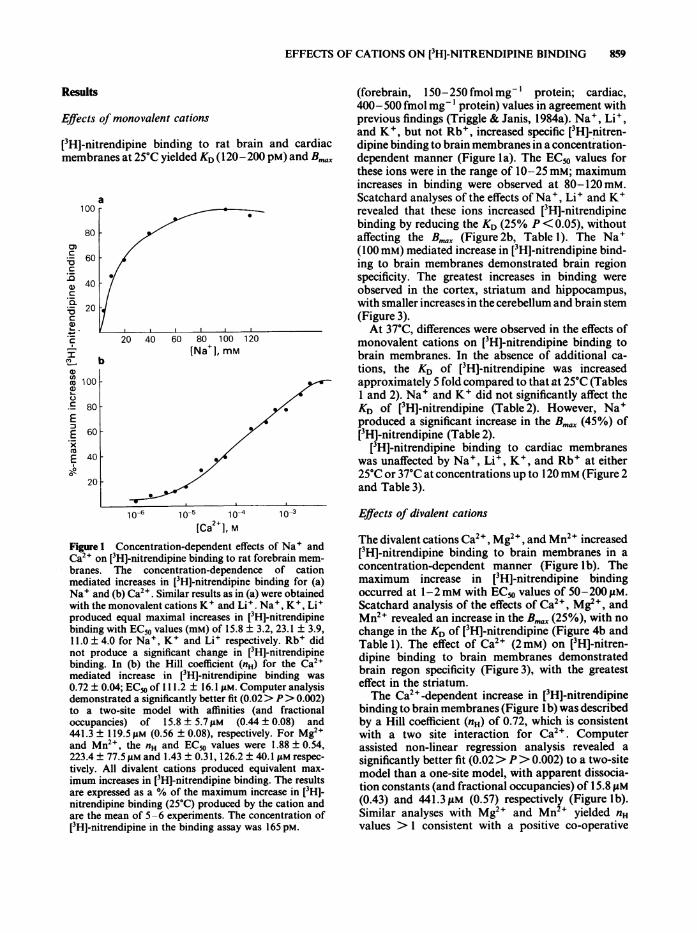
[Ca' ] MFigure 1 Concentration-dependent effects of Na4 andCa2+ on [3H]-nitrendipine binding to rat forebrain mem-branes. The concentration-dependence of cationmediated increases in [3H]-nitrendipine binding for (a)Na+ and (b) Ca24. Similar results as in (a) were obtainedwith the monovalent cations K+ and Li+. Na+, K+, Li+produced equal maximal increases in [3HJ-nitrendipinebinding with EC50 values (mM) of 15.8 ± 3.2, 23.1 ± 3.9,11.0 ± 4.0 for Na+, K4 and Li4 respectively. Rb4 didnot produce a significant change in [3H]-nitrendipinebinding. In (b) the Hill coefficient (nH) for the Ca24mediated increase in [3HJ-nitrendipine binding was
0.72 ± 0.04; EC50 of 1 11.2 ± 16.1 JAM. Computer analysisdemonstrated a significantly better fit (0.02> P> 0.002)to a two-site model with affinities (and fractionaloccupancies) of 15.8 ± 5.7 pM (0.44 ± 0.08) and441.3 ± 119.5 jiM (0.56 ± 0.08), respectively. For Mg24and Mn24, the nH and EC50 values were 1.88 ± 0.54,223.4 ± 77.5 jAM and 1.43 ± 0.31, 126.2 ± 40.1 JAM respec-
tively. All divalent cations produced equivalent max-
imum increases in [3HJ-nitrendipine binding. The resultsare expressed as a % of the maximum increase in [3H]-nitrendipine binding (25°C) produced by the cation andare the mean of 5-6 experiments. The concentration of[3H]-nitrendipine in the binding assay was 165 pM.
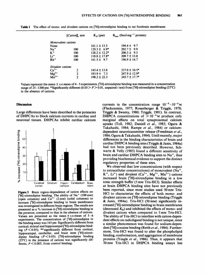
(forebrain, 150-250fmolmg-' protein; cardiac,400- 500 fmol mg-' protein) values in agreement withprevious findings (Triggle & Janis, 1984a). Na4, Li4,and K+, but not Rb4, increased specific [3H]-nitren-dipine binding to brain membranes in a concentration-dependent manner (Figure la). The EC50 values forthese ions were in the range of 10-25 mM; maximumincreases in binding were observed at 80-120 mM.Scatchard analyses of the effects of Na', Li+ and K4revealed that these ions increased [3H]-nitrendipinebinding by reducing the KD (25% P< 0.05), withoutaffecting the Bmax (Figure 2b, Table 1). The Na4(100 mM) mediated increase in [3H]-nitrendipine bind-ing to brain membranes demonstrated brain regionspecificity. The greatest increases in binding wereobserved in the cortex, striatum and hippocampus,with smaller increases in the cerebellum and brain stem(Figure 3).At 37C, differences were observed in the effects of
monovalent cations on [3H]-nitrendipine binding tobrain membranes. In the absence of additional ca-tions, the KD of [3H]-nitrendipine was increasedapproximately 5 fold compared to that at 25°C (Tables1 and 2). Na4 and K4 did not significantly affect theKD of [3H]-nitrendipine (Table 2). However, Na4produced a significant increase in the B... (45%) of[3H]-nitrendipine (Table 2).
[3H]-nitrendipine binding to cardiac membraneswas unaffected by Na+, Li', K+, and Rb4 at either25°C or 37°C at concentrations up to 120mm (Figure 2and Table 3).
Effects of divalent cations
The divalent cations Ca24, Mg2+, and Mn2+ increased[3H]-nitrendipine binding to brain membranes in aconcentration-dependent manner (Figure lb). Themaximum increase in [3H]-nitrendipine bindingoccurred at 1-2mM with ECm values of 50-200 gLM.Scatchard analysis of the effects of Ca24, Mg24, andMn24 revealed an increase in the Bmax (25%), with nochange in the KD of [3H]-nitrendipine (Figure 4b andTable 1). The effect of Ca2+ (2 mM) on [3H]-nitren-dipine binding to brain membranes demonstratedbrain regon specificity (Figure 3), with the greatesteffect in the striatum.The Ca24-dependent increase in [3H]-nitrendipine
binding to brain membranes (Figure lb) was describedby a Hill coefficient (nH) of 0.72, which is consistentwith a two site interaction for Ca24. Computerassisted non-linear regression analysis revealed asignificantly better fit (0.02> P> 0.002) to a two-sitemodel than a one-site model, with apparent dissocia-tion constants (and fractional occupancies) of 15.8 gM(0.43) and 441.3 gM (0.57) respectively (Figure lb).Similar analyses with Mg24 and Mn2+ yielded nHvalues > 1 consistent with a positive co-operative
860 G.T. BOLGER & P. SKOLNICK
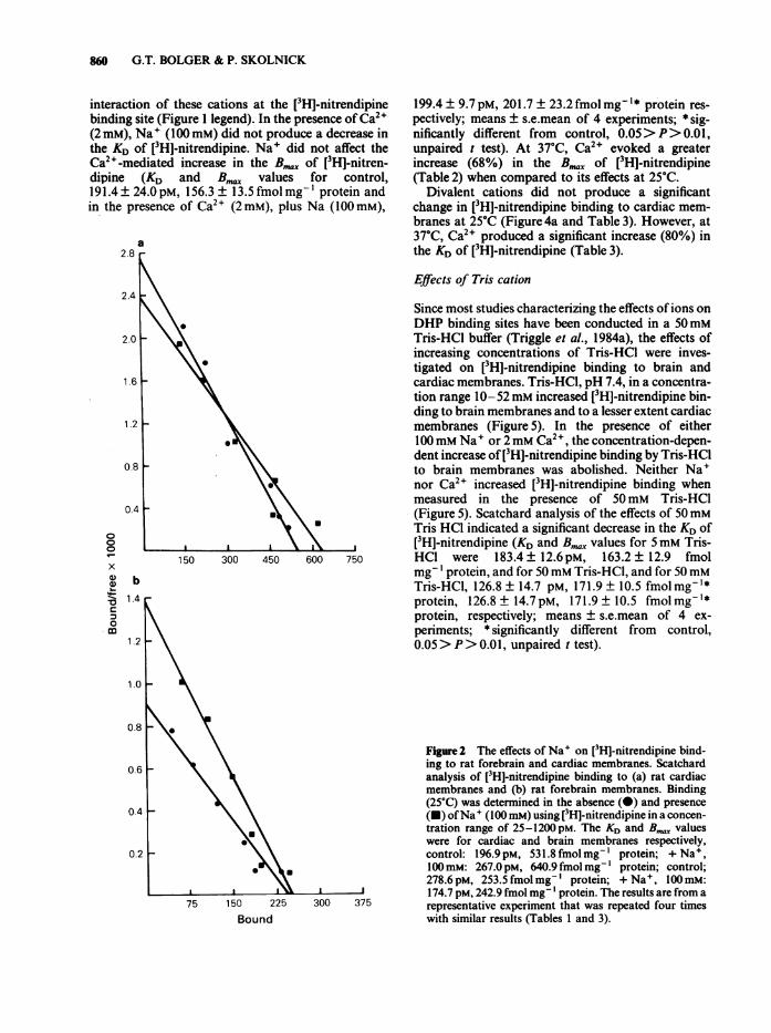
interaction of these cations at the [3H]-nitrendipinebinding site (Figure I legend). In the presence of Ca2"(2mM), Na+ (100mM) did not produce a decrease inthe KD of [3HJ-nitrendipine. Na+ did not affect theCa2"-mediated increase in the B,,..t, of [3H]-nitren-dipine (KD and B,,,, values for control,191.4 ± 24.0 pM, 156.3 ± 13.5 fmol mg-' protein andin the presence of Ca2+ (2mM), plus Na (100mM),
a
000
x
1.b'- 1.4c
0
1.2
1.0
0.8
a
150 300 450 600 750
2.8
2.4
1.2
0.8
0.4
199.4 ± 9.7pM, 201.7 ± 23.2 fmol mg-I* protein res-pectively; means ± s.e.mean of 4 experiments; * sig-nificantly different from control, 0.05> P>0.01,unpaired t test). At 370C, Ca2' evoked a greaterincrease (68%) in the B,,.. of [3H]-nitrendipine(Table 2) when compared to its effects at 25°C.
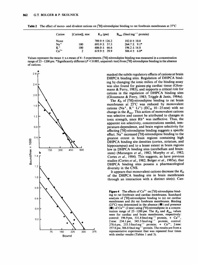
Divalent cations did not produce a significantchange in [3H]-nitrendipine binding to cardiac mem-branes at 25°C (Figure 4a and Table 3). However, at37°C, Ca2+ produced a significant increase (80%) inthe KD of [3H]-nitrendipine (Table 3).
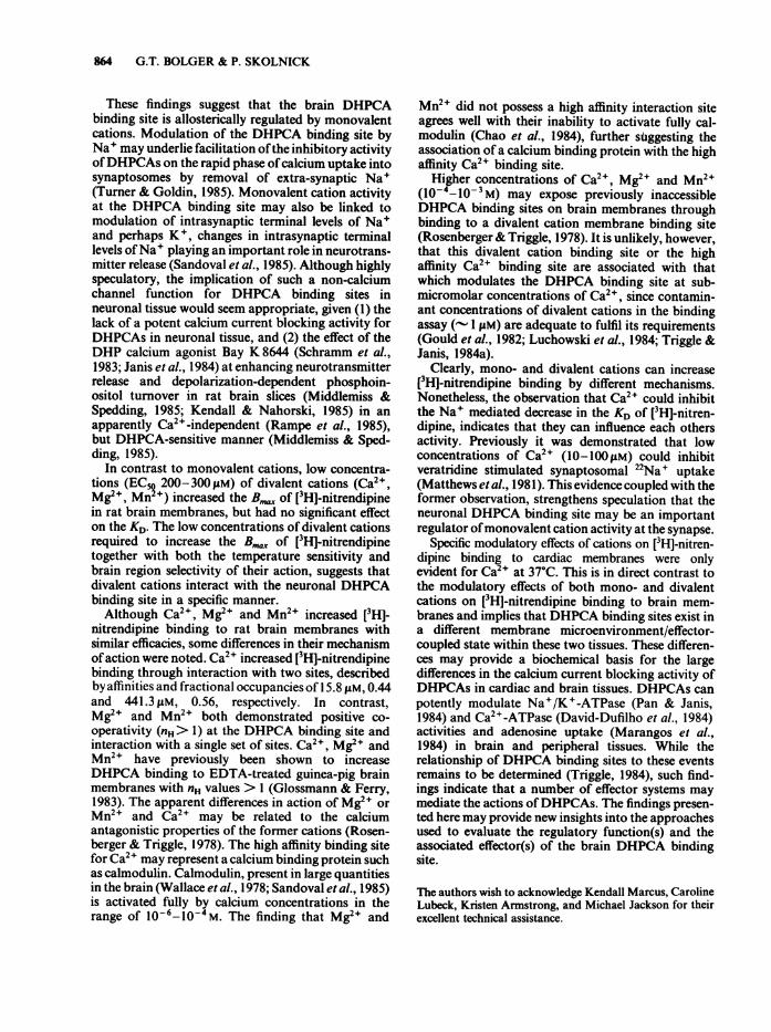
Effects of Tris cation
Since most studies characterizing the effects of ions onDHP binding sites have been conducted in a 50mMTris-HCI buffer (Triggle et al., 1984a), the effects ofincreasing concentrations of Tris-HC1 were inves-tigated on [3H]-nitrendipine binding to brain andcardiac membranes. Tris-HCI, pH 7.4, in a concentra-tion range 10- 52mM increased [3H]-nitrendipine bin-ding to brain membranes and to a lesser extent cardiacmembranes (Figure 5). In the presence of either100mM Na+ or 2mM Ca2+, the concentration-depen-dent increase of [3HJ-nitrendipine binding by Tris-HC1to brain membranes was abolished. Neither Na+nor Ca2+ increased [3H]-nitrendipine binding whenmeasured in the presence of 50mM Tris-HCl(Figure 5). Scatchard analysis of the effects of 50mMTris HCO indicated a significant decrease in the KD of[3H]-nitrendipine (KD and B,,,. values for 5mM Tris-HC1 were 183.4± 12.6PM, 163.2± 12.9 fmolmg 'protein, and for 50mM Tris-HCI, and for 50mMTris-HCl, 126.8 ± 14.7 PM, 171.9 + 10.5 fmolmg-'*protein, 126.8 ± 14.7 pM, 171.9 ± 10.5 fmolmg-l*protein, respectively; means ± s.e.mean of 4 ex-periments; * significantly different from control,0.05> P> 0.01, unpaired t test).
75 150 225
Bound
Figure 2 The effects of Na+ on [3HJ-nitrendipine bind-ing to rat forebrain and cardiac membranes. Scatchardanalysis of [3H]-nitrendipine binding to (a) rat cardiacmembranes and (b) rat forebrain membranes. Binding(25'C) was determined in the absence (0) and presence(U) ofNa+ (100mM) using [3HJ-nitrendipine in a concen-tration range of 25-1200 pM. The KD and B,,. valueswere for cardiac and brain membranes respectively,control: 196.9 pM, 531.8 fmol mg-' protein; + Na+,100 mM: 267.0 pM, 640.9 fmol mg- I protein; control;278.6pM, 253.5 fmolmg-' protein; + Na+, 100mM:174.7 pM, 242.9 fmol mg-' protein. The results are from a
300 375 representative experiment that was repeated four timeswith similar results (Tables 1 and 3).
0.2
EFFECTS OF CATIONS ON [3H]-NITRENDIPINE BINDING
Table I The effect of mono- and divalent cations on [3H]-nitrendipine binding to rat forebrain membranes
[Cation]], mM KD (pM) Bmax (fmol mg-' protein)
Monovalent cationsNoneNa' 100K+ 100Li+ 100Rb+ 100
161.1 ± 13.5120.3 ± 6.9*128.2 ± 12.2*118.8 ± 17.8*161.5 ± 9.7
194.4 ± 9.7202.7 ± 9.9200.3 ± 9.5209.7 ± 13.0196.9 ± 14.7
Divalent cationsCa2+Mg2+Mn2+
2 145.4± 13.82 185.9 ± 7.32 198.2 ± 22.3
Values represent the mean ± s.e.mean of4- 5 experiments. [3H1-nitrendipine binding was measured in a concentrationrange of 25-1200 pM. *Significantly different (0.05> P> 0.01, unpaired t test) from [3HJ-nitrendipine binding (25C)in the absence of cations.
Discussion
Large differences have been described in the potenciesof DHPCAs to block calcium currents in cardiac andneuronal tissues. DHPCAs inhibit cardiac calcium
0)
C
.C_
cjQ
0._
a)
cI
n-a)
.)
C-
Cerebral Striatum Hippo- Cerebellum Braincortex campus stem
Figure 3 Brain region-dependence of cation effects on[3H]-nitrendipine binding. The ability of Na+ (100mM)(open columns) and Ca2+ (2 mM) (solid columns) toincrease [3H]-nitrendipine binding to brain membraneswas investigated in different brain regions. The results arepresented as a % increase in [3H]-nitrendipine binding inthe presence, compared to that in the absence of cations.Values are presented as the mean ± s.e.mean of 5-6experiments. The concentration of [3H]-nitrendipine inthe binding assay was 165 pM. Significantly different from
cortical, striatal and hippocampal [3H]-nitrendipine bind-ing (P<0.05); **significantly different from cortical,hippocampal, cerebellar, and brain stem [3H]-nitren-dipine binding (P <0.05). [3H]-nitrendipine binding(25°C) in the presence of cations was significantly dif-ferent, P< 0.005, from control binding.
currents in the concentration range 10-9_10-6M(Fleckenstein, 1977; Rosenberger & Triggle, 1978;Triggle & Swamy, 1980; Triggle, 1981). In contrast,DHPCA concentrations of > 10-6 M produce onlymarginal effects on total synaptosomal calciumuptake (Toll, 1982; Daniell et al., 1983; Ogura &Takahashi, 1984; Rampe et al., 1984) or calcium-dependent neurotransmitter release (Freedman et al.,1984; Ogura & Takahashi, 1984). Until recently, majordifferences in the binding characteristics of brain andcardiac DHPCA binding sites (Triggle & Janis, 1984a)had not been previously described. However, Sch-wartz & Velly (1985) found a different sensitivity ofbrain and cardiac DHPCA binding sites to Na+, thusproviding biochemical evidence to support the distinctregulatory properties of these sites.We observed that low concentrations (with respect
to extracellular concentrations) of monovalent (Na+,K+, Li+) and divalent (Ca2 , Mg2+, Mn2 ) cationsincreased brain [3H]-nitrendipine binding in a lowionic strength buffer (5mM Tris-HCl). Similar effectsat brain DHPCA binding sites have not previouslybeen reported, since most studies used 50 mM Tris-HCl to characterize the effects of both mono- anddivalent cations on [3H]-nitrendipine binding (Triggle& Janis, 1984a). Tris-HCI (50 mM) significantly in-creased [3H]-nitrendipine binding to brain membranes(decreased KD) and inhibited the effects of mono- anddivalent cations when compared to 5 mM Tris-HCI.The ability ofTris-HCI to interfere with cation-depen-dent effects on radioligand binding is not unique, sincea similar phenomenon was found for sodium-depen-dent [3H]-cocaine binding (Reith et al., 1984). Further-more, Tris-HCl was found to alter the phospholipidbinding, conformation, and aggregation ofmembraneproteins (Yeagle et al., 1986). Thus, it appears that50mM Tris-HCl in DHPCA binding assays has
237.0 ± 10.5*247.9 ± 12.9*245.7 ± 17.7*
861
862 G.T. BOLGER & P. SKOLNICK
Table 2 The effect of mono- and divalent cations on [3H]-nitrendipine binding to rat forebrain membranes at 37°C
Cation [Cation]], mM KD (PM) Bmax (fmol mg-I protein)
None 789.9 ± 126.2 182.0 ± 10.0Na' 100 693.9 ± 57.5 264.7 ± 9.1*K+ 100 606.0 ± 66.6 196.2 ± 16.8Ca2+ 2 619.9 ± 29.9 306.4 ± 6.0*
Values represent the mean ± s.e.mean of4- 5 experiments. [3H]-nitrendipine binding was measured in a concentrationrange of25 -1200 pM. * Significantly different (P < 0.005, unpaired t test) from [3H]-nitrendipine binding in the absenceof cations.
a2.8 - masked the subtle regulatory effects of cations at brain
DHPCA binding sites. Regulation of DHPCA bind-
2.4 - ing by changing the ionic milieu of the binding assaywas also found for guinea-pig cardiac tissue (Gloss-mann & Ferry, 1983), and supports a critical role for
2.0 - cations in the regulation of DHPCA binding sites(Glossmann & Ferry, 1983; Triggle & Janis, 1984a).The Kt, of [3H]-nitrendipine binding to rat brain
1.6 membranes at 25°C was reduced by monovalentcations (Na', K+ Li+) (EC50 10-25mM) with nochange in the B,... This action of monovalent cations
1.2 - was selective and cannot be attributed to changes inionic strength, since Rb+ was ineffective. Thus, theapparent ion selectivity, concentrations needed, tem-
0.8 - perature-dependence, and brain region selectivity foraffecting [3H]-nitrendipine binding suggests a specificeffect. Na+ increased [3H]-nitrendipine binding to the
o o0.4 t greatest extent in brain regions containing higho \ DHPCA binding site densities (cortex, striatum, and
150L* ^ ,\ \ hippocampus) and to a lesser extent in brain regionsa 150 300 450 600 low in DHPCA binding sites (cerebellum and brain-
b stem) (Marangos et al., 1982; Murphy et al., 1982;
n 1.4 - Cortes et al., 1984). This suggests, as have previousC
0 studies (Cortes et al., 1982; Bolger et al., 1985a), thatm 1.2 ~ DHPCA binding sites possess a pharmacological
1.2 - diversity in the CNS.It appears that monovalent cations decrease the KD
1.0 _ of the DHPCA binding site in brain membranesthrough an interaction with a distinct site(s). Can-
0.8 *Figure 4 The effects of Ca2+ on [3H]-nitrendipine bind-ing to rat forebrain and cardiac membranes. Scatchard
0.6 - analysis of [3HJ-nitrendipine binding to (a) rat cardiacmembranes and (b) rat forebrain membranes. Binding(25°C) was determined in the absence (0) and presence
0.4 - (U) of Ca2+ (2 mM) using [3H]-nitrendipine in a concen-tration range of 25-1200 pM. The KD and Bmax valueswere for cardiac and brain membranes, respectively:
0.2 - control: 196.9 pM, 531.8 fmol mg' protein, + Ca2 ,* \m 2 mM: 234.1 pM, 545.5 fmol mg-' protein; control:
278.6pM, 253.5fmolmg-1 protein; + Ca2+, 2 mM:, s ,\ }s 1 257.8 pM, 306.8 fmol mg-' protein. The results are from a
75 150 225 300 375 representative experiment that was repeated four timesRr, nd with similar results (Tables 1 and 3).DLUUI IU
EFFECTS OF CATIONS ON [3H]-NITRENDIPINE BINDING 863
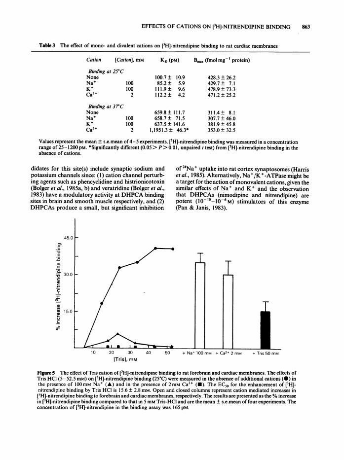
Table 3 The effect of mono- and divalent cations on [3H]-nitrendipine binding to rat cardiac membranes
Cation [Cation], mM KD (pM) Bmax (fmol mgI protein)
Binding at 25°CNone 100.7 ± 10.9Na' 100 85.2 ± 5.9K+ 100 111.9 ± 9.6Ca2+ 2 112.2 ± 4.2
Binding at 37°CNone 659.8 ± 111.7Na+ 100 658.7 ± 71.5K+ 100 637.5± 141.6Ca2+ 2 1,1951.3 ± 46.3*
428.3 ± 26.2429.7 ± 7.1478.9 ± 73.3471.2 ± 25.2
311.4 ± 8.1307.7 ± 46.0381.9 ± 45.8353.0 ± 32.5
Values represent the mean ± s.e.mean of4- 5 experiments. [3H]-nitrendipine binding was measured in a concentrationrange of 25-1200 pM. *Significantly different (0.05> P> 0.01, unpaired t test) from [3HJ-nitrendipine binding in theabsence of cations.
didates for this site(s) include synaptic sodium andpotassium channels since: (1) cation channel perturb-ing agents such as phencyclidine and histrionicotoxin(Bolger et al., 1985a, b) and veratridine (Bolger et al.,1983) have a modulatory activity at DHPCA bindingsites in brain and smooth muscle respectively, and (2)DHPCAs produce a small, but significant inhibition
45.0C0.C
CDC
CLta 30.0-C
a)
I)Cl) 15.01501L.)C
of 24Na' uptake into rat cortex synaptosomes (Harriset al., 1985). Alternatively, Na+/K+-ATPase might bea target for the action ofmonovalent cations, given thesimilar effects of Na' and K+ and the observationthat DHPCAs (nimodipine and nitrendipine) arepotent (10- 10-106M) stimulators of this enzyme(Pan & Janis, 1983).
[Tris], mM
Figure 5 The effect of Tris cation of [3H]-nitrendipine binding to rat forebrain and cardiac membranes. The effects ofTris HCl (5-52.5 mM) on [3H]-nitrendipine binding (25'C) were measured in the absence of additional cations (-) inthe presence of 100mM Na+ (A) and in the presence of 2mM Ca2+ (-). The EC50 for the enhancement of [3H]-nitrendipine binding by Tris HCl is 15.6 ± 2.8 mM. Open and closed columns represent cation mediated increases in[3HJ-nitrendipine binding to forebrain and cardiac membranes, respectively. The results are presented as the % increasein [3HJ-nitrendipine binding compared to that in 5 mm Tris-HCl and are the mean ± s.e.mean offour experiments. Theconcentration of [3H]-nitrendipine in the binding assay was 165 pM.
864 G.T. BOLGER & P. SKOLNICK
These findings suggest that the brain DHPCAbinding site is allosterically regulated by monovalentcations. Modulation of the DHPCA binding site byNa' may underlie facilitation ofthe inhibitory activityofDHPCAs on the rapid phase ofcalcium uptake intosynaptosomes by removal of extra-synaptic Na'(Turner & Goldin, 1985). Monovalent cation activityat the DHPCA binding site may also be linked tomodulation of intrasynaptic terminal levels of Na'and perhaps K+, changes in intrasynaptic terminallevels ofNa+ playing an important role in neurotrans-mitter release (Sandoval et al., 1985). Although highlyspeculatory, the implication of such a non-calciumchannel function for DHPCA binding sites inneuronal tissue would seem appropriate, given (1) thelack of a potent calcium current blocking activity forDHPCAs in neuronal tissue, and (2) the effect of theDHP calcium agonist Bay K 8644 (Schramm et al.,1983; Janis et al., 1984) at enhancing neurotransmitterrelease and depolarization-dependent phosphoin-ositol turnover in rat brain slices (Middlemiss &Spedding, 1985; Kendall & Nahorski, 1985) in anapparently Ca2+-independent (Rampe et al., 1985),but DHPCA-sensitive manner (Middlemiss & Sped-ding, 1985).
In contrast to monovalent cations, low concentra-tions (ECSo 200-30011M) of divalent cations (Ca2+,Mg2+, Mn +) increased the B,, of [3H]-nitrendipinein rat brain membranes, but had no significant effecton the KD. The low concentrations of divalent cationsrequired to increase the Bma of [3H]-nitrendipinetogether with both the temperature sensitivity andbrain region selectivity of their action, suggests thatdivalent cations interact with the neuronal DHPCAbinding site in a specific manner.Although Ca2+, Mg2+ and Mn2+ increased [3H1-
nitrendipine binding to rat brain membranes withsimilar efficacies, some differences in their mechanismofaction were noted. Ca2+ increased [3H]-nitrendipinebinding through interaction with two sites, describedby affinities and fractional occupancies of 15.8 gM, 0.44and 441.3 pM, 0.56, respectively. In contrast,Mg2+ and Mn2+ both demonstrated positive co-operativity (nH> 1) at the DHPCA binding site andinteraction with a single set of sites. Ca2+, Mg2+ andMn2+ have previously been shown to increaseDHPCA binding to EDTA-treated guinea-pig brainmembranes with nH values > I (Glossmann & Ferry,1983). The apparent differences in action of Mg2' orMn2' and Ca2+ may be related to the calciumantagonistic properties of the former cations (Rosen-berger & Triggle, 1978). The high affinity binding sitefor Ca2" may represent a calcium binding protein suchas calmodulin. Calmodulin, present in large quantitiesin the brain (Wallace et al., 1978; Sandoval et al., 1985)is activated fully by calcium concentrations in therange of 10-6-10-4M. The finding that Mg2' and
Mn2" did not possess a high affinity interaction siteagrees well with their inability to activate fully cal-modulin (Chao et al., 1984), further stsggesting theassociation of a calcium binding protein with the highaffinity Ca2" binding site.
Higher concentrations of Ca2 , Mg24 and Mn24(10-4-10-3 M) may expose previously inaccessibleDHPCA binding sites on brain membranes throughbinding to a divalent cation membrane binding site(Rosenberger & Triggle, 1978). It is unlikely, however,that this divalent cation binding site or the highaffinity Ca24 binding site are associated with thatwhich modulates the DHPCA binding site at sub-micromolar concentrations of Ca24, since contamin-ant concentrations of divalent cations in the bindingassay (- I JM) are adequate to fulfil its requirements(Gould et al., 1982; Luchowski et al., 1984; Triggle &Janis, 1984a).
Clearly, mono- and divalent cations can increase[H]-nitrendipine binding by different mechanisms.Nonetheless, the observation that Ca24 could inhibitthe Na4 mediated decrease in the KD of [3H]-nitren-dipine, indicates that they can influence each othersactivity. Previously it was demonstrated that lowconcentrations of Ca24 (10-100IM) could inhibitveratridine stimulated synaptosomal 22Na+ uptake(Matthews et al., 1981). This evidence coupled with theformer observation, strengthens speculation that theneuronal DHPCA binding site may be an importantregulator ofmonovalent cation activity at the synapse.
Specific modulatory effects of cations on [3H]-nitren-dipine binding to cardiac membranes were onlyevident for Ca24 at 37°C. This is in direct contrast tothe modulatory effects of both mono- and divalentcations on [3H]-nitrendipine binding to brain mem-branes and implies that DHPCA binding sites exist ina different membrane microenvironment/effector-coupled state within these two tissues. These differen-ces may provide a biochemical basis for the largedifferences in the calcium current blocking activity ofDHPCAs in cardiac and brain tissues. DHPCAs canpotently modulate Na+/K+-ATPase (Pan & Janis,1984) and Ca24-ATPase (David-Dufilho et al., 1984)activities and adenosine uptake (Marangos et al.,1984) in brain and peripheral tissues. While therelationship of DHPCA binding sites to these eventsremains to be determined (Triggle, 1984), such find-ings indicate that a number of effector systems maymediate the actions ofDHPCAs. The findings presen-ted here may provide new insights into the approachesused to evaluate the regulatory function(s) and theassociated effector(s) of the brain DHPCA bindingsite.
The authors wish to acknowledge Kendall Marcus, CarolineLubeck, Kristen Armstrong, and Michael Jackson for theirexcellent technical assistance.
EFFECTS OF CATIONS ON [3H]-NITRENDIPINE BINDING 865
References
BEAN, B.P. (1984). Nitrendipine block of cardiac calciumchannels: High affinity binding to the inactivated state.Proc. natn. Acad. Sci. U.S.A., 81, 6388-6392.
BELLEMANN, P., FERRY, D., LUBBECKE, F. & GLOSS-MANN, H. (1981). [3H]-nitrendipine, a potent calciumantagonist binds with high affinity to cardiac membranes.Arzheim. Forsch., 31, 2064-2067.
BIRDSALL, N.J.M., BURGEN, A.S.V. & HULME, E.C. (1978).The binding of agonists to brain muscarinic receptors.Molec. Pharmac., 14, 723-732.
BOLGER, G.T., GENGO, P.T., LUCHOWSKI, E.M., SIEGEL, H.& TRIGGLE, D.J. (1982). High affinity binding of acalcium channel antagonist to smooth and cardiac mus-cle. Biochem. Biophys. Res. Commun., 104, 1604-1609.
BOLGER, G.T., GENGO, P., KLOCKOWSKI, R., LUCHOWSKI,E., SEIGEL, H., JANIS, R.A., TRIGGLE, A.M. & TRIGGLE,D.J. (1983). Characterization of the binding of the Ca2+channel antagonist [3H]-Nitrendipine to guinea pig ilealsmooth muscle. J. Pharmac. exp. Ther., 225, 291-309.
BOLGER, G.T., WEISSMANN, B.A. & SKOLNICK, P. (1985a).The behavioral effects of the calcium agonist BAYK 8644 in the mouse: Antagonism by the calciumantagonist nifedipine. Naunyn-Schmiedebergs Arch.Pharmac., 328, 373-377.
BOLGER, G.T., RAFFERTY, M.F. & SKOLNICK, P. (1985b).Phencyclidine increases the affinity of dihydropyridinecalcium channel antagonist binding in rat brain. Naunyn-Schmiedebergs Arch. Pharmac., 330, 227-234.
CHAO, S-H., SUZUKI, Y., ZYSK, J.R. &CHEUNG, W.Y. (1984).Activation of calmodulin by various metal cations as afunction of ionic radius. Molec. Pharmac., 26, 75-82.
CORTES, R., SUPAVILAI, P., KAROBATH, M. & PLACIOUS,J.M. (1983). The effects of lesions in the rat hippocampussuggest the association of calcium channel blocker bind-ing sites with a specific neuronal population. Neurosci.Lett., 42, 249-254.
CORTES, R., SUPAVILAI, P., KAROBATH, M. & PLACIOS,J.M. (1984). Calcium antagonist binding sites in the ratbrain: Quantitative autoradiographic mapping using the1,4-dihydropyridines [3H]PN-200-1 10 and [3H]PY 108-068. J. Neural. Transmission, 60, 169-197.
DANIELL, L.C., BARR, E.M. & LESLIE, S!W. (1983). 45Ca2+-uptake into rat whole synaptosomes unaltered bydihydropyridine calcium antagonists. J. Neurochem., 41,1455-1459.
DAVID-DUFILHO, M. DEVYNCK, M.A., KAZDA, S. &MEYER, P. (1984). Stimulation by nifedipine of calciumtransport by cardiac sarccalemmal vesicles from spontan-eously hypertensive rats. Eur. J. Pharmac., 97, 121-127.
EHLERT, F.J., ITOGA, E., ROESKE, W.R. &YAMAMURA, H.I.(1982). The interaction of [3H~nitrendipine with receptorsfor calcium antagonists in the cerebral cortex and heart ofrats. Biochem. biophys. Res. Commun., 104, 937-943.
FERRY, D. & GLOSSMANN, H. (1982). Identification ofputative calcium channels in skeletal muscle microsomes.FEBS Lett., 148, 331-337.
FLECKENSTEIN, A. (1977). Specific pharmacology of cal-cium in myocardium cardiac pacemakers and vascularsmooth muscle. A. Rev. Pharmac. Tox., 17, 149-166.
FOSSET, M., JAIMOVICH, E. & LAZDUNSKI, M. (1983).[3Hlnitrendipine labelling of the Ca2+-channel in skeletal
muscle. Eur. J. Pharmac., 86, 141-142.FREEDMAN, S.B., DAWSON, G., VILLEREAL, M.L. &
MILLER, R.J. (1984). Identification and characterizationof voltage sensitive calcium channels in neuronal clonalcell lines. J. Neurosci., 4, 1453-1467.
GLOSSMANN, H. & FERRY, D.R. (1983). Molecularapproach to the calcium channel. Drug Development, 9,63-98.
GOLL, A., FERRY, D.R. & GLOSSMANN, H. (1983). Targetsize analysis of Ca2+ channels labelled with [3H]-verapamil. Eur. J. Biol., 83, 177-186.
GOULD, R.J., MURPHY, K.M.M. & SNYDER, S.H. (1982).[3H]-nitrendipine labeled calcium channels discriminateinorganic calcium agonists and antagonists. Proc. natn.Acad. Sci. U.S.A., 79, 3656-3660.
HARRIS, R.A., JONES, S.B., BRUNO, P. & BYLUND, D.B.(1985). Effects of dihydropyridine derivatives andanticonvulsant drugs on [3H]nitrendipine binding andcalcium and sodium fluxes in brain. Biochem. Pharmac.,34, 2187-2191.
HOFFMEISTER, F., BENZ, U., HEISE, A., KRAUSE, H.P. &NEUSER, V. (1982). Behavioral effects of nimodipine inanimals. Arzheim-Forsch/Drug Res., 32, 347-360.
HORNE, P., TRIGGLE, D.J. & VENTER, J.C. (1984). Nitren-dipine and isoproterenol induce phosphorylation of a42,000 Dalton protein that comigrates with the affinitylabeled calcium channel regulatory subunit. Biochem.Biophys. Res. Commun., 121, 890-898.
JANIS, R.A., MAURER, S.C., SARMIENTO, J.G., BOLGER,G.T. & TRIGGLE, D.J. (1982). Binding of [3H~nimodipineto cardiac and smooth muscle membranes. Eur. J.Pharmac., 82, 191-194.
JANIS, R.A., RAMPE, D., SARMIENTO, J.G. & TRIGGLE, D.J.(1984). Specific binding of a calcium channel activatir,[H]BAY K8644, to membranes from cardiac muscle andbrain. Biochem. biophys. Res. Commun., 121, 317-323.
KENDALL, D.A. & NAHORSKI, S.R. (1985). Dihydropyridinecalcium channel activators and antagonists influencedepolarization-evoked inositol phospholipid hydrolysisin brain. Eur. J. Pharmac., 115, 31-36.
LOWRY, O.H., ROSEBROUGH, N.J., FARR, A.L. & RANDALL,R.J. (1951). Protein measurements with the folin phenolreagent. J. biol. Chem., 193, 265-275.
LUCHOWSKI, E.M., YOUSIF, F., TRIGGLE, D.J., MAURER,S.C., SARMIENTO, J.G. & JANIS, R.A. (1984). Effects ofmetal cations and calmodulin antagonists on [3H]nitren-dipine binding in smooth and cardiac muscle. J. Pharmac.exp. Ther., 230, 607-613.
MARANGOS, P.J., PATEL, J., MILLER, C. & MARTINO, A.M.(1982). Specific calcium antagonist binding sites in brain.Life Sci., 31, 1575-1585.
MARANGOS, P.J., FINKEL, M.S., VERMA, A., MATURI, M.F.,PATEL, J. & PATTERSON, R.E. (1984). Adenosine uptakesites in dog heart and brain: Interaction with calciumantagonists. Life Sci., 35, 1109-1116.
MATTHEWS, J.C., WARNICK, J.E., ALBUQUERQUE, E.X. &ELDEFRAWI, M.E. (1981). Characterization of the elec-trogenic sodium channel from rat brain membranes usingneurotoxin dependent 22Na'-uptake. Memb. Biochem., 4,71-104.
MENDELSON, W.B., OWEN, C., SKOLNICK, P., PAUL, S.M.,
866 EFFECTS OF CATIONS ON [3HFNITRENDIPINE BINDING
MARTIN, J.V., KO, G. & WAGNER, R. (1984). Nifedipineblocks sleep induction by flurazepam in the rat. Sleep, 7,64-68.
MIDDLEMISS, D.N. & SPEDDING, M. (1985). A functionalcorrelate for the dihydropyridine binding site in rat brain.Nature, 314, 94-96.
MILLER, G.L. (1959). Protein determination for large num-bers of samples. Anal. Chem., 964, 1959.
MURPHY, K.M.M. & SNYDER, S.H. (1982). Calcium antagon-ists receptor binding sites labeled with [3H]nitrendipine.Eur. J. Pharmac., 77, 201-202.
MURPHY, K.M.M., GOULD, R.J. & SNYDER, S.H. (1982).Autoradiographic visualization of [3Hjnitrendipine bind-ing sites in rat brain: Localization to synaptic zones. Eur.J. Pharmac., 81, 517-519.
OGURA, A. & TAKAHASHI, M. (1984). Differential effect of adihydropyridine derivative to Ca2' entry pathwys inneuronal preparations. Brain Res., 201, 323-330.
PAN, M. & JANIS, R.A. (1984). Stimulation of Na4-K4ATPase of isolated smooth muscle membranes by theCa24 channel inhibitors nimodipine and nitrendipine.Biochem. Pharmac., 33, 787-791.
RAMPE, D., JANIS, R.A. & TRIGGIE, D.J. (1984). BAY K8644,a 1,4-dihydropyridine Ca2+ channel activator. Dissocia-tion of binding and functional effects in brain synap-tosomes. J. Neurochem., 43, 1688-1692.
REITH, M.A.E., MEISLER, B.E., SERSLER, H. & LAJTHA, A.(1984). [3Hlcocaine binding in brain is inhibited by tris(hydroxymethyl) amino methane. J. Neurosci. Methods.,12, 151-154.
RENGASAMY, A., PTASIENSKI, J. & HOSEY, M.M. (1985).Purification of the cardiac 1,4-dihydropyridine receptor/calcium channel complex. Biochem. biophys. Res. Com-mun., 126, 1-7.
ROSENBERGER, L. & TRIGGLE, D.J. (1978). Calcium,calcium translocation and specific calcium antagonists.In Calcium in Drug Action, ed. Weiss, G.B. pp. 3-31, NewYork: Plenum Publishing Co.
SANDOVAL, M.E., AQUINO, G. & CHAVEZ, J.L. (1985).Sodium-dependent, calmodulin-dependent transmitterrelease from synaptosomes. Neurosci. Lett., 56, 271-277.
SCHRAMM, M., THOMAS, G., TOWART, R. & FRANCK-OWIAK, G. (1983). Activation of calcium channels bynovel 1,4-dihydropyridines: A new mechanism for
positive inotropics or smooth muscle stimulants. Arz-neium-Forsch. Drug Res., 33, 1268-1272.
SCHWARTZ, J. & VELLY, J. (1985). Interference of sodiumwith [3H1-nitrendipine binding to cardiac membranes. Br.J. Pharmac., 84, 511-515.
STARKE, K., SPATH, L. & WICHMANN, T. (1984). Effects ofverapamil, diltiazem and ryosidine on the release ofdopamine and acetylcholine in rabbit caudate nucleusslices. Naunyn-Schmiedebergs Arch. Pharmac., 325,124-130.
TOLL, L. (1982). Calcium antagonists, high affinity bindingand inhibition ofcalcium transport in a clonal cell line. J.biol. Chem., 257, 13189-13192.
TRIGGLE, D.J. (1981). Calcium antagonists: Basic chemicaland pharmacological aspects. In New Perspectives onCalcium Antagonists, ed. Weiss, G.B., pp. 1-18, Beth-esda, MD: American Physiological Society.
TRIGGLE, D.J (1984). Ca2+-channels revisited: Problemsand promises. Trends Pharmac. Sci., 5, 4.
TRIGGLE, D.J. & JANIS, R.A. (1984a). Calcium channelantagonists: New perspectives from the radioligand bind-ing assay. In Modern Methods in Pharmacology, ed. Liss,A.R. pp. 1-28, New York: A.R. Liss Inc.
TRIGGLE, D.J & JANIS, R.A.(1984b). The 1,4-dihydrop'ridine receptor: a regulatory component of theCa2" channel. J. cardiovasc. Pharmac, 6, S949-S955.
TRIGGLE, D.J. & SWAMY, V.C. (1980). Pharmacology ofagents that affect calcium. Chest, 78 Suppl., 174-179.
TURNER, T.J. & GOLDIN, S.M. (1985). Calcium channels inrat brain synaptosomes: Identification and phar-macological characterization. J. Neurosci., 5, 841-849.
VENTER, J.C., FRASER, C.M., SCHABER, J.S., JUNG, C.Y.,BOLGER, G. & TRIGGLE, D.J. (1983). Molecular proper-ties of the slow inward calcium channel. J. biol. Chem.,258, 9344-9348.
WALLACE, R.W., THOMAS, J.L., TALLANT, E.A. &CHEUNG,W.Y. (1978). Purification and characterization of aninhibitor protein of brain adenylate cyclase and cyclicnucleotide phosphodiesterase. J. biol. Chem., 254,377-382.
YEAGLE, P.L., SELINSKY, B.S., SPROWL, C. & MESSINA, A.(1986). Modulation by potassium, Tris, and cholesterolof the calcium ATPase of sarcoplasmic reticulum. Bio-phys. J., 49, t-PM-E9.
(Received December 30, 1985.Revised March 27, 1986.Accepted April 8, 1986.)