Embed Size (px)
Citation preview

Regenerative Endodontics
Calcium Hydroxide–induced Proliferation,Migration, Osteogenic Differentiation, andMineralization via the Mitogen-activated ProteinKinase Pathway in Human Dental Pulp Stem Cells
Luoping Chen, MM,* Lisha Zheng, PhD,* Jingyi Jiang, BS,* Jinpeng Gui, BS,* Lingyu Zhang, BS,*Yan Huang, PhD,* Xiaofang Chen, PhD,* Jing Ji, PhD,* and Yubo Fan, PhD*†Abstract
SignificanceCalcium hydroxide products are the gold standardfor direct pulp capping. Our study showed theimportant roles of MAP kinase pathway in calciumhydroxide–induced proliferation, migration, osteo-genic differentiation, and mineralization in humanDPSCs. It shed light on the molecular mechanismof calcium hydroxide regulated DPSCs behavior.It also provided a scientific foundation for futurecell therapy and dental regeneration with DPSCs.
Introduction: Calcium hydroxide has been extensivelyused as the gold standard for direct pulp capping inclinical dentistry. It induces proliferation, migration, andmineralization in dental pulp stem cells (DPSCs), butthe underlying mechanisms are still unclear. The aim ofthis study was to investigate the role of the mitogen-activated protein (MAP) kinase pathway in calciumhydroxide–induced proliferation, migration, osteogenicdifferentiation, and mineralization in human DPSCs.Methods: Human DPSCs between passages 3 and 6were used. DPSCs were preincubated with inhibitors ofMAP kinases and cultured with calcium hydroxide. Thephosphorylated MAP kinases were detected by Westernblot analysis. Cell viability was analyzed via the methyl-thiazol tetrazolium assay. Cell migration was estimatedusing the wound healing assay. Alkaline phosphatase(ALP) expression was analyzed using the ALP stainingassay. Mineralization was studied by alizarin red staininganalysis. Results: Calcium hydroxide significantly pro-moted the phosphorylation of the c-Jun N-terminal kinase(JNK), p38, and extracellular signal–regulated kinase. Theinhibition of JNK and p38 signaling abolished calcium hy-droxide–induced proliferation of DPSCs. The inhibition ofJNK, p38, and extracellular signal–regulated kinasesignaling suppressed the migration, ALP expression,and mineralization of DPSCs. Conclusions: Our studyshowed that the MAP kinase pathway was involved incalcium hydroxide–induced proliferation, migration, oste-ogenic differentiation, and mineralization in humanDPSCs. (J Endod 2016;42:1355–1361)
Key WordsCalcium hydroxide, dental pulp stem cells, migration,mineralization, proliferation
From the *Key Laboratory for Biomechanics and MechanobiologyBeijing, China; and †National Research Center for Rehabilitation Te
Address requests for reprints to Drs Lisha Zheng and Yubo Fan, ScChina 100191. E-mail address: [email protected] or yubofan0099-2399/$ - see front matter
Copyright ª 2016 American Association of Endodontists.http://dx.doi.org/10.1016/j.joen.2016.04.025
JOE — Volume 42, Number 9, September 2016
Dental pulp is oftenexposed because of
fracture of the dentalcrown, dental caries, toothpreparation, or hypoplasia.Direct pulp capping hasbeen used as a solution tomaintain pulp health andfunction. Calcium hydrox-ide products are the goldstandard and have beenextensively used for over
70 years (1, 2). All the applications of calcium hydroxide in pulp capping lie on itsbiological properties. The high alkaline pH of calcium hydroxide solutions induces acoagulation necrosis layer when in direct contact with pulp tissue. It forms amineralized barrier to block the exposed surface (2, 3). The high alkaline pH ofcalcium hydroxide also can solubilize and release some growth factors from dentin,which are responsible for pulp repair (2). Calcium hydroxide can also neutralizethe lactic acid secreted by osteoclasts and prevent mineral tissue destruction (4).Dental pulp stem cells (DPSCs) are postnatal stem cells isolated from dental pulptissue (5). They express similar putative stem cell surface markers as bone marrowstem cell populations and are characterized by clonogenic abilities, a high growthrate, and multilineage potential (6). DPSCs can generate a dentinlike structure linedwith human odontoblast–like cells that surround pulplike interstitial tissue in vivo(5). Studies have shown that DPSCs are capable of osteogenic, dentinogenic, adipo-genic, chondrogenic, myogenic, and neurogenic differentiation and are regarded asa promising tool for tissue regeneration (7–12).
Calcium hydroxide has been reported to increase the recruitment, proliferation, cellmigration, andmineralization of DPSCs (3), but the underlying molecular mechanism hasnot been revealed. The mitogen-activated protein (MAP) kinase pathway controls a broadarray of physiological processes, such as proliferation, migration, gene expression,mitosis, and apoptosis (13). It is composed of 4 signaling families: extracellular signal-regulated kinase (ERK), c-Jun N-terminal kinase (JNK), p38, and the big MAP kinase-1signaling families. MAP kinases are phosphorylated by upstream MAP kinases for subse-quent signal transduction (14). ERK is involved in the regulation of meiosis, mitosis, and
of Ministry of Education, School of Biological Science andMedical Engineering, Beihang University,chnical Aids, Beijing, China.hool of Biological Science andMedical Engineering, XueYuan Road No. 37, Haidian District, Beijing,@buaa.edu.cn
MAP Kinase Pathway in hDPSCs 1355

Regenerative Endodontics
postmitotic functions in differentiated cells (13). It has been reported thatERK involved in bioceramic putty induced migration, and genipin pro-moted odontogenic differentiation in pulp cells (15, 16). JNK ischaracterized by its response to the inhibition of protein synthesis (17)and is involved in lipopolysaccharide (LPS)-induced cell migration inDPSCs (18). p38 regulates the expression of many cytokines. It is also re-ported that p38 plays a role in LPS-promoted odontoblastic differentiationinDPSCs (19). Therefore, the aimof this study was to investigate the role ofMAP kinases in calciumhydroxide–induced proliferation,migration, oste-ogenic differentiation, and mineralization in DPSCs.Materials and MethodsCell Isolation and Culture
Human dental pulp tissues were obtained from healthy young do-nors (14–22 years) who extracted the third molars for orthodontic rea-sons at Peking University People’s Hospital, Beijing, China. This studyconforms to the Declaration of Helsinki, and all processes wereapproved by the Beihang University Ethical Committee, Beijing, China.Informed consent from each patient was obtained. The pulp tissuewas digested with 3 mg/mL collagenase type I (Sigma-Aldrich, St Louis,MO) and 4 mg/mL Dispase (Sigma-Aldrich) for 1 hour at 37�C. Single-cell suspensions were seeded into 96-well plates by 1 to 2 cells/well withalpha modification of Eagle medium (Gibco-BRL, Grand Island, NY)including 10% fetal bovine serum (Gibco-BRL), 100 U/mL penicillin(Amresco, OH, USA), and 100 mg/mL streptomycin (Amresco, Solon,OH) and then incubated in a humidified atmosphere containing 5%CO2 at 37
�C. Over 50 cells/well were scored as colonies and passagedby 0.25% trypsin (Sigma-Aldrich). The cells between passages 3 and 5were used in the following experiments: DPSCs characterized by flowcytometric analysis of CD44, CD146, CD29, STRO-1, CD45, and CD34(Supplemental Figure S1 is available online at www.jendodon.com).The multiple differentiation potentials of osteogenesis and chondrogen-esis were also confirmed.
Figure 1. Calcium hydroxide induced the phosphorylation of the MAP kinase pathwwith DPSCs for 5, 15, 30, or 60 minutes. p-p38, p-JNK, and p-ERK were detected by rEach experiment was repeated more than 3 times. *P < .05. #P < .01.
1356 Chen et al.
Western Blot AnalysisWemixed calcium hydroxide powder with distilled water to obtain
10 mg/mL calcium hydroxide stock solution. The stock solution wasstirred, centrifuged, and filtered using a 0.22-mm filter. The final con-centration of 10 mg/mL calcium hydroxide was made by adding 3 mL of10 mg/mL calcium hydroxide stock into the 3.5-mm culture dish with3 mL media. Calcium hydroxide groups treated with calcium hydroxideor the control group treated with the same volume of solution used todissolve calcium hydroxide were harvested and lysed by a radioimmu-noprecipitation assay (RIPA) lysis buffer (Beyotime, Shanghai, China)on ice. From 40 to 60 mg proteins were separated by 10% sodium do-decyl sulfate polyacrylamide gel electropheresis (SDS-PAGE). Proteinswere transferred to polyvinylidene fluoride (PVDF) membranes (Milli-pore, Bedford, MA) and then blocked by 4% bovine serum albumin(BSA; Sigma-Aldrich) for 2 to 4 hours in room temperature. After themembranes were washed with Tris-buffered saline containing 0.05%Tween-20 (Sigma-Aldrich), primary antibodies with phosphorylatedJNK, phosphorylated p38, phosphorylated ERK, and glyceraldehyde-3-phosphate dehydrogenase (GAPDH) (all from Santa Cruz Biotech,Santa Cruz, CA) at a dilution of 1:1000 in 4% BSA were incubatedwith a PVDF membrane at 4�C for overnight, respectively. Horseradishperoxidase (HRP)-conjugated secondary antibodies and super electro-chemiluminescence (Applygen, Beijing, China) were used to detect thebands of target proteins. The intensity was calculated using Bio-RadQuantity One (Bio-Rad, Hercules, CA). Each result was normalized tocorresponding GAPDH bands.
Methylthiazol Tetrazolium AssayCell viability was analyzed via the methylthiazol tetrazolium (MTT)
assay. Equal amounts of cells were seeded on 96-well plastic dishes.DPSCs were pretreated with dimethyl sulphoxide (DMSO) for the con-trol and calcium hydroxide groups or with 10 mmol/L JNK inhibitor(SP600125), an ERK inhibitor (PD98059), or a p38 inhibitor
ay. Culture medium with or without 10 mg/mL calcium hydroxide was culturedespective phosphorylated antibodies. Relative intensities are shown as columns.
JOE — Volume 42, Number 9, September 2016

Figure 2. MAP kinases involved in calcium hydroxide–induced proliferation and migration in DPSCs. (A) MTT assays were used to analyze the cell viability of calcium-induced proliferation treated or untreated with the inhibitor of JNK (SP: SP600125, 10 mmol/L), p38 kinase (SB: SB203580, 10 mmol/L), or ERK (PD: PD98059,10 mmol/L). (B) The wound healing assays were used to investigate calcium hydroxide–induced migration in DPSCs. Representative data for the results were shown,and the quantitative data are provided in the panels below. Each experiment was performed at least 3 times. Scale bar = 100 mm. *P < .05. #P < .01.
Regenerative Endodontics
JOE — Volume 42, Number 9, September 2016 MAP Kinase Pathway in hDPSCs 1357

Regenerative Endodontics
(SB203580) for 1 hour (all inhibitors were from Santa Cruz Biotech).The medium was changed to fresh medium with or without 10 mg/mLcalcium hydroxide (Amresco). After 24 hours of treatment, DPSCs wereincubated with 5 mg/mL MTT (Amresco) for 4 hours at 37�C. The tetra-zolium salts were dissolved by DMSO after the incubation period. Thesuspension was collected, and the optical density was measured at570 nm. Each experiment was performed at least 3 times. The resultswere normalized to the control group.Wound Healing AssayDPSCs were cultured on 6-well plastic dishes until they grew a
monolayer. A cross area was scratched with a pipette tip, and thefloating cells were moved by phosphate-buffered saline. DPSCs werepretreated with an MAP kinase inhibitor and changed to culture me-dium with or without calcium hydroxide as described previously. Thescratched areas at each time point of 0, 12, and 24 hours were imaged.To estimate the migration of DPSCs, the cell-free area of each cross wascalculated using DP2-BSW software (Olympus, Tokyo, Japan). Eachmigration result was relative to the image of the control group. Therelated wound area (percentage) was used to quantify the data.
Alkaline Phosphatase Staining AssayDPSCs were pretreated with an MAP kinase inhibitor and changed
to normal medium with or without calcium hydroxide for 7, 14, and21 days and incubated with BCIP/NBP reagent (Beyotime) for about10 minutes at room temperature. Color development was stopped bywater. All staining images were taken under a microscope (IX 71,Olympus). Each experiment was performed at least 3 times.
Alizarin Red Staining AssayDPSCs were pretreated with an MAP kinase inhibitor and changed
to normal medium and mineralization induction medium (50 mmol/Lascorbic acid, 10 mmol/L b-glycerophosphate, and 100 nmol/L dexa-methasone) with calcium hydroxide for 7, 14, and 21 days. The controlgroups were preincubated with the same volume of DMSO and changedwith normal medium or mineralization induction medium without cal-
Figure 3. JNK, p38, and ERK played important roles in calcium hydroxide–indu(SP600125, 10 mmol/L), p38 kinase (SB203580, 10 mmol/L), or ERK (PD98059,with or without calcium hydroxide. After 7, 14, and 21 days, the ALP staining assa
1358 Chen et al.
cium hydroxide for 7, 14, and 21 days. DPSCs were added to 40 mmol/Lalizarin red (pH = 4.2; American MasterTech, Lodi, CA) solution andincubated at room temperature for 30 minutes with 50 rpm shaking.Each dish was washed and imaged. For the quantitative assay,10 mmol/L sodium phosphate–10% acetylpyrimidium (pH = 7.0) so-lution was added into the alizarin red–stained samples and incubated atroom temperature for 15 minutes. Then, the solutions were transferredto a 96-well plate and measured at 562 nm.
Statistical AnalysisData were presented with means � standard deviation from at
least 3 experiments. One-way analysis of variance was used for multiplecomparisons using SPSS version 13.0 software (SPSS Inc, Chicago, IL).A P value <.05 was considered statistically significant.
ResultsCalcium Hydroxide–induced Phosphorylationsof MAP Kinases in Human DPSCs
Calcium hydroxide, 10 mg/mL, was added into the medium, andthe same volume of solution that was used to dissolve calcium hydroxidewas added into the control group (3). The phosphorylations of MAP ki-nases were analyzed by Western blot analysis after 5, 10, 30, and 60 mi-nutes, respectively. The results showed that calcium hydroxide inducedMAP kinase phosphorylation rapidly. Phosphorylated-p38 (p-p38) andphosphorylated-JNK (p-JNK) increased after 5 minutes of incubationand lasted for 1 hour. p-ERK increased after 10 minutes of exposureand also lasted for 1 hour (Fig. 1).
JNK and P38 Involved in Calcium Hydroxide–induced Proliferation in DPSCs
The MTT assay was used to investigate the viability of DPSCs(Fig. 2A). Calcium hydroxide increased the cell numbers of DPSCs,which was similar to the research of Ji et al (3). The numbers of cellsdecreased significantly by inhibitors of JNK and p38, and the inhibitionof ERK barely affected calcium hydroxide–induced proliferation. It
ced ALP expression in DPSCs. After preincubation with the inhibitor of JNK10 mmol/L) for 1 hour, the culture medium was changed into normal mediumy was analyzed, and representative data are shown. Scale bar = 100 mm.
JOE — Volume 42, Number 9, September 2016
Regenerative Endodontics
showed that JNK and p38 but not ERK were involved in calcium hydrox-ide–induced proliferation in DPSCs.JNK, p38, and ERK Involved in Calcium Hydroxide–induced Migration in DPSCs
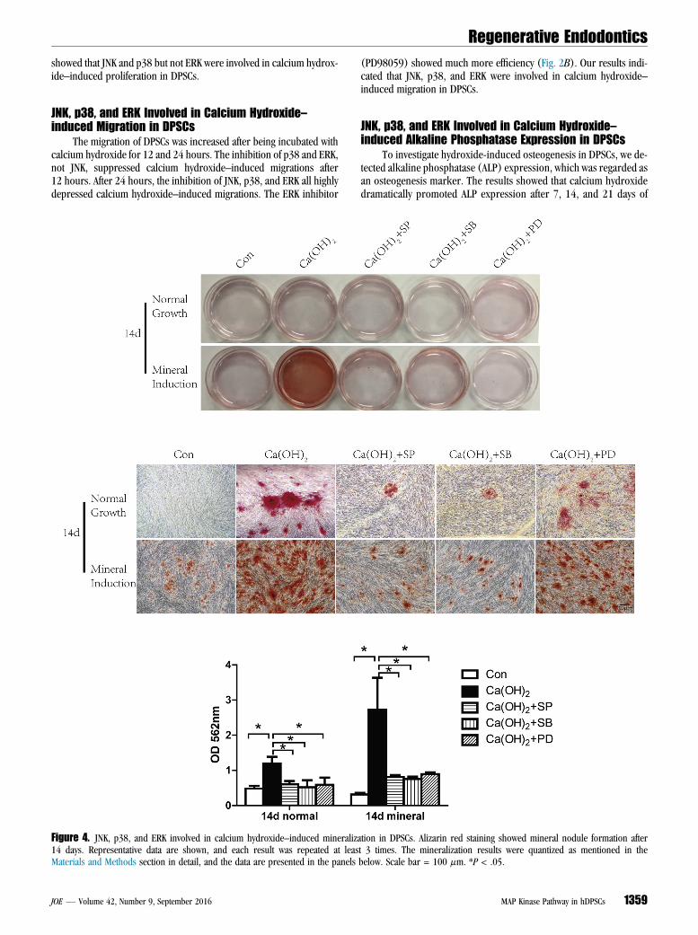
The migration of DPSCs was increased after being incubated withcalcium hydroxide for 12 and 24 hours. The inhibition of p38 and ERK,not JNK, suppressed calcium hydroxide–induced migrations after12 hours. After 24 hours, the inhibition of JNK, p38, and ERK all highlydepressed calcium hydroxide–induced migrations. The ERK inhibitor
Figure 4. JNK, p38, and ERK involved in calcium hydroxide–induced mineraliza14 days. Representative data are shown, and each result was repeated at leasMaterials and Methods section in detail, and the data are presented in the panels
JOE — Volume 42, Number 9, September 2016
(PD98059) showed much more efficiency (Fig. 2B). Our results indi-cated that JNK, p38, and ERK were involved in calcium hydroxide–induced migration in DPSCs.
JNK, p38, and ERK Involved in Calcium Hydroxide–induced Alkaline Phosphatase Expression in DPSCs
To investigate hydroxide-induced osteogenesis in DPSCs, we de-tected alkaline phosphatase (ALP) expression, which was regarded asan osteogenesis marker. The results showed that calcium hydroxidedramatically promoted ALP expression after 7, 14, and 21 days of
tion in DPSCs. Alizarin red staining showed mineral nodule formation aftert 3 times. The mineralization results were quantized as mentioned in thebelow. Scale bar = 100 mm. *P < .05.
MAP Kinase Pathway in hDPSCs 1359

Regenerative Endodontics
incubation. JNK (SP600125) and p38 inhibitors (SB203580)decreased calcium hydroxide–induced ALP expression significantly.However, the ERK inhibitor (PD98059) seemed to affect ALP expres-sion slightly (Fig. 3). It showed that JNK and p38 played more impor-tant roles in calcium hydroxide–induced osteogenic differentiationthan ERK in DPSCs.JNK, p38, and ERK Involved in Calcium Hydroxide–induced Mineralization in DPSCs
The alizarin red assay was used to identify the mineralization ofDPSCs. DPSCs did not show mineralization in normal medium, andmineralization induction medium increased mineralization after 7, 14,and 21 days; 10 mg/mL calcium hydroxide increased mineralizationeven in the normal medium after 7, 14, and 21 days. The mineralizationinduction medium groups have a much higher mineralization reactionthan the normal growth medium groups. The results after 7 days showedthat compared with calcium hydroxide–induced mineralization, JNK(SP600125) and p38 inhibitors (SB203580) inhibited the formation ofmineral nodes remarkably. The ERK inhibitor (PD98059) slightly affectedmineralization, but we could not find a significant difference in the quan-tified data (Supplemental Figure S2 is available online at www.jendodon.com). After 14 days, calcium hydroxide prominently increased mineral-ization, especially in mineralization induction medium. All 3 MAP kinaseinhibitors decreased mineralization significantly (Fig. 4). After 21 days,the JNK (SP600125) and p38 inhibitors (SB203580) seemed to inhibitmineralization more efficiently than the ERK inhibitor (PD98059), butthere was still no significant difference between the 3 inhibitors in thequantified data (Supplemental Figure S3 is available online at www.jendodon.com). The results showed that MAP kinases were involved incalcium hydroxide–induced mineralization in DPSCs.
DiscussionOne of the main goals of vital pulp therapy is to stimulate the pulp
to initiate reparative tertiary dentin formation. This process integratessteps of migration, proliferation, and mineralization of pulp cells(20). As the gold standard of direct pulp capping materials in clinicalvital pulp therapy, calcium hydroxide increases the proliferation ofDPSCs (3). The phosphorylated levels of p38, JNK, and ERK are allappreciably increased in response to calcium hydroxide in 10 to30 minutes. It indicates that activation of the MAP kinase pathway isan early response in DPSCs stimulated by calcium hydroxide. ThisMAP kinase inhibitor experiment further shows that calcium hydrox-ide–induced proliferation was mainly regulated by JNK and p38 butnot by ERK in DPSCs.
Cell migration is an important step in pulpal wound healing. Therepair process includes the migration of DPSCs to the wounded areasbefore they differentiate to form a reparative dentin (21). MAP kinasesplay a crucial role in cell migration (22). JNK phosphorylates paxillin,Spir, DCX, and MAPs and promotes cell migration. The inhibition ofJNK significantly diminishes the migration of cells such as mouse embry-onic fibroblasts and embryonic stem cells (22). p38 regulates actin reor-ganization and cell migration by phosphorylating MAPK-activated proteinkinase 2/3 (MAPK APK2/3), paxillin, and caldesmon (23, 24). ERKregulates membrane protrusions and focal adhesion turnover viaphosphorylating myosin light chain kinase, calpain, and focal adhesionkinase (FAK) (22, 25, 26). Our study showed that the inhibition ofJNK, p38, and ERK significantly impaired calcium hydroxide–inducedmigration in DPSCs. It indicated that the MAP kinase pathway plays animportant role in calcium hydroxide–induced migration in DPSCs.
We studied ALP expression after being incubated with calcium hy-droxide with or without MAP kinase inhibitors. Because ALP is the
1360 Chen et al.
marker of odontogenic/osteogenic differentiation, it showed that calciumhydroxide significantly increased the odontogenic/osteogenic differenti-ation of DPSCs. However, a previous study reports that extracellular Ca2+
inhibited ALP activity (27). It may be because of the different concentra-tion of Ca2+ or calcium salt. MAP kinases have been revealed to regulatedifferentiation in dental pulp cells. p38a is involved in BMP-2–inducedodontoblastic differentiation of human dental pulp cells (28). The bioac-tive calcium phosphate cements (a-tricalcium phosphate based) incor-porating zinc bioglass induce odontogenic differentiation through theMAP kinase pathway in human dental pulp cells (29). Our results indi-cated that the inhibition of JNK, p38, and ERK attenuated calcium hydrox-ide–induced odontogenic/osteogenic differentiation. It suggested that theMAP kinase pathway is involved in calcium hydroxide–induced odonto-genic/osteogenic differentiation in DPSCs.
Dental use of calcium hydroxide relates mainly to its unique poten-tial to stimulate tissuemineralization, but the mechanism is still unclear.It has been suggested that the high pH of calcium hydroxide solutionmay initiate mineralization although other highly alkaline compoundssuch as barium hydroxide and calcium phosphate fail to initiate miner-alization (30, 31). The calcium ions in calcium hydroxide are not partof the mineralized repair tissue. It initiates rather than composes therepaired pulp tissues (1). A recent study (27) showed that Ca2+
induced the mineralization of DPSCs, and Erk1/2 may be involved inthis process. The MAP kinase pathway plays a role in 17b-estradiol–induced mineralization in human dental pulp cells (32); 1a,25(OH)2D3 promoted odontoblastic differentiation and mineralization of hu-man dental pulp cells via modulating ERK activation (33). Our resultsindicated that ERK, especially JNK, and p38, played roles in calcium hy-droxide–induced mineralization in DPSCs.
ConclusionOur findings suggested that MAP kinases involved in calcium hy-
droxide induced proliferation, migration, osteogenic differentiation,and mineralization in human DPSCs. Calcium hydroxide rapidly pro-moted the phosphorylation of JNK, p38, and ERK. JNK and p38 playedroles in calcium hydroxide–induced proliferation. JNK, p38, and ERKwere involved in calcium hydroxide–induced migration, osteogenic dif-ferentiation, and mineralization in human DPSCs. Our work revealedthe mechanism of calcium hydroxide regulated DPSCs and provided ascientific foundation for future cell therapy with DPSCs.
AcknowledgmentsSupported by the National Natural Science Foundation of China
(grant nos. 11572030, 11102015, 61227902, 11120101001,11421202, 11302020, and 11402017), the Fundamental ResearchFunds for the Central Universities, Beijing Higher Education YoungElite Teacher Project, the 111 Project (B13003), grant YWF-15-YG-003 (Beihang University), Specialized Research Fund for the DoctoralProgram of Higher Education (20131102130004), and NationalBasic Research Program of China (973 program, 2011CB710901).
The authors deny any conflicts of interest related to this study.
Supplementary MaterialSupplementary material associated with this article can be
found in the online version at www.jendodon.com (http://dx.doi.org/10.1016/j.joen.2016.04.025).
References1. Foreman PC, Barnes IE. Review of calcium hydroxide. Int Endod J 1990;23:283–97.2. Modena KC, Casas-Apayco LC, Atta MT, et al. Cytotoxicity and biocompatibility of
direct and indirect pulp capping materials. J Appl Oral Sci 2009;17:544–54.
JOE — Volume 42, Number 9, September 2016

Regenerative Endodontics
3. Ji YM, Jeon SH, Park JY, et al. Dental stem cell therapy with calcium hydroxide indental pulp capping. Tissue Eng Part A 2010;16:1823–33.4. Heithersay GS. Calcium hydroxide in the treatment of pulpless teeth with associated
pathology. J Br Endod Soc 1975;8:74–93.5. Gronthos S, Mankani M, Brahim J, et al. Postnatal human dental pulp stem cells
(DPSCs) in vitro and in vivo. Proc Natl Acad Sci U S A 2000;97:13625–30.6. Shi S, Bartold PM, Miura M, et al. The efficacy of mesenchymal stem cells to regen-
erate and repair dental structures. Orthod Craniofac Res 2005;8:191–9.7. Graziano A, d’Aquino R, Laino G, Papaccio G. Dental pulp stem cells: a promising
tool for bone regeneration. Stem Cell Rev 2008;4:21–6.8. Huang GT, Gronthos S, Shi S. Mesenchymal stem cells derived from dental tissues vs.
those from other sources: their biology and role in regenerative medicine. J DentRes 2009;88:792–806.
9. Morsczeck C, Schmalz G, Reichert TE, et al. Somatic stem cells for regenerativedentistry. Clin Oral Investig 2008;12:113–8.
10. Morito A, Kida Y, Suzuki K, et al. Effects of basic fibroblast growth factor on thedevelopment of the stem cell properties of human dental pulp cells. Arch Histol Cytol2009;72:51–64.
11. Ballini A, De Frenza G, Cantore S, et al. In vitro stem cell cultures from humandental pulp and periodontal ligament: new prospects in dentistry. Int J Immunopa-thol Pharmacol 2007;20:9–16.
12. Laino G, d’Aquino R, Graziano A, et al. A new population of human adult dental pulpstem cells: a useful source of living autologous fibrous bone tissue (LAB). J BoneMiner Res 2005;20:1394–402.
13. Johnson GL, Lapadat R. Mitogen-activated protein kinase pathways mediated by ERK,JNK, and p38 protein kinases. Science 2002;298:1911–2.
14. Burotto M, Chiou VL, Lee JM, Kohn EC. The MAPK pathway across different malig-nancies: a new perspective. Cancer 2014;120:3446–56.
15. Zhang J, Zhu LX, Cheng X, et al. Promotion of dental pulp cell migration and pulprepair by a bioceramic putty involving FGFR-mediated signaling pathways. J Dent Res2015;94:853–62.
16. Kwon YS, Lim ES, Kim HM, et al. Genipin, a cross-linking agent, promotes odonto-genic differentiation of human dental pulp cells. J Endod 2015;41:501–7.
17. Kyriakis JM, Banerjee P, Nikolakaki E, et al. The stress-activated protein kinase sub-family of c-Jun kinases. Nature 1994;369:156–60.
18. Li D, Fu L, Zhang Y, et al. The effects of LPS on adhesion and migration of humandental pulp stem cells in vitro. J Dent 2014;42:1327–34.
JOE — Volume 42, Number 9, September 2016
19. He W, Wang Z, Luo Z, et al. LPS promote the odontoblastic differentiation of humandental pulp stem cells via MAPK signaling pathway. J Cell Physiol 2015;230:554–61.
20. Tuna D, Olmez A. Clinical long-term evaluation of MTA as a direct pulp capping ma-terial in primary teeth. Int Endod J 2008;41:273–8.
21. Schroder U. Effects of calcium hydroxide-containing pulp-capping agents on pulp cellmigration, proliferation, and differentiation. J Dent Res 1985;64(Spec No):541–8.
22. Huang C, Jacobson K, Schaller MD. MAP kinases and cell migration. J Cell Sci 2004;117:4619–28.
23. Goncharova EA, Vorotnikov AV, Gracheva EO, et al. Activation of p38 MAP-kinaseand caldesmon phosphorylation are essential for urokinase-induced human smoothmuscle cell migration. Biol Chem 2002;383:115–26.
24. Huang C, Borchers CH, Schaller MD, Jacobson K. Phosphorylation of paxillin byp38MAPK is involved in the neurite extension of PC-12 cells. J Cell Biol 2004;164:593–602.
25. Glading A, Bodnar RJ, Reynolds IJ, et al. Epidermal growth factor activates m-calpain(calpain II), at least in part, by extracellular signal-regulated kinase-mediated phos-phorylation. Mol Cell Biol 2004;24:2499–512.
26. Hunger-Glaser I, Salazar EP, Sinnett-Smith J, Rozengurt E. Bombesin, lysophosphatidicacid, and epidermal growth factor rapidly stimulate focal adhesion kinase phosphor-ylation at Ser-910: requirement for ERK activation. J Biol Chem 2003;278:22631–43.
27. Li S, Hu J, Zhang G, et al. Extracellular Ca2+ promotes odontoblastic differentiationof dental pulp stem cells via BMP2-mediated Smad1/5/8 and Erk1/2 pathways. J CellPhysiol 2015;230:2164–73.
28. Qin W, Lin ZM, Deng R, et al. p38a MAPK is involved in BMP-2-induced odonto-blastic differentiation of human dental pulp cells. Int Endod J 2012;45:224–33.
29. Zhang J, Park YD, Bae WJ, et al. Effects of bioactive cements incorporating zinc-bioglass nanoparticles on odontogenic and angiogenic potential of human dentalpulp cells. J Biomater Appl 2015;29:954–64.
30. Tronstad L, Andreasen JO, Hasselgren G, et al. pH changes in dental tissues after rootcanal filling with calcium hydroxide. J Endod 1981;7:17–21.
31. Mitchell DF, Shankwalker GB. Osteogenic potential of calcium hydroxide and othermaterials in soft tissue and bone wounds. J Dent Res 1958;37:1157–63.
32. Woo SM, Seong KJ, Oh SJ, et al. 17beta-Estradiol induces odontoblastic differenti-ation via activation of the c-Src/MAPK pathway in human dental pulp cells. BiochemCell Biol 2015;93:587–95.
33. Woo SM, Lim HS, Jeong KY, et al. Promotes odontogenic differentiation of humandental pulp cells via ERK activation. Mol Cells 2015;38:604–9.
MAP Kinase Pathway in hDPSCs 1361