Embed Size (px)
Citation preview

Calcium oxalates: fromsynthesis to pathologicalcalcifications
Baptiste Colcombet-Cazenave
Under the supervision of Christian Bonhomme
Internship report - from January 6th , 2014 to January 10th, 2014


Acknowledgements
First of all, I warmly thank Clément Sanchez, head of the LCMCP laboratory, who gave me the
opportunity to perform this internship and Florence Babonneau, the current director.
I am grateful to all the « Sol-gel materials and NMR » team, and especially to my supervisor
Christian Bonhomme.
I would also like to thank Laure Bonhomme, César Leroy and Cristina Coelho who helped me during my
internship.
I would like to offer my gratitude to Gervaise Mosser and Carole Aimé who gave me the opportunity to see a
SEM (Scanning Electron Microscopy) analysis.
I also thank Antoine Tally who has accepted to be my tutor. Finally, I thank Tamara Milosevic and
Alice Demarez who supported me in the organization of the internship.

Calcium oxalates: from synthesisto pathological calcifications
Key words: Synthetic calcium oxalates (oxalates de calcium synthétiques), characterization(caractérisation), solid state NMR (Résonance Magnétique Nucléaire du solide), Infraredspectroscopy IR (spectroscopie infrarouge), powder X-Ray Diffraction, XRD (diffraction des rayonsX sur poudre), Scanning Electron Microscopy, SEM (Microscopie Electronique à Balayage), kidneystones (calculs rénaux).
Abstract
Nephrolithiasis corresponds to a major societal problem. The in depth knowledge of the chemicalstructures involved during the calcification process is a prerequisite for full understanding of thephysico-chemical steps governing the precipitation of inorganic phases (in kidney or tissues).Calcium oxalates play a key role in nephrolithiasis. During the internship, synthetic calciumoxalates were obtained and characterized by various spectroscopic techniques and X-raydiffraction. Among them, 13C CP MAS NMR methods have been implemented. Finally, all thesetechniques were applied for the detailed characterization of natural kidney stones.
Graphical abstract

Table of contents
Introduction 1
Context and background 2
Activity 3
Outcomes 5
Conclusion 12
Supplementary materials 13
References 16
Introduction
One of the main axes of the laboratory's research project is based on the study of kidney stones. At
the very beginning, this is a medical issue. Nephrolithiasis can indeed be lethal for the patient and it may
have different origins. Today, this has turned out to be also an important problem of society because
600 millions of euros are spent every year to diagnose nephrolithiasis and to treat kidney stones in France,
often through a surgical act. In the human body, kidney stones can have many different structures and they
are classified as nanocomposites. Their structures include many proteins, triglycerids and other organic
components but the most important aspect of kidney stone remains calcium oxalates and some
hydroxyapatite. In nature, oxalates can be found in three different phases : monohydrate (CaC2O4.H2O),
dihydrate (CaC2O4.2H2O) and trihydrate (CaC2O4.3H2O). It has been shown that the monohydrate (COM)
represents about 67% of the kidney stones for men, and 75% for women whereas the dihydrate represents
33% for men and 25% for women. On the other hand, the trihydrate phase is really exceptional for both men
and women [1].
Thereby, this study is at the frontier between Biology and Chemistry. It consists in the synthesis of pure
phases of calcium oxalates, mimicking the most important part of the natural components, in order to
characterize each of them deeply. The main methods is solid state NMR, but powder XRD and IR are very
important as well. After having ran some experiments and studied many publications, researchers are no
longer focused only on synthetic materials, but they try now to determine precisely which phases are
involved in real kidney stones (and in which proportions). Following this approach, the goal would be to
explain the interactions between the inorganic crystals and the organic molecules, such as catechin, which
can be found in green tea [2].
I wanted to do my internship in the « Sol-gel materials and NMR » group for many reasons. First of
all, the project is an example of the crossing of scientific fields, which is the most important part of our
studies. Adding to that, it involves new topics of interest, such as crystallography, that we have never
encountered. This internship has been extremely interesting due to all the methods that were used. NMR was
a particular source of curiosity because we had heard so little about it in high school and it had never been
clearly explained. That is one of the reasons why I developed such a strong interest towards these specific
subjects. In fact, in high school, all we did was to quickly use some results from NMR experiments but no
one really tried to describe the fundamental concepts, even in understandable terms. That led to a sort of
frustration caused by using something that I did not fully understand. This internship was a great opportunity
to learn more about NMR, not to get all of it because this is something rather complicated, but to have an
idea of what is this “strange” experiment. Moreover, this internship gave me an overview of laboratory
dynamics, interactions between people working in their own ways on different scientific fields. And finally, I
wanted to discover how a research project is managed by a group of scientists in order to make discoveries or
explain experimental facts we observed but we don't understand yet.
1

Context and background
It all starts in hospitals. Lithiasis is a frequent pathology which affects 10% of the population of
industrialized countries and reach people from 30 years old for men and 25 years old for women [1].
It therefore represents an important amount of patients and so it is an important topic of medicine. Surgeons
have been removing kidney stones from patients for many decades. Pathological calcifications can occur in
various parts of the body such as brain, breast, cartilage, cardiac valves, heart... and not only in the
kidney [3]. The stones collected from surgeries are studied by many techniques in hospital (IR study or SEM
observation) but also in laboratories. The great amount of information obtained leads to a classification of
most of the stones, depending on their crystalline structures or compositions. Then, one uses this
classification as references for the diagnosis of the patient disease. In fact, the medical characteristics of the
patient when surgery occured (metabolic dysfunction or specific food habit etc..) allow to make a link
between some deseases and the type of stones observed.
It is known that calcium oxalates represent 75.7% of the composition of kidney stones. The structure of the
three hydrated phases has been determined by many different experiments including chemical analysis, IR
spectroscopy, XRD and SEM [4]. These experiments were ran on real stones collected from patients but also
on synthetic samples. In a kidney stone, there are two major phases: whewellite (monohydrate form) and
weddellite (dihydrate form) [1]. Adding to that, we know that weddellite and caoxite (trihydrate) forms are
metastable and may transform into the monohydrate one under specific conditions: this could undeniably be
an argument to explain the monohydrate's preponderance. The fact is that researchers have not found yet a
full explaination of the way various factors influence the lithiasis process, the phases involved in
calcifications and the morphology of the crystals.
When I entered the project, the team had done an important part of the characterization of the different
phases, even if it was not entirely completed. Consequently, the researchers in the group were still running
their own experiments and they were also using previous studies about various parts of this topic.
2

Activity report
The project of the « Sol-gel materials and NMR » team was first to synthesize and characterize pure
and mixed phases in order to have solid spectroscopic references. This approach is crucial for an in depth
analysis of natural kidney stones. For that purpose, the group first worked on syntheses which allowed to
study pure calcium oxalate phases. With the studies already performed in various hospitals (such as Tenon in
Paris) on stones and the results obtained by the group on pure phases, they now take advantage of advanced
characterization of calcium oxalate crystals.
Synthesis:
I had the opportunity to synthesize on my own a pure calcium oxalate powder. The protocole for the
synthesis (called thereafter CaOxCL11) is described hereafter.
First of all, different solutions of the salts were prepared by dissolution of chemical components. A solution
of calcium chloride (CaCl2) with a concentration of 0.099 mol.L-1 (0.3386 g of CaCl2 in 30.52 mL of
deionized water) was used. A solution of sodium oxalate (Na2C2O4) with a concentration of 0.00968 mol.L-1
(0.0402 g of Na2C2O4 in 30.97 mL of deionized water). The obtained ratio was: 10eq CaCl2 / 1eq Na2C2O4.
The solution of calcium chloride was put into a beaker in an ice bath (< 7°C). Then, the solution of sodium
oxalate was added drop by drop under magnetic stirring. The obtained solution was stirred for 15 more
minutes in the ice bath. Then, filtering (about 1h30) of the solution with a strainer (P5) was performed.
Finally, the strainer was washed with 15 mL of water. The powder was filtered on paper and finally collected
(mfinal = 0.0395 g). Using this protocol, pure COD is expected.
Characterization techniques:
Solid state NMR: three samples (including COM and two kidney stones – n°1 and n°2) were
inestigated using a Bruker AVANCE III spectrometer, a 2.5 mm Bruker MAS probe, and 2.5 mm zirconia
(ZrO2) rotors. 13C CP (cross polarization) MAS (Magic Angle Spinning at 5 kHz) were performed with 536
scans for COM and 1000 scans for both kidney stones (recycle delay: 2 s). Among all the manipulations,
I filled the rotors and I cleaned them with ethanol after the experiments. I also set up the tune (and match) of
the probe for 1H and 13C.
Powder XRD: CaOxCL11 (see above) and both kidney stones (n°1 and n°2) were analyzed by
powder XRD using a Bruker D8 Advance diffractometer. The wavelenght was 1.5406 Å (6° to 35° in 2-theta
scale mode, and a step of 0.009°). For this specific analysis, I prepared the samples for the experiments by
myself.
IR spectroscopy: CaOxCL11 and both kidney stones (n°1 and n°2) were analyzed by IR
spectroscopy as well using a PerkinElmer Spectrum 400, FT-IR/FT-NIR spectrometer. A Universal ATR
(Attenuated Total Reflectance) sampling accessory, with a diamond ATR crystal, was used.
3

SEM: finally, I discovered the implementation of SEM studies but not on kidney stones or calcium
oxalates samples but on silica-collagen hybrid composites (collaboration with C. Aimé, Hitachi S-3400N
microscope). The samples have been coated with a thin gold film and submited to electronic beam in a
vacuum chamber before the analysis.
4

Outcomes
As mentioned before, three samples were studied during this internship (namely CaOxCL11 and kidney
stones n°1 and n°2). The main results corresponding to the IR, powder XRD and solid state NMR
experiments will be described below.
IR spectroscopy: in IR spectra, each peak corresponds to specific molecular vibrations. According
to the experimental results obtained on pure synthetic phases (COM, COD and COT – see Figure S1) and
data already published in the literature [4, 5], each phase is characterized by specific absorption peaks. For
the COM phase, absorbance peaks between 3000 and 3500 cm-1 correspond to the caracteristic stretching
vibrations of -OH. The peaks at ~1620 cm-1 and ~1315 cm-1 are significant of -COO vibrations. Such peaks
are found also in the spectra corresponding to COD and COT. Most importantly, the peak at 912 cm-1 is truly
characteristic of COD. In Figure 1 (sample CaOxCL11 ), we notice the peak at 913 cm-1, showing the
presence of COD in the sample. However, even if the peaks between 3000 and 3500 cm-1 are not intense
enough to caracterize a COM form, the structuration of the -OH region is much more important than the one
observed for COD (see Figure S1). In conlusion, CaOxCL11 corresponds to a major COD phase and a
minor COM phase.
There are different explanations for this experimental observation. Maybe, some not yet idendified factors
during the synthesis of COD could lead partially to COM. For example, the filtration was performed during a
long time and not necessarily under low temperature conditions. It is also known that COD is metastable and
that the “end phase” is COM. It is possible that the increase of temperature during the filtration led to a
certain amount of COM.
XRD: a powder XRD pattern presents diffraction peaks which are characteristic of the specific
crystalline structure of a given compound. References used here to determine the nature of the involved
phases are extracted from the JCPDS database. The XRD pattern obtained for CaOxCL11 shows a major
COD phase and a minor COM phase (Figure 2). This result is in full agreement with the IR data presented
above.
5

Figure 1: IR spectrum of CaOxCL11 sample.
Figure 2: powder XRD pattern of CaOxCL11.
6

In a second stage, the above mentioned characterization methods were implemented in order to characterize
kidney stones n°1 and n°2.
IR spectroscopy: for both kidney stones, IR analyses show the major presence of COD and the
minor presence of COM even if COD/COM ratio seems to be more important for the kidney stone n°1.
For the kidney stone n°1, we notice also a broad peak (rather intense) at 1029 cm-1, characteristic of
phosphate groups (P=O) (Figure 3).
XRD: the Figure 4 presents the powder XRD patterns corresponding to both kidney stones. Through
comparison with the JCPDS database, one can conclude that the kidney stones n°1 and n°2 contain both
COD and COM forms. In the case of kiney stonee n°2, the COM phase seems to be the major one. At this
stage, the presence of a phase involving phosphates is not clearly established.
13C CP MAS: 13C CP MAS NMR experiments show as well the mixtures of COM/COD in the case
of the kidney stones (Figure 5) [6]. Surprisingly, 43Ca MAS NMR analyses performed in december 2013
showed unambiguously the presence of the unique COD form in both kidney stones (Figure 6). This 43Ca
analysis has been run just after crushing the kidney stone samples: perhaps, such powdering of the samples
extended the surface of the obtained micro-crystals and led to the transformation of part of COD into COM
(as observed later on by 13C NMR spectroscopy!).
7

Figure 3: IR spectra of powdered kidney stones (n°1 and n°2).
8

Figure 4: powder XRD patterns of kidney stones n°1 and n°2.
9
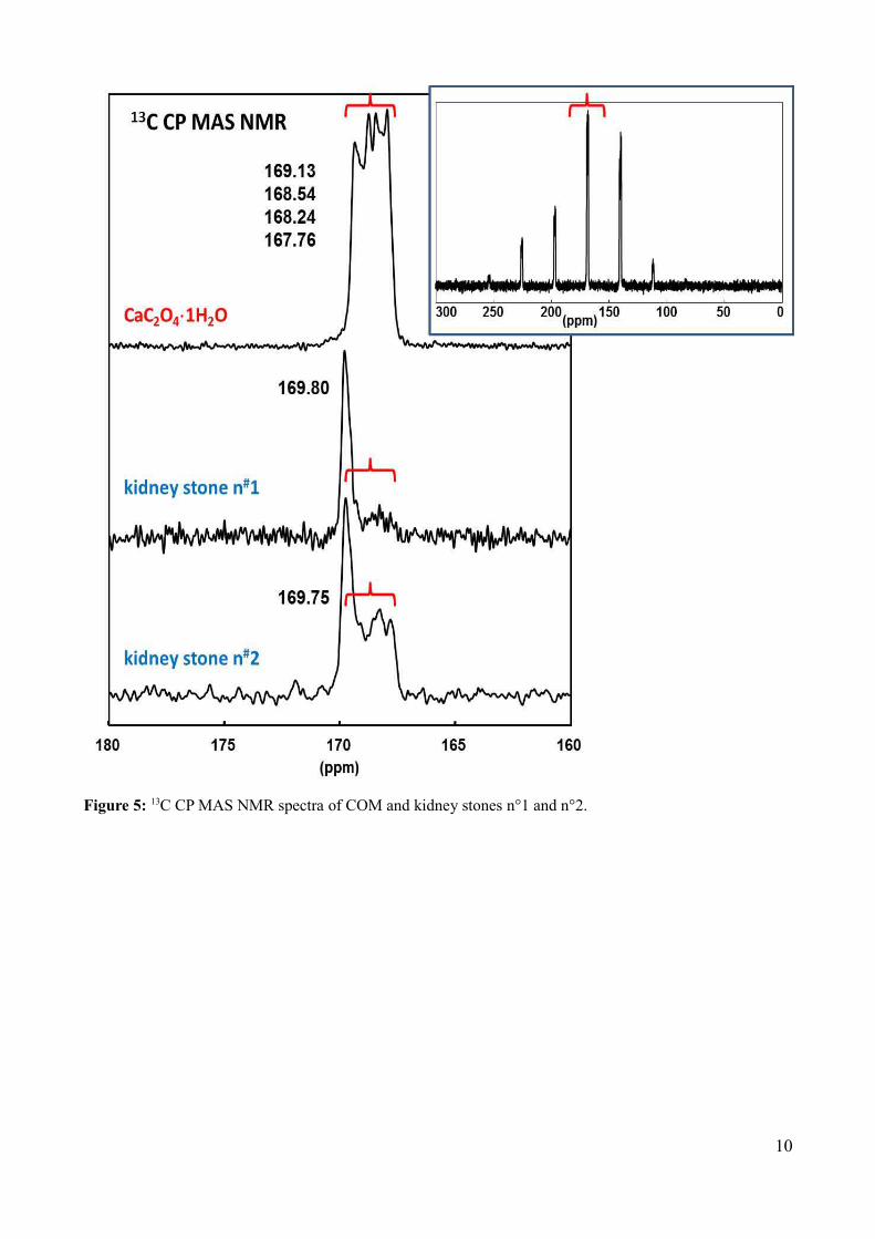
Figure 5: 13C CP MAS NMR spectra of COM and kidney stones n°1 and n°2.
10

Figure 6: 43Ca MAS NMR spectra of COM, COD, COT and kidney stones n°1 and n°2.
11

Conclusion
The work done during my internship highlights the limited stability of COD (and also COT) calcium oxalate
phases. In fact, the experiments on the stones ran this week didn't give us exactly the same results as those
obtained in december, 2013. A possible assumption is that part of the sample evolved chemically after a
certain amount of time.
Moreover, the synthesis we performed presents an unexpected amount of monohydrate form (COM) and we
made the hypothesis that some of the COD transformed into COM during filtering. This can be considered
as another proof of COD's limited stability. The following of the project will to try to explain the impact of
different organic molecules (such as catechine) on the crystallization process and on the morphology of the
kidney stones.
12
About of the author
I am currently in L1 at « Licence Frontières Du Vivant », interdisciplinary bachelor. The last diploma I obtained is
the baccalauréat (2013) when I was in high school at Saint-Michel de Picpus (Saint-Mandé).

Supplementary materials
13

14

13C NMR of calcium oxalate hydrates: COM (1H2O), COD (2H2O) and COT (3H2O).
15

References
[1] - M. Daudon, Annales d'urologie 2005, 39, 209-231. Epidémiologie actuelle de la lithiase rénale enFrance.
[2] - Z. Chen, C. Wang, H. Zhou, L. Sang, and X. Li, Cryst. Eng. Comm. 2010, 12, 845-852. Modulation ofcalcium oxalate crystallization by commonly consumed green tea.
[3] - D. Bazin, M. Daudon, C. Combes, and C. Rey, Chem. Rev. 2012, 112, 5092-5120. Characterization andsome physicochemical aspects of pathological microcalcifications.
[4] - M. Daudon and D. Bazin, Urolithiasis 2012, 683-707. Application of physical methods to kidney stones
and Randall's plaque characterization.
[5] – J-M. Ouyang, L. Duan, B. Tieke, Langmuir 2003, 19, 8980-8985. Effects of carboxilic acids on the
crystal growth of calcium oxalate nanoparticles in lecithin-water liposome systems.
[6] – M. Bak et al., J. Urology 2000, 164, 856-863.
16