Embed Size (px)
Citation preview
Editorial: Calculated Risks 2Sillas Duarte Jr, DDS, MS, PhD
Fluorescence—Mimicking Nature for Ultimate Esthetics in 7 Implant DentistryIñaki Gamborena, DMD, MSD, FID/Markus Blatz, DMD, PhD
The Platinum Foil Technique: History, Indication, Fabrication, and Fit 25Sascha Hein, MDT/Willi Geller, MDT
BIOMATERIALS UPDATE 40Adhesive Resin Cements for Bonding Esthetic Restorations: A ReviewSillas Duarte Jr, DDS, MS, PhD/Neimar Sartori, DDS, MS/Avishai Sadan, DMD/ Jin-Ho Phark, DDS, Dr Med Dent
Optimal Color Control and Layering Protocol for All-Ceramic 67 Restorations in the Esthetic Zone. Part 1: Zirconia Crown and Leucite-Reinforced Feldspathic VeneerTyler P. Lasseigne, DDS, CDT/Luis Infante, DDS/HiroTokutomi, RDT/ Arturo J. Mendez, DDS
Excellence in Dental Esthetics: A Multidisciplinary Challenge 83Luc Rutten, MDT/Patrick Rutten, MDT/Iñaki Gamborena, DMD, MSD, FID
Concepts for an Ultraconservative Approach to Indirect Anterior 103 RestorationsOswaldo Scopin de Andrade, DDS, MS, PhD/Sidney Kina, DDS, MS/ Ronaldo Hirata, DDS, MS, PhD
STATE OF THE ART 120Artistry and CAD/CAM Technology: Predictable Outcomes in Contemporary Implant Dentistry Domenico Cascione, MDT, BS/Anas Aloum, BDS/Kazunari Takanashi, RDT/ Jina Ha, CDT/Mamaly Reshad, DDS, MSc
2011
EDITOR-IN-CHIEFSillas Duarte Jr, DDS, MS, PhDAssociate ProfessorDivision of Restorative SciencesOstrow School of Dentistry University of Southern CaliforniaLos Angeles, California
ASSOCIATE EDITORJin-Ho Phark, DDS, Dr Med DentDivision of Restorative SciencesOstrow School of Dentistry University of Southern CaliforniaLos Angeles, California
EDITORIAL REVIEW BOARDPinhas Adar, CDT, MDT Atlanta, GeorgiaNaoki Aiba, CDT Monterey, CaliforniaAmir Avishai, PhD Cleveland, OhioMike Bellerino, CDT Metairie, LouisianaMarkus B. Blatz, DMD, PhD Philadelphia, PennsylvaniaKaren Bruggers, DDS, MS Cary, North CarolinaGerard J. Chiche, DDS Augusta, GeorgiaShiro Kamachi, DMD Boston, MassachusettsEdward A. McLaren, DDS Los Angeles, CaliforniaServando Ramos, DDS US ArmyAvishai Sadan, DMD Los Angeles, CaliforniaThomas J. Salinas, DDS Rochester, MinnesotaTomizaku Tada, CDT Pasadena, CaliforniaFabiana Varjão, DDS, MS, PhD Los Angeles, CaliforniaAki Yoshida, CDT Weston, Massachusetts
The Essence of the Internal Staining Technique: 133 Application and FabricationNaoto Yuasa, RDT
Experimental Birefringence Photography in Dentistry: 151 Unlocking Infinite Creative PossibilitiesSascha Hein, MDT/Joshua Polansky, BA, MDC
The Importance of Gingival Tissue in 163 Anterior Esthetic Rehabilitations Giovani Gambogi Parreira, MDT/Leandro Medeiros dos Santos, DDS, MS/ Wilson Batista Mendes, DDS, MS, PhD
Opacity, Part 2: Value Control of Adjacent Porcelain Laminate Veneers 175and CrownsHiroki Goto, RDT
Reproduction of Natural Vivid Appearance in Porcelain 185 Crown Restorations: Case Presentations and Fabrication TechniqueNaoto Yuasa, RDT
An Ultraconservative Approach to Porcelain Veneers 193 in the 21st CenturyMamaly Reshad, DDS, MSc/Willi Geller, MDT/Domenico Cascione, MDT
Esthetic Rehabilitation of Tetracycline-Stained and Worn Teeth 200 with Porcelain Laminate VeneersKazunori Matsumoto, RDT
The Concept Crown: An Innovative Treatment Option 207 Using Current TechnologiesPinhas Adar, MDT/Adam Mieleszko, CDT/Stephen Chu, DMD, MSD, CDT
Volume 34
PUBLISHERH.W. Haase
EXECUTIVE VICE-PRESIDENTWilliam G. Hartman
JOURNAL DIRECTORLori A. Bateman
PRODUCTION EDITORPatrick Penney
ADVERTISING SALESWilliam G. Hartman
ADVERTISING/EDITORIAL/ SUBSCRIPTION OFFICEQuintessence Publishing Co, Inc4350 Chandler DriveHanover Park, Illinois 60133Phone: (630) 736-3600Toll-free: (800) 621-0387Fax: (630) 736-3633E-mail: [email protected]
Website: http://www.quintpub.comQDT is published once a year by Quintessence Publishing Co, Inc, 4350 Chandler Drive, Hanover Park, Illinois, 60133. Price per copy: $85.
MANUSCRIPT SUBMISSIONQDT publishes original articles covering dental laboratory techniques and meth-ods. For submission information, contact Lori Bateman ([email protected]).
Copyright © 2011 by Quintessence Pub-lishing Co, Inc. All rights reserved. No part of this publication may be reproduced or transmitted in any form or by any means, electronic or mechanical, including pho-tocopying, recording, or any information and retrieval system, without permission in writing from the publisher. The publisher assumes no responsibility for unsolicited manuscripts. All opinions are those of the authors. Reprints of articles published in QDT can be obtained from the authors.
Permission to photocopy items solely for internal or personal use and for the internal or personal use of specific clients is granted by Quintessence Publishing Co, Inc, for libraries and other users registered with the Copyright Clearance Center (CCC) Transac-tion Reporting Service, provided that the base fee of $5 per article plus $.10 per page is paid directly to the CCC, 222 Rosewood Drive, Danvers, MA 01923 (www.copyright.com). Identify this publication by including with your payment the fee code: 0-86715-375-0/11 $5 + $.10.
Printed in CanadaISSN 0896-6532 / ISBN 978-0-86715-514-3Cover photograph by Sascha Hein
QDT 2011 2
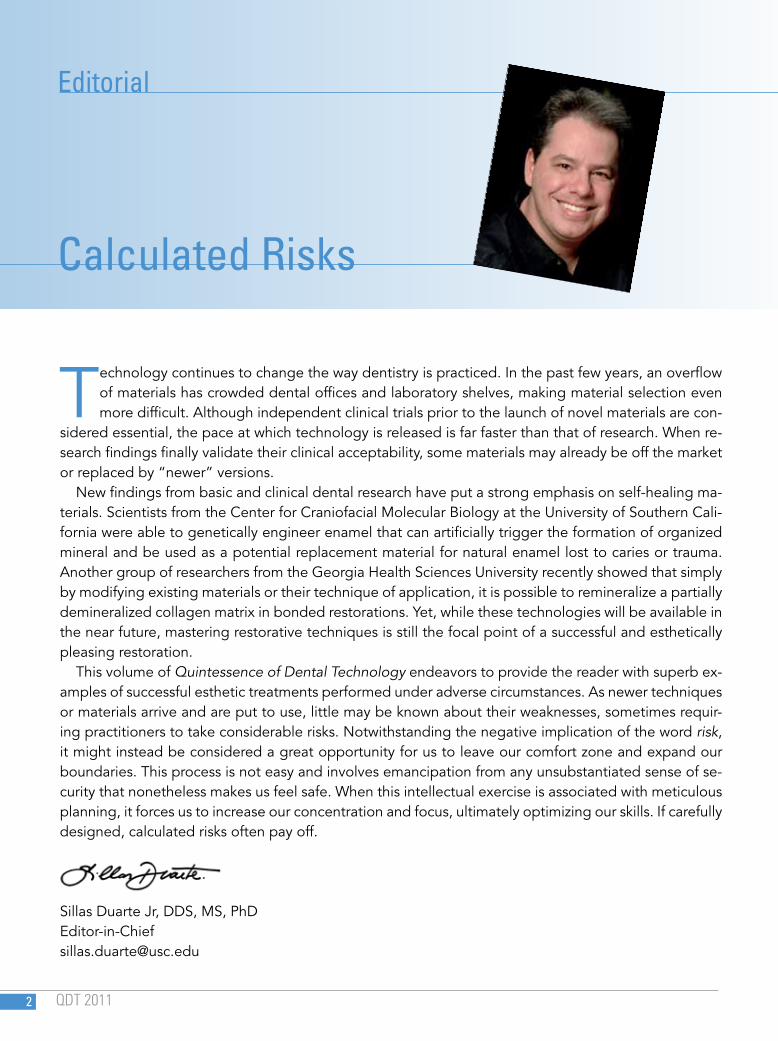
Technology continues to change the way dentistry is practiced. In the past few years, an overflow of materials has crowded dental offices and laboratory shelves, making material selection even more difficult. Although independent clinical trials prior to the launch of novel materials are con-
sidered essential, the pace at which technology is released is far faster than that of research. When re-search findings finally validate their clinical acceptability, some materials may already be off the market or replaced by “newer” versions.
New findings from basic and clinical dental research have put a strong emphasis on self-healing ma-terials. Scientists from the Center for Craniofacial Molecular Biology at the University of Southern Cali-fornia were able to genetically engineer enamel that can artificially trigger the formation of organized mineral and be used as a potential replacement material for natural enamel lost to caries or trauma. Another group of researchers from the Georgia Health Sciences University recently showed that simply by modifying existing materials or their technique of application, it is possible to remineralize a partially demineralized collagen matrix in bonded restorations. Yet, while these technologies will be available in the near future, mastering restorative techniques is still the focal point of a successful and esthetically pleasing restoration.
This volume of Quintessence of Dental Technology endeavors to provide the reader with superb ex-amples of successful esthetic treatments performed under adverse circumstances. As newer techniques or materials arrive and are put to use, little may be known about their weaknesses, sometimes requir-ing practitioners to take considerable risks. Notwithstanding the negative implication of the word risk, it might instead be considered a great opportunity for us to leave our comfort zone and expand our boundaries. This process is not easy and involves emancipation from any unsubstantiated sense of se-curity that nonetheless makes us feel safe. When this intellectual exercise is associated with meticulous planning, it forces us to increase our concentration and focus, ultimately optimizing our skills. If carefully designed, calculated risks often pay off.
Sillas Duarte Jr, DDS, MS, [email protected]
Editorial
Calculated Risks
HEIN/GELLER
QDT 2011 36
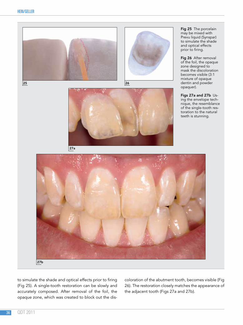
to simulate the shade and optical effects prior to firing (Fig 25). A single-tooth restoration can be slowly and accurately composed. After removal of the foil, the opaque zone, which was created to block out the dis-
coloration of the abutment tooth, becomes visible (Fig 26). The restoration closely matches the appearance of the adjacent tooth (Figs 27a and 27b).
25 26
27a
27b
Fig 25 The porcelain may be mixed with Prevu liquid (Synspar) to simulate the shade and optical effects prior to firing.
Fig 26 After removal of the foil, the opaque zone designed to mask the discoloration becomes visible (3:1 mixture of opaque dentin and powder opaquer).
Figs 27a and 27b Us-ing the envelope tech-nique, the resemblance of the single-tooth res-toration to the natural teeth is stunning.
The Platinum Foil Technique: History, Indication, Fabrication, and Fit
QDT 2011 37
Full Crowns
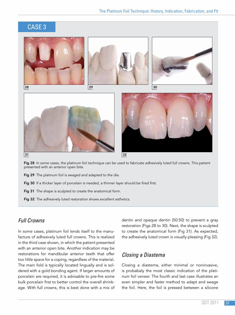
In some cases, platinum foil lends itself to the manu-facture of adhesively luted full crowns. This is realized in the third case shown, in which the patient presented with an anterior open bite. Another indication may be restorations for mandibular anterior teeth that offer too little space for a coping, regardless of the material. The main fold is typically located lingually and is sol-dered with a gold bonding agent. If larger amounts of porcelain are required, it is advisable to pre-fire some bulk porcelain first to better control the overall shrink-age. With full crowns, this is best done with a mix of
dentin and opaque dentin (50:50) to prevent a gray restoration (Figs 28 to 30). Next, the shape is sculpted to create the anatomical form (Fig 31). As expected, the adhesively luted crown is visually pleasing (Fig 32).
Closing a Diastema
Closing a diastema, either minimal or noninvasive, is probabaly the most classic indication of the plati-num foil veneer. The fourth and last case illustrates an even simpler and faster method to adapt and swage the foil. Here, the foil is pressed between a silicone
Fig 28 In some cases, the platinum foil technique can be used to fabricate adhesively luted full crowns. This patient presented with an anterior open bite.
Fig 29 The platinum foil is swaged and adapted to the die.
Fig 30 If a thicker layer of porcelain is needed, a thinner layer should be fired first.
Fig 31 The shape is sculpted to create the anatomical form.
Fig 32 The adhesively luted restoration shows excellent esthetics.
28 29 30
CASE 3
31 32
DUARTE ET AL
QDT 2011 62
19
20a
20b
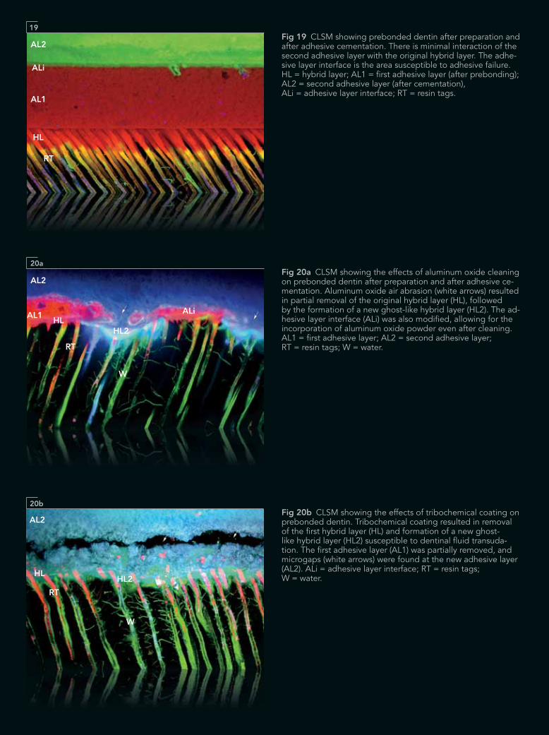
Fig 19 CLSM showing prebonded dentin after preparation and after adhesive cementation. There is minimal interaction of the second adhesive layer with the original hybrid layer. The adhe-sive layer interface is the area susceptible to adhesive failure. HL = hybrid layer; AL1 = first adhesive layer (after prebonding); AL2 = second adhesive layer (after cementation), ALi = adhesive layer interface; RT = resin tags.
Fig 20a CLSM showing the effects of aluminum oxide cleaning on prebonded dentin after preparation and after adhesive ce-mentation. Aluminum oxide air abrasion (white arrows) resulted in partial removal of the original hybrid layer (HL), followed by the formation of a new ghost-like hybrid layer (HL2). The ad-hesive layer interface (ALi) was also modified, allowing for the incorporation of aluminum oxide powder even after cleaning. AL1 = first adhesive layer; AL2 = second adhesive layer; RT = resin tags; W = water.
Fig 20b CLSM showing the effects of tribochemical coating on prebonded dentin. Tribochemical coating resulted in removal of the first hybrid layer (HL) and formation of a new ghost-like hybrid layer (HL2) susceptible to dentinal fluid transuda-tion. The first adhesive layer (AL1) was partially removed, and microgaps (white arrows) were found at the new adhesive layer (AL2). ALi = adhesive layer interface; RT = resin tags; W = water.
AL2
AL1
HL
RT
ALi
AL2
AL1HL
HL2
W
W
RT
ALi
AL2
HL2HL
RT
Adhesive Resin Cements for Bonding Esthetic Restorations: A Review
QDT 2011 63
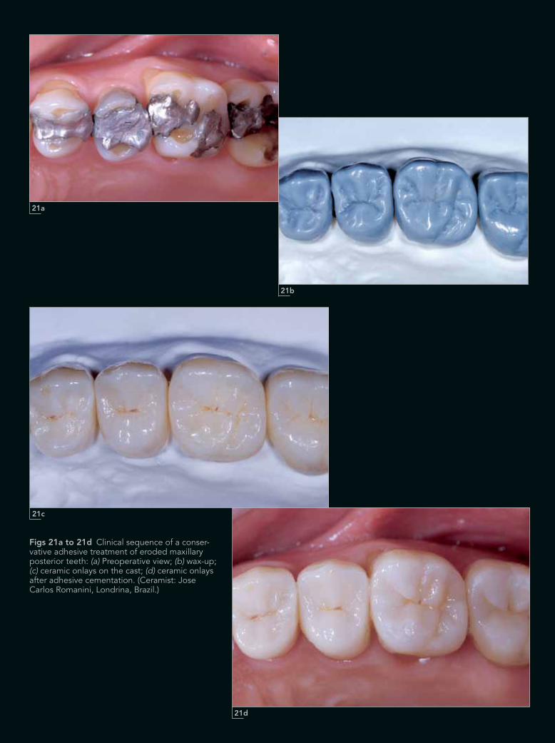
Figs 21a to 21d Clinical sequence of a conser-vative adhesive treatment of eroded maxillary posterior teeth: (a) Preoperative view; (b) wax-up; (c) ceramic onlays on the cast; (d) ceramic onlays after adhesive cementation. (Ceramist: Jose Carlos Romanini, Londrina, Brazil.)
21a
21c
21b
21d
LASSEIGNE ET AL
QDT 2011 76
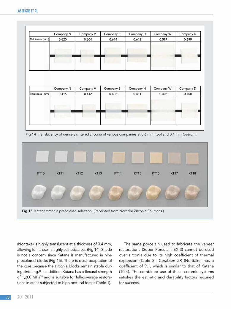
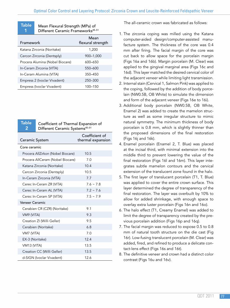
(Noritake) is highly translucent at a thickness of 0.4 mm, allowing for its use in highly esthetic areas (Fig 14). Shade is not a concern since Katana is manufactured in nine precolored blocks (Fig 15). There is close adaptation of the core because the zirconia blocks remain stable dur-ing sintering.55 In addition, Katana has a flexural strength of 1,200 MPa54 and is suitable for full-coverage restora-tions in areas subjected to high occlusal forces (Table 1).
The same porcelain used to fabricate the veneer restorations (Super Porcelain EX-3) cannot be used over zirconia due to its high coefficient of thermal expansion (Table 2). Cerabien ZR (Noritake) has a coefficient of 9.1, which is similar to that of Katana (10.4). The combined use of these ceramic systems satisfies the esthetic and durability factors required for success.
Fig 15 Katana zirconia precolored selection. (Reprinted from Noritake Zirconia Solutions.)
Fig 14 Translucency of densely sintered zirconia of various companies at 0.6 mm (top) and 0.4 mm (bottom).
KT10 KT11 KT12 KT13 KT14 KT15 KT16 KT17 KT18
Company N
Company N
0.620
0.415
0.604
0.412
0.614
0.408
0.612
0.411
0.597
0.405
0.599
0.408
Company V
Company V
Company 3
Company 3
Company H
Company H
Company W
Company W
Company D
Company D
Thickness (mm)
Thickness (mm)
Optimal Color Control and Layering Protocol: Zirconia Crown and Leucite-Reinforced Feldspathic Veneer
QDT 2011 77
The all-ceramic crown was fabricated as follows:
1. The zirconia coping was milled using the Katana computer-aided design/computer-assisted manu-facture system. The thickness of the core was 0.4 mm after firing. The facial margin of the core was cut back to allow space for the porcelain margin (Figs 16a and 16b). Margin porcelain (M. Clear) was applied to the gingival marginal area (Figs 16c and 16d). This layer matched the desired cervical color of the adjacent veneer while limiting light transmission.
2. Internal stain (Cervical 1, Salmon Pink) was applied to the coping, followed by the addition of body porce-lain (NW0.5B, OB White) to simulate the dimension and form of the adjacent veneer (Figs 16e to 16i).
3. Additional body porcelain (NW0.5B, OB White, Enamel 2) was added to create the mamelon struc-ture as well as some irregular structure to mimic natural symmetry. The minimum thickness of body porcelain is 0.8 mm, which is slightly thinner than the proposed dimensions of the final restoration (Figs 16j and 16k).
4. Enamel porcelain (Enamel 2, T. Blue) was placed at the incisal third, with minimal extension into the middle third to prevent lowering the value of the final restoration (Figs 16l and 16m). This layer inte-grates subtle mamelon contours and the cervical extension of the translucent zone found in the halo.
5. The first layer of translucent porcelain (T1, T. Blue) was applied to cover the entire crown surface. This layer determined the degree of transparency of the final restoration. The layer was overbuilt by 10% to allow for added shrinkage, with enough space to overlay extra luster porcelain (Figs 16n and 16o).
6. The halo effect (T1, Creamy Enamel) was added to limit the degree of transparency created by the pre-vious porcelain addition (Figs 16p and 16q).
7. The facial margin was reduced to expose 0.5 to 0.8 mm of natural tooth structure on the die cast (Fig 16r). Low-fusing translucent porcelain (M. Clear) was added, fired, and refined to produce a delicate con-tact-lens effect (Figs 16s and 16t).
8. The definitive veneer and crown had a distinct color contrast (Figs 16u and 16v).
Table
1
Mean Flexural Strength (MPa) of Different Ceramic Frameworks45–51
Mean Framework flexural strength
Katana Zirconia (Noritake) 1,200
Cercon Zirconia (Dentsply) 900–1,000
Procera Alumina (Nobel Biocare) 600–650
In-Ceram Zirconia (VITA) 550–600
In-Ceram Alumina (VITA) 350–450
Empress 2 (Ivoclar Vivadent) 250–300
Empress (Ivoclar Vivadent) 100–150
Table
2
Coefficient of Thermal Expansion of Different Ceramic Systems45–51
Coefficient of Ceramic System thermal expansion
Core ceramic
Procera AllZirkon (Nobel Biocare) 10.5
Procera AllCeram (Nobel Biocare) 7.0
Katana Zirconia (Noritake) 10.4
Cercon Zirconia (Dentsply) 10.5
In-Ceram Zirconia (VITA) 7.7
Cerec In-Ceram ZR (VITA) 7.6 ~ 7.8
Cerec In-Ceram AL (VITA) 7.2 ~ 7.6
Cerec In-Ceram SP (VITA) 7.5 ~ 7.9
Veneer Ceramic
Cerabien CR (CZR) (Noritake) 9.1
VM9 (VITA) 9.3
Creation ZI (Willi Geller) 9.5
Cerabien (Noritake) 6.8
VM7 (VITA) 7.0
EX-3 (Noritake) 12.4
VM13 (VITA) 13.5
Creation CC (Willi Geller) 13.5
d-SIGN (Ivoclar Vivadent) 12.6

103 QDT 2011
The current focus in restorative dentistry is hard tissue preservation. This concept dictates that treatment for an indirect procedure must be as
conservative as possible. In the anterior dentition, it is especially important to preserve the tooth structure regardless of the individual treatment plan.
Composite resin direct restorations are often used for minimally invasive restorations (eg, Class 3 and 4 restorations). With the development of new techniques
for esthetic smile design, composite resin is also used for add-on procedures in the anterior dentition.1,2 How-ever, there is still a lack of scientific evidence regarding the long-term clinical performance of composite resin restorations in the anterior region. When comparing composite resin and ceramic restorations, the latter has better scientific documentation in terms of esthet-ics, color stability, and wear resistance.3–5 This suggests that in cases of failure, the original restoration should be replaced with a ceramic veneer or crown. Before a tooth is prepared for a crown, however, a critical evalu-ation should be carried out to determine which con-servative treatment approach to select.6,7
Among the many conservative techniques available for anterior restorations, the use of laminate veneers has been the most investigated.5,8 The advantage of ceramic laminate veneers compared with traditional crown preparation begins with the amount of tooth structure that is preserved, allowing the restoration to
Concepts for an Ultraconservative Approach to Indirect Anterior Restorations
Concepts for an Ultraconservative Approach to Indirect Anterior Restorations
Oswaldo Scopin de Andrade, DDS, MS, PhD1
Sidney Kina, DDS, MS2
Ronaldo Hirata, DDS, MS, PhD3
Oswaldo Scopin de Andrade, DDS, MS, PhD1
Sidney Kina, DDS, MS2
Ronaldo Hirata, DDS, MS, PhD3
1 Director, Advanced Program in Implant and Esthetic Dentistry, Senac University, São Paulo, Brazil.
2 Director, Advanced Program in Prosthodontics, São Leopoldo Mandic School of Dentistry, São Paulo, Brazil.
3 Director, Advanced Program in Restorative Dentistry, CETAO Institute, São Paulo, Brazil.
Correspondence to: Dr Oswaldo Scopin de Andrade, Rua Barão de Piracicamirim 889 Apt. 61, Piracicaba, São Paulo, Brazil 13.416.005. Email: [email protected]
HEIN/POLANSKY
QDT 2011 156
18
16
17 19
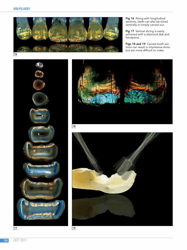
Fig 16 Along with longitudinal sections, teeth can also be sliced vertically or simply carved out.
Fig 17 Vertical slicing is easily achieved with a diamond disk and handpiece.
Figs 18 and 19 Carved tooth sec-tions can result in impressive shots but are more difficult to make.
Experimental Birefringence Photography in Dentistry
QDT 2011 157
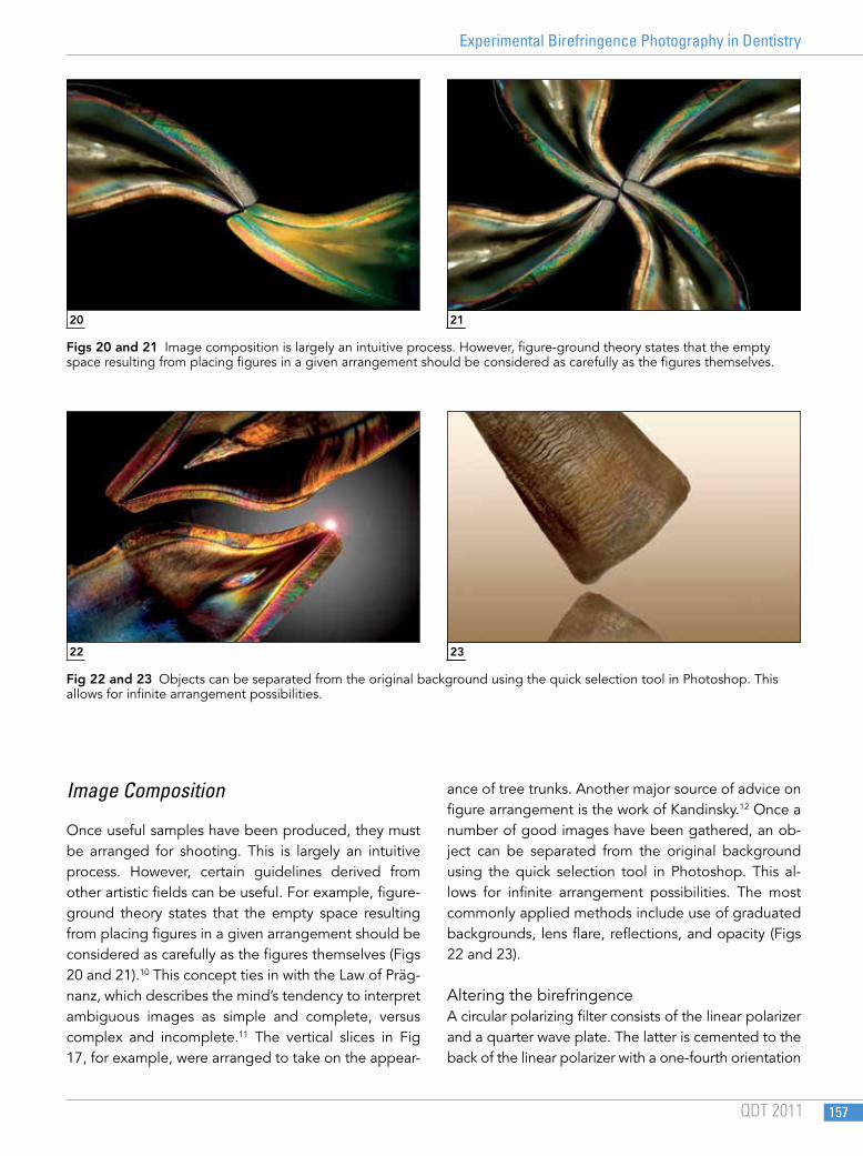
Image Composition
Once useful samples have been produced, they must be arranged for shooting. This is largely an intuitive process. However, certain guidelines derived from other artistic fields can be useful. For example, figure-ground theory states that the empty space resulting from placing figures in a given arrangement should be considered as carefully as the figures themselves (Figs 20 and 21).10 This concept ties in with the Law of Präg-nanz, which describes the mind’s tendency to interpret ambiguous images as simple and complete, versus complex and incomplete.11 The vertical slices in Fig 17, for example, were arranged to take on the appear-
ance of tree trunks. Another major source of advice on figure arrangement is the work of Kandinsky.12 Once a number of good images have been gathered, an ob-ject can be separated from the original background using the quick selection tool in Photoshop. This al-lows for infinite arrangement possibilities. The most commonly applied methods include use of graduated backgrounds, lens flare, reflections, and opacity (Figs 22 and 23).
Altering the birefringenceA circular polarizing filter consists of the linear polarizer and a quarter wave plate. The latter is cemented to the back of the linear polarizer with a one-fourth orientation
20
22
21
23
Figs 20 and 21 Image composition is largely an intuitive process. However, figure-ground theory states that the empty space resulting from placing figures in a given arrangement should be considered as carefully as the figures themselves.
Fig 22 and 23 Objects can be separated from the original background using the quick selection tool in Photoshop. This allows for infinite arrangement possibilities.