Embed Size (px)
Citation preview
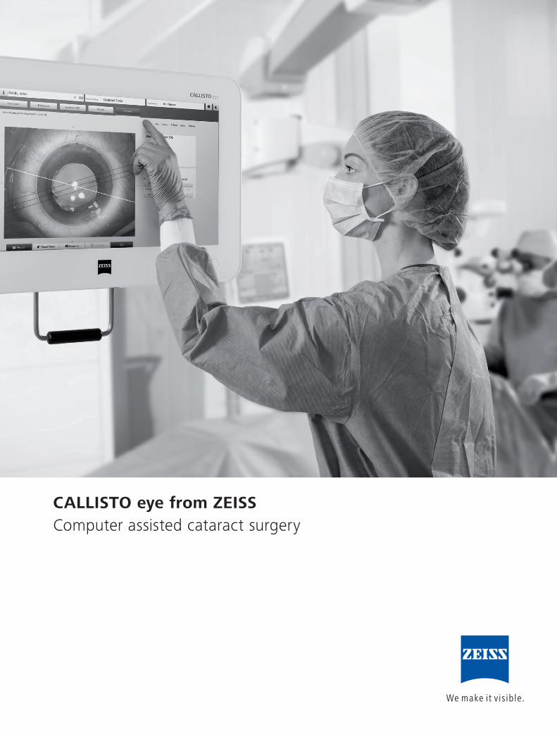
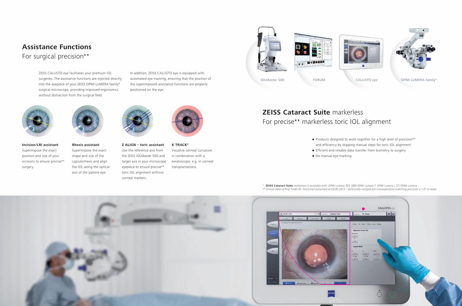
CALLISTO eye from ZEISSComputer assisted cataract surgery
2 3
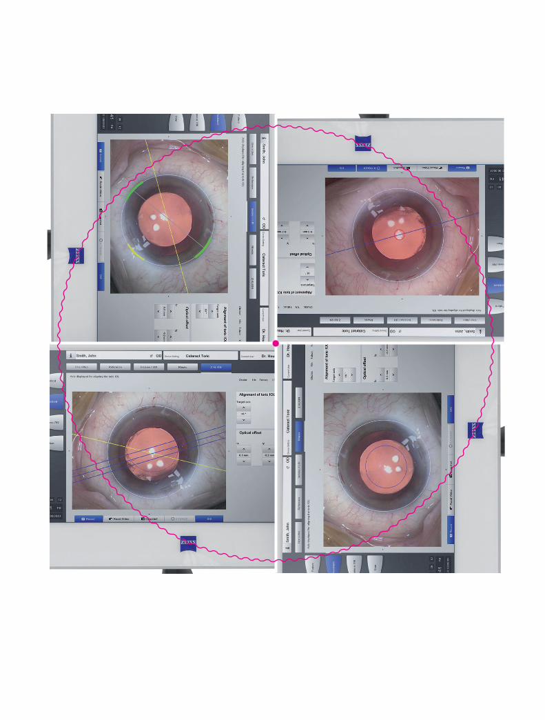
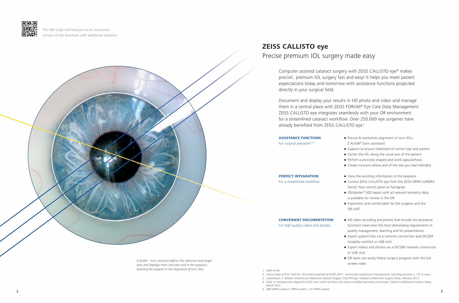
Z ALIGN – toric assistant defines the reference and target axes and displays them onscreen and in the eyepiece, assisting the surgeon in the alignment of toric IOLs.
This QR-Code will lead you to an interactive
version of this brochure with additional features.
ZEISS CALLISTO eyePrecise premium IOL surgery made easy
Computer assisted cataract surgery with ZEISS CALLISTO eye® makes precise1, premium IOL surgery fast and easy! It helps you meet patient expectations today and tomorrow with assistance functions projected directly in your surgical field.
Document and display your results in HD photo and video and manage them in a central place with ZEISS FORUM® Eye Care Data Management. ZEISS CALLISTO eye integrates seamlessly with your OR environment for a streamlined cataract workflow. Over 250.000 eye surgeries have already benefited from ZEISS CALLISTO eye.1
ASSISTAnCE funCTIOnS
For surgical precision2,3,4
PErfECT InTEGrATIOn
For a streamlined workflow
COnvEnIEnT dOCumEnTATIOn
For high quality videos and photos
1. Data on file
2. Clinical data of Prof. Findl / Dr. Hirnschall presented at ESCRS 2013 – technically verifyed pre-/ intraoperative matching precision ± 1.0° in mean3. Lackerbauer, C. Modern Solutions for Refractive Cataract Surgery: CALLISTO eye. Cataract & Refractive Surgery Today. February 2013.4. Findl, O. Intraoperative alignment of IOL and control of rhexis size using a modified operating microscope. Cataract & Refractive Surgery Today.
March 2012.5. S88 / OPMI Lumera T, OPMI Lumera i, S7 / OPMI Lumera
• Precise & markerless alignment of toric IOLs:
Z ALIGN® (toric assistant)
• Support to ensure treatment of correct eye and patient
• Center the IOL along the visual axis of the patient
• Perfom a precisely shaped and sized capsulorhexis
• Create incisions where and of the size you had intended
• View the assisting information in the eyepiece
• Control ZEISS CALLISTO eye from the ZEISS OPMI LUMERA
family5 foot control panel or handgrips
• IOLMaster® 500 report with all relevant biometry data
is available for review in the OR
• Ergonomic and comfortable for the surgeon and the
OR staff
• HD video recording and photos that include the assistance
functions meet even the most demanding requirements in
quality management, teaching and for presentations
• Import patient lists via a network connection and DICOM
modality worklist or USB stick
• Export videos and photos via a DICOM network connection
or USB stick
• OR team can easily follow surgery progress with the full
screen video
4 5
ZEISS Cataract Suite markerlessFor precise** markerless toric IOL alignment
• Products designed to work together for a high level of precision**
and efficiency by skipping manual steps for toric IOL alignment
• Efficient and reliable data transfer from biometry to surgery
• No manual eye marking
OPMI LUMERA family* FORUM CALLISTO eyeIOLMaster 500
* ZEISS Cataract Suite markerless is available with: OPMI Lumera 700, S88 / OPMI Lumera T, OPMI Lumera i, S7 / OPMI Lumera** Clinical data of Prof. Findl / Dr. Hirnschall presented at ESCRS 2013 – technically verifyed pre-/ intraoperative matching precision ± 1.0° in mean
Assistance functionsFor surgical precision**
Incision / LrI assistant
Superimpose the exact
position and size of your
incisions to ensure precise**
surgery.
rhexis assistant
Superimpose the exact
shape and size of the
capsulorhexis and align
the IOL along the optical
axis of the patient eye.
K TrACK®
Visualize corneal curvature
in combination with a
keratoscope, e.g. in corneal
transplantations.
ZEISS CALLISTO eye facilitates your premium IOL
surgeries. The assistance functions are injected directly
into the eyepiece of your ZEISS OPMI LUMERA family*
surgical microscope, providing improved ergonomics
without distraction from the surgical field.
In addition, ZEISS CALLISTO eye is equipped with
automated eye tracking, ensuring that the position of
the superimposed assistance functions are properly
positioned on the eye.
Z ALIGn – toric assistant
Use the reference axis from
the ZEISS IOLMaster 500 and
target axis in your microscope
eyepiece to ensure precise**
toric IOL alignment without
corneal markers.
6
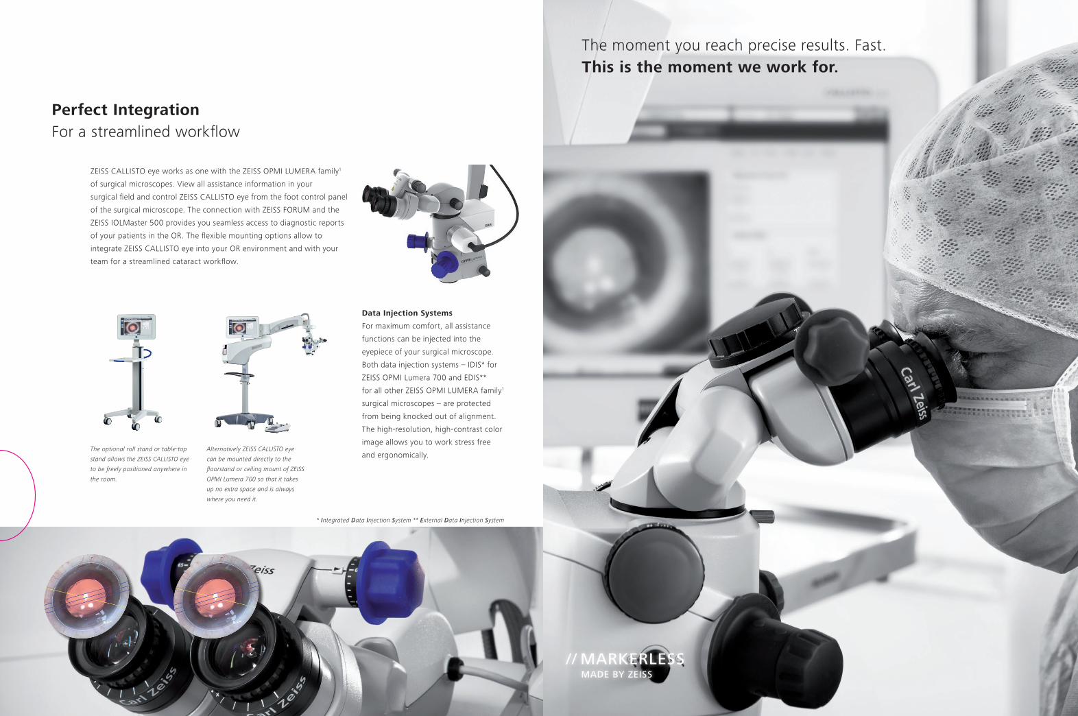
// MARKERLESS MAdE By ZEISS
The moment you reach precise results. Fast. This is the moment we work for.
Perfect IntegrationFor a streamlined workflow
Alternatively ZEISS CALLISTO eye
can be mounted directly to the
floorstand or ceiling mount of ZEISS
OPMI Lumera 700 so that it takes
up no extra space and is always
where you need it.
The optional roll stand or table-top
stand allows the ZEISS CALLISTO eye
to be freely positioned anywhere in
the room.
data Injection Systems
For maximum comfort, all assistance
functions can be injected into the
eyepiece of your surgical microscope.
Both data injection systems – IDIS* for
ZEISS OPMI Lumera 700 and EDIS**
for all other ZEISS OPMI LUMERA family1
surgical microscopes – are protected
from being knocked out of alignment.
The high-resolution, high-contrast color
image allows you to work stress free
and ergonomically.
ZEISS CALLISTO eye works as one with the ZEISS OPMI LUMERA family1
of surgical microscopes. View all assistance information in your
surgical field and control ZEISS CALLISTO eye from the foot control panel
of the surgical microscope. The connection with ZEISS FORUM and the
ZEISS IOLMaster 500 provides you seamless access to diagnostic reports
of your patients in the OR. The flexible mounting options allow to
integrate ZEISS CALLISTO eye into your OR environment and with your
team for a streamlined cataract workflow.
* Integrated Data Injection System ** External Data Injection System
8 9
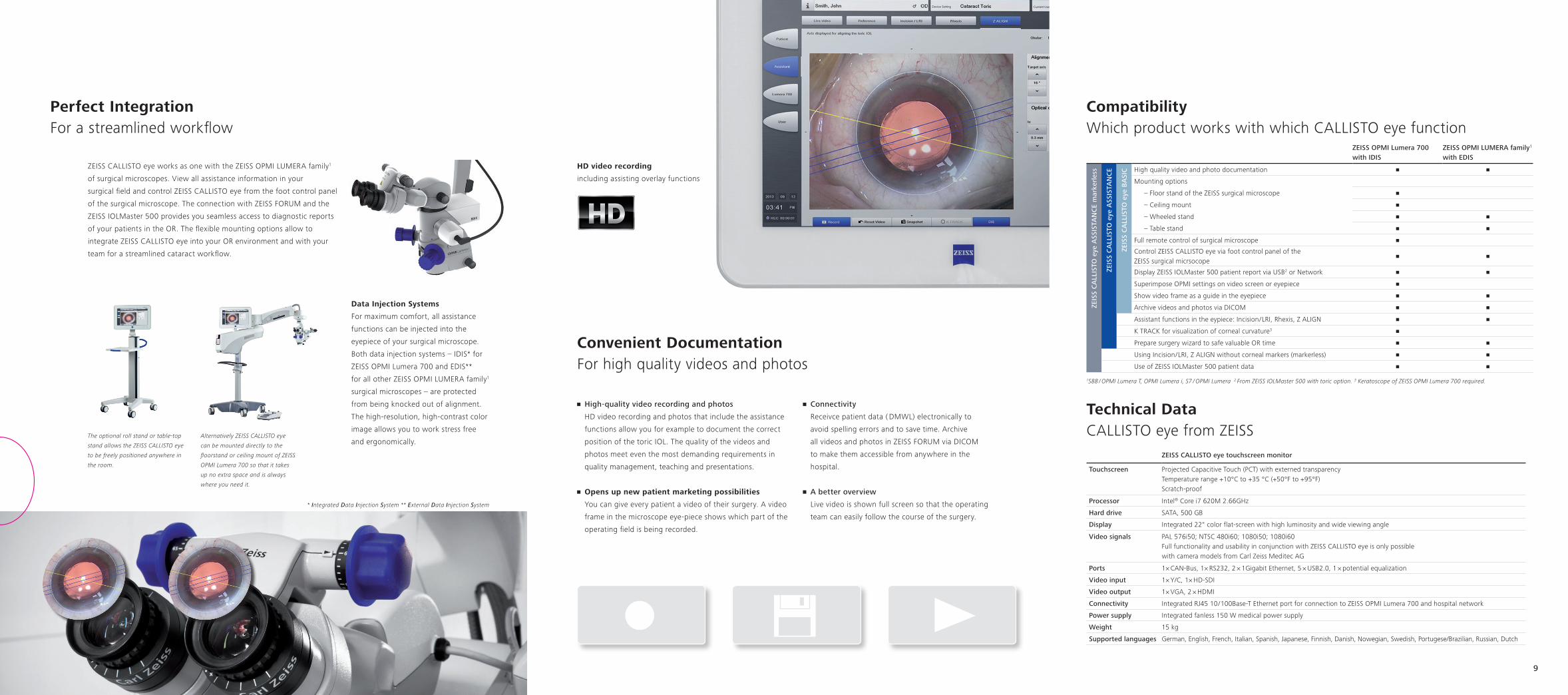
Convenient documentationFor high quality videos and photos
Hd video recording
including assisting overlay functions
• High-quality video recording and photos
HD video recording and photos that include the assistance
functions allow you for example to document the correct
position of the toric IOL. The quality of the videos and
photos meet even the most demanding requirements in
quality management, teaching and presentations.
• Opens up new patient marketing possibilities
You can give every patient a video of their surgery. A video
frame in the microscope eye-piece shows which part of the
operating field is being recorded.
• Connectivity
Receivce patient data ( DMWL) electronically to
avoid spelling errors and to save time. Archive
all videos and photos in ZEISS FORUM via DICOM
to make them accessible from anywhere in the
hospital.
• A better overview
Live video is shown full screen so that the operating
team can easily follow the course of the surgery.
ZEISS CALLISTO eye touchscreen monitor
Touchscreen Projected Capacitive Touch (PCT) with externed transparencyTemperature range +10°C to +35 °C (+50°F to +95°F)Scratch-proof
Processor Intel® Core i7 620M 2.66GHz
Hard drive SATA, 500 GB
display Integrated 22" color flat-screen with high luminosity and wide viewing angle
Video signals PAL 576i50; NTSC 480i60; 1080i50; 1080i60Full functionality and usability in conjunction with ZEISS CALLISTO eye is only possible with camera models from Carl Zeiss Meditec AG
Ports 1× CAN-Bus, 1× RS232, 2 × 1Gigabit Ethernet, 5 × USB2.0, 1 × potential equalization
Video input 1× Y/C, 1× HD-SDI
Video output 1× VGA, 2 × HDMI
Connectivity Integrated RJ45 10 / 100Base-T Ethernet port for connection to ZEISS OPMI Lumera 700 and hospital network
Power supply Integrated fanless 150 W medical power supply
Weight 15 kg
Supported languages German, English, French, Italian, Spanish, Japanese, Finnish, Danish, Nowegian, Swedish, Portugese/Brazilian, Russian, Dutch
CompatibilityWhich product works with which CALLISTO eye function
ZEISS OPMI Lumera 700with IdIS
ZEISS OPMI LUMERA family1 with EdIS
ZEIS
S C
ALL
ISTO
eye
ASS
ISTA
NC
E m
arke
rles
s
ZEIS
S C
ALL
ISTO
eye
ASS
ISTA
NC
E
ZEIS
S C
ALL
ISTO
eye
BA
SIC High quality video and photo documentation • •
Mounting options
– Floor stand of the ZEISS surgical microscope •
– Ceiling mount •
– Wheeled stand • •
– Table stand • •
Full remote control of surgical microscope •
Control ZEISS CALLISTO eye via foot control panel of the ZEISS surgical micrsocope
• •
Display ZEISS IOLMaster 500 patient report via USB2 or Network • •
Superimpose OPMI settings on video screen or eyepiece •
Show video frame as a guide in the eyepiece • •
Archive videos and photos via DICOM • •
Assistant functions in the eypiece: Incision / LRI, Rhexis, Z ALIGN • •
K TRACK for visualization of corneal curvature3 •
Prepare surgery wizard to safe valuable OR time • •
Using Incision / LRI, Z ALIGN without corneal markers (markerless) • •
Use of ZEISS IOLMaster 500 patient data • •
1S88 / OPMI Lumera T, OPMI Lumera i, S7 / OPMI Lumera 2 From ZEISS IOLMaster 500 with toric option. 3 Keratoscope of ZEISS OPMI Lumera 700 required.
Technical dataCALLISTO eye from ZEISS
Perfect IntegrationFor a streamlined workflow
Alternatively ZEISS CALLISTO eye
can be mounted directly to the
floorstand or ceiling mount of ZEISS
OPMI Lumera 700 so that it takes
up no extra space and is always
where you need it.
The optional roll stand or table-top
stand allows the ZEISS CALLISTO eye
to be freely positioned anywhere in
the room.
data Injection Systems
For maximum comfort, all assistance
functions can be injected into the
eyepiece of your surgical microscope.
Both data injection systems – IDIS* for
ZEISS OPMI Lumera 700 and EDIS**
for all other ZEISS OPMI LUMERA family1
surgical microscopes – are protected
from being knocked out of alignment.
The high-resolution, high-contrast color
image allows you to work stress free
and ergonomically.
ZEISS CALLISTO eye works as one with the ZEISS OPMI LUMERA family1
of surgical microscopes. View all assistance information in your
surgical field and control ZEISS CALLISTO eye from the foot control panel
of the surgical microscope. The connection with ZEISS FORUM and the
ZEISS IOLMaster 500 provides you seamless access to diagnostic reports
of your patients in the OR. The flexible mounting options allow to
integrate ZEISS CALLISTO eye into your OR environment and with your
team for a streamlined cataract workflow.
* Integrated Data Injection System ** External Data Injection System

EN_3
0_01
0_01
03III
CALL
ISTO
eye
, OPM
I LUM
ERA,
IOLM
aste
r, Z
ALIG
N, K
TRA
CK a
nd F
ORU
M a
re re
gist
ered
trad
emar
ks o
f Car
l Zei
ss M
edite
c AG
. Th
e co
nten
ts o
f the
bro
chur
e m
ay d
iffer
from
the
curre
nt s
tatu
s of
app
rova
l of t
he p
rodu
ct in
you
r cou
ntry
. Ple
ase
cont
act o
ur re
gion
al re
pres
enta
tive
for m
ore
info
rmat
ion.
Not
for s
ale
in th
e U.
S. C
ALLI
STO
eye
as
part
of th
e ZE
ISS
Cata
ract
Sui
te m
arke
rless
can
not b
e pu
rcha
sed
until
the
valid
regu
lato
ry re
quire
men
ts h
ave
been
met
. Su
bjec
t to
chan
ge in
des
ign
and
scop
e of
del
iver
y an
d as
a re
sult
of o
ngoi
ng te
chni
cal d
evel
opm
ent.
Prin
ted
on e
lem
enta
l chl
orin
e-fre
e bl
each
ed p
aper
.©
201
3 by
Car
l Zei
ss M
edite
c AG
. All
copy
right
s re
serv
ed.
Carl Zeiss Meditec AG Goeschwitzer Strasse 51–5207745 JenaGermanywww.meditec.zeiss.com/contacts
www.meditec.zeiss.com/callisto-eye
Scan the QR Code with your smartphone and experience more about the product. For more information, visit: www.meditec.zeiss.com/callisto-eye