Embed Size (px)
DESCRIPTION
salud
Citation preview
DOI: 10.3322/canjclin.53.3.141 2003;53;141-169 CA Cancer J Clin
Steven Sener Costanza, W. Phil Evans, III, Roger S. Foster, Jr., Edward Hendrick, Harmon J. Eyre and
Robert A. Smith, Debbie Saslow, Kimberly Andrews Sawyer, Wylie Burke, Mary E. American Cancer Society Guidelines for Breast Cancer Screening: Update 2003
This information is current as of May 30, 2011
http://caonline.amcancersoc.org/cgi/content/full/53/3/141the World Wide Web at:
The online version of this article, along with updated information and services, is located on
http://caonline.amcancersoc.org/subscriptions/individuals only): , go to (USCA: A Cancer Journal for CliniciansTo subscribe to the print issue of
ISSN: 1542-4863. OnlineAtlanta GA 30303. (©American Cancer Society, Inc.) All rights reserved. Print ISSN: 0007-9235.
is owned, published, and trademarked by the American Cancer Society, 250 Williams Street NW,CAWiley-Blackwell. A bimonthly publication, it has been published continuously since November 1950.
is published six times per year for the American Cancer Society byCA: A Cancer Journal for Clinicians
by on May 30, 2011 (©
Am
erican Cancer S
ociety, Inc.) caonline.am
cancersoc.orgD
ownloaded from

CA Cancer J Clin 2003;53:141-169
Volume 53 • Number 3 • May/June 2003 141
ABSTRACT In 2003, the American Cancer Society updated its guidelines for early detection
of breast cancer based on recommendations from a formal review of evidence and a recent
workshop. The new screening recommendations address screening mammography, physical
examination, screening older women and women with comorbid conditions, screening
women at high risk, and new screening technologies. (CA Cancer J Clin 2003;54:141-169.)
© American Cancer Society, 2003.
INTRODUCTION
The underlying premise for breast cancer screening is that it allows for thedetection of breast cancers before they become palpable. Breast cancer is aprogressive disease, and small tumors are more likely to be early stage disease, have abetter prognosis, and are more successfully treated.1 In this document, we use theterm screening to refer to the testing of asymptomatic individuals for the detection ofoccult disease. Early detection means the application of a technique or strategy thatresults in earlier diagnosis of nonpalpable, as well as palpable, breast cancers thanotherwise would have occurred.
The efficacy of breast cancer screening has been demonstrated in randomizedcontrolled trials (RCTs) and observational studies; thus,most organizations that issuerecommendations endorse regular mammography as an important part of preventivecare. However, while it is true that screen-detected breast cancers are associated withreduced morbidity and mortality, the majority of women who participate inscreening will not develop breast cancer in their lifetime. Screening also will notbenefit all women who are diagnosed with breast cancer, and it leads to harms inwomen who undergo biopsy for abnormalities that are not breast cancer, as well asthose who are overtreated for ductal carcinoma in situ (DCIS) that might have beennonprogressive. Thus, in addition to benefits, limitations of screening and harmsassociated with screening are addressed in this guideline update.
American Cancer Society Guidelinesfor Breast Cancer Screening: Update 2003Robert A. Smith, PhD; Debbie Saslow, PhD; Kimberly Andrews Sawyer;Wylie Burke, MD, PhD(for the High-Risk Work Group); Mary E. Costanza, MD (for the Screening Older Women WorkGroup);W. Phil Evans III, MD (for the Mammography Work Group); Roger S. Foster, Jr., MD(for the Physical Examination Work Group); Edward Hendrick, PhD (for the New TechnologiesWork Group); Harmon J. Eyre, MD; Steven Sener, MD (for the Breast Cancer Advisory Group)
Dr. Smith is Director, CancerScreening, American CancerSociety, Atlanta, GA.
Dr. Saslow is Director, Breast andGynecologic Cancer, AmericanCancer Society, Atlanta, GA.
Ms. Andrews Sawyer is CancerControl Researcher, AmericanCancer Society, Atlanta, GA.
Dr. Burke is Chair and Professor ofMedical History and Ethics,University of Washington, Seattle,WA.
Dr. Costanza is Professor ofMedicine, University of Mas-sachusetts Medical School,Worcester, MA.
Dr. Evans is Director of BreastImaging, University of TexasSouthwestern Center for BreastCare, Dallas, TX.
Dr. Foster is Wadley R. GlennProfessor of Surgery, Retired,Emory University School ofMedicine, Atlanta, GA.
Dr. Hendrick is ResearchProfessor and Director, BreastImaging Research, Department ofRadiology, Lynn Sage Compre-hensive Breast Center and theNorthwestern Memorial Hospital,Chicago, IL.
Dr. Eyre is Chief Medical Officerand Executive Vice President,Research and Cancer Control,American Cancer Society, Atlanta,GA, and Editor in Chief of CA.
Dr. Sener is Vice Chairman,Department of Surgery, EvanstonNorthwestern Healthcare, Evans-ton, IL.
This article is available online at:http://CAonline.AmCancerSoc.org
Author disclosures: Dr. Runowicz receives speaking fees and research support from Cytyc Corporation (FirstCyte Ductal Lavage). Dr. Rubinstein is on the speaker’s bureau for Myriad Genetic Laboratories, Inc. Dr. D’Orsi isa medical consultant to GE Medical Systems and R2 Technology, Inc. Dr. Feig is on the medical advisory board ofR2 Technology, Inc., a company that sells a computer-aided detection device for mammography; he does not receiveany financial remuneration or grant support from the company. Dr. Giger is a shareholder in R2 Technology, Inc.;she also has received unrestricted research support from the company in the past.
by on May 30, 2011 (©
Am
erican Cancer S
ociety, Inc.) caonline.am
cancersoc.orgD
ownloaded from

In 1997, the American Cancer Society(ACS) updated its guidelines for breast cancerscreening.2 The most notable change in the1997 guideline update was the recom-mendation that women should begin annualscreening at age 40; the previous guideline hadrecommended mammography every one totwo years for women beginning at age 40, andannual mammography for women beginning atage 50.3 The 1997 update also noted that therewas no chronological age at which screeningshould stop, emphasizing that as long as awoman was in good health she likely would benefit from breast cancer screening.Recommendations for clinical breast exam-ination (CBE) were modified by adding theadvice that women 40 and older scheduleannual CBE close to the time of, and before,their annual mammograms.2
Guideline Development
In 2002, the ACS convened an expert panelto review the existing early detection guidelinesbased on evidence that has accumulated sincethe last revision. The panel was divided intowork groups to review recent evidence anddevelop recommendations regarding: (1)mammography; (2) physical examination; (3)screening of older women and women withcomorbid conditions; (4) screening high-riskwomen; and (5) screening with new tech-nologies.
During the current guideline review,literature related to breast cancer screeningpublished between January 1997 andSeptember 2002, including new screening tests,was identified using MEDLINE (NationalLibrary of Medicine), bibliographies ofidentified articles, personal files of panelmembers, and unpublished manuscripts. Expertpanel members reviewed articles using specifiedcriteria and discussed them during a series ofconference calls. Each work group developedrecommendations, rationale, and evidencesummaries, and reviewed the summaries
developed by the other work groups prior to aSeptember 2002 workshop. When evidence was insufficient or lacking, the finalrecommendations incorporated the expertopinions of the panel members. During theconference calls and workshop, consensus wasreached on the key issues within the guidelinerecommendations. Following the workshop,ACS Breast Cancer Advisory Group membersdeliberated over the guideline modifications.Each work group member and workshopattendee was given the opportunity to reviewthe draft of this manuscript. Numerousprofessional, advocacy, and governmentalorganizations also were invited to review thedraft guidelines.
RECOMMENDATIONS, RATIONALE, AND EVIDENCE
Summary of Guidelines
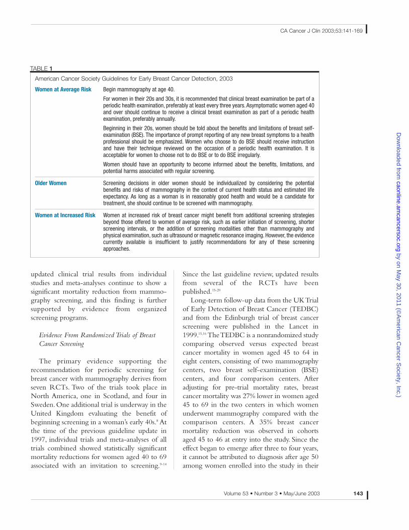
A summary of the update of the ACSguidelines for early breast cancer detection isshown in Table 1.
SCREENING WITH MAMMOGRAPHY
Recommendation
Women at average risk should begin annualmammography at age 40.Women should havean opportunity to become informed about the benefits, limitations, and potential harmsassociated with regular screening.
Rationale and Evidence
Since 1997, there have been updates in the evidence from RCTs of breast cancerscreenings. Several other reports havechallenged the value of screening for breastcancer with mammography,4-7 leading to a surgeof new literature reexamining the underlyingevidence related to breast cancer screening.The
142 CA A Cancer Journal for Clinicians
ACS Guidelines for Breast Cancer Screening: Update 2003
by on May 30, 2011 (©
Am
erican Cancer S
ociety, Inc.) caonline.am
cancersoc.orgD
ownloaded from
updated clinical trial results from individualstudies and meta-analyses continue to show asignificant mortality reduction from mammo-graphy screening, and this finding is furthersupported by evidence from organizedscreening programs.
Evidence From Randomized Trials of BreastCancer Screening
The primary evidence supporting therecommendation for periodic screening forbreast cancer with mammography derives fromseven RCTs. Two of the trials took place inNorth America, one in Scotland, and four inSweden. One additional trial is underway in theUnited Kingdom evaluating the benefit ofbeginning screening in a woman’s early 40s.8 Atthe time of the previous guideline update in1997, individual trials and meta-analyses of alltrials combined showed statistically significantmortality reductions for women aged 40 to 69associated with an invitation to screening.9-14
Since the last guideline review, updated resultsfrom several of the RCTs have beenpublished.15-20
Long-term follow-up data from the UK Trialof Early Detection of Breast Cancer (TEDBC)and from the Edinburgh trial of breast cancerscreening were published in the Lancet in1999.15,16 The TEDBC is a nonrandomized studycomparing observed versus expected breastcancer mortality in women aged 45 to 64 ineight centers, consisting of two mammographycenters, two breast self-examination (BSE)centers, and four comparison centers. Afteradjusting for pre-trial mortality rates, breastcancer mortality was 27% lower in women aged45 to 69 in the two centers in which womenunderwent mammography compared with thecomparison centers. A 35% breast cancermortality reduction was observed in cohortsaged 45 to 46 at entry into the study. Since theeffect began to emerge after three to four years,it cannot be attributed to diagnosis after age 50among women enrolled into the study in their
Volume 53 • Number 3 • May/June 2003 143
CA Cancer J Clin 2003;53:141-169
American Cancer Society Guidelines for Early Breast Cancer Detection, 2003
Women at Average Risk Begin mammography at age 40.
For women in their 20s and 30s, it is recommended that clinical breast examination be part of aperiodic health examination, preferably at least every three years. Asymptomatic women aged 40and over should continue to receive a clinical breast examination as part of a periodic healthexamination, preferably annually.
Beginning in their 20s, women should be told about the benefits and limitations of breast self-examination (BSE). The importance of prompt reporting of any new breast symptoms to a healthprofessional should be emphasized. Women who choose to do BSE should receive instructionand have their technique reviewed on the occasion of a periodic health examination. It isacceptable for women to choose not to do BSE or to do BSE irregularly.
Women should have an opportunity to become informed about the benefits, limitations, andpotential harms associated with regular screening.
Older Women Screening decisions in older women should be individualized by considering the potentialbenefits and risks of mammography in the context of current health status and estimated lifeexpectancy. As long as a woman is in reasonably good health and would be a candidate fortreatment, she should continue to be screened with mammography.
Women at Increased Risk Women at increased risk of breast cancer might benefit from additional screening strategiesbeyond those offered to women of average risk, such as earlier initiation of screening, shorterscreening intervals, or the addition of screening modalities other than mammography andphysical examination, such as ultrasound or magnetic resonance imaging. However, the evidencecurrently available is insufficient to justify recommendations for any of these screeningapproaches.
TABLE 1
by on May 30, 2011 (©
Am
erican Cancer S
ociety, Inc.) caonline.am
cancersoc.orgD
ownloaded from

40s.15 In the Edinburgh trialfollow-up, the investigatorsapplied an improved methodof adjusting for socio-economic status and censoredbreast cancer diagnoses morethan three years after theconclusion of the study, sincecases diagnosed after threeyears were unlikely to havebeen prior false negatives;29% fewer breast cancerdeaths were observed in thegroup invited to screeningcompared with an initialestimate of 13 percent.16 Theinvestigators also reportedthat there was no significantdifference in the observedbenefit based on age atrandomization.
Updated results from theCanadian National BreastCancer Screening Trial(NBSS-1 and NBSS-2) havebeen reported since 1997.17,18
In 2000, Miller, et al. reported13-year follow-up resultsfrom the NBSS-2, whichcompared annual two-viewmammography and CBEwith annual CBE only inwomen aged 50 to 59 atrandomization. The authorsreported no difference in thebreast cancer mortality rate inthe group randomized toreceive an invitation to annualmammography and CBEcompared with the groupinvited to receive CBE only(RR = 1.02), and concludedthat mammography providedno additional advantagecompared with carefullyconducted CBE.17 In 2002,
the Canadian investigators reported updatedresults from the NBSS-1, which comparedannual mammography and CBE with usual carein women aged 40 to 49.18 After 11 to 16 yearsof follow-up there was no difference in thebreast cancer mortality rate in the group invitedto mammography screening compared withCBE with usual care (RR = 0.97).
In 2000, Tabár and colleagues reported 20-year follow-up of the Swedish Two-CountyTrial of breast cancer screening.19 With follow-up through 1998, there was a statisticallysignificant 32% reduction in mortality inwomen aged 40 to 69 (RR = 0.68, 95%confidence interval (CI) 0.59 to 0.80) associatedwith an invitation to screening. A larger,consistent effect was observed in each of the twocounties for women aged 50 to 69. Results forwomen aged 40 to 49 differed between the twocounties, with a substantial reduction in breastcancer mortality in the W-county (RR = 0.76,95% CI 0.42 to 1.40), but not in the E-county(RR = 1.06, 95% CI 0.65 to 1.76).Tabár, et al.have reported previously that this inconsistencyis explained by the observed higher fatality ratesin nonattenders to screening in the invitedgroup in the E-county.21
Swedish investigators recently updated theoverview analysis of the Swedish trials ofmammography screening based on follow-upto 1996.20 With a median follow-up time fromrandomization to the end of follow-up of 15.8years, the investigators observed an overall 21%statistically significant reduction in breast cancermortality among women aged 40 to 74 atrandomization associated with an invitation tomammography (RR = 0.79).
As part of the evidence review of the USPreventive Services Task Force (USPSTF), anew meta-analysis of the RCTs was conduct-ed by Humphrey, et al.22 and publishedsimultaneously with the updated USPSTFguidelines.23 The meta-analysis of trial results(excluding the Edinburgh trial) from all agegroups showed a statistically significant 16%mortality reduction associated with an
144 CA A Cancer Journal for Clinicians
ACS Guidelines for Breast Cancer Screening: Update 2003
High-Risk Work Group: Wylie Burke, MD, PhD, (Chair), Professorand Chair, Department of Medical Historyand Ethics, University of Washington,Seattle, WA; Elizabeth Claus, MD, PhD,Associate Professor, Department ofEpidemiology and Public Health, YaleUniversity School of Medicine, NewHaven, CT; Mary Daly, MD, PhD, Director,Margaret Dyson Family Risk AssessmentProgram and Director, Cancer ControlScience Program, Fox Chase CancerCenter, Philadelphia, PA; Paula Gordon,MD, Clinical Professor, Department ofRadiology, University of British Columbia,Vancouver, BC; Constance D. Lehman,MD, PhD, Director of Breast Imaging,Seattle Cancer Care Alliance andAssociate Professor, Department ofRadiology, University of WashingtonMedical Center, Seattle, WA; OlufunmilayoI. Olopade, MD, Professor of Medicine andDirector, Center for Clinical CancerGenetics, University of Chicago MedicalCenter, Chicago, IL; Wendy S. Rubinstein,MD, PhD, Assistant Professor of Medicine,Northwestern University Feinberg Schoolof Medicine, and Medical Director, Centerfor Medical Genetics, EvanstonNorthwestern Healthcare, and DivisionHead, Medical Genetics, EvanstonHospital, Evanston, IL; Debbie Saslow,PhD, Director, Breast and GynecologicCancer, American Cancer Society, Atlanta,GA; Robert A. Smith, PhD, Director, CancerScreening, American Cancer Society,Atlanta, GA.
Screening Older Women WorkGroup:Mary E. Costanza, MD, (Chair), Professorof Medicine, University of MassachusettsMedical School, Worcester, MA; LodovicoBalducci, MD, Professor of Medicine, H.Lee Moffitt Cancer Center and ResearchInstitute, Tampa, FL; Cheryl Kidd, MPH,Director of Education, Susan G. KomenBreast Cancer Foundation, Dallas, TX;Jeanne Mandelblatt, MD, MPH, Director,Cancer Control Program, and Director ofCancer and Aging Research, LombardiCancer Center, Georgetown UniversityMedical Center, Washington DC; BarbaraMonsees, MD, Professor, MallinckrodtInstitute of Radiology, and Chief, BreastImaging Section, Washington UniversitySchool of Medicine, St. Louis, MO; PeterPressman, MD, Professor of ClinicalSurgery, Weill Medical College of CornellUniversity, New York, NY; William A.Satariano, PhD, MPH, Professor ofEpidemiology, School of Public Health,University of California at Berkeley,Berkeley, CA; Louise C. Walter, MD,Assistant Professor of Medicine, Divisionof Geriatrics, VA Medical Center, SanFrancisco, CA; Debbie Saslow, PhD,Director, Breast and Gynecologic Cancer,
by on May 30, 2011 (©
Am
erican Cancer S
ociety, Inc.) caonline.am
cancersoc.orgD
ownloaded from

invitation to screening (RR = 0.84). Similarmeta-analyses were conducted for women aged40 to 49 at randomization, with results leadingthe authors to conclude that the risk reductionfrom mammography screening does not differsubstantially by age, although absolute benefitsare lower in women under age 50 comparedwith women aged 50 and over.The authors ofthe updated reports from Edinburgh and theTEDBC reached similar conclusions aboutage-specific benefits.15,16
The most recent results from therandomized clinical trials are shown in Table 2.24
While there is variation in the observedmortality reductions, a meta-analysis of themost recent results showed a 24% mortalityreduction associated with an invitation toscreening. Further, although the trials varysomewhat in their design, their results areuniformly consistent with respect to therelationships between the observed shift in stageat diagnosis and reduction in mortality, i.e.,those trials that achieved the greatest reductionin the relative risk of being diagnosed with anode-positive breast cancer also have shown thegreatest mortality reductions.25,26
In October 2001, the Lancet published a
research letter by Ole Olsenand Peter Gøtzsche7 from theNordic Cochrane Centre inCopenhagen. The authorsevaluated the randomizedtrials of breast cancerscreening through meta-analysis, concluding that fiveof the seven trials wereflawed and should not beregarded as providing reliablescientific evidence.Olsen andGøtzsche further suggestedthat breast cancer mortalitywas an unreliable endpoint,and that only comparison ofall-cause mortality betweenthe experimental and controlgroups could serve as anunbiased endpoint. Based ontheir meta-analysis, whichwas restricted to the Malmöand Canadian trials, theauthors found no evidence ofa reduced mortality asso-ciated with an invitation tomammography (RR = 1.0),
Volume 53 • Number 3 • May/June 2003 145
CA Cancer J Clin 2003;53:141-169
Most Recently Published Results of the Breast Cancer Screening Trials on Breast CancerMortality Reduction With Invitation to Screening
Study Age Range Percentage Mortality Reduction (95% CI†)
HIP 40 - 64 24 (7, 38)
Malmö 45 - 69 19 (-8, 39)
Two-County Trial, Sweden 40 - 74 32 (20, 41)
Edinburgh 45 - 64 21 (-2, 40)
Stockholm 40 - 64 26 (-10, 50)
Canada NBSS-1 40 - 49 -3 (-26, 27)
Canada NBSS-2 50 - 59 -2 (-33, 22)
Gothenburg 39 - 59* 16 (-39, 49)
All Trials Combined 39 - 74 24 (18, 30)
TABLE 2
*There are more recent publications from the Gothenburg trial but they refer only to the under-50 age group.†CI = Confidence interval.
American Cancer Society, Atlanta, GA;Robert A. Smith, PhD, Director, CancerScreening, American Cancer Society,Atlanta, GA.
Physical Examination WorkGroup: Roger S. Foster Jr., MD, (Chair), Wadley R.Glenn Professor of Surgery, Retired,Emory University School of Medicine,Atlanta, GA; Cornelia Baines, MD,Professor, Department of Public HealthSciences, University of Toronto, Toronto,Canada; Lynn Erdman, RN, MS, VicePresident, Women’s and Cancer Services,NorthEast Medical Center, Concord, NC;Margaret Rinehart-Ayres, PT, PhD,Associate Professor, Department ofPhysical Therapy, Thomas JeffersonUniversity, Philadelphia, PA; Ruby Senie,PhD, Professor of Epidemiology andSociomedical Science, Mailman Schoolof Public Health, Columbia University,New York, NY; David J. Winchester, MD,Assistant Professor of Surgery, FeinbergSchool of Medicine, NorthwesternUniversity, Chicago, IL; William C. Wood,MD, Professor and Chairman,Department of Surgery, Emory UniversitySchool of Medicine, Atlanta, GA; DebbieSaslow, PhD, Director, Breast andGynecologic Cancer, American CancerSociety, Atlanta, GA; Robert A. Smith,PhD, Director, Cancer Screening,American Cancer Society, Atlanta, GA.
Mammography Work Group:W. Phil Evans III, MD, (Chair), Director ofBreast Imaging, University of TexasSouthwestern Center for Breast Care,Dallas, TX; Linda Warren Burhenne, MD,Clinical Professor, Diagnostic Radiologyand Breast Imaging, University of BritishColumbia, Vancouver, BC; Carl J. D’Orsi,MD, Professor of Radiology andHematology/Oncology and Director,Division of Breast Imaging, The EmoryClinic, Atlanta, GA; Stephen A. Feig, MD,Director of Breast Imaging, Departmentof Radiology, Mount Sinai Hospital, andProfessor of Radiology, Mount SinaiSchool of Medicine, New York, NY; Amy S.Langer, MBA, Executive Director, NationalAlliance of Breast Cancer Organizations(NABCO), New York, NY; A. Marilyn Leitch,MD, Professor, Department of SurgicalOncology, and Medical Director, UTSouthwestern Center for Breast Care,University of Texas Southwestern MedicalCenter at Dallas, TX; Stephen Sener, MD,Vice Chairman, Department of Surgery,Evanston Northwestern Healthcare,Evanston, IL; Steven H. Woolf, MD, MPH,Professor, Department of Family Practice,Virginia Commonwealth University,Fairfax, VA; Bonnie C. Yankaskas, PhD,Professor, Department of Radiology,University of North Carolina, Chapel Hill,
by on May 30, 2011 (©
Am
erican Cancer S
ociety, Inc.) caonline.am
cancersoc.orgD
ownloaded from

and concluded that there wasno reliable evidence thatscreening reduced breastcancer mortality. Severalguideline groups, nationalboards of health, and nu-merous individual authorshave provided formal cri-tiques of the methodologyand conclusions of Olsen andGøtzsche.20,22,24,26-30 The re-views uniformly concludedthat the evidence providedby the Cochrane Review didnot support the claim that methodological short-comings in the conduct ofthe trials were of suchsignificance to invalidate theconclusion that screening forbreast cancer with mam-mography reduces breastcancer mortality.
Evidence From ServiceScreening
The inherent limitations ofthe breast cancer screeningRCTs to estimate the benefitsassociated with exposure tomodern mammography haveled to increased interest in
evaluating the impact of screening in thecommunity setting, also referred to as servicescreening. Service screening evaluation canestimate breast cancer mortality for women whoactually attend community screening programsand for the population as a whole. Servicescreening evaluation can also be used to attributedifferences in mortality over time to screening,improvements in therapy, and increasedawareness.31 However, establishing the relativevalue between screening and nonscreeningfactors is complex and can be only indirectlyestimated.
Blanks, et al. reported on the impact of theUK National Health Service breast cancerscreening program in women aged 55 to 69years between 1990 and 1998,32 estimating a21.3% reduction in breast cancer mortality,with a smaller direct effect of mammography(6.4%) compared with increased awareness andimprovements in therapy (14.9%). Jonsson andcolleagues have reported on service screeningin Sweden for women aged 40 to 49,33 and 50to 69,34 and the investigators concluded that theestimated mortality reductions were consistentwith the estimates from the RCTs.
Two additional investigations from Swedenwere able to classify breast cancer cases beforeand after the introduction to screening on thebasis of exposure to screening.31,35 In the mostrecent report,35 which expanded an earlieranalysis to seven counties in the Uppsalaregion, Duffy and colleagues compared breastcancer mortality in the prescreening andpostscreening periods among women aged 40to 69 in six counties and 50 to 69 in a seventhcounty. Overall, they observed a 44% mortalityreduction in women who underwent screeningand a 39% reduction in overall breast cancermortality after adjustment for selection biasassociated with the policy of offering screeningto the population.35 Because the authors wereable to distinguish between screened andunscreened cohorts, they estimated that abouttwo-thirds of the observed mortality reductionswere attributable to screening, with theremainder due to improvements in therapy andincreased awareness. While Blanks, et al.estimate a smaller benefit of mammography,they acknowledge they were unable to identifywhich among the breast cancer deaths in thepostscreening period were from cases diagnosedbefore screening was health policy.31,35 Thesedata demonstrated that organized screeningwith high rates of attendance in a setting thatachieves a high degree of programmatic qualityassurance did achieve breast cancer mortalityreductions equal to or greater than thoseobserved in the randomized trials.
146 CA A Cancer Journal for Clinicians
ACS Guidelines for Breast Cancer Screening: Update 2003
NC; Debbie Saslow, PhD, Director, Breastand Gynecologic Cancer, AmericanCancer Society, Atlanta, GA; Robert A.Smith, PhD, Director, Cancer Screening,American Cancer Society, Atlanta, GA.
New Technologies Work Group:Edward Hendrick, PhD, (Chair), ResearchProfessor and Director, Breast ImagingResearch, Department of Radiology, LynnSage Comprehensive Breast Center,Northwestern Memorial Hospital,Chicago, IL; Maryellen Giger, PhD,Professor of Radiology, Director ofGraduate Programs in Medical Physics,Department of Radiology, University ofChicago, Chicago, IL; Paula Gordon, MD,Clinical Professor, Department ofRadiology, University of British Columbia,Vancouver, BC; Valerie P. Jackson, MD,John A. Campbell Professor of Radiology,Indiana University School of Medicine,Indianapolis, IN; Constance D. Lehman,MD, PhD, Director of Breast Imaging,Seattle Cancer Care Alliance, andAssociate Professor, Department ofRadiology, University of WashingtonMedical Center, Seattle, WA; JeannePetrek, MD, Director, Surgical Program,Evelyn H. Lauder Breast Center, MemorialSloan-Kettering Cancer Center, New York,NY; Edward Sickles, MD, Professor ofRadiology, University of California Schoolof Medicine, San Francisco, CA; Martin J. Yaffe, PhD, Senior Scientist,Imaging/Bioengineering Research,Sunnybrook and Women’s College Health Sciences Centre, and Professor,Departments of Medical Imaging andMedical Biophysics, University of Toronto,Toronto, Canada; Debbie Saslow, PhD,Director, Breast and Gynecologic Cancer,American Cancer Society, Atlanta, GA;Robert A. Smith, PhD, Director, CancerScreening, American Cancer Society,Atlanta, GA.
by on May 30, 2011 (©
Am
erican Cancer S
ociety, Inc.) caonline.am
cancersoc.orgD
ownloaded from

Breast cancer mortality reductions associat-ed with screening have been reported from the Florence, Italy Screening program, alsocomparing breast cancer mortality amongattenders and nonattenders to screening, and in the population before and after theintroduction of screening between 1990 and1996.36 The incidence-based mortality ratio(i.e., the rate of fatal incident breast cancercases) comparing 1990 to 1996 with 1985 to1986 shows a 50% reduction in the rate ofbreast cancer deaths (RR = 0.50, 95% CI 0.38to 0.66).After excluding the breast cancer casesdiagnosed at the first screening examination(i.e., the prevalent screening round), the rate ofStage II or higher breast cancer cases was 42%lower in screened women compared with thewomen diagnosed with breast cancer that hadnot been invited to screening (RR = 0.58, 95%CI 0.45 to 0.74). The investigators concludedthat breast cancer mortality reductions wereattributable to improvements in therapy and theintroduction of a breast cancer screeningprogram.
These data demonstrate that modern,organized screening programs with high ratesof attendance can achieve breast cancermortality reductions equal to or greater thanthose observed in the RCTs. Insofar asadditional RCTs of breast cancer screening areunlikely, the evaluation of service screeningrepresents an important new development forseveral reasons, specifically by measuring thevalue of modern mammography in thecommunity and measuring the benefit frommammography screening to women whoactually get screened.
Screening Intervals
Mortality reductions for women aged 40 to69 have been observed in trials that screened atintervals of 12 and 24 (and over) months, andthus, some guidelines recommend screening atan interval of one-two years. However, datafrom two trials13,14 and inferential evidence used
to estimate the duration ofthe detectable pre-clinicalphase,1,37 i.e., sojourn time,have provided persuasiveevidence that youngerwomen likely will benefitmore from annual screeningcompared with screening at two-year intervals, aconclusion also reached inthe recent USPSTF evidencereview.22 Further, data fromboth RCTs and from servicescreening programs haveshown that the proportionalincidence of interval cancersin the period after a normalscreening examination ishigher in younger womencompared with olderwomen, suggesting fastergrowth rates in youngerwomen.14,38,39 Tabár andcolleagues have estimatedthat tumor sojourn timeslengthen with increasing age,and using Two-County trialdata have estimated the meansojourn time for women byage as follows: 40 to 49 = 2.4years, 50 to 59 = 3.7 years, 60to 69 = 4.2 years, and 70 to79 = 4 years.19
Modeling data also havesuggested that progressively shorter screeningintervals result in both the detection of tumorsat smaller sizes and in decreased mortality rates.Estimating tumor characteristics associated withscreening intervals of 24, 12, and 6 months,Michaelson, et al. showed that shorter screeningintervals were associated with greaterreductions in the proportion of cases diagnosedwith distant metastases.40 Also, in a subsequentmodeling analysis of 1,352 women from theVan Nuys Breast Cancer Center between 1966and 1990,Michaelson, et al. showed that smaller
Volume 53 • Number 3 • May/June 2003 147
CA Cancer J Clin 2003;53:141-169
Breast Cancer Advisory Group: Stephen Sener, MD, (Chair), ViceChairman, Department of Surgery,Evanston Northwestern Healthcare,Evanston, IL; Barbara Andreozzi, Chair,Montana Breast and Cervical HealthAdvisory Council, CommunityDevelopment Specialist, Montana StateUniversity Extension Service, Anaconda,MT; Lynn Erdman, RN, MS, VicePresident, Women’s and Cancer Services,NorthEast Medical Center, Concord, NC;W. Phil Evans III, MD, Director of BreastImaging, University of TexasSouthwestern Center for Breast Care,Dallas, TX; Herschel W. Lawson, MD,Medical Advisor, Division of CancerPrevention and Control, Centers forDisease Control and Prevention, Atlanta,GA; Jeanne Petrek, MD, Director, SurgicalProgram, Evelyn H. Lauder Breast Center,Memorial Sloan-Kettering Cancer Center,New York, NY; Margaret Rinehart-Ayres,PT, PhD, Associate Professor, Departmentof Physical Therapy, Thomas JeffersonUniversity, Philadelphia, PA; Christy A.Russell, MD, Associate Professor ofMedicine and Co-Director of the LeeBreast Center, University of SouthernCalifornia, USC/Norris ComprehensiveCancer Center, Los Angeles, CA; CarolynD. Runowicz, MD, Vice Chairman,Department of Obstetrics andGynecology, St. Lukes-Roosevelt HospitalCenter, and Professor of ClinicalObstetrics and Gynecology, UniversityHospital of Columbia University College ofPhysicians and Surgeons, and Director,Gynecologic Oncology Research,Women’s Health Service Line ContinuumHealth Partners, Inc., New York, NY;William C. Wood, MD, Professor andChairman, Department of Surgery, EmoryUniversity School of Medicine, Atlanta,GA; Debbie Saslow, PhD, Director, Breastand Gynecologic Cancer, AmericanCancer Society, Atlanta, GA.
by on May 30, 2011 (©
Am
erican Cancer S
ociety, Inc.) caonline.am
cancersoc.orgD
ownloaded from

tumor size was highly correlated with longersurvival time independent of method of cancerdetection.41
While sojourn times lengthen withincreasing age, these data provide only a limitedbasis for establishing screening intervals, and in particular, they provide only a roughapproximation for an interval that should notbe exceeded, since the recommended screeninginterval should always be shorter than theestimated mean sojourn time. Since the goal ofscreening is the reduction in the incidence rateof advanced disease, the screening intervalshould be set for a period of time in whichadherence to routine screening is likely to resultin the detection of the majority of cancerswhile still occult and localized. While annualscreening likely is more beneficial for allwomen,1,40,42 the importance of annualscreening clearly is greater in premenopausalwomen (< 55) compared with postmenopausalwomen. However, given the prognostic value ofsmaller tumors, and the finding that annualscreening results in more favorable tumorcharacteristics in both pre- and postmenopausalwomen, annual screening may offer advantagesover biennial screening well into thepostmenopausal period.43
Limitations of Mammography and Harms
Associated With Screening
As is the case with any screening exam-ination, the goal of breast cancer screening is todetect occult breast cancer in a population ofwomen in which the great majority will nothave breast cancer on the occasion of a regularexamination, and the large majority will notdevelop breast cancer in their lifetime.Althoughthe efficacy of mammography has beendemonstrated, it does not achieve perfectsensitivity or specificity in women undergoingscreening, and as such, the issue of adverseconsequences for women who do and who donot have breast cancer has been a source ofgrowing attention, and has become one of the
core issues in recent debates aboutmammography. False negatives can beattributed to inherent technological limitationsof mammography, quality assurance failures, andhuman error; false positives also can beattributed to these factors as well as toheightened medical-legal concerns over theconsequence of missed cancers. Further, insome instances, a patient’s desire for definitivefindings in the presence of a low-suspicionlesion also contributes to false positives. Theconsequences of these errors include missedcancers, with potentially worse prognosis, aswell as anxiety and harms associated withinterventions for benign or nonobligateprecursor lesions.
This issue of limitations and harms is bothimportant and complex, since mammography’sshortcomings are due to the interplay betweenhost characteristics (age, risk, breast density, andtumor growth rates) and provider factors(technical limitations and quality assurancefailures). Thus, theoretically, there is at leastsome level of limitations and harms that isinherent to breast cancer screening andunavoidable. Beyond this level are potentialimprovements in screening and reductions inharms that could be achieved through varioustechnical and system-related interventions.Thisrelationship between risk, benefit, limitations,and harms is complicated by the fact that notonly is it multifactorial, but also that individualwomen likely will weigh the benefits,limitations, and harms of screening differentlydepending on their age, values, and theirunderstanding of the issues. Still, there isconcern that the rate of false-positivemammography and benign biopsy is excessiveand could be reduced with improvements inscreening quality, although other factors such asmedical-legal pressures and individual anxietyabout uncertainty may partly be outside theinfluence of additional improvements inquality.44 There also is agreement that stepsshould be taken to reduce anxiety associatedwith screening,45 i.e., the waiting time to
148 CA A Cancer Journal for Clinicians
ACS Guidelines for Breast Cancer Screening: Update 2003
by on May 30, 2011 (©
Am
erican Cancer S
ociety, Inc.) caonline.am
cancersoc.orgD
ownloaded from

diagnosis,46,47 and that there should beconscientious efforts applied toward informingwomen about the likelihood of both false-negative and false-positive findings.48
A number of investigations have attempted tomeasure the extent of psychological and physi-cal harms associated with false-positivemammography and, in particular,have attemptedto identify whether or not harms are lasting andhave consequences for psychological well-beingand subsequent screening. In general, theevidence suggests that some women experienceanxiety related to screening, and a greaterpercentage experience anxiety related to false-positive results, but for most womenpsychological distress is short-lived and does nothave lasting consequences on either stress levelsor likelihood of subsequent screening.47,49 Arecent study by Schwartz and colleagues revealedthat women are aware that false-positive resultsoccur, accept false-positive results as a part ofscreening, and do not regard false positives as animportant harm in the context of the underlyinggoal of early breast cancer detection.48 However,women’s awareness of the chance of a false-positive finding and acceptance of false positivesas a cost of screening should not detract fromorganized efforts to provide information aboutthe range of screening outcomes, to achieve anacceptable rate of false-positive results inscreening programs, and to mitigate thespectrum of harms. Further, health professionalsmust become more sensitive to both short-termand long-term effects of false-positive results.
As use of mammography has increased,concerns have been raised about detection andovertreatment of DCIS.50,51 Although thedetection of DCIS is an inevitable consequenceof mammographic screening, the concern thatnot all DCIS is progressive has to be weighedagainst the estimate that a substantial portion isprogressive.The actual fraction is not known,andhistorical estimates from case series may not bedirectly generalizable to cases identified throughmammographic screening.52 However, in oneseries, invasive breast cancer developed in over
half of women with low-grade DCIS lesionsidentified on biopsy but not treated.53 Further,incomplete excision of DCIS or failure to exciseDCIS associated with an invasive cancer is adeterminant for local failure.54,55 As with invasivebreast cancer, the histologic subtype and grade ofDCIS can be regarded as having prognostic valuein relation to risk of recurrence of invasivecancer or DCIS.Thus, there is little question thatsome women benefit from detection andtreatment of DCIS. However, since some DCISis not progressive, diagnostic evaluation andtreatment of DCIS lesions that would notprogress to invasive disease is a harm associatedwith screening, although the extent of harm isuncertain, as is how it might be avoided.Overtreatment of a progressive DCIS lesion thatcould be cured with less aggressive treatment alsorepresents a harm, although it should not beattributed to screening.
At this time, the majority of detected DCISoccurs as a result of identification of smalllesions on a mammogram that are perceived tobe important. It is not possible through imageevaluation to either distinguish DCIS frominvasive breast cancer or progressive DCIS fromnonprogressive DCIS. Schwartz and colleagueshave recommended that information providedto women undergoing mammography shouldinclude a discussion about detection of DCIS.48
Important questions remain about how toidentify those noninvasive ductal cancers thatare most likely to progress to become invasivecancers.
PHYSICAL EXAMINATION
Recommendations
Clinical Breast Examination
For average-risk asymptomatic women intheir 20s and 30s, it is recommended that CBEbe part of a periodic health examination,preferably at least every three years.The exam
Volume 53 • Number 3 • May/June 2003 149
CA Cancer J Clin 2003;53:141-169
by on May 30, 2011 (©
Am
erican Cancer S
ociety, Inc.) caonline.am
cancersoc.orgD
ownloaded from

should include BSE instruction for the purposeof gaining familiarity with breast composition.Information should be provided about thebenefits and limitations of CBE and BSE, and itshould be emphasized that breast cancer risk isvery low for women in their 20s and graduallyincreases with age. The importance of promptreporting of any new symptoms to a healthprofessional also should be emphasized.
Asymptomatic women aged 40 and overshould continue to receive CBE as part of aperiodic health examination, preferablyannually. Beginning at age 40, discussion duringCBE should include information aboutscreening mammography. There may be somebenefit to performing the CBE shortly beforethe mammogram. At the time of CBE, thebenefits and limitations of physical examinationand mammography should be discussed withthe patient.
Breast Self-Examination
Beginning in their 20s, women should betold about the benefits and limitations of BSE.The importance of prompt reporting of anynew breast symptoms to a health professionalshould be emphasized.Women who choose todo BSE should receive instruction and havetheir technique reviewed on the occasion of aperiodic health examination. It is acceptable forwomen to choose not to do BSE or to do BSEirregularly.
Rationale and Evidence
The logic for the earlier detection of clinicalfindings in the breast is analogous to the logicfor detecting breast cancer before a tumor ispalpable.With increasing tumor size, even afterbreast cancers become palpable, the likelihoodof regional and distant metastasis increases.Long-term survival measured either withregistry data or with data from RCTs is poorerwith each incremental 5 mm increase in tumorsize.19,56 For average-risk women under age 40,
earlier detection of palpable tumors with CBEor BSE can lead to earlier therapy. After age 40, CBE and BSE are regarded as adjunctivebecause mammography does not achieveperfect sensitivity. Although there are no directclinical data linking an increased rate of breastpreservation to CBE or BSE, it is plausible thatearlier detection of symptomatic breast cancerresults in a greater probability for a breastconserving approach.
The evidence supporting the value of CBEand BSE as methods of reducing breast cancermortality is limited and mostly inferential,although there is no definitive prospectiveRCT evidence from which to drawconclusions about either exam. Thus, currentrecommendations rely on existing evidence, butalso on expert opinion based on a recognitionthat population-based studies continue to showa relatively large proportion of self-detectedcancers.
Clinical Breast Examination
Today, mammography and clinical breastexamination are recommended to women 40and older because: (1) there are RCT datashowing the combination of mammographyand CBE was associated with lower breastcancer mortality,16,57 and (2) evidence fromthese RCTs and demonstration projectsshowed that some cancers detected by CBEwere not detected by mammography.16,57,58
The USPSTF recommends mammographywith or without CBE, and it has concluded thatthere is insufficient evidence to recommend foror against breast cancer screening with CBEalone.23 Evaluation of CBE as a detectionmodality has generally focused on theperformance characteristics of the test, i.e.,sensitivity, specificity, and positive predictivevalue.57-60 On all aspects, performancecharacteristics are poorer than those ofmammography. Sensitivity of CBE in particularwas estimated in a recent meta-analysis to beonly 54 percent.61 While noting that two trials
150 CA A Cancer Journal for Clinicians
ACS Guidelines for Breast Cancer Screening: Update 2003
by on May 30, 2011 (©
Am
erican Cancer S
ociety, Inc.) caonline.am
cancersoc.orgD
ownloaded from

demonstrated breast cancer mortalityreductions associated with the combination ofmammography and CBE,16,62 the USPSTFconcluded there was insufficient evidence toquantify the incremental benefits of addingCBE to mammography.23 This particularquestion is more pertinent today, since much ofthe RCT data related to the value of CBEcombined with mammography derives from aperiod that predates modern breast imaging.The proportion of breast cancers not visiblewith modern, high-quality mammographyappears to be considerably lower today than inthe past.60,63,64
Based on findings from 752,081 CBEs,Boboand colleagues reported that 6.9 percent of all CBEs were coded as abnormal, and that five cancers were detected per 1,000examinations.60 However, only 5.1 percent ofthe malignancies (193/3,753), or 2.56 per10,000 CBE exams, were detected in womenwith an abnormal CBE and benign findings onthe mammogram. Since women with self-detected breast symptoms were 7.2 times aslikely to have an abnormal exam, it is likely thatsome proportion of these CBE-positive caseswere first detected by women themselves,leading to an even lower rate of breast cancerdetection attributable to CBE alone.Newcomer, et al. recently reported on themode of detection in 2,341 Wisconsin womengreater than or equal to the age of 50 diagnosedwith breast cancer from 1988 to 1991.Womenwere asked how their breast cancer was firstdiscovered—48 percent were self-detected, 41percent were detected by mammography,and 11 percent were detected by CBE.63
Oestreicher, et al. evaluated the sensitivity ofCBE in 468 women diagnosed with breastcancer within a year of a screening CBE.64
Overall sensitivity was estimated to be 35percent, but the majority of these cases (83.6percent) also were detected by mammo-graphy. Among women with false-negativemammograms, 37 percent were detected byCBE, but overall only 5.7 percent (n = 27) of
breast cancers were diagnosed by CBE only.64
Sensitivity ranged from 17.2 percent for tumorsless than or equal to 0.5 cm to 58.3 percent fortumors greater than or equal to 2.1 cm, and waslower in younger women and women withhigher body weight.
At this time, it is unclear what CBEcontributes to detection of breast cancer,although it is likely that in presumablyasymptomatic women the contribution is small.When done prior to mammography, CBE mayidentify an area of suspicion that will not bevisible on mammography and/or provideinformation that guides subsequent imagingexams. As a growing proportion of women arereceiving regular mammograms, the relativecontribution of CBE to early breast cancerdetection and its cost-effectiveness warrantrenewed attention. At this time, in womenscreened with mammography, the cancerdetection rate for CBE appears to be low, andthe evidence for breast cancer mortalityreduction associated with CBE is weak andindirect.
However, apart from some contribution tobreast cancer detection, CBE may serve anadditional, separate function: it can provide theoccasion to raise awareness about breast cancerand to provide accurate education on thevariety of breast cancer-related topics, includinginformation about breast symptoms, genetics,risk factors, and newer cancer detectiontechnologies. The consensus opinion of theACS review panel was that until moreinformative scientific evidence is available,periodic CBE is recommended with theadditional endorsement that the occasion of aCBE should be used to raise awareness aboutthe early detection of breast cancer. Inrendering this opinion, there were some panelmembers who believed that the evidenceagainst the benefit of CBE was not strongenough to abandon the recommendation,while other panel members believed that therecommendation for CBE was not evidence-based and should be eliminated.
Volume 53 • Number 3 • May/June 2003 151
CA Cancer J Clin 2003;53:141-169
by on May 30, 2011 (©
Am
erican Cancer S
ociety, Inc.) caonline.am
cancersoc.orgD
ownloaded from

Breast Self-Examination and Self-Detection
The goal of periodic BSE, as with CBE, is todetect palpable tumors. An additional role ofBSE is to increase awareness of normal breastcomposition, so that there is heightenedawareness of changes that may be detectedduring BSE or at some other time.The value ofheightened awareness, however it may beachieved, is commonly acknowledged65-67 basedon the value of earlier treatment of bothnonpalpable and palpable breast cancers.Stockton, et al. attributed a shift toward morefavorable stage at diagnosis and declining breastcancer mortality in the 1980s in East Anglia toincreasing awareness and prompt reporting ofsigns and symptoms of breast cancer that began before the beginning of a breast cancerscreening program.68
The first studies suggesting possibleeffectiveness of BSE were published in 1978.69,70
These two studies and many additional studiesin the premammography era found that ingeneral, women who reported that they hadbeen BSE performers had their breast cancersdetected at a smaller size and at earlier clinicaland/or pathologic stage. Regular performanceof BSE did not mean that the breast cancer wasnecessarily self-detected during a formal BSEprocedure. Even regular BSE performerscommonly detected their breast cancerincidentally, suggesting that there was acomponent of increased body awareness (orperhaps increased awareness of subtlesymptoms) in addition to the self-performedphysical examination.71
The literature on the effectiveness of BSE asa detection modality has shown mixed results,but recent evidence reviews have focused onthe absence of direct evidence of benefit in twoRCTs,72,73 and data indicating that the rate ofbenign biopsy is higher in women whoregularly perform BSE compared with womenwho do not regularly perform BSE.73 TheUSPSTF concluded that the evidence isinsufficient to recommend for or against
teaching or performing routine BSE.23 TheCanadian Task Force on Preventive HealthCare went further and recommended againstroutine instruction in BSE in periodic healthexaminations on the basis of fair evidence of nobenefit and good evidence of harm (false-positive results).65 The Canadian Task Force didrecommend that women should be taught topromptly report any breast changes orconcerns, and those women who choose topractice BSE should receive careful instructionas well as information about risks, benefits, andlimitations. However, in an accompanyingeditorial, Nekhlyudov and Fletcher argued thatthe existing data do not provide a sound basisfor dismissing the value of BSE based on boththe limitations in the RCT data andobservational studies, and on the basis of theprinciple that when evidence is lacking, it isbest to err on the side of prudence.74
There are a number of methodologicalchallenges to the evaluation of BSE, not theleast of which is the difficulty of measuringadherence and competence. While early andrecent null results from the Shanghai trial73,75 arecommonly cited as evidence that BSE isineffective,66,67 the investigators have cautionedthat the trial was a study of BSE instruction andnot BSE per se, and that it should not beinferred that there would be no reduction inthe risk of dying from breast cancer if womenpracticed BSE competently and frequently.However, the findings do suggest that inpopulations where heightened awareness andprompt reporting of breast symptoms iscommon, BSE may offer less potential forearlier interventions than in populations whereawareness is low and presentation of large,advanced tumors is more common. The valueof BSE may be even lower for women withheightened awareness who are having regularmammograms.
As with CBE, it is unclear what BSEinstruction, irregular BSE, or adherent, highlycompetent BSE contributes to earlier detectionof and reduced mortality from breast cancer.
152 CA A Cancer Journal for Clinicians
ACS Guidelines for Breast Cancer Screening: Update 2003
by on May 30, 2011 (©
Am
erican Cancer S
ociety, Inc.) caonline.am
cancersoc.orgD
ownloaded from

However, given variable adherence to screeningguidelines, imperfect sensitivity, and unevenaccess to mammography, incidental self-detection of breast cancer still accounts for asignificant percentage of incident cases. Forthese and other reasons, women should beencouraged to be aware of how their breastslook and feel in order to be able to recognizeany changes and promptly report them.
As with CBE, there were some panelmembers who believed that the evidenceagainst a benefit of BSE is not strong enough to abandon the recommendation, while otherpanel members believed that there wasinsufficient evidence to continue to recom-mend BSE.
Need for Further Research
The evidence supporting the value of CBEand BSE is largely inferential.The most recentliterature reviews revealed the limitations of thecurrent data for drawing evidence-basedconclusions about the value of physical exams.The ACS review panel recognized that therewere fundamental questions about the value of physical examinations in average-riskasymptomatic women that should be addressedin a research agenda.The actual contributions ofCBE and BSE to the detection of breast cancer,training and performance issues, the importanceof heightened awareness of breast cancer signsand symptoms, as well as understanding howthat awareness is achieved and maintained,represent important areas for research.
MAMMOGRAPHY SCREENING IN OLDER WOMEN
Recommendation
Screening decisions in older women shouldbe individualized by considering the potentialbenefits and risks of mammography in thecontext of current health status and estimated lifeexpectancy.As long as a woman is in reasonably
good health and would be a candidate fortreatment, she should continue to be screenedwith mammography. However, if an individualhas an estimated life expectancy of less than threeto five years, severe functional limitations, and/ormultiple or severe comorbidities likely to limitlife expectancy, it may be appropriate to considercessation of screening. Chronological age aloneshould not be the reason for the cessation ofregular screening.
Rationale and Evidence
The size of the older population is growingexponentially. Persons over age 65 yearscurrently represent approximately one-eighth ofthe US population (35 million), and theirnumbers are expected to double in the next 20years (accounting for one in five Americans).76
Increasing numbers of women and their healthcare providers are faced with questions aboutwhether and when to end breast cancerscreening. They will be required to makejudgments on the balance between the potentialbenefits of screening—reduction of breastcancer morbidity and mortality resulting fromearly detection—and potential harms, whichamong women with comorbidity or limitedlongevity could cause suffering and diminishedquality of life in remaining years withoutappreciable benefit.The balance of this equationshifts with chronological age, life expectancy,comorbidity, and functional limitation.
Disease Burden
Diagnosis of invasive breast cancer in womenaged 65 and older accounts for approximately45 percent of all new breast cancer cases,77 anddiagnosis of breast cancer in women aged 65and older accounts for about 45 percent of allbreast cancer deaths.78 Breast cancer mortalityincreases with advancing age, ranging from 86deaths per 100,000 women aged 65 to 69 yearsto 200 deaths per 100,000 women aged 85years and older.79 Although incidence and
Volume 53 • Number 3 • May/June 2003 153
CA Cancer J Clin 2003;53:141-169
by on May 30, 2011 (©
Am
erican Cancer S
ociety, Inc.) caonline.am
cancersoc.orgD
ownloaded from

mortality rates are higher in older women, thequestion of screening in this population mustbe considered in the context of competing risksof death from comorbid conditions, limitedlongevity, and a woman’s overall health status.
Characteristics of the Disease: Biology of Breast
Cancer in Older Women
Theoretical considerations suggest that olderwomen may have a higher prevalence of lessaggressive tumors than younger women.80 Ingeneral, the growth rate of a tumor is related toits aggressiveness: if less aggressive tumors have longer sojourn times (i.e., a longermammographically detectable preclinicalphase), they are also more likely to becomemanifest later in life and to be more prevalentamong older individuals. Clinical observationssupport this hypothesis. Nixon, et al.81 andLyman,et al.82 have shown that the prevalence ofpoorly differentiated (Grade 3) tumorsdecreases, and the prevalence of hormonereceptor-rich tumors increases as the age of thepatient population increases. Evidence alsosuggests that growth and metastatic spread ofbreast cancer is slower in older womencompared with younger women. In a series of819 Finnish women, Holmberg, et al.83 foundthat for tumors of similar size, the prevalence ofaxillary lymph node involvement decreasedwith the age of the patient after age 55. Tabárand colleagues reported that for any giventumor size, the presence of Grade 3 tumors andthe likelihood of nodal involvement are lower inolder women compared with younger women.19
These data indicate that the prevalence of lessaggressive tumors increases with age.
While overall lower aggressiveness of breastcancers in older women offers greater potentialfor detection at a favorable stage and successfultreatment, it is important to emphasize thatbreast cancer is a potentially lethal disease at anyage. Regardless of patient age, larger tumor sizeis associated with higher nuclear grade, greaterrisk of nodal involvement, and a poorer
outcome. Thus, the greater prevalence of lessaggressive tumors should not result in lessvigilant efforts focused on early detection andtreatment of breast cancers in older women.
Effectiveness of Mammography in Older Women
There are limited data on the efficacy ofscreening mammography in women over theage of 69. Only one RCT included womenolder than 69.21 Published screening studieshave concluded that the performance andeffectiveness of mammography is at least asgood if not better in women aged 70 and oldercompared with younger women.84,85 In theabsence of more definitive data, groups thathave issued screening guidelines have reachedthe same conclusion.23
Rosenberg, et al. used a population-baseddatabase and statewide tumor registry in NewMexico to study the factors (including age)affecting mammography sensitivity and stage atdiagnosis.86 Among women 65 and over(47,000 examinations), sensitivity was 81percent; the sensitivity for women aged 50 to64, 40 to 49, and less than 40 was 78 percent,77 percent, and 54 percent, respectively. Forwomen older than 65 who did not have densebreasts, Rosenberg’s study showed that thesensitivity for the detection of breast cancer wascomparable regardless of whether the womenused hormone replacement therapy (83 percentversus 86 percent). However, for women overage 65 with dense breasts, screeningmammography sensitivity was lower amongwomen on hormone replacement therapycompared with women not on hormonereplacement (64 percent versus 84 percent).86
Because there are many complex issues,unanswered questions, and research needsrelated to hormone replacement therapy andmammographic density, there is insufficientevidence at this time to make a specificrecommendation regarding differential screen-ing for older women who take hormonereplacement therapy and/or who have radio-
154 CA A Cancer Journal for Clinicians
ACS Guidelines for Breast Cancer Screening: Update 2003
by on May 30, 2011 (©
Am
erican Cancer S
ociety, Inc.) caonline.am
cancersoc.orgD
ownloaded from

graphically dense breasts.Faulk87 compared data for women in the age
groups 50 to 64 and 65 and over. Abnormalmammogram interpretations and number ofbiopsies were comparable among the women inboth groups, but positive predictive value,biopsy yield, and rate of cancers per thousandscreens were higher in the older age group aswould be expected on the basis of higherdisease prevalence. In the same study, there alsowas a tendency toward lower stage at diagnosisin the group of older women.
Data from the screening mammographyprogram of British Columbia88 showedcomparable abnormal interpretation rates forwomen aged 70 and above compared withwomen 40 to 69 but higher cancer detectionrates. Medical audit data from the University ofCalifornia, San Francisco,89,90 on a small numberof older women showed similar results. In otherwords, while the likelihood of an abnormalmammogram is similar across age groups, thecancer yield is greater with increasing age.
Smith-Bindman, et al.91 studied femaleCalifornia Medicare beneficiaries aged 66 to 79years. In this series, the risk of detectingmetastatic breast cancer was significantlyreduced among women 60 to 79 years whounderwent screening mammography, with aRR of 0.57 (CI 0.45 to 0.72). Although thesedata are indirect, these findings are consistentwith evidence from the randomized trialsdemonstrating that sensitivity increases due todecreased density, and positive predictive valueincreases due to high prevalence, making yieldhigher in older versus younger women.
Sojourn Time and Screening Interval
As noted earlier, the length of the sojourntime increases with increasing age, and isestimated to be approximately four years forwomen aged 70 to 79.19 Longer sojourn timesin older women have raised the question ofwhether a subset of screen-detected incidencecases represent overdiagnosis, i.e., detection of
cases that would not have been manifestedclinically in the patient’s lifetime. One methodof estimating overdiagnosis is the prevalencescreen predictive index (PSPI), which is theproportion of tumors diagnosed at a prevalencescreen that would have arisen clinically ifscreening had not taken place. From evaluationof Two-County Trial data,Tabár and colleaguesestimated that the percentage of the PSPItumors for women aged 70 to 79 is 87 percent,and for women aged 50 to 69 it is 100percent.19 Thus, there is little or no evidence tosuggest that there is significant overdiagnosis ofbreast cancers by mammographic screening ofolder women.
As noted above, Field, et al. showed thatshorter screening intervals in women aged 65and over also were associated with morefavorable tumor characteristics.43 In this smallstudy, the average tumor size in women whohad undergone annual screening (N = 93) was10.7 mm (median = 9.5 mm), and for womenwho had undergone biennial screening (N =27), the average tumor size was 16.5 mm(median = 15 mm). Seventy-two percent of thewomen who had undergone annual screeninghad a tumor T1bN0 or less, whereas only 44percent of the women who underwent biennialscreening were of comparable stage.
The lengthening of the sojourn time withincreasing age was among the important factorscontributing to greater mortality reductionsobserved in older versus younger women in theRCTs92 and is a basis for offering screening atwider intervals to older women in some servicescreening programs. However, inferentialevidence suggests that annual screening stillresults in more favorable tumor characteristicsat diagnosis compared with screening every twoyears, although the magnitude and importanceof this difference is less for postmenopausalwomen compared with premenopausalwomen. Based on inferential evidence andexpert opinion, the majority of panel membersendorsed continuing to recommend annualscreening for younger and older women.
Volume 53 • Number 3 • May/June 2003 155
CA Cancer J Clin 2003;53:141-169
by on May 30, 2011 (©
Am
erican Cancer S
ociety, Inc.) caonline.am
cancersoc.orgD
ownloaded from

Comorbidity and Breast Cancer
With advancing age, incidence of breastcancer remains high, breast cancer mortality rateincreases, but overall life expectancy decreases.Because the survival benefit from screeningmammography takes several years to emerge,consideration of the effectiveness of screeningmammography in older women must addressissues of comorbidity and life expectancy as wellas questions of test performance.93,94
In the National Health Interview Survey(NHIS), the percentage of women whoreported two or more comorbid conditionsincreased from 45 percent among those aged 60to 69 years to 61 percent for those aged 70 to79 to 70 percent for those aged 80 years andover.95 Results of one national study indicatedthat many older people with cancer areconcurrently being treated for other conditionsthat include arthritis, hypertension, and heartdisease.96 However, these data also revealed thatthere are significant numbers of olderindividuals who are in good health. A centralissue is whether detecting early stage breastcancer confers an advantage among womenwith comorbidity as it does among womenwithout comorbidity. Breast cancer patientswith comorbidity have poorer chances ofsurvival than patients without comorbidity afteradjustment for other prognostic indicators, suchas stage of disease at diagnosis, tumor grade, andhistology.97-102 Diabetes, renal failure, stroke, liverdisease, and a previous cancer were among theconditions that predicted early mortality amongwomen with breast cancer.97,102 Satariano andRagland found that the relative risk of breastcancer death declined, but the relative risk ofdeaths due to other causes increased as thenumber of comorbid conditions increased.97 Astudy by McPherson and colleagues based onbreast cancer cases detected by screeningmammography in women aged 65 to 101 fromthe Upper Midwest Oncology Registry Systemfound a significantly lower relative risk of deathfor women without multiple comorbidities
than for women with multiple comorbidities.However, women with multiple or severecomorbidities comprised only 13 percent of5,186 women.85 It is important to rememberthat many women in older age groups will nothave comorbidity severe enough to negate thebenefits in survival and quality of life derivedfrom screening.
Life Expectancy
At age 70, the average life expectancy for awoman in the United States is 15.4 years.Indeed, even women at very advanced ages maybe expected to have considerable additionalyears of life, as is shown in Table 3.
Cancer screening decisions in older womenare complicated by the heterogeneity in healthstatus of this population. As noted above, thereis great variation in number and severity ofcomorbidities, suggesting that screeningguidelines based solely on chronological age areincomplete. Figure 1 shows the distribution oflife expectancy for US women according to theupper, middle, and lower quartiles of lifeexpectancy at each age. For example,approximately 25 percent of 75-year-oldwomen will live more than 17 years, 50 percentwill live at least 11.9 years, and 25 percent willlive less than 6.8 years.93
In using Figure 1 to anchor life expectancy
156 CA A Cancer Journal for Clinicians
ACS Guidelines for Breast Cancer Screening: Update 2003
Average Life Expectancy for Women Aged 65and Over*
Age Average Life Expectancy
65 19.1
70 15.4
75 12.1
80 9.1
85 6.6
90 4.8
95 3.5
TABLE 3
*Life Table for Females: United States, 1999, National VitalStatistics Report, Vol. 50, No. 6, March 21, 2002.
by on May 30, 2011 (©
Am
erican Cancer S
ociety, Inc.) caonline.am
cancersoc.orgD
ownloaded from

estimates, physicians can assess many clinicalvariables to determine whether a woman istypical of someone in the lower quartile of lifeexpectancy for her age or is more likely to bein the middle or upper quartile.103 For example,conditions such as congestive heart failure(Class III or IV), end-stage renal disease ondialysis, oxygen-dependent chronic obstructivepulmonary disease, or moderate to severedementia104,105 would result in a life expectancyin the lowest 25th percentile. Figure 1 showsthat women 80 years old and older in thelowest quartile have life expectancies of lessthan five years, so the likelihood that they willbenefit from screening mammography iscomparatively lower than women in upper
quartiles.96 Conversely, up until age 85, mostwomen have a life expectancy exceeding fiveyears, as do some very healthy 90-year-oldwomen.These women potentially may benefitfrom screening mammography. These datareveal that estimating longevity requires con-sideration not only of chronological age butalso the presence of chronic conditions.
Finally, some older women with physical or cognitive problems may be particularlyvulnerable to the burdens, discomfort, andanxiety associated with screening.93 However,there is evidence that some physiciansoverestimate physical, cognitive, and evenfinancial burdens of screening,106,107 and may failto refer older women for mammography based
Volume 53 • Number 3 • May/June 2003 157
CA Cancer J Clin 2003;53:141-169
0
5
10
15
20
25
70 75 80 85 90 95
15.7
21.3
9.5
17
11.9
6.8
13
8.6
4.6
9.6
5.9
2.9
6.8
3.9
1.8
4.8
2.7
1.1
Top 25th Percentile
50th Percentile
Lowest 25th Percentile
Life
Exp
ecta
ncy
for
Wo
men
(yea
rs)
Age (years)
*Data from the Life Tables of the United States 1997. Adapted from Walter LC, Covinsky KE. Cancer screening in elderly patients: A framework forindividualized decision making. JAMA 2001;285:2750-2756.
FIGURE 1
Upper, Middle, and Lower Quartiles of Life Expectancy for Women at Selected Ages*
by on May 30, 2011 (©
Am
erican Cancer S
ociety, Inc.) caonline.am
cancersoc.orgD
ownloaded from
on the anticipation that the patient will refuseto be screened. These are challengingcircumstances for the physician and the patient,and there will be individuals where foregoingscreening is appropriate.Talking to patients andif appropriate, with an involved caregiver, aboutpersonal preferences and the balance of benefitto harm can lead to a shared decision that isappropriate.93
Though estimations of life expectancy thatincorporate severity of comorbidity andfunctional impairments are imperfect predictorsof longevity, they allow for a more completeconsideration of the potential benefits andharms of screening mammography than simplyfocusing on chronological age.
ACCEPTABILITY, QUALITY OF LIFE, AND HARMS
ASSOCIATED WITH SCREENING
Although the majority of womenunderstand that false-positive test results are an inevitable part of screening,48 abnormalmammogram results can result in significantshort-term psychological distress as well asincreased health care utilization and costs.49,108,109
Previous work has identified a number ofdomains, particularly psychosocial, that areaffected during the interval from notification ofan abnormal mammogram to determinationthat cancer is absent.Though the overwhelmingmajority of these studies have been conductedin younger women, there is no reason to believethat the effects of a false-positive screeningresult will vary substantially by age. However,these effects, when noted, are generallytransient,47,110-112 have no effect on endocrineand immunological function,113 and areinversely related to the time from abnormalnotification to resolution with a result ofbenign pathology.47 The short-term experiencesfollowing false-positive mammogram resultshave not been consistently linked to futurescreening behaviors.114
One concern frequently raised aboutscreening older women with a life expectancy offive to ten years is the detection and treatment,including overtreatment, of DCIS. As statedearlier, detection of DCIS is an inevitableconsequence of mammography screening. Therate of screen-detected DCIS increases with agebut is a lower proportion of screen-detectedbreast cancers in older women compared withyounger women.115 It is important to note thatthe incidence rate of invasive disease dwarfs thatof DCIS, and it is not currently possible toidentify which in situ cancers will progress tobecome invasive.While it would be shortsightedto forgo screening older women because of thepossibility of detecting DCIS, treatment decisionsfor older women with DCIS should includeconsideration of life expectancy and health status.
EARLY DETECTION OF BREAST CANCER IN
WOMEN AT INCREASED RISK
Recommendation
Women at increased risk of breast cancermight benefit from additional screeningstrategies beyond those offered to women ofaverage risk, such as earlier initiation ofscreening, shorter screening intervals, or theaddition of screening modalities (such asultrasound or magnetic resonance imaging[MRI]) other than mammography and physicalexamination. However, the evidence cur-rently available is insufficient to justifyrecommendations for any of these screeningapproaches. In lieu of recommendations, pointsof discussion have been developed for womenat increased risk and their health care providerswhen considering screening options. Thesepoints are based on the limited availableevidence and expert opinion. Decisions aboutscreening options for women at increased riskof breast cancer should be based on shareddecision-making after a review of potential
158 CA A Cancer Journal for Clinicians
ACS Guidelines for Breast Cancer Screening: Update 2003
by on May 30, 2011 (©
Am
erican Cancer S
ociety, Inc.) caonline.am
cancersoc.orgD
ownloaded from

benefits, limitations, and harms of differentscreening strategies and the degree ofuncertainty about each. In order to pursueanswers to unresolved questions, importantelements of a research agenda are identified, andefforts to collect needed outcome data areencouraged.
Identification of Women at Significantly
Increased Risk
Over the years, a number of risk factors havebeen identified for breast cancer.116-125 Overall,the most important risk factors are age and sex.Although approximately one percent of all casesare in males, the remainder is in females, andrisk increases with age.After controlling for age,the greatest increase in risk has generally beenassociated with a family history of breast and/orovarian cancer, with the number, type, and ageat onset of affected relatives being importantdeterminants of risk.124,125 Within the group ofwomen with a family history of breast and/orovarian cancer, a relatively small subset ofwomen with inherited mutations deservesspecial mention. Over the past decade, twobreast/ovarian cancer susceptibility genes havebeen identified, named BRCA1 andBRCA2.126,127 Women who are known carriersof mutations in either of these two genes haveparticularly high risks of breast and ovariancancer. Although only laboratory testing canconfirm that a woman carries a deleteriousmutation in one of these genes, genetic andepidemiologic studies document several familyhistory characteristics that suggest an increasedrisk of breast cancer.These reflect the autosomaldominant mode of inheritance128-130 andinclude:• Two or more relatives with breast or ovarian
cancer;• Breast cancer occurring before age 50 in an
affected relative;• Relatives with both breast and ovarian
cancer;
• One or more relatives with two cancers(breast and ovarian cancer or two independentbreast cancers);• Male relatives with breast cancer;• A family history of breast or ovarian cancer
and Ashkenazi Jewish heritage.In general, the likelihood of inherited breast
cancer risk is higher when the biologicrelationship of the affected relative is closer, e.g.,when cancer occurs in a first-degree relative(such as a mother or sister) rather than in asecond-degree relative (such as a grandmotheror aunt). However, risk can be inherited equallyfrom maternal and paternal sides of the family,and when risk is inherited from the paternalside, there may be no apparent affected first-degree relatives.
Several statistical models exist that attempt topredict the risk of breast cancer for women withidentifiable factors associated with thedisease.125,129,131-134 A quantitative evaluation offamily history to determine the likelihood ofBRCA1/2 mutations and to estimate lifetimerisk of breast cancer can be accomplished withthe BRCAPRO statistical model.129,133The Clausstatistical model can be used to estimate eithershort-term or lifetime risk of breast cancerbased on family history and age of cancer onset,and is particularly useful for women withaffected first- and second-degree relatives.125,134
The Gail model131 can also be used to estimateshort-term and lifetime risk of breast cancer.This model estimates five-year and lifetime riskof breast cancer based on age of menarche, ageat birth of first child, number of breast biopsies,the presence of atypical hyperplasia discoveredby breast biopsies, and family history (scored aszero, one, or two first-degree relatives withbreast cancer). Because the Gail model useslimited family history information, it should notbe used if the patient’s family history is theprimary source of risk.135
Each of these models has strengths andweaknesses, and an individual woman’s riskestimates may vary with different models.136 In
Volume 53 • Number 3 • May/June 2003 159
CA Cancer J Clin 2003;53:141-169
by on May 30, 2011 (©
Am
erican Cancer S
ociety, Inc.) caonline.am
cancersoc.orgD
ownloaded from

addition, it has been shown that while thesemodels are accurate for groups of women witha particular risk factor, they are less successful inestimating risk for an individual woman.137,138
Thus, the risk estimates generated from thesemodels should not be considered precise butrather a means to identify subsets of women atincreased risk.
The threshold for defining a woman ashaving elevated risk of breast cancer is based onexpert opinion.Any woman with a BRCA1 orBRCA2 mutation should be considered at highrisk. If mutation testing is not available, or hasbeen done and is noninformative, pedigreecharacteristics suggesting high risk (as notedabove) are also considered to be an indicator ofincreased risk. Risk assessment is likely to offerthe greatest potential benefit for women agedless than 40.
Additional factors that increase the risk ofbreast cancer and thus may warrant earlier ormore frequent screening include previoustreatment with chest irradiation (e.g., forHodgkin disease), a personal history of breastcancer, or a family history of diseases known tobe associated with hereditary breast cancer suchas Li-Fraumeni or Cowdens Syndromes.
Screening Options for Women at Increased Risk
Four screening options may be consideredfor women at increased risk of breast cancer:• Initiation of mammography screening at age
30 or, rarely, at younger ages;• Shorter mammography screening intervals,
e.g., every six months;• Addition of MRI screening;• Addition of ultrasound screening.
Initiation of Mammography Screening at Age 30
The age at which screening should beinitiated for women at high risk is not wellestablished.The argument for early screening is
based on the cumulative risk of breast cancer inwomen with BRCA1 mutations and a strongfamily history of early breast cancer, which isestimated to be three percent by age 30 and 19percent by age 40.139 Population-based data alsoindicate that risk for early breast cancer isincreased by a family history of early breastcancer.134,140 Based on these observations, someexperts have suggested that breast cancerscreening begin five to ten years prior to theearliest previous breast cancer in the family. In1997, an expert panel suggested that screeningbe initiated at some time between the ages of25 and 35 for women with a BRCA1 orBRCA2 mutation.141 Because these recom-mendations were based on limited obser-vational data, the decision regarding when toinitiate screening should be based on shareddecision-making, taking into considerationindividual circumstances and preferences.
Mammography
There are no RCT data and few obser-vational data to assess mammography screeningin high-risk women younger than age 40.Although a number of observational studies haveevaluated screening in young women atincreased risk, most of the subjects in thesestudies have been between the ages of 40 and 50.However, the breast cancer incidence observedin these studies confirms that family historyindicators and BRCA1/2 mutation status canidentify women at increased risk of breast cancer,and that mammography has performancecharacteristics in high-risk young women similarto those in women from the general populationat older ages.90,142 High-risk women under age 50who have regular screening are more likely to bediagnosed at earlier stages and tend to have morefavorable tumor characteristics than those whodo not have regular screening.Thus, initiation ofmammography screening under age 40 maypermit the identification of early breast cancer inwomen at significantly elevated risk, in
160 CA A Cancer Journal for Clinicians
ACS Guidelines for Breast Cancer Screening: Update 2003
by on May 30, 2011 (©
Am
erican Cancer S
ociety, Inc.) caonline.am
cancersoc.orgD
ownloaded from

particular, women with BRCA1 and BRCA2mutations.143
Sensitivity and specificity of mammographyare not well established in women under age40. In general, accuracy measured by bothsensitivity and specificity is lower in youngerwomen compared with older women but still isfavorable, and both sensitivity and specificityimprove incrementally as women get older.90
Several studies have provided evidence for anincreased risk of breast cancer after therapeuticradiation exposure or multiple exposures todiagnostic radiation.144-146 Overall risk fromsingle and cumulative diagnostic exposures issmall, but risk increases with the amount ofexposure and with younger age atexposure.146,147 Thus, it is theoretically possiblethat cumulative radiation exposure associatedwith screening mammography increases therisk of breast cancer.148 It has also beenhypothesized that some women at increasedinherited risk for breast cancer may also haveincreased radiation sensitivity, which couldincrease their risk for radiation-induced breastcancer. This hypothesis may be plausiblebecause studies of BRCA1 and BRCA2 suggestthat these genes code for functions related torepair of radiation damage to DNA.149-153
However, in a report from a multi-institutionalstudy, there was no evidence of increasedradiation sensitivity in 71 BRCA1/2 carriersreceiving radiotherapy after breast-conservingsurgery, within the five-year follow-up of thestudy.154,155 In addition, in a second study, familyhistory did not influence treatment outcomeamong women who received breast-conservingsurgery and radiation therapy.156 Even if thehighest estimates of increased risk of radiation-induced cancers from low mammographicdoses beginning at a younger age are true, therisk-benefit equation still favors mammographyscreening for most or all women, particularly ifradiation exposure from the screening process iskept as low as possible.148 However, givenconcerns and uncertainties about possible
radiation risk, it is important not to screenyoung women for whom there is not a firmbasis for assigning high risk, and to limitradiation exposure during the screening processto the lowest level that still ensures a favorableimage. Further, as part of a decision-makingprocess, women should be informed about theuncertain but still unlikely potential forradiation-induced cancers as a possible harmassociated with regular screening at young ages(e.g., age 30 or, rarely, younger).
Clinician Examination and Breast Self-Examination
There are limited data on the effectiveness ofCBE and BSE in high-risk women. A recentstudy from Memorial Sloan-Kettering CancerCenter suggested a value for BSE in that fivebreast cancers were detected by BSE less than ayear after a previous screen among a cohort ofhigh-risk women (as compared with onecancer detected by clinical exam and 11 cancersdetected as a result of mammographicscreening). However, it was not clear in thisstudy whether the detection of interval cancersoccurred through deliberate self-examinationaccording to instruction or whether discoveryoccurred during the course of normalactivities.157,158 There are no systematic studies inhigh-risk women that evaluate harms(including anxiety, false-positive work-ups, andcomplacency) associated with physical exam,and there has been no systematic comparison ofdifferent screening intervals.
Mammography Screening Intervals of Less Than One Year
There are no known studies that evaluate asemi-annual versus annual screening interval,although those comparing annual versusbiennial favor annual screening. Recom-mendations for shorter intervals have generallybeen based on the incidence of interval cancers
Volume 53 • Number 3 • May/June 2003 161
CA Cancer J Clin 2003;53:141-169
by on May 30, 2011 (©
Am
erican Cancer S
ociety, Inc.) caonline.am
cancersoc.orgD
ownloaded from
and modeling data.40,157 An important potentialresearch question relates to what extent theincidence of interval cancer diagnoses shouldbe used as a basis for recommending morefrequent screening. It is not clear whetherinterval cancers in younger women at increasedrisk are attributable to faster tumor growth rates and shorter sojourn times or to greaterdifficulty in imaging dense parenchyma.40 Datafrom the Two-County trial showed that womenwith family histories of breast cancer hadtumors with faster growth rates than thosewithout family histories of breast cancer, butthe difference was not large.159
Alternative Screening Modalities for Women at Increased Risk
Several groups of investigators have evaluatedthe relative contributions of mammography,MRI, and ultrasound in women with either afamily history of breast cancer or a documentedBRCA1/2 mutation. In addition to thepublished studies, there are several ongoingMRI high-risk screening studies throughout theworld, including the United States, Canada,England, Germany, the Netherlands, France, andItaly. These studies suggest that MRI orultrasound may be beneficial if used as anadjunct to mammography for women with anincreased risk of breast cancer, but not as stand-alone tests. The following discussion pertainsonly to women at increased risk; alternativescreening modalities for women at average riskare covered in the next section on newtechnologies.
Studies of screening MRI in younger high-risk women have found that sensitivities andcancer yields were improved over those ofmammography. Specificity of MRI variedaccording to follow-up management but wasgenerally lower than that of screen-filmmammography.160-163 In studies in which bothprevalent (first) and incident (subsequent)screens were performed, the higher yield ofcancers detected with MRI was true for bothprevalent and incident screens.
Even as studies report high sensitivity withMRI, there are substantial concerns about costsand limited access for women with familial risk.In addition, MRI-guided biopsies are notwidely available. Since false-positive resultsappear to be common,more data are needed onfactors associated with lower specificity rates.Among higher risk women undergoing MRI,there are no data on anxiety and quality of lifeeffects related to false-positive results.
There have not been any studies ofultrasound as a screening modality in high-riskwomen. (See discussion below regarding theuse of ultrasound in average-risk women.)
Need for Further Research
In order to address unanswered clinicalresearch questions, whenever feasible, women atincreased risk should be enrolled in protocolsassessing early screening, more intensivescreening, and the use of new screeningmodalities. Screening MRI should take place incenters with extensive experience in diagnosticMRI and that have the capacity to performMRI-guided biopsies. Many women atincreased risk are currently being screened withMRI and ultrasound outside of clinical trials.Collection of observational data anddevelopment of a national MRI screeningregistry should be strongly encouraged. Whilesome experts have expressed concerns aboutthese tests being used outside of clinical trialsbefore they are shown to be efficacious forscreening, others are concerned about limitingthese tests only to those women at increasedrisk who have access to these trials.
CURRENT STATUS OF NEW TECHNOLOGIES FOR
BREAST CANCER SCREENING FOR WOMEN OF
AVERAGE RISK
Imaging Technologies
Screen-film mammography (SFM) is thecurrent gold standard for breast cancer
162 CA A Cancer Journal for Clinicians
ACS Guidelines for Breast Cancer Screening: Update 2003
by on May 30, 2011 (©
Am
erican Cancer S
ociety, Inc.) caonline.am
cancersoc.orgD
ownloaded from

screening.164 New technologies proposed forbreast cancer screening must equal or,preferably, exceed the performance of SFM inorder to find acceptance as screening tools.New technologies for breast cancer screeningshould aim at identifying more of the cancersthat are missed by SFM, identifying a higherfraction of early stage cancers (Stages 0 and I),and identifying which early stage cancersrequire aggressive treatment or no treatment. Inaddition, any new technology should meet thegoals of an ideal screening tool, i.e., it should besimple to perform, noninvasive, cost-effective,and widely available and acceptable to patients.
A list of potential new technologies for breastcancer screening is included in Table 4. Severalof these modalities have been Food and DrugAdministration (FDA) approved for clinical use,but in most cases, not explicitly for breast cancerscreening. One recently approved technologythat also has been approved for breast screeningand diagnostic use is full-field digitalmammography (FFDM). To obtain FDAapproval, FFDM manufacturers had todemonstrate that their digital mammographysystems were not inferior to screen-filmmammography in terms of sensitivity, specificity,and receiver-operator characteristic (ROC)curve areas. Three FFDM manufacturers havereceived FDA approval. As of September 2002,there were approximately 300 FFDM systems inclinical use in the United States.
The only completed study comparingFFDM to SFM in a screening cohort was doneon a single manufacturer’s prototype system attwo sites.165,166 FFDM had a significantly lowerrecall rate (11.8% versus 14.9%, P > 0.001) andsignificantly lower biopsy rate than SFM (94versus 143 out of 6,736 exams, P < 0.001).However, FFDM had insignificantly lowersensitivity.166
Computer-Aided Detection and Diagnosis
Over the last two decades, computer-aideddetection and diagnosis (CAD) has beendeveloped to aid radiologists in detecting
mammographic abnormalities suspicious forbreast cancer.167,168 Three commercial systemsdesigned to digitize screen-film mammogramsand analyze them for suspicious lesions havereceived FDA approval for clinical use. Thereare approximately 500 CAD systems installed inthe United States. Only one commercial CADsystem has been approved by the FDA for usewith digital mammography. Several clinicalstudies have been conducted to evaluate theeffectiveness of commercial CAD systems inaiding radiologists in the performance andinterpretation of screening mammography.169-171
In the largest clinical series to date,170
radiologists reading with CAD increased theiroverall screening recall rate from 6.5 percent to7.7 percent (an 18.5 percent increase), whileincreasing the number of detected cancers from41 percent to 49 percent (a 19.5 percentincrease) compared with interpretation withoutCAD. Use of CAD increased overall detectionrate from 3.2 to 3.8 cancers per 1,000 womenscreened. These results suggest that CADsystems may aid the average radiologist bysubstantially improving detection of early stagemalignancies with no more than aproportionate increase in recall rate.
With the potential use of ultrasound andbreast MRI for screening (see below),development of CAD systems is underway toaid breast ultrasound and breast MRIinterpretations. Most CAD methods for thesetwo modalities have focused oncharacterization of identified breast lesions.172,173
Recently, however, new methods have beendeveloped for the detection of lesions onultrasound.174
Ultrasound
Ultrasound has become a valuable diagnosticadjunct to mammography because it is widelyavailable and relatively inexpensive to perform.Usually, however, breast ultrasound is usedclinically as a targeted exam, limiting scanningto the focal area of concern.175 Recentimprovements in breast ultrasound technology
Volume 53 • Number 3 • May/June 2003 163
CA Cancer J Clin 2003;53:141-169
by on May 30, 2011 (©
Am
erican Cancer S
ociety, Inc.) caonline.am
cancersoc.orgD
ownloaded from

and its application have demonstrated thatultrasound can help distinguish not onlybetween cyst and solid masses but also betweenbenign and malignant masses.176 Ultrasoundmay also be used to perform image-guided
biopsy of a mass, which allows histologicand/or cytologic assessment for abnormalitiesdetected through the screening process.Prevalence screening studies in women withradiographically dense breasts have reported
164 CA A Cancer Journal for Clinicians
ACS Guidelines for Breast Cancer Screening: Update 2003
Potential New Imaging Technologies for Breast Cancer Detection*
Current Level of Evidence FDA Approval FDA ApprovalSupporting Use in for General Specifically for
Technology Screening Clinical Use Screening
Screen-film mammography (SFM) A Yes Yes
Full-field digital mammography (FFDM) B Yes Yes
Computer-aided detection with SFM B Yes Yes
Computer-aided detection with FFDM B Yes Yes
Ultrasound (US) B Yes No
Magnetic resonance imaging (MRI) B Yes No
Novel ultrasound methods (Doppler, 3D, compound scanning, etc.) C No No
Computer-aided detection with US C No No
Computer-aided detection with MRI C No No
X-ray computer tomography (CT) C Yes No
Scintimammography C Yes No
Positron emission tomography (PET) C Yes No
Elastography (MR and US) C No No
Magnetic resonance spectroscopy C No No
Optical imaging C No No
Optical spectroscopy C No No
Electrical potential measurements C No No
Electrical impedance imaging C Yes No
Electronic palpation C No No
Dedicated breast CT (x-ray, US, optical, thermoacoustic) C No No
Thermography D Yes No
Magnetomammography E No No
Microwave imaging E No No
Hall effect imaging E No No
TABLE 4
Key:A = Strong clinical evidence for effectiveness in screening; technology is routinely used for screening.B = Some clinical evidence for effectiveness or equivalence to screen-film mammography for screening. C = Preclinical data suggest possible promise, but clinical data are sparse or nonexistent; more study is needed.D = Clinical evidence indicates that modality is ineffective as a screening tool.E = Technology is not at the stage that data are available. *Adapted with additions and minor changes from Table 2-1, Institute of Medicine Report on New Technologies inBreast Imaging, 2001.
by on May 30, 2011 (©
Am
erican Cancer S
ociety, Inc.) caonline.am
cancersoc.orgD
ownloaded from

three to four breast cancers per 1,000 womenthat were detected by ultrasound only.175-180
Despite these findings, breast ultrasound hasknown limitations as a screening tool. Breastultrasound requires a skilled operator, and thenumbers of radiologists and technologiststrained to perform the exam is limited. Otherconcerns include the lack of standardized examtechniques and interpretation criteria, theinability of breast ultrasound to detectmicrocalcifications, the variability of equip-ment, and preliminary data suggesting asubstantially higher rate of false-positive examsthan mammography.175,178,179 Like mammo-graphy, ultrasound has a lower specificity inyounger women.181
Studies of ultrasound imaging have shown an ability to find cancers not found onmammography but with sensitivity inferior tothat of MRI.The value of ultrasound is greatestfor women with significant breast density.Ultrasound is less sensitive than MRI, but hasthe advantage of being more widely availableand considerably less expensive.
Magnetic Resonance Imaging
Over the past decade, MRI of the breast hasbecome a useful diagnostic adjunct tomammography and breast ultrasound forevaluation of breast cancer. When used withintravenous injection of an FDA-approved MRcontrast agent, gadolinium DTPA, breast MRIhas been shown to be sensitive to 83 to 100percent of breast cancers above a fewmillimeters in size.182 A summary analysis ofbreast MRI cases showed an overall sensitivityto breast cancer of 96 percent.182 The highsensitivity of breast MRI suggests that it mightalso be useful in screening for breast cancer.However, current concerns about the potentialof MRI as a screening test include costs, thelack of standardized exam techniques andinterpretation criteria, the inability of MRI todetect microcalcifications, the ultimatesensitivity of the test, variability of equipment,and preliminary data suggesting a higher rate of
false-positive exams than mammography.Furthermore, as noted in the previous section,MRI-guided biopsies are not widely available,so it may not currently be feasible to follow upfindings detected by MRI that cannot bevisualized by other imaging technologies.
Other Imaging Technologies
Table 4 also includes potential newtechnologies that are being investigatedprimarily as diagnostic adjuncts to mam-mography. Some, such as scintimammography,positron emission tomography, and electricalimpedance imaging, have received FDA approvalas diagnostic adjuncts to mammography. Noneof these new technologies has successfullyundergone clinical testing that would justify itsuse in screening for breast cancer. Othertechnologies on the list are still beinginvestigated in the laboratory setting and are notyet ready to begin clinical evaluation.
Nonimaging Tests: Ductal Lavage
Ductal lavage is a procedure targeted toasymptomatic women who are assessed to be atsignificantly increased risk for breast cancer.183 Itwas developed in order to collect breast ductepithelial cells for cytologic analysis. There arecurrently insufficient data to recommend theuse of ductal lavage either as an independentscreening modality or in combination withscreening mammography.
CONCLUSION
The guideline panel reaffirmed the value ofbreast cancer screening based on updated resultsfrom RCTs and new evidence published sincethe last ACS guideline review in 1997.However, each of the screening methods thatwas considered has limitations, and there arepotential harms associated with false-positivefindings. Thus, women should be informedboth about the benefits and limitations of
Volume 53 • Number 3 • May/June 2003 165
CA Cancer J Clin 2003;53:141-169
by on May 30, 2011 (©
Am
erican Cancer S
ociety, Inc.) caonline.am
cancersoc.orgD
ownloaded from

REFERENCES1. Tabár L, Duffy SW,Vitak B, et al. The naturalhistory of breast carcinoma:What have we learnedfrom screening? Cancer 1999;86:449-462.2. Leitch AM, Dodd GD, Costanza M, et al.American Cancer Society guidelines for the earlydetection of breast cancer: Update 1997. CACancer J Clin 1997;47:150-153.3. Dodd GD.American Cancer Society guidelineson screening for breast cancer. An overview.Cancer 1992;69:1885-1887.4. Gøtzsche PC, Olsen O. Is screening for breastcancer with mammography justifiable? Lancet2000;355:129-134.5. Olsen O, Gøtzsche PC. Systematic review ofscreening for breast cancer mammography.Available at http://image.thelancet.com/lancet/extra/fullreport.pdf.Accessed April 2003.6. Olsen O, Gøtzsche PC. Screening for breastcancer with mammography. Cochrane DatabaseSyst Rev 2001;4:CD001877.7. Olsen O, Gøtzsche PC. Cochrane review onscreening for breast cancer with mammography.Lancet 2001;358:1340-1342.8. Moss S.A trial to study the effect on breast can-cer mortality of annual mammographic screeningin women starting at age 40.Trial Steering Group.J Med Screen 1999;6:144-148.9. Hendrick RE, Smith RA, Rutledge JH 3rd, etal. Benefit of screening mammography in womenaged 40-49: A new meta-analysis of randomizedcontrolled trials. J Natl Cancer Inst Monogr1997;22:87-92.10. Kerlikowske K. Efficacy of screening mam-mography among women aged 40 to 49 years and50 to 69 years: Comparison of relative andabsolute benefit. J Natl Cancer Inst Monogr1997;22:79-86.
11. Shapiro S. Periodic screening for breast cancer:The HIP Randomized Controlled Trial. HealthInsurance Plan. J Natl Cancer Inst Monogr1997;22:27-30.12.Tabár L, Chen HH, Fagerberg G, et al. Recentresults from the Swedish Two-County Trial: Theeffects of age, histologic type, and mode of detec-tion on the efficacy of breast cancer screening. JNatl Cancer Inst Monogr 1997;22:43-47.13.Andersson I, Janzon L. Reduced breast cancermortality in women under age 50: Updated resultsfrom the Malmo Mammographic ScreeningProgram. J Natl Cancer Inst Monogr 1997;22:63-67.14. Bjurstam N, Bjorneld L, Duffy SW, et al.TheGothenburg breast screening trial: First results onmortality, incidence, and mode of detection forwomen ages 39-49 years at randomization.Cancer 1997;80:2091-2099.15. UK Trial of Early Detection of Breast Cancergroup. 16-year mortality from breast cancer in theUK Trial of Early Detection of Breast Cancer.Lancet 1999;353:1909-1914.16. Alexander FE, Anderson TJ, Brown HK, et al.14 years of follow-up from the Edinburgh ran-domised trial of breast-cancer screening. Lancet1999;353:1903-1908.17. Miller AB, To T, Baines CJ, et al. CanadianNational Breast Screening study-2: 13-year resultsof a randomized trial in women aged 50-59 years.J Natl Cancer Inst 2000;92:1490-1499.18. Miller AB,To T, Baines CJ, et al.The CanadianNational Breast Screening Study-1: Breast cancermortality after 11 to 16 years of follow-up.A ran-domized screening trial of mammography inwomen age 40 to 49 years. Ann Intern Med2002;137:305-312.19.Tabár L,Vitak B, Chen HH, et al.The SwedishTwo-County Trial twenty years later. Updatedmortality results and new insights from long-term
follow-up. Radiol Clin North Am 2000;38:625-651.20. Nystrom L, Andersson I, Bjurstam N, et al.Long-term effects of mammography screening:Updated overview of the Swedish randomised tri-als. Lancet 2002;359:909-919.21.Tabár L, Fagerberg G, Chen HH, et al. Efficacyof breast cancer screening by age. New resultsfrom the Swedish Two-County Trial. Cancer1995;75:2507-2517.22. Humphrey LL, Helfand M, Chan BK, et al.Breast cancer screening: A summary of the evi-dence for the U.S. Preventive Services Task Force.Ann Intern Med 2002;137:347-360.23. U.S. Preventive Services Task Force. Screeningfor breast cancer: Recommendations and ration-ale.Ann Intern Med 2002;137:344-346.24. Duffy SW,Tabár L, Smith RA.The mammo-graphic screening trials: Commentary on therecent work by Olsen and Gøtzsche. CA Cancer JClin 2002;52:68-71.25. Smith RA. Breast cancer screening amongwomen younger than age 50:A current assessmentof the issues. CA Cancer J Clin 2000;50:312-336.26. Tabár L, Smith RA, Duffy SW. Update oneffects of screening mammography. Lancet2002;360:337; author reply:339-340.27.Veronese U, Forrest P,Wood W, et al. Statementfrom the chair: Global Summit on Mam-mographic Screening, 3rd-5th June, 2002:European Institute of Oncology, 2002.28. Swedish Board of Health and Welfare. VilkaEffekter Har Mammografiscreening? Referat avett expertmöte anordnat av Socialstyrelsen ochCancerfonden i Stockholm den 15 februari 2002,2002.29. Health Council of the Netherlands.The ben-efit of population screening for breast cancer withmammography. Health Council of theNetherlands, 2002.
screening and the possibility of harmsassociated with false-positive findings.
Areas for future research in breast cancerscreening have been identified, includingdistinguishing aggressive from nonaggressivecancers, identifying better screening methodsfor high-risk women, understanding theimpact of hormone replacement treatment on breast density and screening efficacy,clarifying the role of physical exam in cancerdetection, and improving the efficacy ofscreening mammography.
During the last decade, breast cancerscreening gained wide acceptance by women
and physicians, data supporting the efficacy ofscreening mammography have undergoneintensive re-examination, and the benefits ofregular mammography have been affirmed.Now, breast cancer screening is at yet anothercritical juncture due to multiple factors thatmay be eroding access, including declininginterest in performing the procedure byradiologists, low reimbursement, and highmalpractice exposure.164,184 At a time when thesize of the population at risk for breast cancer isincreasing every year, we must not allow theability to provide timely, high-quality breastimaging to decline. CA
166 CA A Cancer Journal for Clinicians
ACS Guidelines for Breast Cancer Screening: Update 2003
by on May 30, 2011 (©
Am
erican Cancer S
ociety, Inc.) caonline.am
cancersoc.orgD
ownloaded from

Volume 53 • Number 3 • May/June 2003 167
CA Cancer J Clin 2003;53:141-169
30. Duffy SW. Interpretation of breast screeningtrials: A commentary on the recent paper byGøtzsche and Olsen. Breast 2001;10:209-212.31.Tabár L,Vitak B,Tony HH, et al. Beyond ran-domized controlled trials: Organized mammo-graphic screening substantially reduces breast car-cinoma mortality. Cancer 2001;91:1724-1731.32. Blanks RG, Moss SM, McGahan CE, et al.Effect of NHS breast screening programme onmortality from breast cancer in England andWales, 1990-8: Comparison of observed with pre-dicted mortality. BMJ 2000;321:665-669.33. Jonsson H, Tornberg S, Nystrom L, et al.Service screening with mammography inSweden—evaluation of effects of screening onbreast cancer mortality in age group 40-49 years.Acta Oncol 2000;39:617-623.34. Jonsson H, Nystrom L, Tornberg S, et al.Service screening with mammography of womenaged 50-69 years in Sweden: Effects on mortalityfrom breast cancer. J Med Screen 2001;8:152-160.35. Duffy S,Tabár L, Chen HH, et al.The impactof organized mammographic service screening onbreast carcinoma mortality in seven Swedishcounties. Cancer 2002;95:458-469.36. Paci E, Duffy SW, Giorgi D, et al.Quantification of the effect of mammographicscreening on fatal breast cancers: The FlorenceProgramme 1990-96. Br J Cancer 2002;87:65-69.37. Duffy SW, Day NE, Tabár L, et al. Markovmodels of breast tumor progression: Some age-specific results. J Natl Cancer Inst Monogr1997;22:93-97.38.Tabár L, Faberberg G, Day NE, et al.What isthe optimum interval between mammographicscreening examinations? An analysis based on thelatest results of the Swedish two-county breastcancer screening trial. Br J Cancer 1987;55:547-551.39. Burhenne HJ, Burhenne LW, Goldberg F, et al.Interval breast cancers in the ScreeningMammography Program of British Columbia:Analysis and classification. Am J Roentgenol1994;162:1067-1071; discussion 1072-1075.40. Michaelson JS, Halpern E, Kopans DB. Breastcancer: Computer simulation method for estimat-ing optimal intervals for screening. Radiology1999;212:551-560.41. Michaelson JS, Silverstein M, Wyatt J, et al.Predicting the survival of patients with breast car-cinoma using tumor size. Cancer 2002;95:713-723.42. Michaelson J, Satija S, Moore R, et al.The pat-tern of breast cancer screening utilization and itsconsequences. Cancer 2002;94:37-43.43. Field LR, Wilson TE, Strawderman M, et al.Mammographic screening in women more than64 years old:A comparison of 1- and 2-year inter-vals.Am J Roentgenol 1998;170:961-965.44. Sickles EA. Successful methods to reducefalse-positive mammography interpretations.Radiol Clin North Am 2000;38:693-700.45. Fine MK, Rimer BK, Watts P. Women’sresponses to the mammography experience. J AmBoard Fam Pract 1993;6:546-555.46. Chang SW, Kerlikowske K, Napoles-SpringerA, et al. Racial differences in timeliness of follow-up after abnormal screening mammography.Cancer 1996;78:1395-1402.47. Lampic C, Thurfjell E, Bergh J, et al. Short-
and long-term anxiety and depression in womenrecalled after breast cancer screening. Eur J Cancer2001;37:463-469.48. Schwartz LM,Woloshin S, Sox HC, et al. USwomen’s attitudes to false-positive mammographyresults and detection of ductal carcinoma in situ:Cross sectional survey. BMJ 2000;320:1635-1640.49. Steggles S, Lightfoot N, Sellick SM.Psychological distress associated with organizedbreast cancer screening. Cancer Prev Control1998;2:213-220.50. Ernster VL, Barclay J, Kerlikowske K, et al.Incidence of and treatment for ductal carcinomain situ of the breast JAMA 1996;275:913-918.51. Sox H. Screening mammography for youngerwomen: Back to basics. Ann Intern Med2002;137:361-362.52. Morrow M, Harris JR. Ductal carcinoma insitu and microinvasive carcinoma. In: Harris JR,Lippman ME, Morrow M, Osborne CK, eds.Diseases of the Breast, ed. 2nd Philadelphia:Lippincott Williams & Wilkins, 2000:383-402.53. Page DL, Dupont WD, Rogers LW, et al.Continued local recurrence of carcinoma 15-25years after a diagnosis of low grade ductal carci-noma in situ of the breast treated only by biopsy.Cancer 1995;76:1197-1200.54. Connolly JL, Boyages J, Nixon AJ, et al.Predictors of breast recurrence after conservativesurgery and radiation therapy for invasive breastcancer. Mod Pathol 1998;11:134-139.55. Page DL, Simpson JF. Ductal carcinoma insitu—the focus for prevention, screening, andbreast conservation in breast cancer. N Engl J Med1999;340:1499-1500.56. Ries LAG, Henson DE, Harras A. Survivalfrom breast cancer according to tumor size andnodal status. Surgical Oncology Clin North Am1994;3:35-50.57. Shapiro S, Venet W, Strax P, et al. PeriodicScreening for Breast Cancer: The HealthInsurance Plan Project and its Sequelae.Baltimore: Johns Hopkins Press, 1988.58. Seidman H, Gelb SK, Silverberg E, et al.Survival experience in the Breast CancerDetection Demonstration Project. CA Cancer JClin 1987;37:258-290.59. Alexander FE, Anderson TJ, Brown HK, et al.The Edinburgh randomised trial of breast cancerscreening: Results after 10 years of follow-up. Br JCancer 1994;70:542-548.60. Bobo J, Lee N. Factors associated with accu-rate cancer detection during a clinical breastexamination.Ann Epidemiol 2000;10:463.61. Barton MB, Harris R, Fletcher SW. Therational clinical examination. Does this patienthave breast cancer? The screening clinical breastexamination: Should it be done? How? JAMA1999;282:1270-1280.62. Shapiro S, Strax P,Venet L. Periodic breast can-cer screening in reducing mortality from breastcancer. JAMA 1971;215:1777-1785.63. Newcomer LM, Newcomb PA, Trentham-Dietz A, et al. Detection method and breast carci-noma histology. Cancer 2002;95:470-477.64. Oestreicher N, White E, Lehman CD, et al.Predictors of sensitivity of clinical breast examina-tion (CBE). Breast Cancer Res Treat 2002;76:73-81.
65. Baxter N. Preventive health care, 2001 update:Should women be routinely taught breast self-examination to screen for breast cancer? CMAJ2001;164:1837-1846.66. Harris R, Kinsinger LS. Routinely teachingbreast self-examination is dead. What does thismean? J Natl Cancer Inst 2002;94:1420-1421.67. Austoker J. Breast self examination. BMJ2003;326:1-2.68. Stockton D, Davies T, Day N, et al.Retrospective study of reasons for improved sur-vival in patients with breast cancer in east Anglia:Earlier diagnosis or better treatment BMJ1997;314:472-475; [Published erratum appears inBMJ 1997;314:721].69. Foster RS Jr., Lang SP, Costanza MC, et al.Breast self-examination practices and breast-can-cer stage. N Engl J Med 1978;299:265-270.70. Greenwald P, Nasca PC, Lawrence CE, et al.Estimated effect of breast self-examination androutine physician examinations on breast-cancermortality. N Engl J Med 1978;299:271-273.71. Philip J, Harris WG, Flaherty C, et al. Breastself-examination: Clinical results from a popula-tion-based prospective study. Br J Cancer1984;50:7-12.72. Semiglazov VF, Moiseenko VM, Manikhas AG,et al. [Interim results of a prospective randomizedstudy of self-examination for early detection ofbreast cancer (Russia/St.Petersburg/WHO)].VoprOnkol 1999;45:265-271.73. Thomas DB, Gao DL, Ray RM, et al.Randomized trial of breast self-examination inShanghai: Final results. J Natl Cancer Inst2002;94:1445-1457.74. Nekhlyudov L, Fletcher SW. Is it time to stopteaching breast self-examination? CMAJ2001;164:1851-1852.75. Thomas DB, Gao DL, Self SG, et al.Randomized trial of breast self-examination inShanghai: Methodology and preliminary results. JNatl Cancer Inst 1997;89:355-365.76. Administration on Aging. A Profile of OlderAmericans 2001. Available at: http://www.aoa.gov/aoa/stats/profile/2001/1.html. Accessed4/25/02.77. American Cancer Society SurveillanceProgram. Estimated new cancer cases by sex andage Atlanta:American Cancer Society, 2003.78. National Cancer Institute. SEER* Stat 4/2,ed. 4.2 Bethesda, MD: National Cancer Institute,2002.79. Ries LAG, Eisner MP, Kosary CL, et al. (eds)SEER Cancer Statistics Review, 1973-1999.National Cancer Institute. Bethesda, MD,Available at: http://seer.cancer.gov/csr/1973_1999/.Accessed December 2, 2002.80. Smith-Bindman R, Kerlikowske K. Is there adownside to elderly women undergoing screeningmammography? J Natl Cancer Inst 1998;90:1322-1323.81. Nixon AJ, Neuberg D, Hayes DF, et al.Relationship of patient age to pathologic featuresof the tumor and prognosis for patients with StageI or II breast cancer. J Clin Oncol 1994;12:888-894.82. Lyman GH, et al. Breast cancer in the olderwoman: a comparative study of younger and olderwomen with breast cancer. J Gerontol. In press.
by on May 30, 2011 (©
Am
erican Cancer S
ociety, Inc.) caonline.am
cancersoc.orgD
ownloaded from

168 CA A Cancer Journal for Clinicians
ACS Guidelines for Breast Cancer Screening: Update 2003
83. Holmberg L, Lindgren A, Norden T, et al.Ageas a determinant of axillary node involvement ininvasive breast cancer. Acta Oncol 1992;31:533-538.84. McCarthy EP, Burns RB, Freund KM, et al.Mammography use, breast cancer stage at diagno-sis, and survival among older women. J AmGeriatr Soc 2000;48:1226-1233.85. McPherson CP, Swenson KK, Lee MW. Theeffects of mammographic detection and comor-bidity on the survival of older women with breastcancer. J Am Geriatr Soc 2002;50:1061-1068.86. Rosenberg RD, Hunt WC,Williamson MR, etal. Effects of age, breast density, ethnicity, andestrogen replacement therapy on screening mam-mographic sensitivity and cancer stage at diagno-sis: Review of 183,134 screening mammograms inAlbuquerque, New Mexico. Radiology 1998;209:511-518.87. Faulk RM, Sickles EA, Sollitto RA, et al.Clinical efficacy of mammographic screening inthe elderly. Radiology 1995;194:193-197.88. Kan L, Olivotto IA,Warren Burhenne LJ, et al.Standardized abnormal interpretation and cancerdetection ratios to assess reading volume and read-er performance in a breast screening program.Radiology 2000;215:563-567.89. Sickles EA, Ominsky SH, Sollitto RA, et al.Medical audit of a rapid-throughput mammogra-phy screening practice: Methodology and resultsof 27,114 examinations. Radiology 1990;175:323-327.90. Kerlikowske K, Grady D, Barclay J, et al.Positive predictive value of screening mammogra-phy by age and family history of breast cancer.JAMA 1993;270:2444-2450.91. Smith-Bindman R, Kerlikowske K,Gebretsadik T, et al. Is screening mammographyeffective in elderly women? Am J Med2000;108:112-119.92. Report of the Organizing Committee andCollaborators, Falun Meeting, Falun, Sweden.Breast cancer screening with mammography inwomen aged 40-49 years. Int J Cancer1996;68:693-699.93.Walter LC, Covinsky KE. Cancer screening inelderly patients: A framework for individualizeddecision making. JAMA 2001;285:2750-2756.94. Walter LC, Brand RJ, Counsell SR, et al.Development and validation of a prognostic indexfor 1-year mortality in older adults after hospital-ization. JAMA 2001;285:2987-2994.95. Havlik RJ, Yancik R, Long S, et al. TheNational Institute on Aging and the NationalCancer Institute SEER collaborative study oncomorbidity and early diagnosis of cancer in theelderly. Cancer 1994;74:2101-2106.96.Yancik R, Havlik RJ,Wesley MN, et al. Cancerand comorbidity in older patients: A descriptiveprofile.Ann Epidemiol 1996;6:399-412.97. Satariano WA, Ragland DR. The effect ofcomorbidity on 3-year survival of women withprimary breast cancer. Ann Intern Med1994;120:104-110.98. West DW, Satariano WA, Ragland DR, et al.Comorbidity and breast cancer survival: A com-parison between black and white women. AnnEpidemiol 1996;6:413-419.99. Piccirillo JF, Feinstein AR. Clinical symptoms
and comorbidity: Significance for the prognosticclassification of cancer. Cancer 1996;77:834-42.100. Newschaffer CJ, Bush TL, Penberthy LE, etal. Does comorbid disease interact with cancer?An epidemiologic analysis of mortality in a cohortof elderly breast cancer patients. J Gerontol A BiolSci Med Sci 1998;53:M372-378.101. Fleming ST, Rastogi A, Dmitrienko A, et al.A comprehensive prognostic index to predict sur-vival based on multiple comorbidities:A focus onbreast cancer. Med Care 1999;37:601-614.102.Yancik R,Wesley MN, Ries LA, et al. Effectof age and comorbidity in postmenopausal breastcancer patients aged 55 years and older. JAMA2001;285:885-892.103. Fried LP, Kronmal RA, Newman AB, et al.Risk factors for 5-year mortality in older adults:The cardiovascular health study. JAMA 1998;279:585-592.104. Standards and Accreditation CommitteeMedical Guidelines Task Force. MedicalGuidelines for Determining Prognosis in SelectedNon-Cancer Diseases. 2nd ed. Arlington, VA:National Hospice Organization; 1996.105.Wolfson C,Wolfson DB,Asgharian M, et al.Areevaluation of the duration of survival after theonset of dementia. N Engl J Med 2001;344:1111-1116.106.Weinberger M, Saunders AF, Samsa GP, et al.Breast cancer screening in older women: Practicesand barriers reported by primary care physicians.J Am Geriatr Soc 1991;39:22-29.107. Fox SA, Roetzheim RG, Kington RS.Barriers to cancer prevention in the older person.Clin Geriatr Med 1997;13:79-95.108. Lidbrink E, Elfving J, Frisell J, et al. Neglectedaspects of false positive findings of mammographyin breast cancer screening:Analysis of false positivecases from the Stockholm trial. BMJ1996;312:273-276.109. Barton MB, Elmore JG, Fletcher SW. Breastsymptoms among women enrolled in a healthmaintenance organization: Frequency, evaluation,and outcome. Ann Intern Med 1999;130:651-657.110. Fentiman IS. Psychological sequelae ofscreening women with a family history of breastcancer. Eur J Cancer 1998;34:1991-1992.111. Gilbert FJ, Cordiner CM, Affleck IR, et al.Breast screening: The psychological sequelae offalse-positive recall in women with and without afamily history of breast cancer. Eur J Cancer1998;34:2010-2014.112. Lowe JB, Balanda KP, Del Mar C, et al.Psychologic distress in women with abnormalfindings in mass mammography screening. Cancer1999;85:1114-1118.113. Lidbrink E, Levi L, Pettersson I, et al. Single-view screening mammography: Psychological,endocrine and immunological effects of recallingfor a complete three-view examination. Eur JCancer 1995;31A:932-933.114. Lerman C, Daly M, Sands C, et al.Mammography adherence and psychological dis-tress among women at risk for breast cancer. J NatlCancer Inst 1993;85:1074-1080.115. Ernster VL, Ballard-Barbash R, Barlow WE,et al. Detection of ductal carcinoma in situ inwomen undergoing screening mammography. J
Natl Cancer Inst 2002;94:1546-1554.116. Kelsey JL, Gammon MD. Epidemiology ofbreast cancer. Epidemiol Rev 1990;12:228-240.117. Ottman R, Pike MC, King MC, et al.Familial breast cancer in a population-basedseries.Am J Epidemiol 1986;123:15-21.118. Anderson DE, Badzioch MD. Risk of famil-ial breast cancer. Cancer 1985;56:383-387.119. Sattin RW, Rubin GL, Webster LA, et al.Family history and the risk of breast cancer. JAMA1985;253:1908-1913.120. Schwartz AG, King MC, Belle SH, et al. Riskof breast cancer to relatives of young breast cancerpatients. J Natl Cancer Inst 1985;75:665-668.121. Slattery ML, Kerber RA. A comprehensiveevaluation of family history and breast cancer risk.The Utah Population Database. JAMA1993;270:1563-1568.122. Colditz GA, Willett WC, Hunter DJ, et al.Family history, age, and risk of breast cancer.Prospective data from the Nurses’ Health StudyJAMA 1993;270:338-343; [Published erratumappears in JAMA 1993;270:1548].123. Claus EB, Risch NJ,Thompson WD. Age atonset as an indicator of familial risk of breast can-cer.Am J Epidemiol 1990;131:961-972.124. Lynch HT,Watson P, Conway T, et al. Breastcancer family history as a risk factor for earlyonset breast cancer. Breast Cancer Res Treat1988;11:263-267.125. Claus EB, Risch N,Thompson WD. Geneticanalysis of breast cancer in the cancer and steroidhormone study. Am J Hum Genet 1991;48:232-242.126. Miki Y, Swensen J, Shattuck-Eidens D, et al.Astrong candidate for the breast and ovarian cancersusceptibility gene BRCA1. Science 1994;266:66-71.127.Wooster R, Neuhausen SL, Mangion J, et al.Localization of a breast cancer susceptibility gene,BRCA2, to chromosome 13q12-13. Science1994;265:2088-2090.128. Hoskins KF, Stopfer JE, Calzone KA, et al.Assessment and counseling for women with afamily history of breast cancer. A guide for clini-cians. JAMA 1995;273:577-585.129. Parmigiani G, Berry D, Aguilar O.Determining carrier probabilities for breast can-cer-susceptibility genes BRCA1 and BRCA2.AmJ Hum Genet 1998;62:145-158.130. Begg CB. On the use of familial aggregationin population-based case probands for calculatingpenetrance. J Natl Cancer Inst 2002;94:1221-1226.131. Gail MH, Brinton LA, Byar DP, et al.Projecting individualized probabilities of develop-ing breast cancer for white females who are beingexamined annually. J Natl Cancer Inst1989;81:1879-1886.132. Colditz GA, Rosner B. Cumulative risk ofbreast cancer to age 70 years according to risk fac-tor status: Data from the Nurses’ Health Study.AmJ Epidemiol 2000;152:950-964.133. Berry DA, Parmigiani G, Sanchez J, et al.Probability of carrying a mutation of breast-ovar-ian cancer gene BRCA1 based on family history.J Natl Cancer Inst 1997;89:227-238.134. Claus EB, Risch N, Thompson WD.Autosomal dominant inheritance of early-onset
by on May 30, 2011 (©
Am
erican Cancer S
ociety, Inc.) caonline.am
cancersoc.orgD
ownloaded from

Volume 53 • Number 3 • May/June 2003 169
CA Cancer J Clin 2003;53:141-169
breast cancer. Implications for risk prediction.Cancer 1994;73:643-651.135. Benichou J, Gail MH, Mulvihill JJ. Graphs toestimate an individualized risk of breast cancer. JClin Oncol 1996;14:103-110.136. McTiernan A, Kuniyuki A, Yasui Y, et al.Comparisons of two breast cancer risk estimatesin women with a family history of breast cancer.Cancer Epidemiol Biomarkers Prev 2001;10:333-338.137. Rockhill B, Spiegelman D, Byrne C, et al.Validation of the Gail et al. model of breast cancerrisk prediction and implications for chemopre-vention. J Natl Cancer Inst 2001;93:358-366.138. Euhus DM, Smith KC, Robinson L, et al.Pretest prediction of BRCA1 or BRCA2 muta-tion by risk counselors and the computer modelBRCAPRO. J Natl Cancer Inst 2002;94:844-851.139. Easton DF, Ford D, Bishop DT. Breast andovarian cancer incidence in BRCA1-mutationcarriers. Breast Cancer Linkage Consortium.Am JHum Genet 1995;56:265-271.140. Familial breast cancer: Collaborative reanaly-sis of individual data from 52 epidemiologicalstudies including 58,209 women with breast can-cer and 101,986 women without the disease.Lancet 2001;358:1389-1399.141. Burke W, Daly M, Garber J, et al.Recommendations for follow-up care of individ-uals with an inherited predisposition to cancer. II.BRCA1 and BRCA2. Cancer Genetics StudiesConsortium. JAMA 1997;277:997-1003.142. Brekelmans CT, Seynaeve C, Bartels CC, etal. Effectiveness of breast cancer surveillance inBRCA1/2 gene mutation carriers and womenwith high familial risk. J Clin Oncol 2001;19:924-930.143. Easton DF, Narod SA, Ford D, et al. Thegenetic epidemiology of BRCA1. Breast CancerLinkage Consortium. Lancet 1994;344:761.144. Hildreth NG, Shore RE, Dvoretsky PM.Therisk of breast cancer after irradiation of the thymusin infancy. N Engl J Med 1989;321:1281-1284.145. Hoffman DA, Lonstein JE, Morin MM, et al.Breast cancer in women with scoliosis exposed tomultiple diagnostic x-rays. J Natl Cancer Inst1989;81:1307-1312.146. Land CE. Radiation and breast cancer risk.Prog Clin Biol Res 1997;396:115-124.147. Boice JD Jr, Harvey EB, Blettner M, et al.Cancer in the contralateral breast after radiother-apy for breast cancer. N Engl J Med1992;326:781-785.148. Law J. Cancers detected and induced inmammographic screening: New screening sched-ules and younger women with family history. Br JRadiol 1997;70:62-69.149. Callebaut I, Mornon JP. From BRCA1 toRAP1:A widespread BRCT module closely asso-ciated with DNA repair. FEBS Lett 1997;400:25-30.150. Connor F, Bertwistle D, Mee PJ, et al.Tumorigenesis and a DNA repair defect in micewith a truncating BRCA2 mutation. Nat Genet1997;17:423-430.151. Gowen LC,Avrutskaya AV, Latour AM, et al.BRCA1 required for transcription-coupled repairof oxidative DNA damage. Science 1998;281:1009-1012.152. Brugarolas J, Jacks T. Double indemnity: p53,
BRCA and cancer. p53 mutation partially rescuesdevelopmental arrest in BRCA1 and BRCA2 nullmice, suggesting a role for familial breast cancergenes in DNA damage repair. Nat Med1997;3:721-722.153. Morimatsu M, Donoho G, Hasty P. Cellsdeleted for BRCA2 COOH terminus exhibithypersensitivity to gamma-radiation and prema-ture senescence. Cancer Res 1998;58:3441-3447.154. Pierce LJ, Strawderman M, Narod SA, et al.Effect of radiotherapy after breast-conservingtreatment in women with breast cancer andgermline BRCA1/2 mutations. J Clin Oncol2000;18:3360-3369.155. Pierce LJ, Butler JB, Martel MK, et al.Postmastectomy radiotherapy of the chest wall:Dosimetric comparison of common techniques.Int J Radiat Oncol Biol Phys 2002;52:1220-1230.156. Chabner E, Nixon A, Gelman R, et al. Familyhistory and treatment outcome in young womenafter breast-conserving surgery and radiation ther-apy for early-stage breast cancer. J Clin Oncol1998;16:2045-2051.157. Scheuer L, Kauff N, Robson M, et al.Outcome of preventive surgery and screening forbreast and ovarian cancer in BRCA mutation car-riers. J Clin Oncol 2002;20:1260-1268.158. DeMichele A,Weber BL. Risk managementin BRCA1 and BRCA2 mutation carriers:Lessons learned, challenges posed. J Clin Oncol2002;20:1164-1166.159. Duffy SW,Tabár L, Smith RA, et al. Risk ofbreast cancer and risks with breast cancer: Therelationship between histologic type and conven-tional risk factors, disease progression, and sur-vival. Seminars in Breast Disease 1999.160. Kuhl CK, Schmutzler RK, Leutner CC, et al.Breast MR imaging screening in 192 womenproved or suspected to be carriers of a breast can-cer susceptibility gene: Preliminary results.Radiology 2000;215:267-279.161. Tilanus-Linthorst MM, Obdeijn IM, BartelsKC, et al. First experiences in screening women athigh risk for breast cancer with MR imaging.Breast Cancer Res Treat 2000;63:53-60.162. Warner E, Plewes DB, Shumak RS, et al.Comparison of breast magnetic resonance imag-ing, mammography, and ultrasound for surveil-lance of women at high risk for hereditary breastcancer. J Clin Oncol 2001;19:3524-3531.163. Stoutjesdijk MJ, Boetes C, Jager GJ, et al.Magnetic resonance imaging and mammographyin women with a hereditary risk of breast cancer.J Natl Cancer Inst 2001;93:1095-1102.164. Committee on technologies for the earlydetection of breast cancer. Mammography andBeyond: Developing Technologies for the EarlyDetection of Breast Cancer. Washington, D.D.:National Academy Press, 2001.165. Lewin JM, Hendrick RE, D’Orsi CJ, et al.Comparison of full-field digital mammographywith screen-film mammography for cancer detec-tion: Results of 4,945 paired examinations.Radiology 2001;218:873-880.166. Lewin JM, D’Orsi CJ, Hendrick RE, et al.Clinical comparison of full-field digital mammog-raphy and screen-film mammography for detec-tion of breast cancer. Am J Roentgenol2002;179:671-677.167.Vyborny CJ, Giger ML. Computer vision and
artificial intelligence in mammography. Am JRoentgenol 1994;162:699-708.168. Doi K, MacMahon H, Katsuragawa S, et al.Computer-aided diagnosis in radiology: Potentialand pitfalls. Eur J Radiol 1999;31:97-109.169.Warren Burhenne LJ,Wood SA, D’Orsi CJ, etal. Potential contribution of computer-aideddetection to the sensitivity of screening mammog-raphy. Radiology 2000;215:554-562.170. Freer TW, Ulissey MJ. Screening mammogra-phy with computer-aided detection: Prospectivestudy of 12,860 patients in a community breastcenter. Radiology 2001;220:781-786.171. Zheng B, Ganott MA, Britton CA, et al. Soft-copy mammographic readings with differentcomputer-assisted detection cuing environments:Preliminary findings. Radiology 2001;221:633-640.172. Horsch K, Giger ML, Venta LA, et al.Computerized diagnosis of breast lesions on ultra-sound. Med Phys 2002;29:157-164.173. Penn AI, Bolinger L, Schnall MD, et al.Discrimination of MR images of breast masseswith fractal-interpolation function models. AcadRadiol 1999;6:156-163.174. Drukker K, Giger ML, Horsch K, et al.Computerized lesion detection on breast ultra-sound. Med Phys 2002;29:1438-1446.175. Gordon PB. Ultrasound for breast cancerscreening and staging. Radiol Clin North Am2002;40:431-441.176. Stavros AT, Thickman D, Rapp CL, et al.Solid breast nodules: Use of sonography to distin-guish between benign and malignant lesions.Radiology 1995;196:123-134.177. Gordon PB, Goldenberg SL. Malignant breastmasses detected only by ultrasound. A retrospec-tive review. Cancer 1995;76:626-630.178. Kolb TM, Lichy J, Newhouse JH. Occultcancer in women with dense breasts: Detectionwith screening ultrasound—diagnostic yield andtumor characteristics. Radiology 1998;207:191-198.179. Buchberger W, DeKoekkoek-Doll P, SpringerP, et al. Incidental findings on sonography of thebreast: Clinical significance and diagnosticworkup.Am J Roentgenol 1999;173:921-927.180. Kolb TM, Lichy J, Newhouse JH.Comparison of the performance of screeningmammography, physical examination, and breastUS and evaluation of factors that influence them:An analysis of 27,825 patient evaluations.Radiology 2002;225:165-175.181. O’Driscoll D, Warren R, MacKay J, et al.Screening with breast ultrasound in a populationat moderate risk due to family history. J MedScreen 2001;8:106-109.182. Davis PL, McCarty KS Jr. Sensitivity ofenhanced MRI for the detection of breast cancer:New, multicentric, residual, and recurrent. EurRadiol 1997;7:289-298.183. Dooley WC, Ljung BM, Veronesi U, et al.Ductal lavage for detection of cellular atypia inwomen at high risk for breast cancer. J NatlCancer Inst 2001;93:1624-1632.184. Enzmann DR,Anglada PM, Haviley C, et al.Providing professional mammography services:Financial analysis. Radiology 2001;219:467-473.
by on May 30, 2011 (©
Am
erican Cancer S
ociety, Inc.) caonline.am
cancersoc.orgD
ownloaded from