Embed Size (px)
Citation preview
Can postoperative analgesia be improved by the choice and timing of
analgesics? Studies on nonsteroidal analgesics and opioids with special
focus on opioid-induced hyperalgesia
Thesis for the degree Ph.D.
Cand.med.
Harald Lenz
Department of Anaesthesiology
Oslo University Hospital
2010
FACULTY OF MEDICINE UNIVERSITY OF OSLO
© Harald Lenz, 2010 Series of dissertations submitted to the Faculty of Medicine, University of Oslo No. 995 ISBN 978-82-8072-610-0 All rights reserved. No part of this publication may be reproduced or transmitted, in any form or by any means, without permission. Cover: Inger Sandved Anfinsen. Printed in Norway: AiT e-dit AS. Produced in co-operation with Unipub. The thesis is produced by Unipub merely in connection with the thesis defence. Kindly direct all inquiries regarding the thesis to the copyright holder or the unit which grants the doctorate.
Harald Lenz
Can postoperative analgesia be improved by the choice and timing of
analgesics? Studies on nonsteroidal analgesics and opioids with special
focus on opioid-induced hyperalgesia

3
Contents
1. Acknowledgements ................................................................................................................ 5
2. Abbreviations ......................................................................................................................... 7
3. List of papers.......................................................................................................................... 9
4. Introduction .......................................................................................................................... 10
4.1 Pain pathways ................................................................................................................. 10
4.2 Postoperative pain and analgesia.................................................................................... 12
4.3 Nonsteroidal anti-inflammatory drugs (NSAIDs) and opioids ...................................... 13
4.4 Opioid-induced hyperalgesia (OIH) ............................................................................... 22
4.5 Measurement of pain in clinical trials ............................................................................ 31
5. Aims of the thesis................................................................................................................. 34
6. Materials and methods ......................................................................................................... 36
6.1 Study design, approval and registration ......................................................................... 36
6.2 Patients and volunteers ................................................................................................... 37
6.3 Surgery and anaesthesia in paper I-III ............................................................................ 37
6.4 Measurement of pain ...................................................................................................... 38
6.5 Statistical methods.......................................................................................................... 39
7. Results .................................................................................................................................. 41
7.1 Paper II and paper III ..................................................................................................... 41
7.2 Paper I and paper IV....................................................................................................... 42
8. Discussion ............................................................................................................................ 44
8.1 Paper II and paper III, different COX-inhibitors and opioids ........................................ 44
8.2 Paper I and paper IV, opioid-induced hyperalgesia ....................................................... 50
9. Conclusions .......................................................................................................................... 54
9.1 Paper II and paper III ..................................................................................................... 54

4
9.2 Paper I and paper IV..................................................................................................... 54
10. Future perspectives ............................................................................................................ 56
10.1 NSAIDs ...................................................................................................................... 56
10.2 Oxycodone ................................................................................................................. 56
10.3 Opioid-induced hyperalgesia...................................................................................... 57
11. References .......................................................................................................................... 58
5
1. Acknowledgements
First of all, I would like to thank my supervisor, Professor Johan C. Ræder, for his
encouragement and enthusiasm. Johan always has a solution when things sometimes get
difficult. He is always optimistic, and has never said “no” when I have asked for help. He has
also been of great help during the writing process. Thanks also to my second supervisor,
Audun Stubhaug, who helped me with the last trial. Without his valuable help I would not
have been able to learn the electrical pain model from Professor Martin Schmelz in
Mannheim.
This thesis is based on work carried out at the following institutions:
Department of anaesthesia and Department of orthopaedics, Storgata, Ullevaal
University Hospital.
Department of ambulatory surgery, Ullevaal University Hospital.
Department of anaesthesia and Department of gynaecology and obstetrics, Ullevaal
University Hospital.
Department of anaesthesia, Rikshospitalet.
I am very grateful to all the staff, nurses and doctors who have made it possible to carry out
the clinical trials. I am also very thankful to all the patients and volunteers who have
participated in the trials.
Special thanks to Anna Söderstam and Tomas Drægni for their help with the drugs in trial
number 3. Tomas has also been of great help in carrying out trial number 4. We had a
memorable trip to Mannheim and Martin Schmelz in October 2008.
Thanks to Siv C. Høymork, co-author of my first article, who was a great support at the
beginning of my scientific career.
6
Professor Leiv Sandvik has been of great support and help with the statistics in several of my
studies.
Sometimes one can feel quite lonely as a Ph.D. candidate, but fortunately, I had the pleasure
to share office with Svein Landsverk. We have had interesting, professional discussions, but
also nice conversations about daily life’s trivialities.
Thanks also to my sister-in-law, Elin Try, for her invaluable help in editing and trying to
improve my English in all four articles.
Finally, I want to thank Carl Eivind Bjerkelund, Fridtjof Heyerdahl, Erik Qvigstad and
Martin Schmelz for their important contributions to my work.
7
2. Abbreviations
ACL Anterior Cruciate Ligament
ASA American Society of Anaesthesiologists Physical Status
BBB Blood-Brain Barrier
BIS Bispectral Index
BMI Body Mass Index
CNS Central Nervous System
CPT Cold Pressor Test
COX Cyclooxygenase
DRG Dorsal Root Ganglion
ECG Electrocardiogram
EPT Electrical Pain Threshold
GABA Gamma-Amino Butyric Acid
HR Heart Rate
i.v. Intravenous
LMA Laryngeal Mask
LSH Laparoscopic Supracervical Hysterectomy
MAP Mean Arterial Pressure
MEPT Maximum Electrical Pain Threshold
NCA Nurse-Controlled Analgesia
NMDA N-methyl-D-aspartase
NO Nitric Oxide
N2O Nitrous Oxide
NRM Nucleus Raphe Magnus
8
NSAID Nonsteroidal Anti-Inflammatory Drug
OIH Opioid-Induced Hyperalgesia
PAG Periaquaductal Gray Matter
PCA Patient-Controlled Analgesia
PKC Protein Kinase C
PM Painmatcher
PONV Postoperative Nausea or Vomiting
RCT Randomized Controlled Trial
RVM Rostral Ventral Medulla
SBP Systolic Blood Pressure
SpO2 Pulse Oximetry
TCI Target Controlled Infusion
TLH Total Laparoscopic Hysterectomy
VAS Visual Analog Scale
VRS Verbal Rating Score
9
3. List of papers
Paper I
Lenz H, Raeder J, Hoymork SC: Administration of fentanyl before remifentanil-based
anaesthesia has no influence on post-operative pain or analgesic consumption. Acta
Anaesthesiol Scand 2008; 52:149-54.
Paper II
Lenz H, Raeder J: Comparison of etoricoxib vs. ketorolac in postoperative pain relief. Acta
Anaesthesiol Scand 2008; 52:1278-84.
Paper III
Lenz H, Sandvik L, Qvigstad E, Bjerkelund CE, Raeder J: A comparison of intravenous
oxycodone and intravenous morphine in patient-controlled postoperative analgesia after
laparoscopic hysterectomy. Anesth Analg 2009; 109:1279-83.
Paper IV
Lenz H, Raeder J, Draegni T, Heyerdahl F, Schmelz M, Stubhaug A: Modulation of
remifentanil-induced postinfusion hyperalgesia by parecoxib or ketorolac in humans.
(Submitted)
10
4. Introduction
4.1 Pain pathways
A consequence of a surgical procedure is tissue damage and direct mechanical nerve
stimulation. Tissue damage leads to the release of inflammatory mediators such as histamine,
substance P, bradykinin and prostaglandins with systemic effects on the central nervous
system (CNS) and pain modulation. The inflammatory mediators also activate the peripheral
nociceptors locally, and the nociceptive signals are transmitted to the CNS through the Aβ,
Aδ and C nerve fibres. Aβ fibres have a large diameter and are highly myelinated. They have
a low activation threshold, and are therefore responsible for tactile information. Aδ fibres
have a higher activation threshold. Their myelin sheath is thinner than in Aβ fibres, and their
conduction speed is therefore slower. C fibres are unmyelinated, and the slowest conductive
nerve fibres. They have a high activation threshold, and therefore need strong stimuli to be
activated. Such stimuli are often harmful as they may result in tissue damage. Aδ and C fibres
respond to noxious stimuli like mechanical, thermal or chemical stimuli.1 The noxious signals
from peripheral somatic and visceral sites end in the dorsal horn of the spinal cord (fig. 1).
In the dorsal horn, there is an integration of peripheral nerve transmission and descending
pain modulatory signals.
The noxious signals from the dorsal horn are then transmitted on the contralateral part of the
medulla through the spinothalamic, the spinoreticularis and spinomesencephalic tracts to the
brainstem. The noxious signals are further transmitted through the thalamus, and the signals
finally end in the cortex and result in a conscious perception of pain. In the brainstem there
are collateral branches to the periaquaductal gray matter (PAG) where a regulation of
nociception takes place. From the periaquaductal gray matter there are branches to the
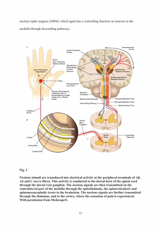
11
nucleus raphe magnus (NRM), which again has a controlling function on neurons in the
medulla through descending pathways.
Fig. 1
Noxious stimuli are transduced into electrical activity at the peripheral terminals of Aβ, Aδ and C nerve fibres. This activity is conducted to the dorsal horn of the spinal cord through the dorsal root ganglion. The noxious signals are then transmitted on the contralateral part of the medulla through the spinothalamic, the spinoreticularis and spinomesencephalic tracts to the brainstem. The noxious signals are further transmitted through the thalamus, and to the cortex, where the sensation of pain is experienced. With permission from Medscape®.
12
A continuous feedback mechanism regulates the nociceptive activity between the connections
in the brainstem and the medulla. In the brainstem there are also connections to the centres
which regulate the blood pressure, heart rate and respiration. These connections form the
basis for the immediate increase in blood pressure, heart rate and respiration rate in the case
of acute noxious stimulation.
4.2 Postoperative pain and analgesia
Pain after surgery has a major effect on patient recovery, and may have implications on
patient safety, the perceived patient care quality as well as health care economics. The most
common complications of ineffective pain control include immobilization, poor wound
healing, chronic pain, deep vein thrombosis, pneumonia, coronary ischemia and insomnia.2-4
Persistent pain in patients after surgery is reported to be between 10-50%, and major chronic
pain is reported in about 2-10% of these patients.4
Postoperative pain is one of the most common concerns and fears among surgical patients.5
Even though there has been a focus on postoperative pain the last decades, a national survey
in the United States demonstrated that 86% of the patients reported moderate, severe or
extreme pain after surgery.5
Pain and pain complications have both medical and financial implications; the patients have
to stay longer in the hospital, and for some readmission to hospital is necessary, which may
lead to dissatisfaction with the medical care.6
There may be a lot of reasons for why postoperative analgesia still is suboptimal in many
patients: poor understanding of pain physiology, lack of efficient analgesics or other
preventive and therapeutic measures as well as suboptimal application of present knowledge
into the individual patient. Poor application of present knowledge and treatment options may
13
be a result of insufficient training of healthcare personnel and patients, fear of serious adverse
effects and economic restrictions on drug and personnel expenses.
The development of hyperalgesia during a surgical procedure has recently attained much
interest in the field of pain physiology; what causes hyperalgesia and may it be prevented?
Early multimodal analgesia also seems to be beneficial for enhanced quality of recovery.7
Paracetamol, nonsteroidal anti-inflammatory drugs, local anaesthesia and opioids play an
important role in multimodal analgesia.7 Opioids are still useful in general anaesthetic
techniques and as treatment of severe postoperative pain, but their role in the development of
hyperalgesia as well as their bothersome adverse effects makes the use of them controversial.
Our focus has been to look into different aspects of basic pain physiology, i.e. opioid-induced
hyperalgesia (OIH) and possibilities to prevent OIH, as well as optimal use of some present
analgesic drugs, i.e. opioids and cyclooxygenase inhibitors.
4.3 Nonsteroidal anti-inflammatory drugs (NSAIDs) and opioids
Nonsteroidal anti-inflammatory drugs (NSAIDs)
NSAIDs are widely used in postoperative pain management, and their analgesic efficacy is
well documented.8-10
NSAIDs have demonstrated opioid-sparing effects after surgery,11-13 and have also an
additive effect when administered in combination with paracetamol.14;15
The opioid-sparing effects of NSAIDs has been demonstrated in a systematic review of
COX-2 inhibitors.16 On average, the opioid consumption in this review was significantly
reduced by 35%, but a significant reduction in opioid-related adverse effects was not shown.
The authors of this review describe the reporting quality with respect to opioid-related
adverse effects as poor. This is interesting, because in a clinical setting the points of interest
are the adverse effects leading to torments for the patients, longer stay in the recovery unit
14
etc., not the total amount of opioids used per se. In a systematic review Marret E et al.
demonstrated that NSAIDs significantly decreased postoperative nausea, vomiting and
sedation.17 In this review, the morphine consumption was positively correlated with the
incidence of nausea and vomiting.
Therefore, it is important that clinical trials do not only register opioid consumption and
analgesic effect, but also opioid-related adverse effects.
The primary analgesic mechanism of NSAIDs is through inhibition of the cyclooxygenase
(COX) enzyme. Cyclooxygenase facilitates the production of prostaglandins, which are
important mediators of inflammation and pain (fig. 2).
Two cyclooxygenase enzymes have been discovered; COX-1 and COX-2.18 COX-1
facilitates the platelet aggregation19 and gastric mucosal protection,20 and COX-2 is an
important mediator of inflammation, pain and fever.21;22
This has resulted in the development of selective COX-2 inhibitors. The advantage of COX-2
inhibitors is the possibility to avoid the tendency for bleeding, which may be very important
in some types of surgery. In addition, gastrointestinal wounds and bleedings are less frequent
with the use of COX-2 inhibitors compared to non-selective NSAIDs.23-26 A selective COX-2
inhibition results in a more specific action on inflammation and pain. Numerous studies have
confirmed that selective COX-2 inhibitors and non-selective NSAIDs have similar analgesic
efficacy.27-30
Traditionally, NSAIDs are considered to be peripherally acting drugs, but evidence has also
clearly demonstrated analgesic efficacy through spinal COX-inhibition.31;32
However, the concept of COX-2 as the major factor in NSAID-induced analgesia has been
challenged. In rats, experimental studies on incisional pain have demonstrated activation of
COX-1 in the spinal cord, and less pain behaviour after intrathecal injection of ketorolac
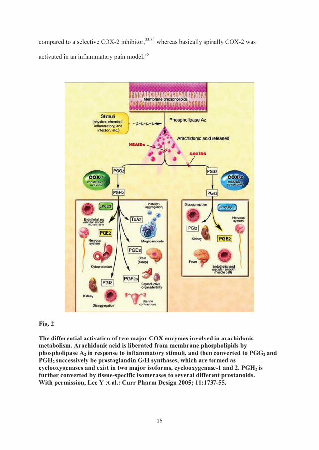
15
compared to a selective COX-2 inhibitor,33;34 whereas basically spinally COX-2 was
activated in an inflammatory pain model.35
Fig. 2
The differential activation of two major COX enzymes involved in arachidonic metabolism. Arachidonic acid is liberated from membrane phospholipids by phospholipase A2 in response to inflammatory stimuli, and then converted to PGG2 and PGH2 successively be prostaglandin G/H synthases, which are termed as cyclooxygenases and exist in two major isoforms, cyclooxygenase-1 and 2. PGH2 is further converted by tissue-specific isomerases to several different prostanoids. With permission, Lee Y et al.: Curr Pharm Design 2005; 11:1737-55.
16
This may indicate that COX activation is different in different pain models. The analgesic
efficacy of various cyclooxygenase inhibitors seems to be more complex than previously
thought, and hopefully further investigations can result in a more detailed clarification of the
analgesic mechanisms of cyclooxygenase inhibitors.
In this thesis two NSAIDs with different selectivity in inhibiting the COX-1 and COX-2
enzymes were compared to investigate the possibility of improving postoperative pain in a
day-surgery unit (paper II). COX-2 inhibitors are presumed to have similar efficacy as
traditional NSAIDs, but whether timing of nonsteroidal anti-inflammatory drugs is of
importance in improving postoperative pain is still a question that needs to be answered. In
paper II, the NSAIDs were administered ahead of surgery to ensure maximum effect before
incision (preemptive analgesia). Preemptive analgesia remains controversial in managing
postoperative pain. In a meta-analysis Ong et al.36 demonstrated that preemptive NSAIDs
reduce analgesic consumption and delay the time to the first rescue analgesic request
postoperatively. There was no improvement in postoperative pain scores. Another review did
not find any significant evidence of better postoperative pain relief by administering NSAIDs
as preemptive medication.37
As described above, experimental studies in rats after incisional pain have demonstrated
activation of COX-1 in the spinal cord, and less pain behaviour after intrathecal injection of
ketorolac before incision compared to a selective COX-2 inhibitor. Therefore, it would be
interesting to investigate, in a clinical setting, if there are any differences between a COX-1
inhibitor and a COX-2 inhibitor administered as preemptive medication with respect to
postoperative pain and analgesic consumption.
Opioids
The opium poppy is known from Mesopotamia since 3-4000 years ago. All antique
civilizations have descriptions of the use of the opium poppy, especially as a tranquillizer and
17
sleep-inducing drug. The name opium has its origin in the Greek word opos meaning juice. In
1806, a pure substance was isolated from the opium poppy and named after the Greek God of
dreams: Morpheus.
Opioids are a generic term for all compounds with the ability to bind the opioid receptors.
Opiats are natural opium alkaloids isolated from the opium poppy, like morphine and
codeine.
Opioids can be classified in several ways:
1) By the way they are produced:
- Naturally occurring: Morphine and codeine.
- Semisynthetic: Heroin, oxycodone and buprenorphine.
- Synthetic: Alfentanil, fentanyl, ketobemidone, methadone, pethidine, remifentanil and
sufentanil.
2) By the type of receptor action:
- Agonists: Alfentanil, codeine, fentanyl, ketobemidone, methadone, morphine,
oxycodone, pethidine, remifentanil and sufentanil.
- Mixed agonists/antagonists: Buprenorphine and nalbuphine.
- Antagonists: Naloxone and metylnaltrexone.
3) By their origin:
- Endogenous opioids: Enkephalin, endorphin and dynorphin.
- Exogenous: Opioid drugs.
There are several different opioid receptors: μ (my), δ (delta), κ (kappa) and ORL1
(nociceptin/orphanin FQ).
The most common opioids are selective µ-opioid receptor agonists: alfentanil, codeine,
fentanyl, ketobemidone, methadone, morphine, remifentanil and oxycodone. Opioid receptors
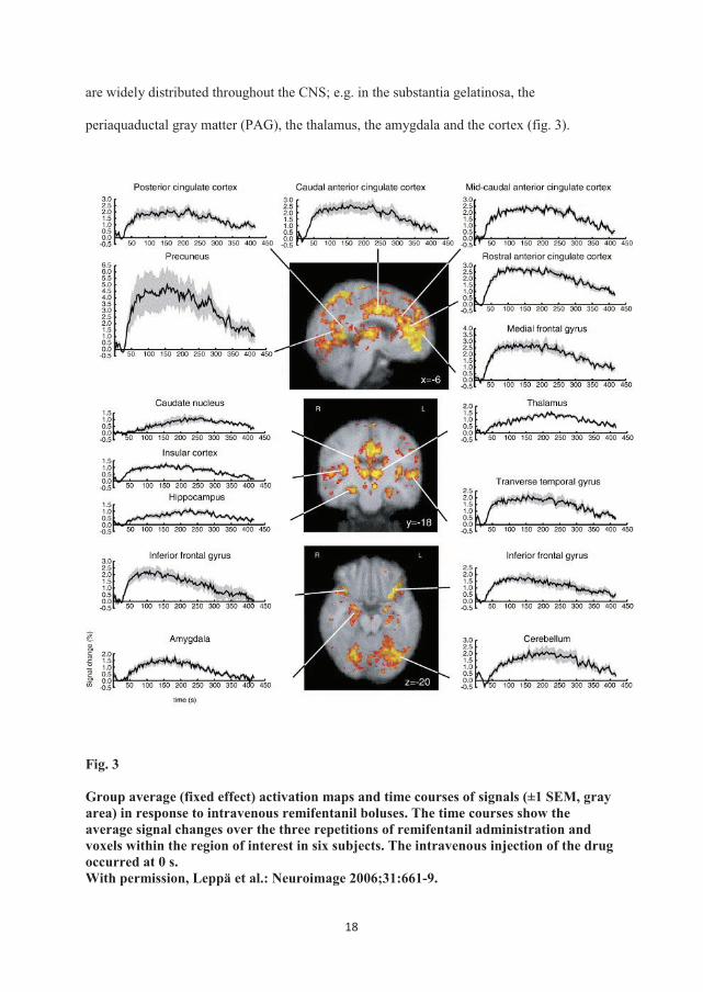
18
are widely distributed throughout the CNS; e.g. in the substantia gelatinosa, the
periaquaductal gray matter (PAG), the thalamus, the amygdala and the cortex (fig. 3).
Fig. 3
Group average (fixed effect) activation maps and time courses of signals (±1 SEM, gray area) in response to intravenous remifentanil boluses. The time courses show the average signal changes over the three repetitions of remifentanil administration and voxels within the region of interest in six subjects. The intravenous injection of the drug occurred at 0 s. With permission, Leppä et al.: Neuroimage 2006;31:661-9.
19
There are also opioid receptors in the gut38;39 and in inflammatory tissue.40
The CNS is the main target for opioids. Opioids inhibit the ascending transmission of noxious
stimuli in the dorsal horn. Opioids can also activate the pain inhibitory system by inhibiting
the activity of GABAergic neurons. This is mediated in the PAG through descending
pathways in the rostral ventral medulla, and inhibits nociceptive responses in the spinal dorsal
horn.
Opioids have a large number of adverse effects like nausea, vomiting, itching, dizziness,
sedation, hallucinations, urinary retention, sleep disturbances, respiratory depression etc. It is
therefore important that the patients are not “overloaded” with opioids. An enhanced use of
opioids increases the risk of adverse effects.
Different opioids have different pharmacological properties, and choosing the right opioid for
the specific surgical procedure may be of importance to achieve better postoperative
analgesia and less adverse effects.
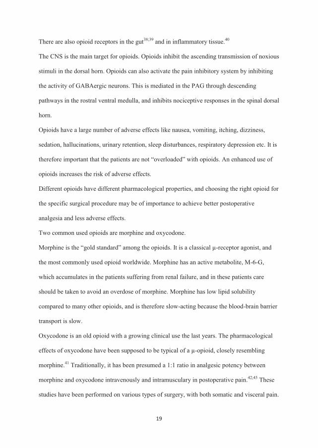
Two common used opioids are morphine and oxycodone.
Morphine is the “gold standard” among the opioids. It is a classical µ-receptor agonist, and
the most commonly used opioid worldwide. Morphine has an active metabolite, M-6-G,
which accumulates in the patients suffering from renal failure, and in these patients care
should be taken to avoid an overdose of morphine. Morphine has low lipid solubility
compared to many other opioids, and is therefore slow-acting because the blood-brain barrier
transport is slow.
Oxycodone is an old opioid with a growing clinical use the last years. The pharmacological
effects of oxycodone have been supposed to be typical of a µ-opioid, closely resembling
morphine.41 Traditionally, it has been presumed a 1:1 ratio in analgesic potency between
morphine and oxycodone intravenously and intramusculary in postoperative pain.42;43 These
studies have been performed on various types of surgery, with both somatic and visceral pain.
20
However, one study on patients undergoing abdominal surgery demonstrated a 2:3 ratio in
analgesic potency between oxycodone and morphine intravenously. In addition, the patients
who received oxycodone were less sedated.44 This may suggest a potential better analgesic
efficacy of oxycodone in visceral pain with less adverse effects. In a multi-modal, tissue-
differentiated experimental pain model in humans, equipotent doses of oxycodone and
morphine demonstrated that oxycodone had a better analgesic effect in visceral pain.45;46 Ev
Even though these studies may indicate that oxycodone is more efficient in the treatment of
visceral pain compared to morphine, this question is still unsettled.hTheref
In paper III, these two opioids were compared with respect to postoperative pain and adverse
effects. The approach in this study was to investigate whether one of those opioids was more
efficient in the treatment of postoperative pain presumed to be dominated by visceral pain
stimulation.
The pharmacological characteristics and differences between oxycodone and morphine are
listed in table 1.
21
Table 1
Oxycodone Morphine
Chemical structure
Receptor binding profile µ-receptor agonist47;48 µ-receptor agonist
Receptor affinity (to µ-receptor) > 20 times less than morphine49 Higher affinity to the µ-receptor than oxycodone49
Receptor activation 3-8 times higher concentration is needed to activate the G-protein compared to morphine
([35S]GTPγ assay)49-51
Lower concentration to activate the G-protein than oxycodone49-51
Analgesic efficacy in postoperative pain
Intravenous: Between 1:1 and 1:1.542-44;52 Epidural: Less potent compared to morphine (8.4-9.8:1)53 Oral: Controlled-release (CR) oxycodone 1.8 times more potent than CR morphine in total effect54
Intravenous: Between 1:1 and 1.5:142-44;52 Epidural: More potent compared to oxycodone53 Oral: Less potent than oxycodone54
Adverse effects Classical µ-opioid-related adverse effects, but less hallucinations55-57 and itching55 (less
histamine release) than morphine
Classical µ-opioid-related adverse effects. More hallucinations and itching than oxycodone55-57
Metabolites O-demethylation in the liver to oxymorphone (no analgesic efficacy)58 and N-demethylation
to noroxycodone (no analgesic efficacy)50
Metabolized in the liver to M-6-G (analgesic efficacy), M-3-G (no analgesic efficacy) and normorphine (analgesic efficacy).59
Volume of distribution 2-3 L/kg60 1-4 L/kg59;61
Elimination Excreted in urine. Oxycodone and noroxycodone as unconjugated, and
oxymorphone as conjugated62
Excreted mainly through urine59
Peak plasma concentration following i.v. administration
Within 25 min41;60 Within 20-30 min63;64
T ½ following intravenous administration
Approximately 2-3 hours60 Approximately 2-3 hours61;65
Protein binding (in vitro) Approximately 38%66 Approximately 31%66
Lipid solubility Similar to morphine66 Low lipid solubility compared to most others opioids (e.g. fentanyl)66
Blood-Brain Barrier transport Active influx of oxycodone into the CNS?67;68 None known active influx
Oral bioavailability Approximately 60%62 18-24%69;70
22
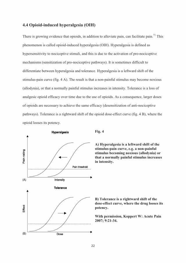
4.4 Opioid-induced hyperalgesia (OIH)
There is growing evidence that opioids, in addition to alleviate pain, can facilitate pain.71 This
phenomenon is called opioid-induced hyperalgesia (OIH). Hyperalgesia is defined as
hypersensitivity to nociceptive stimuli, and this is due to the activation of pro-nociceptive
mechanisms (sensitization of pro-nociceptive pathways). It is sometimes difficult to
differentiate between hyperalgesia and tolerance. Hyperalgesia is a leftward shift of the
stimulus-pain curve (fig. 4 A). The result is that a non-painful stimulus may become noxious
(allodynia), or that a normally painful stimulus increases in intensity. Tolerance is a loss of
analgesic opioid efficacy over time due to the use of opioids. As a consequence, larger doses
of opioids are necessary to achieve the same efficacy (desensitization of anti-nociceptive
pathways). Tolerance is a rightward shift of the opioid dose-effect curve (fig. 4 B), where the
opioid looses its potency.
Fig. 4
A) Hyperalgesia is a leftward shift of the stimulus-pain curve, e.g. a non-painful stimulus becoming noxious (allodynia) or that a normally painful stimulus increases in intensity. B) Tolerance is a rightward shift of the dose-effect curve, where the drug looses its potency. With permission, Koppert W: Acute Pain 2007; 9:21-34.
23
Opioid-induced hyperalgesia is explained as when patients receiving opioids become more
sensitive to pain as a consequence of the opioid therapy itself. The evidence of OIH and the
possibility to prevent OIH can be divided into four types of studies:
1. Studies on former opioid addicts
2. Animal experimental studies
3. Human experimental studies
4. Studies on surgery patients
1. Studies on former opioid addicts
Several studies on current and former opioid addicts have been carried out.72-75
Three of these studies have compared either former addicts on methadone to former addicts
free of methadone,72 or former addicts on methadone to a control group (healthy
volunteers).73;75
The subjects were tested for electrical pain and cold pressor pain. The subjects on methadone
detected pain significantly earlier and had significantly lower pain tolerance than the control
group or former addicts free of opioids.73;75
From this, one may conclude that current opioid addicts are hyperalgesic, and that the
condition is reversible with the withdrawal of the opioid therapy (methadone).
2. Animal experimental studies
Several studies with different models in rats have clearly demonstrated that opioids like
heroin, morphine and fentanyl induced hyperalgesia, and that activation of the NMDA
receptor in the medulla seems to play a key role in the development of opioid-induced
hyperalgesia.76-84
In these studies, treatment with NMDA receptor antagonists like ketamine and MK-
801(dizocilpine maleate) prevented the development of hyperalgesia. It has also been
24
demonstrated that opioids, like remifentanil, activates the NMDA receptor in the dorsal horn
neurons.85
Prostaglandins, like PGE2, can stimulate glutamate release from astrocytes and from the
spinal cord dorsal horns with subsequent activation of the NMDA receptors.86;87
Subsequently, cyclooxygenase inhibitors were found to antagonize this NMDA receptor
activation.88;89 Therefore, it is not surprising that COX-2 inhibition attenuated the level of
thermal hyperalgesia induced by paw carrageenan injection.35
In rats, OIH induced by subcutaneous fentanyl was prevented by administration of
intraperitoneal or intrathecal gabapentin.90 Gabapentin has a binding site to the alpha-2-
subunit of the calcium channel,91 and the antinociceptive efficacy of gabapentin seems to
depend on the affinity to this receptor site.92 The mechanism by which gabapentin prevents
OIH is not totally understood. The underlying hypothesis is that gabapentin inhibits the
glutamate release by binding to the alpha-2-delta subunit of spinal voltage-gated calcium
channels. As opioids increase the level of presynaptic glutamate, this may explain way
gabapentin has the ability to reduce OIH.
In rats, magnesium also seems to prevent OIH after fentanyl administration.93 The main
mechanism seems to be through a voltage-gated antagonist action at the NMDA receptor, and
magnesium is therefore considered as a NMDA receptor antagonist.94
Nitrous oxide (N2O) is a NMDA antagonist,95 and in rats treated with N2O, fentanyl-induced
hyperalgesia in inflammatory and incisional pain models was reduced.96 Sevoflurane also
seems to have a weak anti-hyperalgesic effect.97
3. Human experimental studies
Different experimental pain models have been performed in human volunteers.
a) Short-term infusion of remifentanil with electrical pain. To provoke pain and secondary
hyperalgesia, two microdialysis fibres equipped with internal stainless steel wires are inserted
25
intradermally in the central volar forearm, and a constant current stimulator then provokes
pain. This model has been proven to create a stable area of secondary hyperalgesia to
punctate stimuli by an activation of primarily mechanoinsensitive “silent” C-nociceptors.98
In this model, remifentanil induced postinfusion hyperalgesia,99;100 and different drugs have
been tested to investigate the possibility to prevent/diminish this postinfusion hyperalgesia.
Drugs like ketamine99;101 and parecoxib102 have reduced the area of secondary hyperalgesia to
punctate stimuli.
b) Short-term infusion of remifentanil with heat-capsaicin model. Sensitization is induced by
heating the forearm skin with a thermode at 45 °C. Capsaicin cream applied on the skin
induces pain and primary and secondary hyperalgesia. The area of secondary hyperalgesia
and allodynia can then be measured before, during and after remifentanil infusion. This
model demonstrated that during remifentanil infusion, the area of secondary hyperalgesia was
reduced,103 and after withdrawal, the areas of secondary hyperalgesia and allodynia were
enlarged.104
c) Short-term infusion of remifentanil with electrical pain (applied on the anterior tibial
muscle) and pressure pain test. In this study, remifentanil induced hyperalgesia and tolerance
only in the pressure pain test and was not suppressed by ketamine.105
d) Morphine has demonstrated hyperalgesia in the cold pressor test.106
Summary of the results from experimental pain models on OIH in humans:
Both the electrical model, the cold pressor model, the heat model and the mechanical
(pressure) model demonstrate remifentanil-induced hyperalgesia. OIH in humans can be
prevented by administration of ketamine, a NMDA receptor antagonist. COX-2 inhibition
also seems to have the possibility to prevent OIH. The mechanism behind this is probably
through the NMDA receptor; prostaglandins, like PGE2, can stimulate glutamate release from
26
astrocytes and from the spinal cord dorsal horns with subsequent activation of the NMDA
receptors.86;87 Subsequently, cyclooxygenase inhibitors can antagonize this NMDA receptor
activation.88;89
4. Studies on surgery patients
In clinical trials, it is difficult to demonstrate OIH directly. This is due to the lack of good
models to differentiate between OIH and acute tolerance. The studies that have been
performed demonstrate a more indirect evidence of OIH. In surgery patients, several studies
have been performed comparing a “low dose” opioid group vs. a “high dose” opioid group.
Three of these studies demonstrated higher opioid consumption and higher pain scores
postoperatively in the “high dose” group.107-109 One study did not demonstrate increased
postoperative opioid consumption or pain scores in the group receiving remifentanil, when
compared to a group receiving sevoflurane.110 An important objection to this study is that
both groups received 50% nitrous oxide in oxygen. Nitrous oxide is a NMDA antagonist,95
and may therefore have protected the remifentanil group from developing OIH.
These four studies did not differentiate between OIH and acute tolerance, as both the amount
of postoperative opioid consumption and pain scores was used as main result parameters. To
address this problem another clinical trial has tried to estimate the area of hyperalgesia and
allodynia around the wound postoperatively. The investigators demonstrated that the “high
dose” opioid group had a significantly larger area of pinprick hyperalgesia and allodynia near
the wound compared to the “low dose” opioid group.111 In this study, there was also a third
group receiving a sub-analgesic infusion of ketamine in addition to “high dose” opioid.
Ketamine prevented peri-incisional allodynia and hyperalgesia, and enhanced the opioid
consumption postoperatively.
One study has demonstrated that lornoxicam, a non-selective NSAID, significantly
diminished the acute opioid tolerance and/or hyperalgesia caused by fentanyl.112
27
Interestingly, this study suggests that administration of only three boluses of fentanyl 3 µg/kg
i.v. during approximately 45 min leads to enhanced postoperative opioid consumption
compared to placebo (0.9% NaCl).
The mechanisms of OIH:
It seems that OIH can be induced by peripheral, spinal and systemic administration of
opioids.71 Most of the studies on OIH have been done with µ-opioid receptor agonists. The
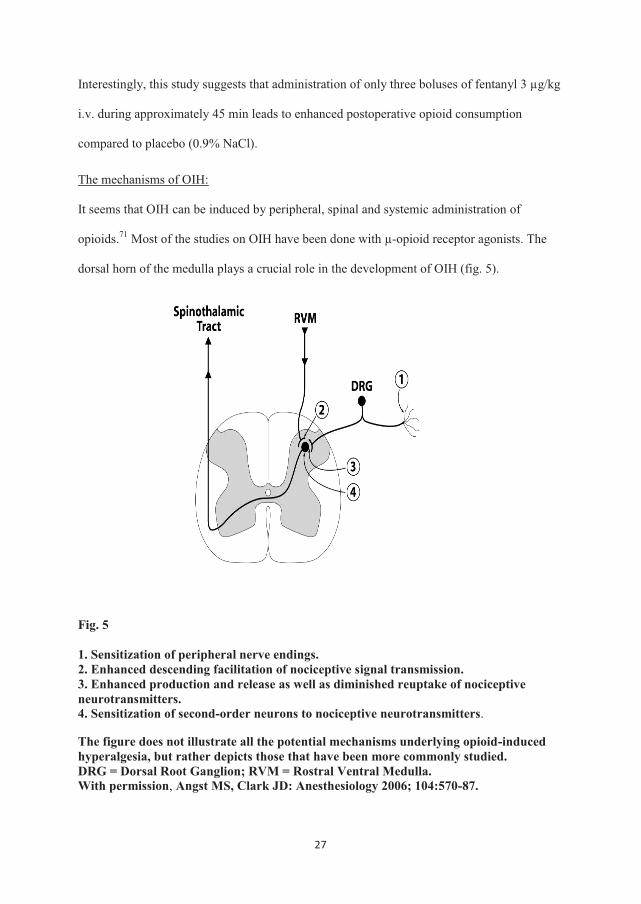
dorsal horn of the medulla plays a crucial role in the development of OIH (fig. 5).
Fig. 5
1. Sensitization of peripheral nerve endings. 2. Enhanced descending facilitation of nociceptive signal transmission. 3. Enhanced production and release as well as diminished reuptake of nociceptive neurotransmitters. 4. Sensitization of second-order neurons to nociceptive neurotransmitters.
The figure does not illustrate all the potential mechanisms underlying opioid-induced hyperalgesia, but rather depicts those that have been more commonly studied. DRG = Dorsal Root Ganglion; RVM = Rostral Ventral Medulla. With permission, Angst MS, Clark JD: Anesthesiology 2006; 104:570-87.
28
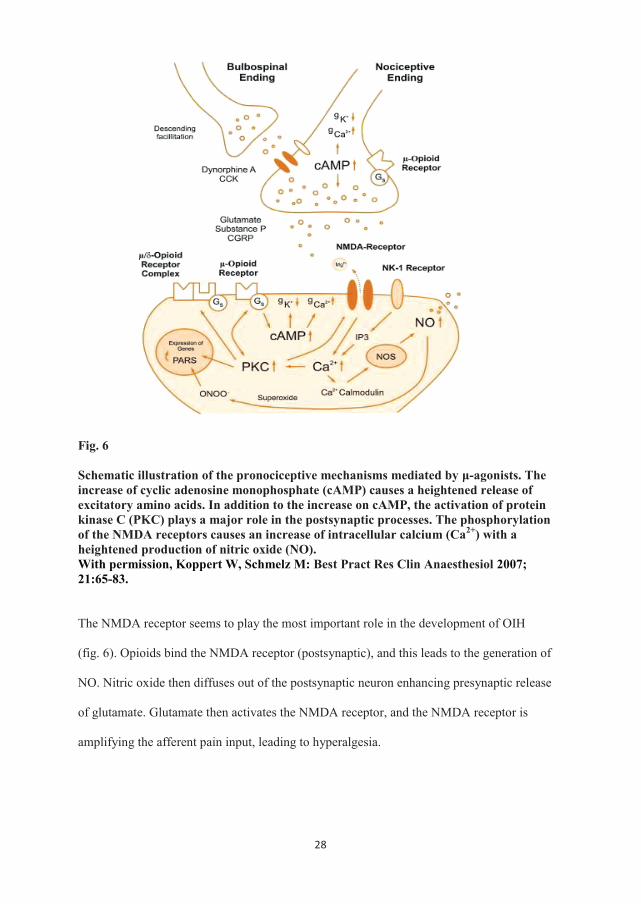
Fig. 6
Schematic illustration of the pronociceptive mechanisms mediated by μ-agonists. The increase of cyclic adenosine monophosphate (cAMP) causes a heightened release of excitatory amino acids. In addition to the increase on cAMP, the activation of protein kinase C (PKC) plays a major role in the postsynaptic processes. The phosphorylation of the NMDA receptors causes an increase of intracellular calcium (Ca2+) with a heightened production of nitric oxide (NO). With permission, Koppert W, Schmelz M: Best Pract Res Clin Anaesthesiol 2007; 21:65-83.
The NMDA receptor seems to play the most important role in the development of OIH
(fig. 6). Opioids bind the NMDA receptor (postsynaptic), and this leads to the generation of
NO. Nitric oxide then diffuses out of the postsynaptic neuron enhancing presynaptic release
of glutamate. Glutamate then activates the NMDA receptor, and the NMDA receptor is
amplifying the afferent pain input, leading to hyperalgesia.
29
Based on current knowledge, how do we deal with OIH in surgery patients today?
In a practical, clinical setting it is difficult to differentiate between OIH and acute tolerance in
surgery patients. Still, it is important for the clinician to know that with the administering of
large doses of opioids the patient is likely to experience more pain and needs more opioids
postoperatively, which is not a desirable situation. If the planned surgical procedure is very
painful or will last for several hours, there is an increased likelihood that large doses of
opioids will be used, and strategies to reduce opioid dosing are necessary.
Strategies to reduce peroperative opioid consumption:
a) If possible, inhalation gases should be used.
Inhalation gases has an analgesic effect,113 and by using inhalation anaesthetics, the opioid
consumption may be reduced.
b) Use of local and regional anaesthesia.
Use of local anaesthetics infiltration or regional blocks may reduce peroperative opioid
consumption,114 and thereby reducing the risk of postoperative OIH.
Preincisional intervention with epidural reduced postoperative opioid consumption and
hyperalgesia definited by the von Frey pain threshold near the wound.115 A more aggressive
use of epidural anaesthesia during surgery seems therefore to be preferable. The systemic use
of intraoperative opioids can be reduced by using epidural anaesthesia actively as an
important analgesic component during surgery.
c) Use of N2O and sevoflurane.
Nitrous oxide is a NMDA receptor antagonist,95 and has demonstrated reduced hyperalgesia
in opioid-exposed rats.96;116 There is also evidence that sevoflurane has antihyperalgesic
effect.97 As far as we know, no clinical study on surgery patients investigating N2O with
respect to OIH has been performed.
30
d) Use of ketamine.
Ketamine is the most potent NMDA receptor antagonist in clinical use. Joly et al.111
administered ketamine 0.5 mg/kg as a bolus, followed by an 5 µg/kg/min infusion during
surgery and 2 μg/kg/min until 48 h after the end of surgery in a group receiving “high dose”
remifentanil. This regimen prevented increased postoperative morphine consumption.
On the other hand, intraoperative infusion of low dose ketamine infusion alone did not
prevent remifentanil-induced acute tolerance and/or hyperalgesia.117;118
More studies are needed before it is possible to put forth a recommendation of a practical use
of ketamine to prevent OIH in surgery patients.
e) Use of NSAIDs.
In an experimental pain model in humans, NSAIDs seem to prevent OIH.102 One clinical
study indicates that NSAIDs may also have this potential in surgery patients.112
f) Use of magnesium.
Magnesium is a NMDA receptor antagonist, and seems to prevent OIH after fentanyl
administration in rats.93 One clinical study has also demonstrated less postoperative morphine
consumption with infusion of magnesium,119 but this study was not designed to investigate
opioid-induced hyperalgesia and/or tolerance.
g) Opioid rotation
Opioid rotation has been suggested as a treatment to avoid/reduce OIH.71 There are several
reports of cancer patients with escalating opioid doses and pain supposed to be hyperalgesia,
who has profited when changing from one opioid to another.120 Changing from phenantren to
peperidin derivatives has been suggested as a treatment to reduce OIH.71
31
Conclusion:
The strongest and most consistent evidences of OIH are derived from studies on normal
volunteers and experimental studies in rats exposed to opioids. There is limited evidence for
the development of OIH in surgery patients,121 however several clinical trials indirectly
indicate that OIH might be a relevant problem in surgery patients.107-109;111;112
In this thesis we have tried to investigate the possibility of reducing postoperative pain and
analgesic consumption by pretreating surgery patients with fentanyl before remifentanil-
based anaesthesia. This is a kind of opioid rotation or shifting of opioids to avoid possible
postoperative opioid-induced hyperalgesia (paper I).
COX-2 inhibitors also seem to prevent/reduce OIH.102 In paper IV, two different
experimental pain models were used to investigate OIH in humans. COX-1 and COX-2
preferring inhibitors were used as pretreatment to investigate any differences in preventing
OIH between COX-1 and COX-2 inhibition.
4.5 Measurement of pain in clinical trials
When measuring pain in clinical trials, it is important that the trauma is similar in all patients.
This is achieved by only including patients scheduled for the same surgical procedure and
involving as few surgeons as possible.
As several factors are involved in the perception of pain, the measurement of pain is
complex. Physiological factors,122 psychological factors,123;124 etnicity,125 gender,126 age127
and earlier experience with pain128 are some of the factors having an impact on postoperative
pain. In RCTs, confounding factors like these must be eliminated as far as possible.
Cultural factors and traditions for pain behaviour are also factors with impact on pain.125 In
Norway, the population is quite conformable compared to many other countries, e.g. the
United States. We included patients with a good understanding of the Norwegian language.
32
This means that the patients in our studies are mostly Caucasians with the same cultural
background.
Gender has an impact on postoperative pain, as women are known to experience more
postoperative pain compared to men.126 When both women and men are included in the same
RCT, it is important that the gender distribution is similar in the comparing groups.
Age also has an impact on postoperative pain; the postoperative opioid consumption
decreases with increased age.127
Patients with chronic pain and/or regular use of pain medication should not be included in
basic clinical pain trials. These patients often have more postoperative pain and need more
opioids to achieve pain relief. If these patients are included and are imbalanced in parallel
groups, the results may be unpredictable.
To avoid many of the known confounding factors with respect to postoperative pain, the ideal
setting in a clinical trial is the same, standardized procedure with only one gender. In
addition, the age distribution should not be too wide. Reduced number of confounders may
increase the statistical power and sensitivity in pain studies, but the conclusions may be less
valid when extrapolated to groups of patients not being included in the study.
In the three clinical trials (paper I-III), the patients were well medicated with analgesic (local
anaesthetics, NSAIDs and paracetamol) and antiemetics (droperidol, ondansetron and
propofol anaesthesia). Of course, good basic pain prophylaxis makes it more difficult to
actually find any differences in pain and adverse effects like nausea and vomiting between
the groups. It is possible that actual differences are not discovered. On the other hand, if
differences are found, this may give a stronger indication of actual differences between the
comparing groups. It may be argued that differences which only appear in a comparison with
inferior pain regimens in placebo groups are not as relevant in clinical practice. In clinical
practice we have to look for improvements when new modalities are administered on the top
33
of present pain regimens. The use of placebo group has also an ethical aspect. Some patients
may receive a less efficient pain treatment, and this could be a problem when applying for
approval from the ethical committee. This is especially true if the analgesic efficacy has been
demonstrated in previous placebo controlled studies, and when the goal of the new study is to
put this into a clinical context.
In the clinical studies we chose to mimic the pain treatment that are actually used in our
hospital. This makes it easier to extrapolate the results to a real clinical situation.
34
5. Aims of the thesis
The overall aim of the thesis was to investigate clinical and experimental aspects of
improving postoperative analgesia by testing different analgesic drugs and by looking for
means of modulation of postoperative opioid-induced hyperalgesia.
In paper II and paper III, two NSAIDs and two opioids, respectively, were compared to
investigate the possibility of improving postoperative pain control.
In paper I and paper IV, the aim was to investigate opioid-induced hyperalgesia, and the
possibility to reduce or prevent postoperative pain or hyperalgesia by pretreating the patients
with opioid (fentanyl) or NSAIDs before remifentanil infusion.
The following hypotheses were tested:
In paper II, etoricoxib (a predominantly COX-2 inhibitor) vs. ketorolac (a predominantly
COX-1 inhibitor) was compared. The primary hypothesis was that etoricoxib would provide
similar maximum early postoperative analgesia as ketorolac. The secondary hypothesis was
that etoricoxib would provide a better analgesic effect after discharge from the hospital.
In paper III, comparison of the analgesic potency, pain scores and adverse effects of
intravenous oxycodone vs. morphine was done in a clinical model of postoperative visceral
pain. The main hypothesis was that oxycodone and morphine were equipotent as analgesics,
in terms of doses measured in mg and with similar efficacy/adverse effect profile.
In paper I, the hypothesis was that pretreatment with fentanyl prior to induction of
remifentanil-based anaesthesia would decrease the self-rated pain scores and opioid
consumption in the postoperative period.
In paper IV, two different experimental human pain models were used to investigate the
possibility of demonstrating remifentanil-induced postinfusion hyperalgesia; a model using
35
electrically evoked pain and a cold pressor test. At the same time, the subjects were
pretreated with parecoxib or ketorolac to investigate the possibility of preventing opioid-
induced hyperalgesia with different types of COX-inhibitors. The main hypothesis was that
remifentanil would induce postinfusion hyperalgesia in both experimental models. The
secondary hypothesis was that parecoxib and ketorolac would prevent or diminish this
postinfusion hyperalgesia in a similar way in both experimental models. By comparing
parecoxib (a predominantly COX-2 inhibitor) against ketorolac (a predominantly COX-1
inhibitor), we had the opportunity to investigate if there were any differences in preventing
remifentanil-induced hyperalgesia by inhibiting either the COX-1 or the COX-2 enzyme.
36
6. Materials and methods
6.1 Study design, approval and registration
The study design in all four studies was prospective, randomized and double-blind.
In the clinical trials (paper I-III), two groups were compared.
The last trial was also placebo-controlled with a crossover design including healthy
volunteers (paper IV). In this trial, each subject went through four sessions: control,
remifentanil, parecoxib + remifentanil and ketorolac + remifentanil. Only males were
included because the pain threshold level is fluctuating in women depending on where in the
menstrual cycle they are. As four sessions were performed, the pain threshold in women may
vary from one session to another, and this is a confounding factor.
In all four papers the randomization was based on computer-generated codes stored in
sequentially numbered, sealed envelopes. A person, who did not participate in the handling or
the assessment of the patients/volunteers, was responsible for preparing these envelopes, and
for opening the envelopes and preparing the medication. The appearance of the syringes (or
tablets) was the same, and the investigator (and the patient/volunteer) was therefore blinded.
Before start, the studies were approved by the Regional Committee for Medical Research
Ethics in South-Eastern Norway and by the local health institution’s privacy protection
representative. The last trial (paper IV) was also approved by the Norwegian Medicines
Agency before start, as the study involved the use of drugs in volunteers and not for an
approved clinical indication with approved doses.
All trials have been registered in ClinicalTrials.gov. ClinicalTrials.gov is a service of the U.S.
National Institutes of Health. It is a registry of clinical trials conducted around the world, and
it is currently compulsory to register clinical trials before start. If not, the possibility to
publish the results is limited.129;130
37
6.2 Patients and volunteers
In the clinical trials (paper I-III), adult persons (18-70 yr) were recruited after a written
informed consent had been obtained. The exclusion criteria were regular use of paracetamol,
NSAIDs, corticosteroids, antiemetics or opioids. Contraindications for
NSAIDs, obesity (paper I) and pregnancy (paper II) were also exclusion criteria. All the
patients were ASA physical status I–II.
In paper IV, volunteers were recruited after a written informed consent had been obtained.
Most of these volunteers were healthy male students, with no known drug allergy or no use of
medication during the experiment. Alcohol or drug abuse was also exclusion criteria.
6.3 Surgery and anaesthesia in paper I-III
In paper I, all patients underwent ACL repair. Both men and women were included.
In paper II, women scheduled for ambulatory, laparoscopic gynaecological surgery were
included. All kinds of minor to moderate day surgical procedures were performed.
In paper III, women scheduled for LSH and TLH were included. All patients had benign
conditions.
Anaesthesia in the clinical trials (paper I-III) was TCI propofol and remifentanil, except from
paper I where µg/kg/min for remifentanil infusion was used. All patients received
paracetamol as basic pain prophylaxis.
In paper II and III, the patients also received a NSAID either before or during surgery. Local
anaesthesia (bupivacain) was administered in all the clinical trials. In the trials only including
women, the patients received antiemetic prophylaxis (droperidol, ondansetron).
38
6.4 Measurement of pain
In the three clinical trials (paper I-III), either VAS and/or VRS was used. VAS is a 100 mm
long visual analog scale, where 0 mm corresponds to no pain, and 100 mm corresponds to the
worst pain imaginable. VRS is a five-point verbal rating scale; 0: no pain, 1: slight pain, 2:
moderate pain, 3: severe pain, and 4: most intense pain.
The measurement of pain is complex (see chapter 4.5). However, we used quite simple
methods to estimate pain postoperatively. VAS and VRS are well established methods, and
their reliability is well documented.131-134 These methods are easy to understand and to use in
a postoperative setting.
In paper I-III, patients with chronic pain and/or regular use of analgesics were excluded.
In paper I and III, PCA for postoperative pain management was used. PCA makes the
patients more autonomous, and this can also be used to tell something about pain indirectly,
as more pain will result in more opioid use.
In paper II, NCA was used for postoperative pain management. In this trial, the patient had to
ask for rescue medication or score VAS ≥ 30 mm to get opioid medication. This might be a
more inaccurate way to treat pain, but in a day surgery unit where the patients often do not
suffer from strong pain, it is a practical way of handling postoperative pain instead of
cumbersome pumps and algorithms. It has been shown that with dedicated nurses, like in our
trial, NCA is experienced as an equally satisfactory method of treating pain compared to
PCA.135;136
In paper III, we tried to create an “ideal” setting to avoid confounding factors with respect to
pain. Laparoscopic hysterectomy is a standardized procedure, performed by few surgeons. In
addition, all the patients are women at approximately the same age. Painmatcher® was used
to discover possible differences in preoperative pain threshold between the two groups. A
possible difference in pain threshold between the two groups would have been a confounding
39
factor. The principle of use of Painmatcher® is to grasp the left hand side electrodes between
the left thumb and the index finger. When the patients push down the electrodes, this initiates
a continuously ascending electrical stimulus.137 Painmatcher® has been found highly reliable
in evaluating pre- and postoperative pain.138 In the first test, the patient had to keep the
pressure until she felt pain (EPT- electrical pain threshold). In the second test, the patient had
to keep the pressure to the maximum tolerable pain (maximum electrical pain threshold -
MEPT).
In paper IV, we induced pain in healthy volunteers. For practical reasons, a verbal rating
scale (0-10) was used in both experimental models (electrical pain and cold pain). During the
experiments, both hands were “tied up” - with ongoing electrical pain at one underarm, and
the other arm exposed to ice water. The use of a linear visual analog scale would have been
difficult, because the participant then had to use one hand to move the pointer.
6.5 Statistical methods
Two parallel groups were compared in the clinical trials (paper I-III). Data were analyzed
using an independent sample t-test for parametric data, the Mann-Whitney U test for non-
parametric data and the χ2-test for categorical data. Bonferroni’s correction was performed on
repeated pain scores values in paper I and II. In paper III, repeated measures analysis of
variance (ANOVA) and area under the curve (AUC) were used for VAS scores. Repeated
measures ANOVA were also used for the sedation scores in this trial.
Paper IV was a randomized, double-blind, placebo-controlled trial with a crossover design.
Previous trials of the electrical pain model have demonstrated that the area of pinprick
hyperalgesia decreases from one session to another.139 Thus, data regarding areas of
secondary hyperalgesia were normalized to achieve the same point of reference in the
participants from all of the four treatments by setting the mean of both baseline
measurements (15 and 30 min) of pinprick hyperalgesia after onset of electrical stimulation,
40
to 100%. The changes from this baseline were transformed to areas under the curve (AUC)
for each period.
We compared the infusion period (30-60 min), the postinfusion period with stimulation (60-
150 min), and the postinfusion period without electrical stimulation (150-210 min). The
changes in percentage were not normally distributed, therefore non-parametric; two related
samples Wilcoxon tests were performed. We also calculated the AUC of VRS scores in the
electrical pain and the cold pressor test. These results were normally distributed, and paired
samples T-tests for each group were performed. Paired samples T-tests were performed on
MAP, HR and SpO2. All data were processed in the SPSS statistical software version 14.0
and 16.0 (SPSS Inc., Chicago, IL). The significance level was set to 0.05.
41
7. Results
7.1 Paper II and paper III
In paper II, one hundred thirty-three women scheduled for ambulatory, laparoscopic
gynaecological surgery were included in this randomized, double-blind study. Group E
received etoricoxib 120 mg orally as premedication and Group K received ketorolac 30 mg
i.v. after induction of anaesthesia.
The first four hours postoperatively, the opioid consumption in Group K was significantly
less than in Group E (Group K 83 ± 65 µg and Group E 123 ± 91 µg fentanyl [mean (SD), P
= 0.004]).
VAS was significantly lower in Group K 30 min after the end of surgery (Group K 31.3 ±
19.7 mm and Group E 43.8 ± 16.9 mm [mean (SD), P < 0.001]). Discharge readiness was
significantly shorter in Group K (222 ± 40 min) compared to Group E (244 ± 47 min) [mean
(SD), P = 0.004]. There were no differences in pain scores or rescue pain medication at 24
and 48 h postoperatively. Group E had less nausea in the 4-24 h period, 9 vs. 22 patients (P =
0.023).
In paper III, ninety-one women admitted for LSH or TLH were randomized to either
intravenous oxycodone or morphine before the end of laparoscopic hysterectomy, and then
continued with patient-controlled analgesia (oxycodone or morphine) for 24 h
postoperatively.
Preoperative electrical pain threshold (EPT) and maximum electrical pain threshold (MEPT)
were similar in the two groups.
The accumulated opioid consumption was significantly less in the oxycodone group
compared to the morphine group; 13.3 ± 10.4 mg vs. 22.0 ± 13.1 mg (P = 0.001). The pain
42
scores were significantly lower in the oxycodone the first hour postoperatively (P = 0.037),
and sedation was significantly lower during the 24 h postoperative period (P = 0.006).
7.2 Paper I and paper IV
In paper I, one-hundred patients admitted for anterior cruciate ligament (ACL) repair were
included and randomized in a double-blind study. Group Pre received fentanyl 1.5 µg/kg i.v.
and Group Post received placebo prior to the remifentanil infusion. At the end of surgery,
Group Pre received fentanyl 1.5 µg/kg and Group Post received fentanyl 3.0 µg/kg i.v.
There were no differences in postoperative pain or analgesic consumption between the two
groups during the first four hours postoperatively. Group Post had significantly less pain in
the 4-24 h period after surgery, with a median VRS score of ‘slight pain’ vs. ‘moderate pain’
in Group Pre (P < 0.05). In this period, the opioid consumption was similar in both groups.
In paper IV, sixteen male volunteers were enrolled to demonstrate remifentanil-induced
postinfusion hyperalgesia in an electrical pain and a cold pain model.
Pinprick hyperalgesia (electrical pain): All groups developed stable and similar pinprick
hyperalgesia areas during the first 30 min of electrical stimulation. During the infusion-
period, the areas under the curve (AUC) for all treatment groups receiving remifentanil were
significantly smaller compared to AUC in the control group.
This antihyperalgesic effect was only present during the infusion. In the postinfusion period,
the areas of pinprick hyperalgesia exceeded control values in the remifentanil group (P =
0.039). Pretreatment with parecoxib prevented remifentanil-induced postinfusion
hyperalgesia compared to the remifentanil group (P = 0.044). Pretreatment with ketorolac did
not prevent postinfusion hyperalgesia compared to the remifentanil group (P = 0.53).
After the electrical stimulation was switched off, there were no significant differences
between the four groups.
43
Cold pressor test (CPT): The AUC values in the first CPT (before any drugs were
administered) were similar in all groups. The CPT at the end of the infusion demonstrated
significantly lower VRS scores in all three groups receiving remifentanil compared to
placebo (P < 0.001). The CPT 1 h after the end of the infusion demonstrated significantly
higher VRS scores in the remifentanil group compared to the control group (P = 0.017).
Pretreatment with ketorolac resulted in significantly lower VRS scores compared to the
remifentanil group (P = 0.046). Pretreatment with parecoxib demonstrated not significant
lower VRS scores compared to Group R (P = 0.18).
44
8. Discussion
8.1 Paper II and paper III, different COX-inhibitors and opioids
In these two clinical trials, different COX-inhibitors and opioids were compared in an attempt
to improve postoperative analgesia in specific clinical models. The choice of different
analgesic agents, included in the multimodal concept of postoperative analgesia, has become
more patient and procedure tailored in recent years (www.postoppain.org). The choice of
analgesics depend on several different factors. The type of surgery is an important
determinant; the extent and type of tissue and cell damage involved, the potential of strong
nerve stimulation or damage and the risk of bleeding. Patient related aspects are also
important: the age and gender of the patient as well as the individual risks of specific adverse
effects such as asthma, gastrointestinal ulcer, renal dysfunction etc. The surgical setting has
also an impact; an inpatient will have access to intravenous administration and professional
surveillance, this is not the case with an ambulatory patient after discharge.
In these two studies paracetamol and local anaesthetics (bupivacain) were used to mimic a
realistic clinical situation with new drugs administered on the top of established routines. The
use of proper, basic non-opioid analgesics in all patient groups will reduce the need of opioid
rescue medication and the risk of potentially serious adverse effects. However, as mentioned
previously, this pragmatic approach may reduce the sensitivity for detecting differences
between the study groups.
Hypothesis paper II:
The primary hypothesis was that etoricoxib would provide similar maximum early
postoperative analgesia as ketorolac. The secondary hypothesis was that etoricoxib would
provide a better analgesic effect after discharge from the hospital.
45
Presurgical injection of ketorolac 30 mg i.v. resulted in less accumulated opioid consumption
during the first 4 h postoperatively, compared to etoricoxib 120 mg orally administered at
least 1 h before surgery. The patients receiving ketorolac also experienced less pain at 30 min
after the end of surgery.
Previous studies comparing etoricoxib to non-selective or predominantly COX-1 selective
NSAIDs in clinical postoperative pain management have demonstrated similar efficacy of the
different drugs.27;28;140
Zhu et al. used an experimental model of incisional pain in rats, which would be similar to a
postoperative clinical situation. They demonstrated significant activation of COX-1 in the
spinal cord with incisional trauma, and less pain behaviour after preoperative intrathecal
injection of ketorolac than of a selective COX-2 inhibitor.33;34 In most studies comparing
non-selective NSAIDs to COX-2 inhibitors, the drugs were administered after surgery. It is
possible that presurgical administration of a COX-1 preferring NSAID, like ketorolac, can
slow down the central sensitization by inhibiting COX-1 in the dorsal horn of the medulla
resulting in less pain postoperatively.33 To our knowledge three, randomized, double-blind
studies with such preoperative administration of either a non-selective NSAID or a COX-2
inhibitor have been conducted.141-143
Pickering et al. compared paracetamol in combination with either rofecoxib, ibuprofen or
placebo.143 Ibuprofen/paracetamol significantly reduced the need of early supplementary
analgesics compared to rofecoxib/paracetamol. Pain scores were also significantly lower in
the ibuprofen group at the time of administration of rescue pain medication.
Morse et al.141 compared ibuprofen 400 mg vs. rofecoxib 40 mg orally as premedication for
mandibular third molar surgery. The ibuprofen group had lower pain scores every time they
scored the patients (not significant; n.s.), and 25% of the patients in the ibuprofen vs. 50% in
the rofecoxib group needed rescue medication (n.s.).
46
Ng et al.142 administered ketorolac 30 mg or parecoxib 40 mg at induction of anaesthesia in
patients undergoing laparoscopic sterilization. The pain scores were lower in the ketorolac
group on awakening and at 1 h postoperatively, but no differences in the need of rescue
medication were seen.
These studies, including our work, may indicate that pretreating patients with a COX-1
preferring NSAID before surgery may lead to lower pain scores and less need of rescue
medication in the early postoperative phase.
We compared one drug administered orally and one drug administered intravenously, and this
may be criticized. Intravenous administration is a more predictable way of achieving a rapid
and more adequate plasma concentration than oral administration. However, the oral
medication in our trial was administered at a mean of 116 min before the start of surgery, and
none of the patients received the tablets less than 60 min before the start of surgery, which
would ensure full absorption and efficacy.144 However, in some of the patients anaesthesia
actually started less than 60 min after the administration of etoricoxib. Anaesthesia per se
may lead to delayed emptying of the stomach, and thus to a potential delayed maximum
plasma concentration of etoricoxib in some patients.
In paper II, the setting is ambulatory surgery where the timing aspects are crucial. The time
from the arrival of the patient in the hospital to the start of anaesthesia and surgery is short,
and administration of premedication at the right time is often difficult to achieve. To ensure
that premedication tablets are completely absorbed and have maximum effect before the start
of anaesthesia, may be important. For etoricoxib, maximum plasma concentration is reached
after 60 min,144 but there are probably important individual differences. Even though almost 2
h (mean) elapsed from premedication to the start of surgery in this trial, the patients receiving
etoricoxib had more pain and needed more opioids postoperatively compared to those
47
receiving ketorolac. The administration of premedication more that two hours before the start
of anaesthesia in a day-surgery unit is unrealistic.
Etoricoxib 120 mg orally and ketorolac 30 mg i.v. were chosen, as these are the
recommended maximal doses according to the approval of the Norwegian government
medicines agency.
For etoricoxib it has been demonstrated that no stronger effect is seen by increasing the oral
dose beyond 120 mg in adults.140 For ketorolac, the initial dosing after marketing of the drug
was 40-60 mg i.v as a singel dose with a possibility to repeat the dose, but after reports of
severe adverse reactions and deaths the maximum dose was set to 90 mg i.v. /day (30 mg
every 8th hour) in adults.145
Surprisingly, there were no differences in pain scores or need of rescue medication at 24 h
and 48 h postoperatively. The recommendation for ketorolac is dosing every 8 h, for
etoricoxib every 24 h. Thus, with 2 doses of each drug administered according to the
recommendations, ketorolac should not be expected to have efficacy throughout the first
night and during the subsequent 24-48 h interval. The second ketorolac dose was
administered just before discharge because of the need of an i.v. access. In our trial, that
corresponded to 3-6 h after the first dose, and this should be even less prone to last until the
registration at 24 h. The lack of prolonged effect of etoricoxib may be explained by a low
baseline pain score and a low amount of rescue medication in both groups. In addition, all
patients received paracetamol 1 g x 4 daily during the study period as a part of a basic pain
prophylaxis.
The etoricoxib group had significantly less nausea 24 h after surgery. This result is difficult to
explain based on pain scores and use of rescue medication in the corresponding time period.
It can be due to subtle non-significant differences in pain, mobilization, hydration or other
PONV risk factors between the groups, or this result may be a coincidence.
48
We demonstrated significantly less time to discharge from the hospital in the ketorolac group
compared to the etoricoxib group. We interpret this as a result of less pain and less need of
opioid rescue medication in the ketorolac group during this part of the postoperative periode.
Hypothesis paper III:
The main hypothesis was that oxycodone and morphine were equipotent analgesics in a study
of clinical visceral pain, in terms of doses measured in mg and with similar effect/adverse
effect profile.
In this clinical model of visceral pain, the patients in the oxycodone group needed
significantly less accumulated oxycodone compared to the accumulated morphine
consumption in the morphine group (13 mg vs. 22 mg). This 2:3 ratio between oxycodone
and morphine has been demonstrated previously in an other clinical model of visceral pain.44
In contrast, clinical studies comparing these two drugs in a postoperative setting with both
somatic and visceral pain have found a 1:1 ratio.42;43
Our findings are supported by experimental studies in humans, which confirm that
oxycodone is superior to morphine in visceral pain.45;46
Oxycodone is clearly a µ-receptor agonist, as discribed in chapter 4.3., table 1. Studies in rats
suggest that oxycodone also has κ-opioid receptor agonist properties,146-148 even though this
is highly disputed,149 and not demonstrated in humans. The κ-opioid receptor is involved in
visceral pain,150;151 and if oxycodone has κ-opioid receptor agonist properties this may
explain why oxycodone has some advantages in the treatment of visceral pain compared to
morphine.
In addition, we found a significantly longer time to the first PCA oxycodone request after the
end of surgery. Based on this, we may conclude that the effect of the first standard dose of
oxycodone administered by the end of surgery lasts longer, or that the drug is more potent, or
both.
49
Oxycodone’s affinity to the μ-opioid receptor is > 20 times less than morphine.49
The oxycodone concentration needed to activate the G-protein as measured by the [35S]GtyS
agonist-stimulated binding is 3-8 times higher compared to morphine.49;50 In spite of this, in
our trial it seems that oxycodone is more potent than morphine, and has less adverse effects
in terms of less sedation.
Recent studies in rats indicate that oxycodone is actively transported through the BBB
(blood-brain barrier).67;68 With the same unbound blood concentrations of oxycodone and
morphine, the unbound concentration of oxycodone in the brain is six times higher than
morphine.68
This may explain why oxycodone seems to be more potent than morphine, but it does not
explain the different ratio between oxycodone and morphine in patients undergoing surgery
with mainly visceral pain compared to patients undergoing surgery with both somatic and
visceral pain.
The first hour postoperatively, the pain scores were significantly lower in the oxycodone
group in spite of the use of PCA. This may again be explained by the possibility that
oxycodone might be more potent that morphine in visceral pain, or that the onset of analgesia
in the morphine group after PCA dosing is slower. Morphine is generally considered to be a
slow-acting drug, but the few clinical studies on this issue suggest a fairly similar time to
onset (about 5-8 min) and peak effect (about 20-30 min) for both drugs administered as i.v.
bolus.152;153 In a clinical postoperative pain study, Kalso et al. demonstrated that oxycodone
achieved faster pain relief and lasted longer than morphine.44 This is in accordance with our
results, indicating a faster onset of analgesia with oxycodone than morphine.
After two hours the differences in pain scores between the oxycodone and morphine groups
disappeared. As oxycodone passes the blood-brain barrier faster than morphine,67;68 this
might explain the gap between pain scores during the first postoperative hour. As morphine
50
slowly penetrates into the CNS, it would take some time before the pain scores are reduced to
the same level as in the oxycodone group.
8.2 Paper I and paper IV, opioid-induced hyperalgesia
Opioid-induced hyperalgesia might influence postoperative pain in terms of stronger pain and
a higher need of analgesics.107-109;111 It is therefore important to explore different approaches
to reduce OIH.
Paper I did not elucidate OIH development, as there was no control group with less potential
of developing OIH. To demonstrate that these patients actually developed OIH/acute
tolerance, a third group with inhalation anaesthetic instead of opioid, or on top of low-dose
opioid should have been included. However, when considering the induction of OIH after
only 30 min of low dose remifentanil in paper IV, it is likely that the patients in both our
groups in paper I developed OIH as they received high doses of remifentanil for
approximately 90 min.
Hypothesis paper I:
Pretreatment with fentanyl before induction of remifentanil-based anaesthesia would
decrease the self-rated pain scores and opioid consumption in the postoperative period.
The idea of pretreatment with one opioid before using another opioid during anaesthesia to
avoid OIH/acute tolerance was based partly on studies on opioid rotation in cancer
patients,71;120 as described in chapter 4.4. Opioid rotation of pure agonists has been
demonstrated to be successful strategies in decreasing OIH.154
The question remains as to whether the use of a pure opioid agonist, with different receptor-
binding properties and a longer duration of action than remifentanil, would be beneficial in
this context. The initiation of study I was also encouraged by widespread, non-documented
practice in many Norwegian departments to administer fentanyl before remifentanil in order
51
to reduce the remifentanil dose, smoothen the recovery and potentially reduce opioid-induced
hyperalgesia.
We did not manage to demonstrate reduced postoperative pain or rescue opioid consumption
after remifentanil-based anaesthesia when pretreating the patients with fentanyl 1.5 µg/kg i.v.
This negative result may be due to the fact that the dose of fentanyl was too low. The
pretreatment dose of 1.5 µg/kg did not have any effect on remifentanil consumption during
surgery compared to the group which did not receive fentanyl before start. The higher
fentanyl dose (Group Pre: 1.5 µg/kg vs. Group Post: 3.0 µg/kg) at the end of surgery resulted
in less pain 24 hours postoperatively in Group Post, which is beyond the expected duration of
effect from such a dose. One explaination may be a potentially better immediate analgesic
protection with the higher dose, which reduce the wind-up and activation of pain
enhancement mechanisms otherwise seen in the postoperative period.
Changing from phenantren to peperidin derivatives has been suggested as a treatment to
reduce OIH.71 McDonnell et al. tried to pretreat patients with morphine (phenantren
derivative) 150 µg/kg before remifentanil (peperidin derivative)-based anaesthesia. They did
not manage to demonstrate reduced pain scores or analgesic consumption postoperatively,
even though morphine is a longer-acting opioid compared to fentanyl.155
Paper I demonstrates that the practice of pretreating the patients with low-dose fentanyl has
no beneficial impact on postoperative pain or analgesic consumption.
Hypothesis paper IV:
The main hypothesis was that remifentanil would induce postinfusion hyperalgesia in both
experimental models. The secondary hypothesis was that parecoxib and ketorolac would
prevent or diminish this postinfusion hyperalgesia in both experimental models.
Two different experimental models (electrical model and cold pressor model) were used to
demonstrate OIH. As NSAIDs are widely used in clinical postoperative pain, it is interesting
52
to investigate whether these drugs can prevent OIH, as OIH may increase postoperative pain
and analgesic consumption.
Parecoxib, a relatively selective COX-2 inhibitor,156 blocked the opioid-induced enlargement
of a hyperalgesic area, suggesting that OIH may be stimulated by spinal COX-2 activity in
humans.102
Both COX-1 and COX-2 are expressed in the spinal cord.34;157;158 There is growing evidence
suggesting that spinal COX-inhibition plays an important role in producing antinociception
and reducing hypersensitivity.34;88;159 There is also evidence for an up-regulation of
prostaglandin E2 in the CNS after surgery.88;160 Therefore, spinal COX-inhibition may be of
importance in preventing acute hyperalgesia after surgery. It would be of interest to
investigate whether a COX-1 preferring inhibitor, like ketorolac,161 also would reduce OIH.
The timing of the administration of a COX-inhibitor seems to be important in reducing OIH.
Parecoxib was either administered just before remifentanil infusion or approximately 30 min
before remifentanil infusion in the study of Tröster et al.102 Only the parecoxib dose
administered 30 min before infusion reduced the postinfusion hyperalgesia.102
Therefore, in our study, ketorolac and parecoxib were administered 15 min before the
electrical stimulus was switched on, and 45 min before remifentanil infusion (fig. 1 in paper
IV). Ketorolac 30 mg i.v. and parecoxib 40 mg i.v. were chosen, as these are the
recommended maximum doses according to the approval from the Norwegian government
medicines agency.
This trial demonstrated that remifentanil induced postinfusion hyperalgesia in both
experimental pain models (electrical pain and cold pain).
Administration of parecoxib before remifentanil infusion, prevented remifentanil-induced
hyperalgesia only in the electrical pain model. This has been demonstrated in an previous
53
study.102 Surprisingly, ketorolac prevented remifentanil-induced hyperalgesia only in the cold
pain model.
While the cutaneous hyperalgesia is mediated by cutaneous “silent” mechano-insensitive C-
nociceptors,162 the cold pressor pain is mediated by nociceptors in the veins.163
This different peripheral activation may explain the different effect of the two NSAIDs in the
two modalities.
The pain scores in the cold pressor test (postinfusion) are relatively similar between
parecoxib and ketorolac, even though only the pain scores in the ketorolac group were
significantly lower compared to the remifentanil group. These results may suggest that COX-
2 rather than COX-1 inhibition may be most important in reducing opioid-induced central
sensitization in these models of experimental pain. However, we do not know to which extent
these experimental pain models can be extrapolated to a situation with clinical postoperative
pain.
It is also interesting to note that hyperalgesia was produced with a fairly low dose of
remifentanil administrated for a short period. Although this was an experimental model, the
results may be added to the ongoing discussion as to how much and how long remifentanil
needs to be administered during clinical anaesthesia for significant hyperalgesia to develop. It
is also interesting to note that in our experimental model the hyperalgesia was shortlasting
(fig. 4 paper IV) and modest. Thus, the impact on pain in a clinical postoperative situation
may not be evident or measurable unless a very sensitive and non-confounding study design
is employed.
54
9. Conclusions
9.1 Paper II and paper III
In paper II, administration of ketorolac 30 mg i.v. before the start of surgery resulted in
significantly less pain 30 min after the end of surgery, significantly less opioid consumption
during the first 4 h postoperatively, and significantly faster postoperative discharge readiness
compared to oral etoricoxib 120 mg administered as premedication.
With a repeated dose of ketorolac before discharge, there were no differences in pain or
rescue analgesic consumption until the next day. Administration of etoricoxib 120 mg in
Group E at 24 h postoperatively did not reduce pain scores or need of rescue medication in
the subsequent 24-48 h period compared to ketorolac, but less nausea was observed in the 4-
24 h period. Thus, in terms of clinical analgesic effect there is no rationale to change from
ketorolac to etoricoxib, although other aspects of adverse effects, costs and mode of
administration are also valid in this context.
In paper III, the accumulated oxycodone consumption was significantly lower compared to
the accumulated morphine consumption. In addition, the oxycodone group had less pain
during the first postoperative hour, and was less sedated throughout the entire postoperative
period. It is our conclusion that PCA oxycodone is a better alternative than PCA morphine in
terms of clinical analgesic effect and less sedation after this kind of procedure.
9.2 Paper I and paper IV
Pretreatment with fentanyl 1.5 µg/kg i.v. (Group Pre) did not reduce postoperative pain or
analgesic consumption (0-4 h) after 90 min of remifentanil-based anaesthesia with
remifentanil 0.43 µg/kg/min. Group Post had significantly less pain in the 4-24 h period after
55
surgery, with a median VRS score of ‘slight pain’ vs. ‘moderate pain’ in Group Pre (P <
0.05). The oxycodone consumption was similar in both groups.
We conclude that there is no rationale to use low dose fentanyl as pretreatment before
remifentanil infusion in this setting, but rather that fentanyl should be administered in an
adequate dose at the end of the surgical procedure.
In paper IV, postinfusion remifentanil-induced hyperalgesia was demonstrated in both
experimental pain models. Parecoxib prevented hyperalgesia in the electrical model, and
ketorolac prevented hyperalgesia in the cold pressor test. Our results may suggest that COX-2
rather than COX-1 inhibition is of major importance to reduce opioid-induced central
sensitization.
56
10. Future perspectives
10.1 NSAIDs
Non-selective NSAIDs and COX-2 inhibitors have been investigated in a large number of
studies with respect to perioperative analgesia. In 2009, Scott Reuben, a leading investigator
was disclosed for data fabrication,164 and a large number of his articles has been retracted.165
Several of Reuben’s works are of controversial topics related to NSAIDs and COX-2
inhibitors.
Reuben has reported that NSAIDs may have a preemptive effect, but this has been questioned
by other investigator groups.37 Further studies are therefore needed to either disprove or
certify this allegation.
The clinical effect of NSAIDs and COX-2 inhibitors on bone and tissue healing is another
important topic that needs clarification. One of Reuben’s articles demonstrated no differences
in the bone fusion rate after 1 year, with the use of perioperative celecoxib.166 This topic
needs more investigation even though another group have also demonstrated no differences in
bone fusion rates with the use of ketorolac.167
The role of NSAIDs and multimodal analgesic regimens in preventing chronic pain after
orthopaedic surgery also remain unsettled,4;168 and needs further studies.
10.2 Oxycodone
Oxycodone is an old opioid which has gained new interest during the last decade.41 Its
possible superior effect in visceral pain has particularly been investigated.45;46 Human studies
have clearly demonstrated that oxycodone is a selective µ-opioid receptor agonist,49;50;169 but
its affinity to the µ-opioid receptor is 20 times less than the affinity of morphine.49 Based on
this, it is difficult to explain that oxycodone and morphine have approximately the same
57
potency in different clinical settings. A Swedish group has demonstrated a potential active
transport mechanism of oxycodone through the BBB,67;68 which may in part explain this gap
between the weak affinity to the µ-opioid receptor and oxycodone’s potency in the clinic.
However, it does not explain the different ratio between oxycodone and morphine in patients
undergoing visceral surgery vs. somatic surgery.43;44;52 Whether this is a real difference or
not, is difficult to conclude from these studies, as they have quite different design.
To rule out this doubt, a future study comparing morphine and oxycodone should actually
contain two clinical trials with equal design. One study with somatic pain postoperatively and
one study with visceral pain postoperatively.
10.3 Opioid-induced hyperalgesia
The strongest evidence that opioids induce hyperalgesia in humans originates from studies of
healthy volunteers exposed to opioid infusion.121 The main problem is to demonstrate OIH in
clinical studies, where there might be a lot of confounding factors compared to the controlled
situation in an experimental design. We also need to get a better understanding of which
experimental pain models that are most relevant for clinical post-surgical pain. It is also
important to document the potential clinical implications of OIH. The main challenge in the
future is do develop a good model being able to differentiate OIH from acute tolerance in a
clinical setting. If this is achieved, we would hopefully be able to develop different types of
clinical interventions to reduce or avoid OIH.
58
11. References
1. Costigan M, Woolf CJ: Pain: Molecular mechanisms. J Pain 2000; 1:35-44.
2. Breivik H: Postoperative pain management: why is it difficult to show that it
improves outcome? Eur J Anaesth 1998; 15:748-51.
3. Carr DB, Goudas LC: Acute pain. Lancet 1999; 353:2051-8.
4. Kehlet H, Jensen TS, Woolf CJ: Persistent postsurgical pain: risk factors and
prevention. Lancet 2006; 367:1618-25.
5. Apfelbaum JL, Chen C, Mehta SS, Gan TJ: Postoperative pain experience: results
from a national survey suggest postoperative pain continues to be undermanaged.
Anesth Analg 2003; 97:534-40.
6. Twersky R, Fishman D, Homel P: What happens after discharge? Return hospital
visits after ambulatory surgery. Anesth Analg 1997; 84:319-24.
7. Kehlet H, Dahl JB: Anaesthesia, surgery, and challenges in postoperative recovery.
Lancet 2003; 362:1921-8.
8. Dahl V, Raeder JC, Drosdal S, Wathne O, Brynildsrud J: Prophylactic oral ibuprofen
or ibuprofen-codeine versus placebo for postoperative pain after primary hip
arthroplasty. Acta Anaesthesiol Scand 1995; 39:323-6.
9. Gillberg LE, Harsten AS, Stahl LB: Preoperative diclofenac sodium reduces post-
laparoscopy pain. Can J Anaesth 1993; 40:406-8.
10. Comfort VK, Code WE, Rooney ME, Yip RW: Naproxen premedication reduces
postoperative tubal ligation pain. Can J Anaesth 1992; 39:349-52.
11. Chen JY, Ko TL, Wen YR, Wu SC, Chou YH, Yien HW, Kuo CD: Opioid-sparing
effects of ketorolac and its correlation with the recovery of postoperative bowel
function in colorectal surgery patients: a prospective randomized double-blinded
study. Clin J Pain 2009; 25:485-9.
12. Cepeda MS, Carr DB, Miranda N, Diaz A, Silva C, Morales O: Comparison of
morphine, ketorolac, and their combination for postoperative pain: results from a
large, randomized, double-blind trial. Anesthesiology 2005; 103:1225-32.
13. Hodsman NB, Burns J, Blyth A, Kenny GN, McArdle CS, Rotman H: The morphine
sparing effects of diclofenac sodium following abdominal surgery. Anaesthesia 1987;
42:1005-8.
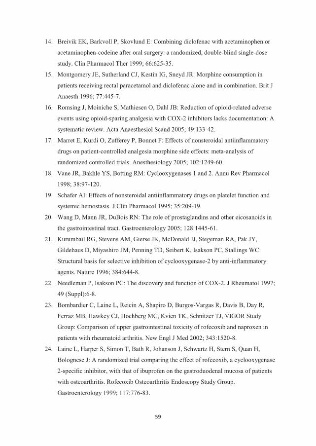
59
14. Breivik EK, Barkvoll P, Skovlund E: Combining diclofenac with acetaminophen or
acetaminophen-codeine after oral surgery: a randomized, double-blind single-dose
study. Clin Pharmacol Ther 1999; 66:625-35.
15. Montgomery JE, Sutherland CJ, Kestin IG, Sneyd JR: Morphine consumption in
patients receiving rectal paracetamol and diclofenac alone and in combination. Brit J
Anaesth 1996; 77:445-7.
16. Romsing J, Moiniche S, Mathiesen O, Dahl JB: Reduction of opioid-related adverse
events using opioid-sparing analgesia with COX-2 inhibitors lacks documentation: A
systematic review. Acta Anaesthesiol Scand 2005; 49:133-42.
17. Marret E, Kurdi O, Zufferey P, Bonnet F: Effects of nonsteroidal antiinflammatory
drugs on patient-controlled analgesia morphine side effects: meta-analysis of
randomized controlled trials. Anesthesiology 2005; 102:1249-60.
18. Vane JR, Bakhle YS, Botting RM: Cyclooxygenases 1 and 2. Annu Rev Pharmacol
1998; 38:97-120.
19. Schafer AI: Effects of nonsteroidal antiinflammatory drugs on platelet function and
systemic hemostasis. J Clin Pharmacol 1995; 35:209-19.
20. Wang D, Mann JR, DuBois RN: The role of prostaglandins and other eicosanoids in
the gastrointestinal tract. Gastroenterology 2005; 128:1445-61.
21. Kurumbail RG, Stevens AM, Gierse JK, McDonald JJ, Stegeman RA, Pak JY,
Gildehaus D, Miyashiro JM, Penning TD, Seibert K, Isakson PC, Stallings WC:
Structural basis for selective inhibition of cyclooxygenase-2 by anti-inflammatory
agents. Nature 1996; 384:644-8.
22. Needleman P, Isakson PC: The discovery and function of COX-2. J Rheumatol 1997;
49 (Suppl):6-8.
23. Bombardier C, Laine L, Reicin A, Shapiro D, Burgos-Vargas R, Davis B, Day R,
Ferraz MB, Hawkey CJ, Hochberg MC, Kvien TK, Schnitzer TJ, VIGOR Study
Group: Comparison of upper gastrointestinal toxicity of rofecoxib and naproxen in
patients with rheumatoid arthritis. New Engl J Med 2002; 343:1520-8.
24. Laine L, Harper S, Simon T, Bath R, Johanson J, Schwartz H, Stern S, Quan H,
Bolognese J: A randomized trial comparing the effect of rofecoxib, a cyclooxygenase
2-specific inhibitor, with that of ibuprofen on the gastroduodenal mucosa of patients
with osteoarthritis. Rofecoxib Osteoarthritis Endoscopy Study Group.
Gastroenterology 1999; 117:776-83.
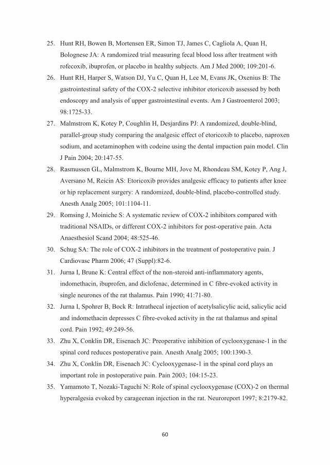
60
25. Hunt RH, Bowen B, Mortensen ER, Simon TJ, James C, Cagliola A, Quan H,
Bolognese JA: A randomized trial measuring fecal blood loss after treatment with
rofecoxib, ibuprofen, or placebo in healthy subjects. Am J Med 2000; 109:201-6.
26. Hunt RH, Harper S, Watson DJ, Yu C, Quan H, Lee M, Evans JK, Oxenius B: The
gastrointestinal safety of the COX-2 selective inhibitor etoricoxib assessed by both
endoscopy and analysis of upper gastrointestinal events. Am J Gastroenterol 2003;
98:1725-33.
27. Malmstrom K, Kotey P, Coughlin H, Desjardins PJ: A randomized, double-blind,
parallel-group study comparing the analgesic effect of etoricoxib to placebo, naproxen
sodium, and acetaminophen with codeine using the dental impaction pain model. Clin
J Pain 2004; 20:147-55.
28. Rasmussen GL, Malmstrom K, Bourne MH, Jove M, Rhondeau SM, Kotey P, Ang J,
Aversano M, Reicin AS: Etoricoxib provides analgesic efficacy to patients after knee
or hip replacement surgery: A randomized, double-blind, placebo-controlled study.
Anesth Analg 2005; 101:1104-11.
29. Romsing J, Moiniche S: A systematic review of COX-2 inhibitors compared with
traditional NSAIDs, or different COX-2 inhibitors for post-operative pain. Acta
Anaesthesiol Scand 2004; 48:525-46.
30. Schug SA: The role of COX-2 inhibitors in the treatment of postoperative pain. J
Cardiovasc Pharm 2006; 47 (Suppl):82-6.
31. Jurna I, Brune K: Central effect of the non-steroid anti-inflammatory agents,
indomethacin, ibuprofen, and diclofenac, determined in C fibre-evoked activity in
single neurones of the rat thalamus. Pain 1990; 41:71-80.
32. Jurna I, Spohrer B, Bock R: Intrathecal injection of acetylsalicylic acid, salicylic acid
and indomethacin depresses C fibre-evoked activity in the rat thalamus and spinal
cord. Pain 1992; 49:249-56.
33. Zhu X, Conklin DR, Eisenach JC: Preoperative inhibition of cyclooxygenase-1 in the
spinal cord reduces postoperative pain. Anesth Analg 2005; 100:1390-3.
34. Zhu X, Conklin DR, Eisenach JC: Cyclooxygenase-1 in the spinal cord plays an
important role in postoperative pain. Pain 2003; 104:15-23.
35. Yamamoto T, Nozaki-Taguchi N: Role of spinal cyclooxygenase (COX)-2 on thermal
hyperalgesia evoked by carageenan injection in the rat. Neuroreport 1997; 8:2179-82.
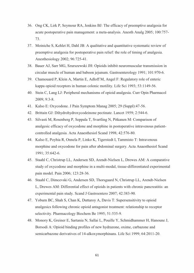
61
36. Ong CK, Lirk P, Seymour RA, Jenkins BJ: The efficacy of preemptive analgesia for
acute postoperative pain management: a meta-analysis. Anesth Analg 2005; 100:757-
73.
37. Moiniche S, Kehlet H, Dahl JB: A qualitative and quantitative systematic review of
preemptive analgesia for postoperative pain relief: the role of timing of analgesia.
Anesthesiology 2002; 96:725-41.
38. Bauer AJ, Sarr MG, Szurszewski JH: Opioids inhibit neuromuscular transmission in
circular muscle of human and baboon jejunum. Gastroenterology 1991; 101:970-6.
39. Chamouard P, Klein A, Martin E, Adloff M, Angel F: Regulatory role of enteric
kappa opioid receptors in human colonic motility. Life Sci 1993; 53:1149-56.
40. Stein C, Lang LJ: Peripheral mechanisms of opioid analgesia. Curr Opin Pharmacol
2009; 9:3-8.
41. Kalso E: Oxycodone. J Pain Symptom Manag 2005; 29 (Suppl):47-56.
42. Brittain GJ: Dihydrohydroxycodeinone pectinate. Lancet 1959; 2:544-6.
43. Silvasti M, Rosenberg P, Seppala T, Svartling N, Pitkanen M: Comparison of
analgesic efficacy of oxycodone and morphine in postoperative intravenous patient-
controlled analgesia. Acta Anaesthesiol Scand 1998; 42:576-80.
44. Kalso E, Poyhia R, Onnela P, Linko K, Tigerstedt I, Tammisto T: Intravenous
morphine and oxycodone for pain after abdominal surgery. Acta Anaesthesiol Scand
1991; 35:642-6.
45. Staahl C, Christrup LL, Andersen SD, Arendt-Nielsen L, Drewes AM: A comparative
study of oxycodone and morphine in a multi-modal, tissue-differentiated experimental
pain model. Pain 2006; 123:28-36.
46. Staahl C, Dimcevski G, Andersen SD, Thorsgaard N, Christrup LL, Arendt-Nielsen
L, Drewes AM: Differential effect of opioids in patients with chronic pancreatitis: an
experimental pain study. Scand J Gastroentero 2007; 42:383-90.
47. Yoburn BC, Shah S, Chan K, Duttaroy A, Davis T: Supersensitivity to opioid
analgesics following chronic opioid antagonist treatment: relationship to receptor
selectivity. Pharmacology Biochem Be 1995; 51:535-9.
48. Monory K, Greiner E, Sartania N, Sallai L, Pouille Y, Schmidhammer H, Hanoune J,
Borsodi A: Opioid binding profiles of new hydrazone, oxime, carbazone and
semicarbazone derivatives of 14-alkoxymorphinans. Life Sci 1999; 64:2011-20.
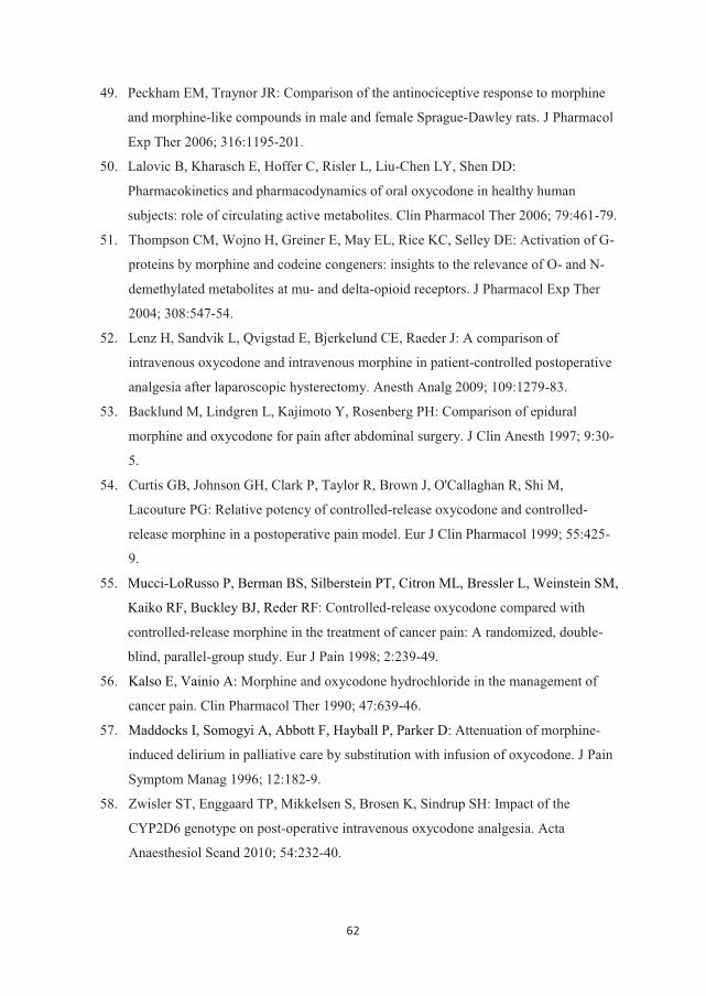
62
49. Peckham EM, Traynor JR: Comparison of the antinociceptive response to morphine
and morphine-like compounds in male and female Sprague-Dawley rats. J Pharmacol
Exp Ther 2006; 316:1195-201.
50. Lalovic B, Kharasch E, Hoffer C, Risler L, Liu-Chen LY, Shen DD:
Pharmacokinetics and pharmacodynamics of oral oxycodone in healthy human
subjects: role of circulating active metabolites. Clin Pharmacol Ther 2006; 79:461-79.
51. Thompson CM, Wojno H, Greiner E, May EL, Rice KC, Selley DE: Activation of G-
proteins by morphine and codeine congeners: insights to the relevance of O- and N-
demethylated metabolites at mu- and delta-opioid receptors. J Pharmacol Exp Ther
2004; 308:547-54.
52. Lenz H, Sandvik L, Qvigstad E, Bjerkelund CE, Raeder J: A comparison of
intravenous oxycodone and intravenous morphine in patient-controlled postoperative
analgesia after laparoscopic hysterectomy. Anesth Analg 2009; 109:1279-83.
53. Backlund M, Lindgren L, Kajimoto Y, Rosenberg PH: Comparison of epidural
morphine and oxycodone for pain after abdominal surgery. J Clin Anesth 1997; 9:30-
5.
54. Curtis GB, Johnson GH, Clark P, Taylor R, Brown J, O'Callaghan R, Shi M,
Lacouture PG: Relative potency of controlled-release oxycodone and controlled-
release morphine in a postoperative pain model. Eur J Clin Pharmacol 1999; 55:425-
9.
55. Mucci-LoRusso P, Berman BS, Silberstein PT, Citron ML, Bressler L, Weinstein SM,
Kaiko RF, Buckley BJ, Reder RF: Controlled-release oxycodone compared with
controlled-release morphine in the treatment of cancer pain: A randomized, double-
blind, parallel-group study. Eur J Pain 1998; 2:239-49.
56. Kalso E, Vainio A: Morphine and oxycodone hydrochloride in the management of
cancer pain. Clin Pharmacol Ther 1990; 47:639-46.
57. Maddocks I, Somogyi A, Abbott F, Hayball P, Parker D: Attenuation of morphine-
induced delirium in palliative care by substitution with infusion of oxycodone. J Pain
Symptom Manag 1996; 12:182-9.
58. Zwisler ST, Enggaard TP, Mikkelsen S, Brosen K, Sindrup SH: Impact of the
CYP2D6 genotype on post-operative intravenous oxycodone analgesia. Acta
Anaesthesiol Scand 2010; 54:232-40.
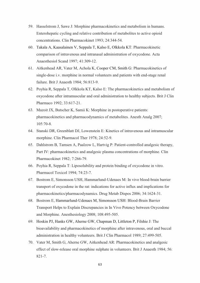
63
59. Hasselstrom J, Sawe J: Morphine pharmacokinetics and metabolism in humans.
Enterohepatic cycling and relative contribution of metabolites to active opioid
concentrations. Clin Pharmacokinet 1993; 24:344-54.
60. Takala A, Kaasalainen V, Seppala T, Kalso E, Olkkola KT: Pharmacokinetic
comparison of intravenous and intranasal administration of oxycodone. Acta
Anaesthesiol Scand 1997; 41:309-12.
61. Aitkenhead AR, Vater M, Achola K, Cooper CM, Smith G: Pharmacokinetics of
single-dose i.v. morphine in normal volunteers and patients with end-stage renal
failure. Brit J Anaesth 1984; 56:813-9.
62. Poyhia R, Seppala T, Olkkola KT, Kalso E: The pharmacokinetics and metabolism of
oxycodone after intramuscular and oral administration to healthy subjects. Brit J Clin
Pharmaco 1992; 33:617-21.
63. Mazoit JX, Butscher K, Samii K: Morphine in postoperative patients:
pharmacokinetics and pharmacodynamics of metabolites. Anesth Analg 2007;
105:70-8.
64. Stanski DR, Greenblatt DJ, Lowenstein E: Kinetics of intravenous and intramuscular
morphine. Clin Pharmacol Ther 1978; 24:52-9.
65. Dahlstrom B, Tamsen A, Paalzow L, Hartvig P: Patient-controlled analgesic therapy,
Part IV: pharmacokinetics and analgesic plasma concentrations of morphine. Clin
Pharmacokinet 1982; 7:266-79.
66. Poyhia R, Seppala T: Liposolubility and protein binding of oxycodone in vitro.
Pharmacol Toxicol 1994; 74:23-7.
67. Bostrom E, Simonsson USH, Hammarlund-Udenaes M: In vivo blood-brain barrier
transport of oxycodone in the rat: indications for active influx and implications for
pharmacokinetics/pharmacodynamics. Drug Metab Dispos 2006; 34:1624-31.
68. Bostrom E, Hammarlund-Udenaes M, Simonsson USH: Blood-Brain Barrier
Transport Helps to Explain Discrepancies in In Vivo Potency between Oxycodone
and Morphine. Anesthesiology 2008; 108:495-505.
69. Hoskin PJ, Hanks GW, Aherne GW, Chapman D, Littleton P, Filshie J: The
bioavailability and pharmacokinetics of morphine after intravenous, oral and buccal
administration in healthy volunteers. Brit J Clin Pharmacol 1989; 27:499-505.
70. Vater M, Smith G, Aherne GW, Aitkenhead AR: Pharmacokinetics and analgesic
effect of slow-release oral morphine sulphate in volunteers. Brit J Anaesth 1984; 56:
821-7.
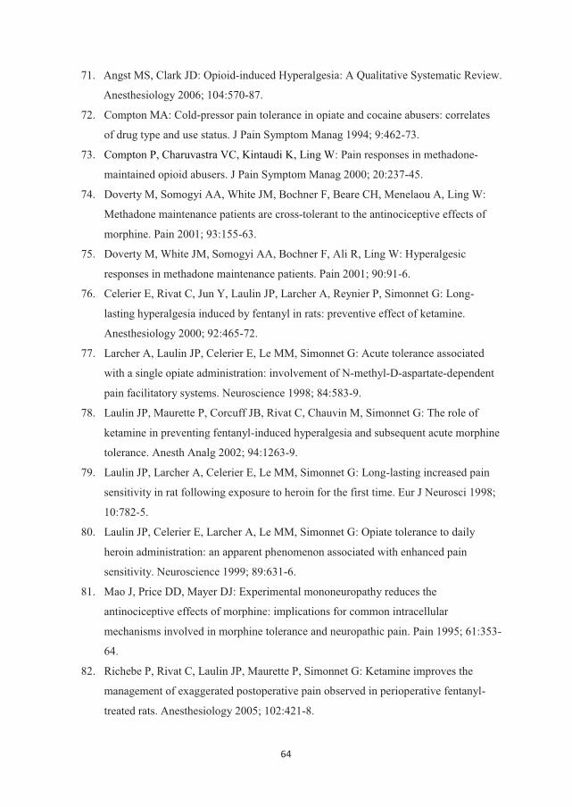
64
71. Angst MS, Clark JD: Opioid-induced Hyperalgesia: A Qualitative Systematic Review.
Anesthesiology 2006; 104:570-87.
72. Compton MA: Cold-pressor pain tolerance in opiate and cocaine abusers: correlates
of drug type and use status. J Pain Symptom Manag 1994; 9:462-73.
73. Compton P, Charuvastra VC, Kintaudi K, Ling W: Pain responses in methadone-
maintained opioid abusers. J Pain Symptom Manag 2000; 20:237-45.
74. Doverty M, Somogyi AA, White JM, Bochner F, Beare CH, Menelaou A, Ling W:
Methadone maintenance patients are cross-tolerant to the antinociceptive effects of
morphine. Pain 2001; 93:155-63.
75. Doverty M, White JM, Somogyi AA, Bochner F, Ali R, Ling W: Hyperalgesic
responses in methadone maintenance patients. Pain 2001; 90:91-6.
76. Celerier E, Rivat C, Jun Y, Laulin JP, Larcher A, Reynier P, Simonnet G: Long-
lasting hyperalgesia induced by fentanyl in rats: preventive effect of ketamine.
Anesthesiology 2000; 92:465-72.
77. Larcher A, Laulin JP, Celerier E, Le MM, Simonnet G: Acute tolerance associated
with a single opiate administration: involvement of N-methyl-D-aspartate-dependent
pain facilitatory systems. Neuroscience 1998; 84:583-9.
78. Laulin JP, Maurette P, Corcuff JB, Rivat C, Chauvin M, Simonnet G: The role of
ketamine in preventing fentanyl-induced hyperalgesia and subsequent acute morphine
tolerance. Anesth Analg 2002; 94:1263-9.
79. Laulin JP, Larcher A, Celerier E, Le MM, Simonnet G: Long-lasting increased pain
sensitivity in rat following exposure to heroin for the first time. Eur J Neurosci 1998;
10:782-5.
80. Laulin JP, Celerier E, Larcher A, Le MM, Simonnet G: Opiate tolerance to daily
heroin administration: an apparent phenomenon associated with enhanced pain
sensitivity. Neuroscience 1999; 89:631-6.
81. Mao J, Price DD, Mayer DJ: Experimental mononeuropathy reduces the
antinociceptive effects of morphine: implications for common intracellular
mechanisms involved in morphine tolerance and neuropathic pain. Pain 1995; 61:353-
64.
82. Richebe P, Rivat C, Laulin JP, Maurette P, Simonnet G: Ketamine improves the
management of exaggerated postoperative pain observed in perioperative fentanyl-
treated rats. Anesthesiology 2005; 102:421-8.
65
83. Rivat C, Laulin JP, Corcuff JB, Celerier E, Pain L, Simonnet G: Fentanyl
Enhancement of Carrageenan-induced Long-lasting Hyperalgesia in Rats: Prevention
by the N-methyl-d-aspartate Receptor Antagonist Ketamine. Anesthesiology 2002;
96:381-91.
84. Van Elstraete AC, Sitbon P, Trabold F, Mazoit JX, Benhamou D: A single dose of
intrathecal morphine in rats induces long-lasting hyperalgesia: the protective effect of
prior administration of ketamine. Anesth Analg 2005; 101:1750-6.
85. Zhao M, Joo DT: Enhancement of spinal N-methyl-D-aspartate receptor function by
remifentanil action at delta-opioid receptors as a mechanism for acute opioid-induced
hyperalgesia or tolerance. Anesthesiology 2008; 109:308-17.
86. Bezzi P, Carmignoto G, Pasti L, Vesce S, Rossi D, Rizzini BL, Pozzan T, Volterra A:
Prostaglandins stimulate calcium-dependent glutamate release in astrocytes. Nature
1998; 391:281-5.
87. O'Rielly DD, Loomis CW: Increased expression of cyclooxygenase and nitric oxide
isoforms, and exaggerated sensitivity to prostaglandin E2, in the rat lumbar spinal
cord 3 days after L5-L6 spinal nerve ligation. Anesthesiology 2006; 104:328-37.
88. Malmberg AB, Yaksh TL: Hyperalgesia mediated by spinal glutamate or substance P
receptor blocked by spinal cyclooxygenase inhibition. Science 1992; 257:1276-9.
89. Malmberg AB, Yaksh TL: Spinal actions of non-steroidal anti-inflammatory drugs:
Evidence for a central role of prostanoids in nociceptive processing. Progr Pharmacol
Clin Pharmacol 1993; 10:91-110.
90. Van Elstraete AC, Sitbon P, Mazoit JX, Benhamou D: Gabapentin prevents delayed
and long-lasting hyperalgesia induced by fentanyl in rats. Anesthesiology 2008;
108:484-94.
91. Gee NS, Brown JP, Dissanayake VU, Offord J, Thurlow R, Woodruff GN: The novel
anticonvulsant drug, gabapentin (Neurontin), binds to the alpha2delta subunit of a
calcium channel. J Biol Chem 1996; 271:5768-76.
92. Field MJ, Hughes J, Singh L: Further evidence for the role of the alpha(2)delta
subunit of voltage dependent calcium channels in models of neuropathic pain. Brit J
Pharmacol 2000; 131:282-6.
93. Van Elstraete AC, Sitbon P, Mazoit JX, Conti M, Benhamou D: Protective effect of
prior administration of magnesium on delayed hyperalgesia induced by fentanyl in
rats. Can J Anaesth 2006; 53:1180-5.
66
94. Fawcett WJ, Haxby EJ, Male DA: Magnesium: physiology and pharmacology. Brit J
Anaesth 1999; 83:302-20.
95. Jevtovic-Todorovic V, Todorovic SM, Mennerick S, Powell S, Dikranian K, Benshoff
N, Zorumski CF, Olney JW: Nitrous oxide (laughing gas) is an NMDA antagonist,
neuroprotectant and neurotoxin. Nat Med 1998; 4:460-3.
96. Richebe P, Rivat C, Creton C, Laulin JP, Maurette P, Lemaire M, Simonnet G:
Nitrous oxide revisited: evidence for potent antihyperalgesic properties.
Anesthesiology 2005; 103:845-54.
97. Richebe P, Rivalan B, Rivat C, Laulin JP, Janvier G, Maurette P, Simonnet G: Effects
of sevoflurane on carrageenan- and fentanyl-induced pain hypersensitivity in
Sprague-Dawley rats. Can J Anaesth 2009; 56:126-35.
98. Schmelz M, Schmid R, Handwerker HO, Torebjork HE: Encoding of burning pain
from capsaicin-treated human skin in two categories of unmyelinated nerve fibres.
Brain 2000; 123:560-71.
99. Angst MS, Koppert W, Pahl I, Clark DJ, Schmelz M: Short-term infusion of the mu-
opioid agonist remifentanil in humans causes hyperalgesia during withdrawal. Pain
2003; 106:49-57.
100. Koppert W, Dern SK, Sittl R, Albrecht S, Schuttler J, Schmelz M: A new model of
electrically evoked pain and hyperalgesia in human skin: the effects of intravenous
alfentanil, S(+)-ketamine, and lidocaine. Anesthesiology 2001; 95:395-402.
101. Koppert W, Sittl R, Scheuber K, Alsheimer M, Schmelz M, Schuttler J: Differential
modulation of remifentanil-induced analgesia and postinfusion hyperalgesia by S-
ketamine and clonidine in humans. Anesthesiology 2003; 99:152-9.
102. Troster A, Sittl R, Singler B, Schmelz M, Schuttler J, Koppert W: Modulation of
remifentanil-induced analgesia and postinfusion hyperalgesia by parecoxib in
humans. Anesthesiology 2006; 105:1016-23.
103. Petersen KL, Jones B, Segredo V, Dahl JB, Rowbotham MC: Effect of remifentanil
on pain and secondary hyperalgesia associated with the heat-capsaicin sensitization
model in healthy volunteers. Anesthesiology 2001; 94:15-20.
104. Hood DD, Curry R, Eisenach JC: Intravenous Remifentanil Produces Withdrawal
Hyperalgesia in Volunteers with Capsaicin-Induced Hyperalgesia. Anesth Analg
2003; 97:810-5.
67
105. Luginbuhl M, Gerber A, Schnider TW, Petersen-Felix S, Arendt-Nielsen L, Curatolo
M: Modulation of remifentanil-induced analgesia, hyperalgesia, and tolerance by
small-dose ketamine in humans. Anesth Analg 2003; 96:726-32.
106. Compton P, Athanasos P, Elashoff D: Withdrawal Hyperalgesia after Acute Opioid
Physical Dependence in Nonaddicted Humans: A Preliminary Study. J Pain 2003;
4:511-9.
107. Chia YY, Liu K, Wang JJ, Kuo MC, Ho ST: Intraoperative high dose fentanyl induces
postoperative fentanyl tolerance. Can J Anaesth 1999; 46:872-7.
108. Guignard B, Bossard AE, Coste C, Sessler DI, Lebrault C, Alfonsi P, Fletcher D,
Chauvin M: Acute opioid tolerance - Intraoperative remifentanil increases
postoperative pain and morphine requirement. Anesthesiology 2000; 93:409-17.
109. Hansen EG, Duedahl TH, Romsing J, Hilsted KL, Dahl JB: Intra-operative
remifentanil might influence pain levels in the immediate post-operative period after
major abdominal surgery. Acta Anaesthesiol Scand 2005; 49:1464-70.
110. Cortinez LI, Brandes V, Munoz HR, Guerrero ME, Mur M: No clinical evidence of
acute opioid tolerance after remifentanil-based anaesthesia. Brit J Anaesth 2001; 87:
866-9.
111. Joly V, Richebe P, Guignard B, Fletcher D, Maurette P, Sessler DI, Chauvin M:
Remifentanil-induced postoperative hyperalgesia and its prevention with small-dose
ketamine. Anesthesiology 2005; 103:147-55.
112. Xuerong Y, Yuguang H, Xia J, Hailan W: Ketamine and Lornoxicam for Preventing a
Fentanyl-Induced Increase in Postoperative Morphine Requirement. Anesth Analg
2008; 107:2032-7.
113. Petersen-Felix S, Luginbuhl M, Schnider TW, Curatolo M, Arendt-Nielsen L,
Zbinden AM: Comparison of the analgesic potency of xenon and nitrous oxide in
humans evaluated by experimental pain. Brit J Anaesth 1998; 81:742-7.
114. Van Lancker P, Abeloos JV, De Clercq CA, Mommaerts MY: The effect of
mandibular nerve block on opioid consumption, nausea and vomiting in bilateral
mandibular osteotomies. Acta Anaesthesiol Belg 2003; 54: 223-6.
115. Katz J, Cohen L, Schmid R, Chan VW, Wowk A: Postoperative morphine use and
hyperalgesia are reduced by preoperative but not intraoperative epidural analgesia:
implications for preemptive analgesia and the prevention of central sensitization.
Anesthesiology 2003; 98:1449-60.
68
116. Bessiere B, Richebe P, Laboureyras E, Laulin JP, Contarino A, Simonnet G: Nitrous
oxide (N2O) prevents latent pain sensitization and long-term anxiety-like behavior in
pain and opioid-experienced rats. Neuropharmacology 2007; 53:733-40.
117. Ganne O, Abisseror M, Menault P, Malhiere S, Chambost V, Charpiat B, Ganne C,
Viale JP: Low-dose ketamine failed to spare morphine after a remifentanil-based
anaesthesia for ear, nose and throat surgery. Eur J Anaesth 2005; 22:426-30.
118. Engelhardt T, Zaarour C, Naser B, Pehora C, De Ruiter J, Howard A, Crawford MW:
Intraoperative Low-Dose Ketamine Does Not Prevent a Remifentanil-Induced
Increase in Morphine Requirement After Pediatric Scoliosis Surgery. Anesth Analg
2008; 107:1170-5.
119. Tramer MR, Schneider J, Marti RA, Rifat K: Role of magnesium sulfate in
postoperative analgesia. Anesthesiology 1996; 84:340-7.
120. Sjogren P, Jensen NH, Jensen TS: Disappearance of morphine-induced hyperalgesia
after discontinuing or substituting morphine with other opioid agonists. Pain 1994;
59:313-6.
121. Fishbain DA, Cole B, Lewis JE, Gao J, Rosomoff RS: Do opioids induce hyperalgesia
in humans? An evidence-based structured review. Pain Med 2009; 10:829-39.
122. Craft RM, Mogil JS, Aloisi AM: Sex differences in pain and analgesia: the role of
gonadal hormones. Eur J Pain 2004; 8:397-411.
123. Kut E, Schaffner N, Wittwer A, Candia V, Brockmann M, Storck C, Folkers G:
Changes in self-perceived role identity modulate pain perception. Pain 2007; 131:191-
201.
124. De Cosmo G, Congedo E, Lai C, Primieri P, Dottarelli A, Aceto P: Preoperative
psychologic and demographic predictors of pain perception and tramadol
consumption using intravenous patient-controlled analgesia. Clin J Pain 2008; 24:399-
405.
125. Tan EC, Lim Y, Teo YY, Goh R, Law HY, Sia AT: Ethnic differences in pain
perception and patient-controlled analgesia usage for postoperative pain. J Pain 2008;
9:849-55.
126. Rosseland LA, Stubhaug A: Gender is a confounding factor in pain trials: women
report more pain than men after arthroscopic surgery. Pain 2004; 112:248-53.
127. Macintyre PE, Jarvis DA: Age is the best predictor of postoperative morphine
requirements. Pain 1996; 64:357-64.
69
128. Rollman GB, Abdel-Shaheed J, Gillespie JM, Jones KS: Does past pain influence
current pain: biological and psychosocial models of sex differences. Eur J Pain 2004;
8:427-33.
129. Bretthauer M, Haug C: Without registration, no publishing! [Norwegian].
Tidsskr Nor Legeforen 2009; 129:733.
130. Haug C, Gotzsche PC, Schroeder TV: Registries and registration of clinical trials.
New Engl J Med 2005; 353:2811-2.
131. Bijur PE, Silver W, Gallagher EJ: Reliability of the visual analog scale for
measurement of acute pain. Acad Emerg Med 2001; 8:1153-7.
132. Breivik EK, Bjornsson GA, Skovlund E: A comparison of pain rating scales by
sampling from clinical trial data. Clin J Pain 2000; 16:22-8.
133. Collins SL, Moore RA, McQuay HJ: The visual analogue pain intensity scale: what is
moderate pain in millimetres? Pain 1997; 72:95-7.
134. DeLoach LJ, Higgins MS, Caplan AB, Stiff JL: The visual analog scale in the
immediate postoperative period: intrasubject variability and correlation with a
numeric scale. Anesth Analg 1998; 86:102-6.
135. Choiniere M, Rittenhouse BE, Perreault S, Chartrand D, Rousseau P, Smith B, Pepler
C: Efficacy and costs of patient-controlled analgesia versus regularly administered
intramuscular opioid therapy. Anesthesiology 1998; 89:1377-88.
136. Munro AJ, Long GT, Sleigh JW: Nurse-administered subcutaneous morphine is a
satisfactory alternative to intravenous patient-controlled analgesia morphine after
cardiac surgery. Anesth Analg 1998; 87:11-5.
137. Lundeberg T, Lund I, Dahlin L, Borg E, Gustafsson C, Sandin L, Rosen A, Kowalski
J, Eriksson SV: Reliability and responsiveness of three different pain assessments. J
Rehabil Med 2001; 33:279-83.
138. Stener-Victorin E, Kowalski J, Lundeberg T: A new highly reliable instrument for the
assessment of pre- and postoperative gynecological pain. Anesth Analg 2002; 5:151-
7.
139. Koppert W, Filitz J, Troster A, Ihmsen H, Angst M, Flor H, Schuttler J, Schmelz M:
Activation of naloxone-sensitive and -insensitive inhibitory systems in a human pain
model. J Pain 2005; 6:757-64.
140. Malmstrom K, Sapre A, Coughlin H, Agrawal NGB, Mazenko RS, Fricke JR:
Etoricoxib in acute pain associated with dental surgery: A randomized, double-blind,
70
placebo- and active comparator-control led dose-ranging study. Clin Ther 2004;
26:667-79.
141. Morse Z, Tump A, Kevelham E: Ibuprofen as a pre-emptive analgesic is as effective
as rofecoxib for mandibular third molar surgery. Odontology 2006; 94:59-63.
142. Ng A, Temple A, Smith G, Emembolu J: Early analgesic effects of parecoxib versus
ketorolac following laparoscopic sterilization: a randomized controlled trial. Brit J
Anaesth 2004; 92:846-9.
143. Pickering AE, Bridge HS, Nolan J, Stoddart PA: Double-blind, placebo-controlled
analgesic study of ibuprofen or rofecoxib in combination with paracetamol for
tonsillectomy in children. Brit J Anaesth 2002; 88:72-7.
144. Cochrane DJ, Jarvis B, Keating GM: Etoricoxib. Drugs 2002; 62:2637-51.
145. Choo V, Lewis S: Ketorolac doses reduced. Lancet 1993; 342:109.
146. Nielsen CK, Ross FB, Lotfipour S, Saini KS, Edwards SR, Smith MT: Oxycodone
and morphine have distinctly different pharmacological profiles: radioligand binding
and behavioural studies in two rat models of neuropathic pain. Pain 2007; 132:289-
300.
147. Nozaki C, Saitoh A, Tamura N, Kamei J: Antinociceptive effect of oxycodone in
diabetic mice. Eur J Pharmacol 2005; 524:75-9.
148. Ross FB, Smith MT: The intrinsic antinociceptive effects of oxycodone appear to be
kappa-opioid receptor mediated. Pain 1997; 73:151-7.
149. Kalso E: How different is oxycodone from morphine? Pain 2007; 132:227-8.
150. Eisenach JC, Carpenter R, Curry R: Analgesia from a peripherally active kappa-
opioid receptor agonist in patients with chronic pancreatitis. Pain 2003; 101:89-95.
151. Black DT, Trevethick M: The kappa opioid receptor is associated with the perception
of visceral pain. Gut 1998; 43:312-3.
152. Leow KP, Smith MT, Watt JA, Williams BE, Cramond T: Comparative oxycodone
pharmacokinetics in humans after intravenous, oral, and rectal administration. Ther
Drug Monit 1992; 14:479-84.
153. Tveita T, Thoner J, Klepstad P, Dale O, Jystad A, Borchgrevink PC: A controlled
comparison between single doses of intravenous and intramuscular morphine with
respect to analgesic effects and patient safety. Acta Anaesthesiol Scand 2008; 52:920-
5.
154. Koppert W: Opioid-induced hyperalgesia-Pathophysiology and clinical relevance.
Acute Pain 2007; 9:21-34.
71
155. McDonnell C, Zaarour C, Hull R, Thalayasingam P, Pehora C, Ahier J, Crawford
MW: Pre-treatment with morphine does not prevent the development of remifentanil-
induced hyperalgesia. Can J Anaesth 2008; 55:813-8.
156. Cheer SM, Goa KL: Parecoxib (parecoxib sodium). Drugs 2001; 61:1133-41.
157. Ebersberger A, Grubb BD, Willingale HL, Gardiner NJ, Nebe J, Schaible HG: The
intraspinal release of prostaglandin E2 in a model of acute arthritis is accompanied by
an up-regulation of cyclo-oxygenase-2 in the spinal cord. Neuroscience 1999; 93:775-
81.
158. Willingale HL, Gardiner NJ, McLymont N, Giblett S, Grubb BD: Prostanoids
synthesized by cyclo-oxygenase isoforms in rat spinal cord and their contribution to
the development of neuronal hyperexcitability. Brit J Pharmacol 1997; 122:1593-604.
159. Southall MD, Michael RL, Vasko MR: Intrathecal NSAIDS attenuate inflammation-
induced neuropeptide release from rat spinal cord slices. Pain 1998; 78:39-48.
160. Buvanendran A, Kroin JS, Berger RA, Hallab NJ, Saha C, Negrescu C, Moric M,
Caicedo MS, Tuman KJ: Upregulation of prostaglandin E2 and interleukins in the
central nervous system and peripheral tissue during and after surgery in humans.
Anesthesiology 2006; 104:403-10.
161. Warner TD, Giuliano F, Vojnovic I, Bukasa A, Mitchell JA, Vane JR: Nonsteroid
drug selectivities for cyclo-oxygenase-1 rather than cyclo- oxygenase-2 are associated
with human gastrointestinal toxicity: A full in vitro analysis. Proc Natl Acad Sci U S
A 1999; 96:7563-82.
162. Weidner C, Schmelz M, Schmidt R, Hansson B, Handwerker HO, Torebjork HE:
Functional attributes discriminating mechano-insensitive and mechano- responsive C
nociceptors in human skin. J Neurosci 1999; 19:10184-90.
163. Klement W, Arndt JO: The role of nociceptors of cutaneous veins in the mediation of
cold pain in man. J Physiol 1992; 449:73-83.
164. Eisenach JC: Data fabrication and article retraction: how not to get lost in the woods.
Anesthesiology 2009; 110:955-6.
165. White PF, Kehlet H, Liu S: Perioperative analgesia: what do we still know? Anesth
Analg 2009; 108:1364-7.
166. Reuben SS, Ekman EF, Raghunathan K, Steinberg RB, Blinder JL, Adesioye J: The
effect of cyclooxygenase-2 inhibition on acute and chronic donor-site pain after
spinal-fusion surgery.[Retraction in Neal JM. Reg Anesth Pain Med. 2009 Mar-
Apr;34(2):184; PMID: 19288610]. Reg Anesth Pain Med 2006; 31:6-13.
72
167. Pradhan BB, Tatsumi RL, Gallina J, Kuhns CA, Wang JC, Dawson EG: Ketorolac
and spinal fusion: does the perioperative use of ketorolac really inhibit spinal fusion?
Spine 2008; 33:2079-82.
168. Brennan TJ, Kehlet H: Preventive analgesia to reduce wound hyperalgesia and
persistent postsurgical pain: not an easy path. Anesthesiology 2005; 103:681-3.
169. Narita M, Nakamura A, Ozaki M, Imai S, Miyoshi K, Suzuki M, Suzuki T:
Comparative Pharmacological Profiles of Morphine and Oxycodone under a
Neuropathic Pain-Like State in Mice: Evidence for Less Sensitivity to Morphine.
Neuropsychopharmacol 2008; 33:1097-112.

I


II


III


IV

1
Title
Modulation of remifentanil-induced postinfusion hyperalgesia by parecoxib or ketorolac in
humans.
Authors
Harald Lenz M.D., University of Oslo and Oslo University Hospital, Department of
Anesthesia, Ullevaal, Oslo, Norway.
Johan Raeder M.D., Ph.D., Professor at University of Oslo and Oslo University Hospital,
Department of Anesthesia, Ullevaal, Oslo, Norway.
Tomas Draegni CRNA, Oslo University Hospital, Department of Anesthesia, Ullevaal, Oslo,
Norway.
Fridtjof Heyerdahl M.D., Ph.D., Oslo University Hospital, Department of Anesthesia,
Ullevaal, Oslo, Norway.
Martin Schmelz, M.D., Ph.D., Professor at University of Heidelberg, Department of
Anesthesia, Mannheim, Germany.
Audun Stubhaug M.D., Ph.D., Oslo University Hospital, Department of Anesthesia,
Rikshospitalet, Oslo, Norway.
Acknowledgement
We would like to express our gratitude to Leiv Sandvik MSc, PhD, Professor in Biostatistics
at University of Oslo, and Center for Clinical Research, Oslo University Hospital, Oslo,
Norway, for his help with the statistics.
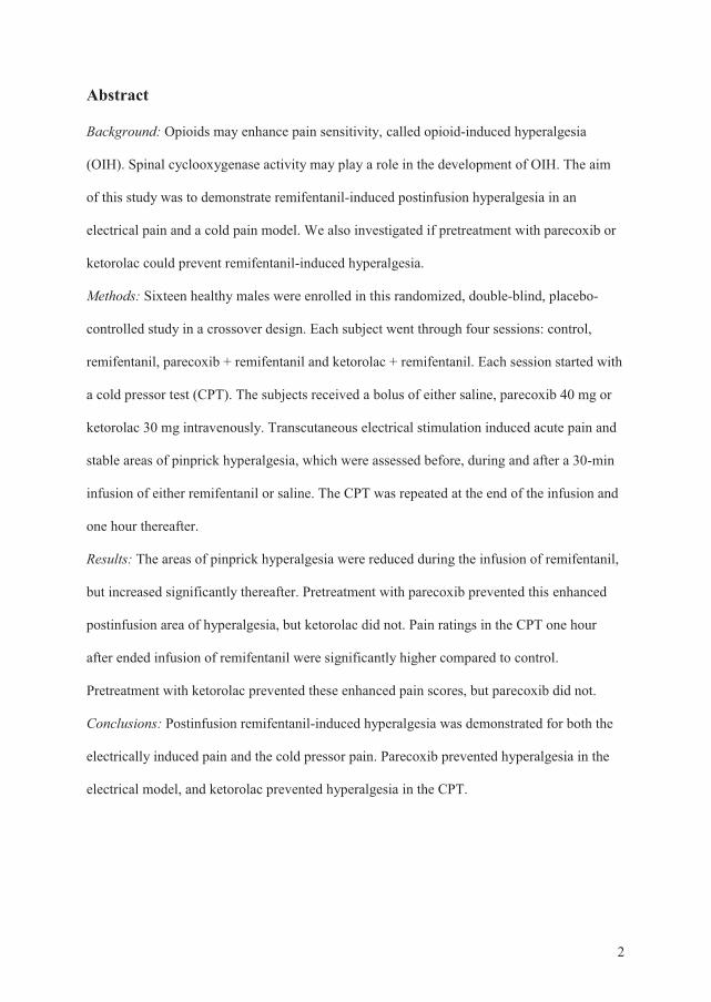
2
Abstract
Background: Opioids may enhance pain sensitivity, called opioid-induced hyperalgesia
(OIH). Spinal cyclooxygenase activity may play a role in the development of OIH. The aim
of this study was to demonstrate remifentanil-induced postinfusion hyperalgesia in an
electrical pain and a cold pain model. We also investigated if pretreatment with parecoxib or
ketorolac could prevent remifentanil-induced hyperalgesia.
Methods: Sixteen healthy males were enrolled in this randomized, double-blind, placebo-
controlled study in a crossover design. Each subject went through four sessions: control,
remifentanil, parecoxib + remifentanil and ketorolac + remifentanil. Each session started with
a cold pressor test (CPT). The subjects received a bolus of either saline, parecoxib 40 mg or
ketorolac 30 mg intravenously. Transcutaneous electrical stimulation induced acute pain and
stable areas of pinprick hyperalgesia, which were assessed before, during and after a 30-min
infusion of either remifentanil or saline. The CPT was repeated at the end of the infusion and
one hour thereafter.
Results: The areas of pinprick hyperalgesia were reduced during the infusion of remifentanil,
but increased significantly thereafter. Pretreatment with parecoxib prevented this enhanced
postinfusion area of hyperalgesia, but ketorolac did not. Pain ratings in the CPT one hour
after ended infusion of remifentanil were significantly higher compared to control.
Pretreatment with ketorolac prevented these enhanced pain scores, but parecoxib did not.
Conclusions: Postinfusion remifentanil-induced hyperalgesia was demonstrated for both the
electrically induced pain and the cold pressor pain. Parecoxib prevented hyperalgesia in the
electrical model, and ketorolac prevented hyperalgesia in the CPT.
3
Introduction
Opioids are the cornerstone in the treatment of moderate to severe pain. In addition to
alleviate pain, there is growing evidence that μ-opioids may reduce the pain threshold,
resulting in enhanced pain when the opioid effect diminishes after end of administration.1-5
This phenomen is known as opioid-induced hyperalgesia (OIH). Large doses of μ-opioids
during surgery seem to enhance pain after emergence from anesthesia and the patients need
more opioids postoperatively.6-8
Activation of the N-methyl-d-aspartate (NMDA)-receptor complex by μ-receptor agonists
seems to be one underlying mechanism of this hyperalgesia development,4,9,10 and OIH can
be prevented by administrating ketamine, an NMDA receptor antagonist.2,4,5,11
Prostaglandins, like PGE2, can stimulate glutamate release from astrocytes and from the
spinal cord dorsal horns with subsequent activation of the NMDA receptors.12,13
Subsequently, cyclooxygenase inhibitors were found to antagonize this NMDA receptor
activation.14,15 Since the NMDA receptor is involved in OIH, it may be possible to reduce
OIH by cyclooxygenase (COX) inhibitors. Parecoxib, a relatively selective COX-2
inhibitor,16 blocked the opioid-induced enlargement of a hyperalgesic area, suggesting that
OIH may be stimulated by spinal COX-2 activity in humans.17 Both the COX-1 and COX-2
variant of the COX-enzyme are expressed in the spinal cord, and COX-1 activation has also
been shown to be important in pain development after surgery.18-20 Therefore, it would be of
interest to investigate whether a COX-1 preferring inhibitor, like ketorolac,21 also could
reduce OIH.
In this study we investigated whether parecoxib and ketorolac could prevent OIH. We used
two different experimental pain models; one model using electrical stimulation to induce pain
and secondary mechanical hyperalgesia22,23 and another model inducing pain from cold
stimuli (cold pressor test - CPT).24

4
The cold pressor pain has been shown to be opioid sensitive.25 By adding CPT as a second
pain stimulus, we wanted to study whether postinfusion hyperalgesia also include acute pain
ratings to this tonic, painful stimulus and could be modulated by COX-antagonists.
5
Materials and Methods
The protocol of this randomized, double-blind, placebo-controlled study with a crossover
design in volunteers was reviewed and approved by the Regional Committee for Medical
Research Ethics in Eastern Norway and the Norwegian Medicines Agency. The study was
registered in ClinicalTrails.gov (ID: NCT 00785863).
Sixteen healthy male subjects were enrolled. None of the subjects had any known drug
allergy or used any other kind of medication before or during the experiments. Alcohol or
drug abuse were exclusion criteria. All subjects were familiarized with the experimental
models by a pre-study session.
To provoke pain and secondary hyperalgesia we used a well established intra-dermal
electrical pain model.2,4,17,22 This model has been proven to create a stable area of secondary
hyperalgesia to punctate stimuli by an activation of primarily mechanoinsensitive “silent” C-
nociceptors.23 These nociceptors are activated at high current densities.26
Two microdialysis fibers equipped with internal stainless steel wires were inserted
intradermally in the central volar forearm. The intradermal length of each fiber was
approximately 10 mm, and the distance between the fibers about 5 mm.
We applied monophasic, rectangular electrical pulses of 0.5 ms duration with alternating
polarity via a constant current stimulator, Digitimer DS7A (Digitimer, Hertfordshire, UK) at
2 Hz. The current intensity was gradually increased during the first 15 min of the stimulus
administration to induce a pain score of 6 on a verbal rating score (VRS) going from 0 to 10
(0 = no pain, 10 = worst pain imaginable). The subjects were allowed to use a decimal to
indicate one level between integers (i.e. 0.5, 1.5, etc.), making it a 21-points scale.
The current was adjusted so that the subject scored VRS = 6 after 15 min, and this current
intensity was kept constant for the rest of the stimulation period; totally 150 min (fig. 1).
6
Each subject went through four separate sessions with an interval of at least one week
between each session.
The control group (Group C) received a 0.9% NaCl bolus intravenously and an infusion of
0.9% NaCl (fig. 1). The remifentanil group (Group R) received a 0.9% NaCl bolus
intravenously and an infusion of remifentanil. The parecoxib + remifentanil group (Group
PR) received a 40 mg parecoxib bolus intravenously and an infusion of remifentanil. The
ketorolac + remifentanil group (Group KR) received a 30 mg ketorolac bolus intravenously
and an infusion of remifentanil.
10 ml syringes for bolus of 0.9% NaCl, parecoxib or ketorolac were used. The parecoxib and
ketorolac syringes were diluted with 0.9% NaCl, so all syringes contained the same amount
of clear liquid (10 ml).
The infusion time of remifentanil or saline was 30 min. 50 ml syringes with the same amount
of clear liquid were used. We used TCI-effect site target control infusion, Minto-model,27,28
starting with 1.0 ng/ml for the first 2 min, thereafter 2.5 ng/ml for the rest of the infusion time
of 30 min (fig. 1). By using effect-TCI we reached a steady-state level faster than using
µg/kg/min infusion, which earlier studies have used.2,4,17
During the infusion of remifentanil or 0.9% NaCl the investigator asked about possible opioid
side effects (e.g. nausea, dizziness, pruritus).
Non-invasive blood pressure, electrocardiography and pulse oximetry were monitored
continuously during the study.
During the ongoing electrical pain stimulus, the subjects scored VRS every 5 min. In
addition, the area of pinprick-hyperalgesia was determined by using a 26 g von Frey filament.
The hyperalgesia areas were determined by measuring in four linear paths parallel and
orthogonally to the axis of the forearm from distant starting points toward the stimulation site
(step size 5 mm), until the subject reported increased pain sensation. In the measuring of the
7
area of secondary hyperalgesia, both diameters (D/2 × d/2 × π) were used. Area of secondary
hyperalgesia was determined every 15 min, from the start of the electrical stimulation (time =
0 min) until one hour after the end of the electrical stimulation period (210 min).
The subjects also underwent the CPT,24 and pain was reported using VRS, as in the electrical
pain model. They performed 3 CPTs for each of the four sessions: one test before any drugs
were administered, one test just before the end of the remifentanil/saline infusion, and one
test one hour after the end of the remifentanil/saline infusion (fig. 1). The last CPT was
performed one hour after the end of the infusion, because presumably there should be
virtually no remifentanil left in the circulation or in the brain, based on the elimination
algorithm in the Minto model.27,28
The water in the CPTs had a temperature between 0.5-1.5 °C. The hand was kept in the water
for a maximum of 120 sec at each test. The subjects were asked to score pain (VRS) every 10
sec. If the subject was unable to keep the hand in the water for 2 min (the pain was
unbearable), the rest of the values were scored to 10.
Statistics
Sample size was calculated based on results from a previous study on area of post-infusion
hyperalgesia.17 Assuming a SD for the difference between treatments of 10%, a paired t-test
(alfa = 0.05, two-sided) with 16 paired observations would yield 96% power to detect a mean
difference of 10% , and 80% power to detect a difference of 7.5% (SamplePower 2.0). Based
on these calculations, we decided to include 16 subjects.
Earlier studies using this electrical pain model have demonstrated that the area of pinprick
hyperalgesia decreases from one session to another session despite the same intensity in
electrical stimulation and pain ratings.29
8
Therefore, data regarding areas of secondary hyperalgesia were normalized to achieve the
same point of reference in subjects from all of the four treatments by setting the mean of both
baseline measurements of pinprick hyperalgesia, i.e. 15 and 30 min after onset of electrical
stimulation, to 100%. The changes from this baseline were calculated as areas under the
curve (AUC) for different time periods.
We compared the infusion period (30-60 min), the postinfusion period (60-150 min), and the
postinfusion period without electrical stimulus (150-210 min). The areas under the curve for
change in area of hyperalgesia (percentage changes) were not normally distributed, therefore
non-parametric, two-sided Wilcoxon tests were performed.
AUC of VRS for the electrical pain and the CPT were also calculated. These results were
normally distributed, and paired samples T-tests for each group were performed.
Paired samples T-tests were performed on MAP, HR and SpO2.
Data were analyzed in SPSS 16.0. The significance level was set to 0.05.
9
Results
All subjects participated in all sessions as planned without dropouts or interfering medication.
There were no correlation between mean electrical current (mA) needed for VRS = 6 after 15
min and mean VRS for the first cold pressor test in the individual test person (P = 0.52, by
Pearson correlation test).
Side effects and vital function monitoring values
The vital function monitoring values (SpO2, MAP and HR) are shown in fig. 2. All subjects
developed side effects during remifentanil infusion (table 2). The side effects never interfered
with the subjects’ ability to answer the questions from the investigators.
Mean arterial blood pressure measured just after the second CPT increased significantly
compared to MAP measured before the CPT in Group C, but the increase was significantly
reduced in the three groups receiving remifentanil (fig. 2). The post-infusion CPT (third CPT)
induced unchanged blood pressure increase in Group C, but in Group R and Group PR the
increase remained significantly lower. Only in Group KR the blood pressure increased to a
similar level as in Group C (fig. 2).
Pain ratings
To achieve a pain rating of VRS 6, the average current was increased to 31.2 ± 22.5 mA
(mean ± SD) during the first 15 min of the electrical stimulation (table 1). After keeping the
current constant, the pain ratings decreased significantly at 30 min, to VRS 5.1 ± 0.07 (mean
± SEM). No significant differences were found between the four groups (fig. 3).
During infusion of remifentanil/saline, the pain ratings decreased significantly in the groups
receiving remifentanil compared Group C (control), P < 0.001 for all three groups (fig. 3).
There were no significant differences in VRS between the three groups receiving remifentanil
10
during the 30 min of infusion. After the end of the infusion, the pain ratings increased and
reached control values. In the Group KR (ketorolac + remifentanil), the pain ratings (VRS)
exceeded the ratings in Group C and became significantly higher during the rest of the
electrical stimulation (P = 0.036, by paired samples T-test).
Pinprick hyperalgesia
All groups developed stable pinprick hyperalgesia areas during the first 30 min of the
electrical stimulation. The areas measured 65.3 (10.9) cm2 in Group C, 71.2 (8.2) cm2 in
Group R, 73.3 (5.9) cm2 in Group PR and 72.7 cm2 (6.9) in Group KR (mean ± SEM). There
were no significant differences between the groups (paired samples T-test).
During the remifentanil/saline infusion-period the areas under the curve (AUC) for all three
treatment groups receiving remifentanil became significantly smaller compared to control (P
< 0.001 in Group PR and Group KR, P = 0.025 in Group R, fig. 4). There were no significant
differences between the groups receiving remifentanil, albeit the reduction of hyperalgesic
areas appeared to be more pronounced when COX-inhibitors were co-administered.
This antihyperalgesic effect was only present during the infusion. After the end of the
infusion, the areas of pinprick hyperalgesia exceeded control values in Group R, P = 0.039
(60-150 min – post infusion period). Pretreatment with parecoxib prevented postinfusion
remifentanil-hyperalgesia compared to Group R, P = 0.044. Pretreatment with ketorolac did
not prevent postinfusion hyperalgesia compared to Group R, P = 0.53.
After the electrical stimulation was switched off (150-210 min – stop electric stimulus
period), there were no significant differences in the areas of pinprick hyperalgesia between
the four groups (fig. 4). The remifentanil induced hyperalgesia (Group R) has returned to
baseline values (Group C).
11
Cold pressor test
The AUC values of the first CPT (before any drugs were administered) remained similar
between all sessions (fig. 5A). In the Group C, there were no significant differences between
the three consecutive CPTs.
The CPT at the end of the infusion (second CPT) demonstrated similar and significant lower
VRS in all three groups receiving remifentanil compared to second CPT in Group C (P <
0.001, fig. 5B).
The CPT 1 h after the end of infusion showed significantly higher VRS in Group R compared
to Group C (P = 0.017, fig. 5C). Pretreatment with ketorolac (Group KR) resulted in
significantly lower VRS compared to Group R (P = 0.046), whereas pretreatment with
parecoxib did not (P = 0.18).
There were no significant differences between Group C, Group PR and Group KR at the third
CPT.
12
Discussion
We were able to reproduce significant hyperalgesia after cessation of a 30 min infusion of
remifentanil as assessed by increased area of secondary mechanical hyperalgesia as
demonstrated in earlier studies.2,4,17 In parallel, pain ratings to noxious cold stimuli increased
in the remifentanil group compared to control, reflecting hyperalgesia in the cold pressor test.
Increased pain after cessation of opioid application can be interpreted as acute tolerance or
OIH.1 The duration of remifentanil application in our study was too short to investigate
tolerance.30 Previous studies in healthy volunteers did not observe remifentanil-induced
tolerance even at application times of 90 min.2 Thus, in our study, post-infusion hyperalgesia
developed as measured by pain ratings to electrical stimulation and area of secondary
mechanical hyperalgesia. Hyperalgesia for electrical pain has been demonstrated previously,2-
4,17 but not for heat stimuli.2
While confirming that remifentanil induced increase of secondary mechanical hyperalgesia,
we additionally found post-infusion hyperalgesia in the CPT, as might have been expected
according to previous results.30,31
Our results confirm that low-dose remifentanil (target 2.5 ng/ml) and short infusion (30 min)
result in detectable hyperalgesia. However, the magnitude of hyperalgesia seems to be limited
and the duration shortlasting, as the areas of hyperalgesia returned to control values after the
electrical pain was swift off (fig. 4).
Opioid-induced NMDA activation or internalization of µ-opioid receptors have been
suggested as the etiology of OIH.32
Recently, intracellular recordings of dorsal horn neurons have shown that withdrawal of
remifentanil can acutely induce long-term potentiation (LTP) in spino-thalamic tract
projection neurons that was sensitive to NMDA blockers.33 Increased pain ratings to CPT and
13
electrical stimuli as well as larger areas of secondary mechanical hyperalgesia are in
accordance with these results.
Administering parecoxib before remifentanil infusion, prevented remifentanil induced
hyperalgesia only in the electrical pain model. This has been demonstrated in an earlier
study.17 Surprisingly, ketorolac prevented remifentanil induced hyperalgesia only in the cold
pain model.
The secondary mechanical hyperalgesia is induced by sensitizing spinal input from “silent”
mechano-insensitive C-nociceptors,26 that renders the dorsal horn hypersensitive to A-delta
nociceptor input.34 In contrast, the cold pressor pain is mediated by tonic discharge of
nociceptors in the veins.35 This different peripheral activation may explain the different effect
of the two NSAIDs on the two modalities.
Both COX-1 and COX-2 are expressed in the spinal cord.18-20 There is growing evidence
suggesting that spinal COX-inhibition plays an important role in producing antinociception
and reducing hypersensitivity.14,18,36,37 There is also evidence for an up-regulation of
prostaglandin E2 in CNS after surgery.14,38 Therefore, spinal COX-inhibition may be of
importance in preventing acute hyperalgesia after surgery.
So far, there are few studies on OIH in a clinical incision pain model, but one study
demonstrated increased pain sensitivity assessed by peri-incisional allodynia and
hyperalgesia after large-dose intraoperative remifentanil.11 In that study ketamine prevented
peri-incisional allodynia and hyperalgesia.
To our knowledge no clinical study has tried to do the same with a COX-inhibitor. However,
one study has demonstrated that lornoxicam, a combined COX-1 and COX-2 inhibitor,
significantly diminished the acute opioid tolerance and/or hyperalgesia caused by fentanyl.39
The ratings of electrically induced pain after end of remifentanil infusions in the study from
Tröster et al.17 exceeded the pain scores compared to their control group. This hyperalgesic
14
effect remained stable for the rest of the observation period. We could only demonstrate this
hyperalgesic effect in the ketorolac group (fig. 3). We hypothesize that the CPTs may have
disturbed the pain ratings from the electrical pain scores, accordning to the classical gate-
theory.40 The CPT is a painful test, and many of the subjects had considerably lowered scores
from the stable electrical pain stimulation after the CPTs.
An interesting observation is that, in addition to higher electrical pain scores in Group KR
after remifentanil-infusion, this group also had a significantly higher MAP after the last CPT
compared to Group PR and Group R (fig. 2). Blood pressure control and descending pain
inhibition share common pathways and mechanisms in a complex and not fully understood
way. The fact that the ketorolac group had higher MAP after the last CPT (DNIC-diffuse
noxious inhibitory control-procedure) and higher electrical pain scores after infusion
compared with the remifentanil and the parecoxib group indicates that ketorolac may block
descending inhibition induced by cold pressor test and that mechanisms shared with blood
pressure control is involved (eg. NA-mechanisms).
There was no correlation between mean electrical current (mA) needed for VRS = 6 after 15
min and mean VRS for the first cold pressor test in the individual test person. This is in
accordance with results from an animal study, which demonstrates a lack of correlation of
sensitivity to nociception in different pain models.41
An important question is the relevance of our results for clinical postoperative pain. The
measurement of pain to cold-, electrical-, heat- and pressure-stimuli are only experimental
tests that will not ideally mimic clinical pain conditions; on the other hand, using surgical
incision is un-feasible in volunteers.
An animal, incisional pain model was linked to spinal COX-1 activation,18 whereas COX-2
was activated in an inflammatory pain model.42 Comparable data from humans do not exist.

15
Certainly more research should be done in this area, preferably with clinical models of real
surgical pain.
In conclusion, we have demonstrated postinfusion remifentanil-hyperalgesia in electrically
induced pain and cold pressor pain. Our results may suggest that COX-2 rather than COX-1
inhibition is of major importance to reduce opioid-induced central sensitization.
16
References
1. Angst MS, Clark DJ: Opioid-induced Hyperalgesia: A Qualitative Systematic Review.
ANESTHESIOLOGY 2006; 104:570-87
2. Angst MS, Koppert W, Pahl I, Clark JD, Schmelz M: Short-term infusion of the mu-
opioid agonist remifentanil in humans causes hyperalgesia during withdrawal. Pain
2003; 106:49-57
3. Koppert W, Angst MS, Alsheimer M, Sittl R, Albrecht S, Schuttler J, Schmelz M:
Naloxone provokes similar pain facilitation as observed after short-term infusion of
remifentanil in humans. Pain 2003; 106:91-9
4. Koppert W, Sittl R, Scheuber K, Alsheimer M, Schmelz M, Schuttler J: Differential
modulation of remifentanil-induced analgesia and postinfusion hyperalgesia by S-
ketamine and clonidine in humans. ANESTHESIOLOGY 2003; 99:152-9
5. Luginbuhl M, Gerber A, Schnider TW, Petersen-Felix S, Arendt-Nielsen L, Curatolo
M: Modulation of remifentanil-induced analgesia, hyperalgesia, and tolerance by small-
dose ketamine in humans. Anesth Analg 2003; 96:726-32
6. Hansen EG, Duedahl TH, Romsing J, Hilsted KL, Dahl JB: Intra-operative remifentanil
might influence pain levels in the immediate post-operative period after major
abdominal surgery. Acta Anaesthesiol Scand 2005; 49:1464-70
7. Guignard B, Bossard AE, Coste C, Sessler DI, Lebrault C, Alfonsi P, Fletcher D,
Chauvin M: Acute opioid tolerance - Intraoperative remifentanil increases postoperative
pain and morphine requirement. ANESTHESIOLOGY 2000; 93:409-17
17
8. Chia YY, Liu K, Wang JJ, Kuo MC, Ho ST: Intraoperative high dose fentanyl induces
postoperative fentanyl tolerance. Can J Anaesth 1999; 46:872-7
9. Celerier E, Rivat C, Jun Y, Laulin JP, Larcher A, Reynier P, Simonnet G: Long-lasting
hyperalgesia induced by fentanyl in rats: preventive effect of ketamine.
ANESTHESIOLOGY 2000; 92:465-72
10. Rivat C, Laulin JP, Corcuff JB, Celerier E, Pain L, Simonnet G: Fentanyl Enhancement
of Carrageenan-induced Long-lasting Hyperalgesia in Rats: Prevention by the N-
methyl-d-aspartate Receptor Antagonist Ketamine. ANESTHESIOLOGY 2002; 96:381-91
11. Joly V, Richebe P, Guignard B, Fletcher D, Maurette P, Sessler DI, Chauvin M:
Remifentanil-induced postoperative hyperalgesia and its prevention with small-dose
ketamine. ANESTHESIOLOGY 2005; 103:147-55
12. Bezzi P, Carmignoto G, Pasti L, Vesce S, Rossi D, Rizzini BL, Pozzan T, Volterra A:
Prostaglandins stimulate calcium-dependent glutamate release in astrocytes. Nature
1998; 391:281-5
13. O'Rielly DD, Loomis CW: Increased expression of cyclooxygenase and nitric oxide
isoforms, and exaggerated sensitivity to prostaglandin E2, in the rat lumbar spinal cord
3 days after L5-L6 spinal nerve ligation. ANESTHESIOLOGY 2006; 104:328-37
14. Malmberg AB, Yaksh TL: Hyperalgesia mediated by spinal glutamate or substance P
receptor blocked by spinal cyclooxygenase inhibition. Science 1992; 257:1276-9
15. Malmberg AB, Yaksh TL: Spinal actions of non-steroidal anti-inflammatory drugs:
Evidence for a central role of prostanoids in nociceptive processing. Prog Pharmacol
Clin Pharmacol 1993; 10:91-110
18
16. Cheer SM, Goa KL: Parecoxib (parecoxib sodium). Drugs 2001; 61:1133-41
17. Troster A, Sittl R, Singler B, Schmelz M, Schuttler J, Koppert W: Modulation of
remifentanil-induced analgesia and postinfusion hyperalgesia by parecoxib in humans.
ANESTHESIOLOGY 2006; 105:1016-23
18. Zhu X, Conklin DR, Eisenach JC: Cyclooxygenase-1 in the spinal cord plays an
important role in postoperative pain. Pain 2003; 104:15-23
19. Ebersberger A, Grubb BD, Willingale HL, Gardiner NJ, Nebe J, Schaible HG: The
intraspinal release of prostaglandin E2 in a model of acute arthritis is accompanied by
an up-regulation of cyclo-oxygenase-2 in the spinal cord. Neuroscience 1999; 93:775-
81
20. Willingale HL, Gardiner NJ, McLymont N, Giblett S, Grubb BD: Prostanoids
synthesized by cyclo-oxygenase isoforms in rat spinal cord and their contribution to the
development of neuronal hyperexcitability. Br J Pharmacol 1997; 122:1593-604
21. Warner TD, Giuliano F, Vojnovic I, Bukasa A, Mitchell JA, Vane JR: Nonsteroid drug
selectivities for cyclo-oxygenase-1 rather than cyclo- oxygenase-2 are associated with
human gastrointestinal toxicity: A full in vitro analysis. Proc Natl Acad Sci U S A
1999; 96:7563-8
22. Koppert W, Dern SK, Sittl R, Albrecht S, Schuttler J, Schmelz M: A new model of
electrically evoked pain and hyperalgesia in human skin: the effects of intravenous
alfentanil, S(+)-ketamine, and lidocaine. ANESTHESIOLOGY 2001; 95:395-402
19
23. Schmelz M, Schmidt R, Handwerker HO, Torebjork HE: Encoding of burning pain
from capsaicin-treated human skin in two categories of unmyelinated nerve fibres.
Brain 2000; 123:560-71
24. Wolff S, Hardy JD: Studies on pain; observations on pain due to local cooling and on
factors involved in the cold pressor effect. J Clin Invest 1941; 20:521-33
25. Eckhardt K, Ammon S, Hofmann U, Riebe A, Gugeler N, Mikus G: Gabapentin
enhances the analgesic effect of morphine in healthy volunteers. Anesth Analg 2000;
91:185-91
26. Weidner C, Schmelz M, Schmidt R, Hansson B, Handwerker HO, Torebjork HE:
Functional attributes discriminating mechano-insensitive and mechano- responsive C
nociceptors in human skin. J Neurosci 1999; 19:10184-90
27. Minto CF, Schnider TW, Shafer SL: Pharmacokinetics and pharmacodynamics of
remifentanil. II. Model application. ANESTHESIOLOGY 1997; 86:24-33
28. Minto CF, Schnider TW, Egan TD, Youngs E, Lemmens HJ, Gambus PL, Billard V,
Hoke JF, Moore KH, Hermann DJ, Muir KT, Mandema JW, Shafer SL: Influence of
age and gender on the pharmacokinetics and pharmacodynamics of remifentanil. I.
Model development. ANESTHESIOLOGY 1997; 86:10-23
29. Koppert W, Filitz J, Troster A, Ihmsen H, Angst M, Flor H, Schuttler J, Schmelz M:
Activation of naloxone-sensitive and -insensitive inhibitory systems in a human pain
model. J Pain 2005; 6:757-64
20
30. Chu LF, Clark DJ, Angst MS: Opioid tolerance and hyperalgesia in chronic pain
patients after one month of oral morphine therapy: A preliminary prospective study. J
Pain 2006; 7:43-8
31. Compton P, Athanasos P, Elashoff D: Withdrawal Hyperalgesia after Acute Opioid
Physical Dependence in Nonaddicted Humans: A Preliminary Study. J Pain 2003;
4:511-9
32. Chu LF, Angst MS, Clark DJ: Opioid-induced hyperalgesia in humans: molecular
mechanisms and clinical considerations. Clin J Pain 2008; 24:479-96
33. Drdla R, Gassner M, Gingl E, Sandkuhler J: Induction of synaptic long-term
potentiation after opioid withdrawal. Science 2009; 325:207-10
34. Magerl W, Fuchs PN, Meyer RA, Treede RD: Roles of capsaicin-insensitive
nociceptors in cutaneous pain and secondary hyperalgesia. Brain 2001; 124:1754-64
35. Klement W, Arndt JO: The role of nociceptors of cutaneous veins in the mediation of
cold pain in man. J Physiol 1992; 449:73-83
36. Southall MD, Michael RL, Vasko MR: Intrathecal NSAIDS attenuate inflammation-
induced neuropeptide release from rat spinal cord slices. Pain 1998; 78:39-48
37. Zhu X, Conklin DR, Eisenach JC: Preoperative inhibition of cyclooxygenase-1 in the
spinal cord reduces postoperative pain. Anesth Analg 2005; 100:1390-3
38. Buvanendran A, Kroin JS, Berger RA, Hallab NJ, Saha C, Negrescu C, Moric M.
Caicedo MS, Tuman KJ: Upregulation of prostaglandin E2 and interleukins in the
central nervous system and peripheral tissue during and after surgery in humans.
ANESTHESIOLOGY 2006; 104:403-10
21
39. Xuerong Y, Yuguang H, Xia J, Hailan W: Ketamine and Lornoxicam for Preventing a
Fentanyl-Induced Increase in Postoperative Morphine Requirement. Anesth Analg
2008; 107:2032-7
40. Melzack R, Wall PD: Pain mechanisms: a new theory. Science 1965; 150:971-9
41. Mogil JS, Wilson SG, Bon K, Lee SE, Chung K, Raber P, Pieper JO, Hain HS, Belknap
JK, Hubert L, Elmer GI, Chung JM, Devor M : Heritability of nociception I: responses
of 11 inbred mouse strains on 12 measures of nociception. Pain 1999; 80;67-82
42. Yamamoto T, Nozaki-Taguchi N, Yamamoto T, Nozaki-Taguchi N: Role of spinal
cyclooxygenase (COX)-2 on thermal hyperalgesia evoked by carageenan injection in
the rat. Neuroreport 1997; 8:2179-82
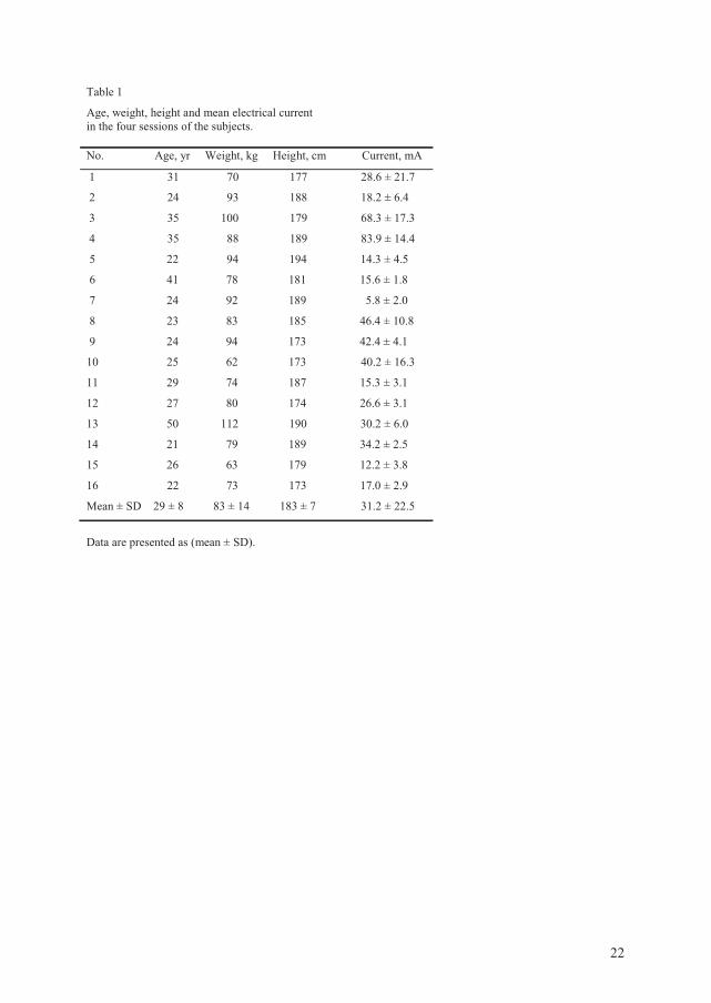
22
Table 1 Age, weight, height and mean electrical current in the four sessions of the subjects. No. Age, yr Weight, kg Height, cm Current, mA
1 31 70 177 28.6 ± 21.7
2 24 93 188 18.2 ± 6.4
3 35 100 179 68.3 ± 17.3
4 35 88 189 83.9 ± 14.4
5 22 94 194 14.3 ± 4.5
6 41 78 181 15.6 ± 1.8
7 24 92 189 5.8 ± 2.0
8 23 83 185 46.4 ± 10.8
9 24 94 173 42.4 ± 4.1
10 25 62 173 40.2 ± 16.3
11 29 74 187 15.3 ± 3.1
12 27 80 174 26.6 ± 3.1
13 50 112 190 30.2 ± 6.0
14 21 79 189 34.2 ± 2.5
15 26 63 179 12.2 ± 3.8
16 22 73 173 17.0 ± 2.9
Mean ± SD 29 ± 8 83 ± 14 183 ± 7 31.2 ± 22.5
Data are presented as (mean ± SD).

23
Table 2
Side effects during drug infusions. Saline Remifentanil Parecoxib + Remifentanil Ketorolac + Remifentanil
Sedation 0 14 11 12
Pruritus 0 10 9 8
Nausea 0 1 3 1
Dizziness 0 6 7 9
Number of subjects who reported side effects (n = 16).
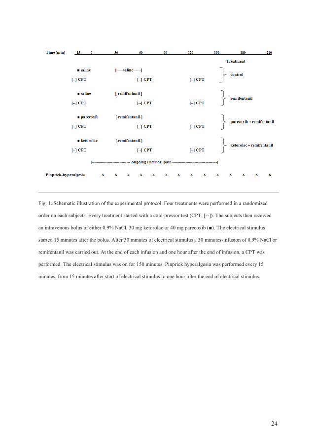
24
Fig. 1. Schematic illustration of the experimental protocol. Four treatments were performed in a randomized
order on each subjects. Every treatment started with a cold-pressor test (CPT, [--]). The subjects then received
an intravenous bolus of either 0.9% NaCl, 30 mg ketorolac or 40 mg parecoxib (■). The electrical stimulus
started 15 minutes after the bolus. After 30 minutes of electrical stimulus a 30 minutes-infusion of 0.9% NaCl or
remifentanil was carried out. At the end of each infusion and one hour after the end of infusion, a CPT was
performed. The electrical stimulus was on for 150 minutes. Pinprick hyperalgesia was performed every 15
minutes, from 15 minutes after start of electrical stimulus to one hour after the end of electrical stimulus.
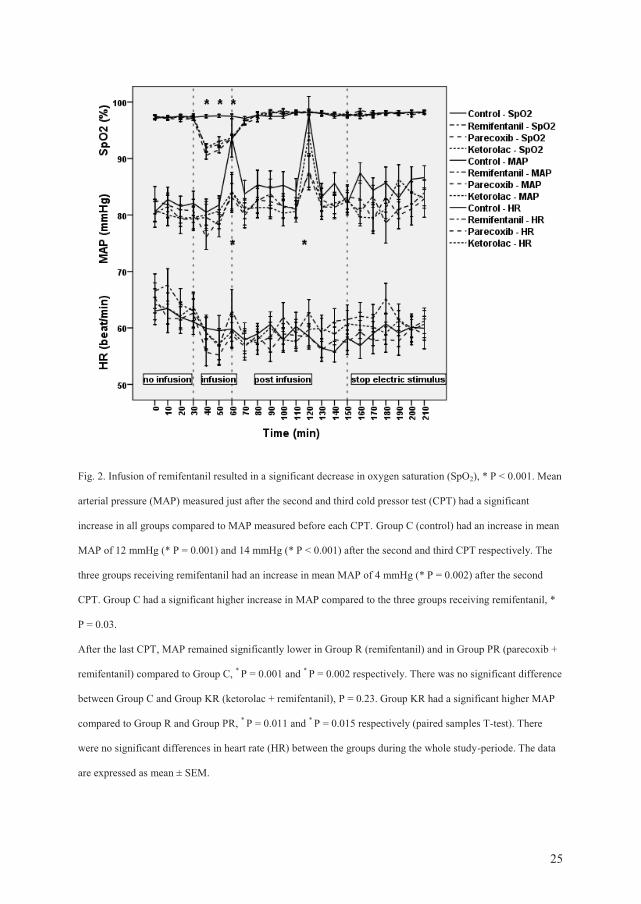
25
Fig. 2. Infusion of remifentanil resulted in a significant decrease in oxygen saturation (SpO2), * P < 0.001. Mean
arterial pressure (MAP) measured just after the second and third cold pressor test (CPT) had a significant
increase in all groups compared to MAP measured before each CPT. Group C (control) had an increase in mean
MAP of 12 mmHg (* P = 0.001) and 14 mmHg (* P < 0.001) after the second and third CPT respectively. The
three groups receiving remifentanil had an increase in mean MAP of 4 mmHg (* P = 0.002) after the second
CPT. Group C had a significant higher increase in MAP compared to the three groups receiving remifentanil, *
P = 0.03.
After the last CPT, MAP remained significantly lower in Group R (remifentanil) and in Group PR (parecoxib +
remifentanil) compared to Group C, * P = 0.001 and * P = 0.002 respectively. There was no significant difference
between Group C and Group KR (ketorolac + remifentanil), P = 0.23. Group KR had a significant higher MAP
compared to Group R and Group PR, * P = 0.011 and * P = 0.015 respectively (paired samples T-test). There
were no significant differences in heart rate (HR) between the groups during the whole study-periode. The data
are expressed as mean ± SEM.
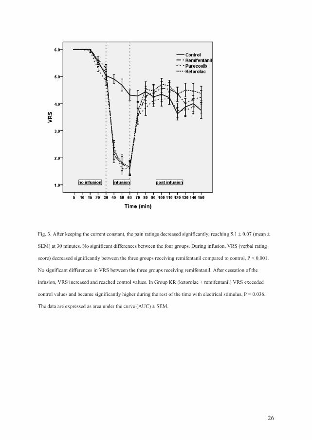
26
Fig. 3. After keeping the current constant, the pain ratings decreased significantly, reaching 5.1 ± 0.07 (mean ±
SEM) at 30 minutes. No significant differences between the four groups. During infusion, VRS (verbal rating
score) decreased significantly between the three groups receiving remifentanil compared to control, P < 0.001.
No significant differences in VRS between the three groups receiving remifentanil. After cessation of the
infusion, VRS increased and reached control values. In Group KR (ketorolac + remifentanil) VRS exceeded
control values and became significantly higher during the rest of the time with electrical stimulus, P = 0.036.
The data are expressed as area under the curve (AUC) ± SEM.
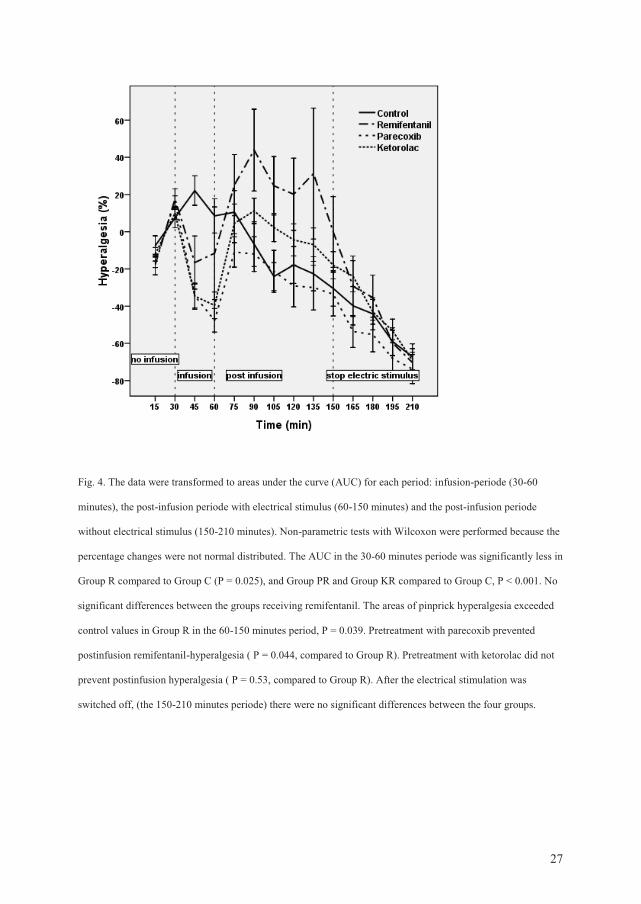
27
Fig. 4. The data were transformed to areas under the curve (AUC) for each period: infusion-periode (30-60
minutes), the post-infusion periode with electrical stimulus (60-150 minutes) and the post-infusion periode
without electrical stimulus (150-210 minutes). Non-parametric tests with Wilcoxon were performed because the
percentage changes were not normal distributed. The AUC in the 30-60 minutes periode was significantly less in
Group R compared to Group C (P = 0.025), and Group PR and Group KR compared to Group C, P < 0.001. No
significant differences between the groups receiving remifentanil. The areas of pinprick hyperalgesia exceeded
control values in Group R in the 60-150 minutes period, P = 0.039. Pretreatment with parecoxib prevented
postinfusion remifentanil-hyperalgesia ( P = 0.044, compared to Group R). Pretreatment with ketorolac did not
prevent postinfusion hyperalgesia ( P = 0.53, compared to Group R). After the electrical stimulation was
switched off, (the 150-210 minutes periode) there were no significant differences between the four groups.
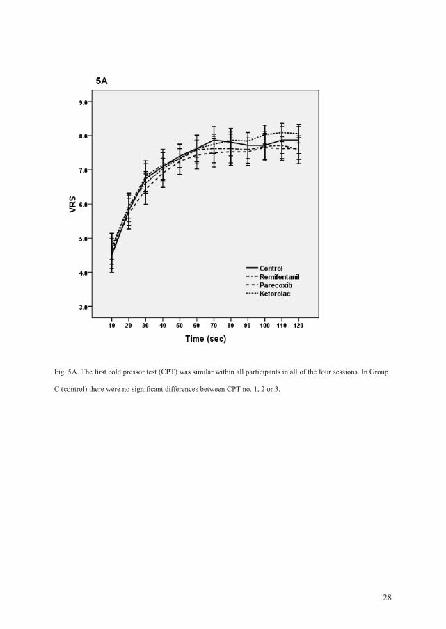
28
Fig. 5A. The first cold pressor test (CPT) was similar within all participants in all of the four sessions. In Group
C (control) there were no significant differences between CPT no. 1, 2 or 3.

29
Fig. 5B. The CPT at the end of the infusion demonstrated significantly lower VRS in all three groups receiving
remifentanil compared to control, P < 0.001. No significant difference between the three groups receiving
remifentanil.
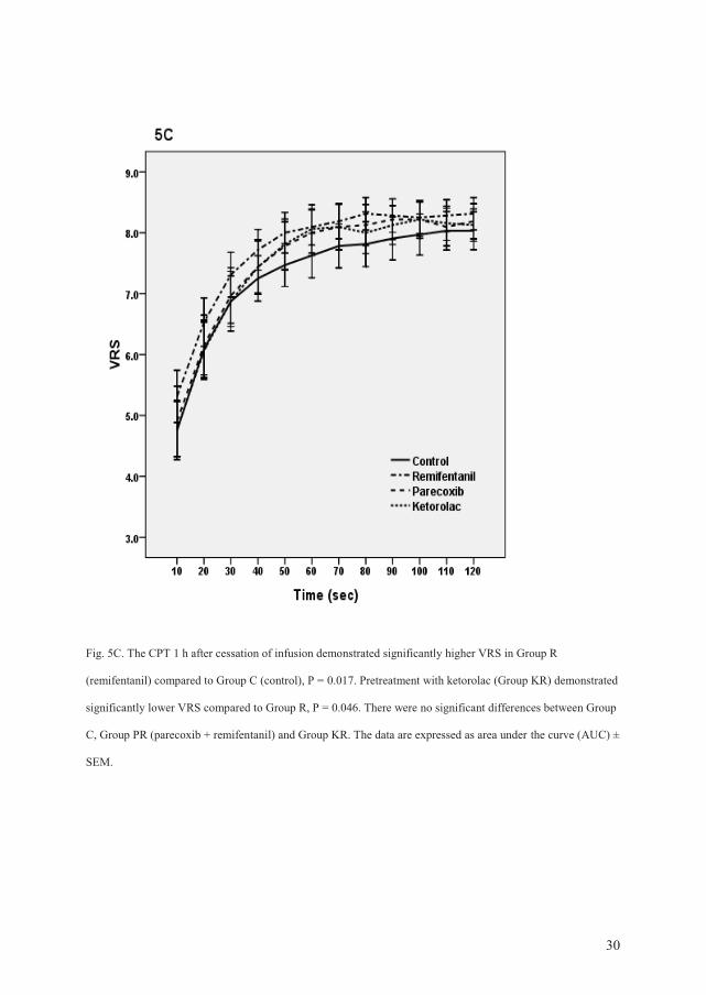
30
Fig. 5C. The CPT 1 h after cessation of infusion demonstrated significantly higher VRS in Group R
(remifentanil) compared to Group C (control), P = 0.017. Pretreatment with ketorolac (Group KR) demonstrated
significantly lower VRS compared to Group R, P = 0.046. There were no significant differences between Group
C, Group PR (parecoxib + remifentanil) and Group KR. The data are expressed as area under the curve (AUC) ±
SEM.


![Postoperative analgesia using fentanyl plus …...clinical pathway, is composed of various elements to improve perioperative management 3]. Although [ERAS could decrease the complication](https://img.pdfslide.net/doc/110x75/5e3141f21a5d817b0a149d7e/postoperative-analgesia-using-fentanyl-plus-clinical-pathway-is-composed-of.jpg)