Embed Size (px)
Citation preview
CANCER GENOMICS
Developmental and oncogenicprograms in H3K27M gliomasdissected by single-cell RNA-seqMariella G. Filbin,1,2,3,4,5* Itay Tirosh,3,4,6* Volker Hovestadt,1,3,4*McKenzie L. Shaw,1,3,4
Leah E. Escalante,1,3,4 Nathan D. Mathewson,7 Cyril Neftel,1,3,4,8 Nelli Frank,9
Kristine Pelton,10 Christine M. Hebert,1,3,4 Christine Haberler,11 Keren Yizhak,4
Johannes Gojo,5 Kristof Egervari,1 Christopher Mount,12 Peter van Galen,1,3,4
Dennis M. Bonal,13 Quang-De Nguyen,13 Alexander Beck,1 Claire Sinai,2,10 Thomas Czech,14
Christian Dorfer,14 Liliana Goumnerova,2 Cinzia Lavarino,15 Angel M. Carcaboso,15
JaumeMora,15 Ravindra Mylvaganam,1 Christina C. Luo,1 Andreas Peyrl,5 Mara Popović,16
Amedeo Azizi,5 Tracy T. Batchelor,17 Matthew P. Frosch,1 Maria Martinez-Lage,1
Mark W. Kieran,2 Pratiti Bandopadhayay,2,4 Rameen Beroukhim,4,18 Gerhard Fritsch,9
Gad Getz,1,4 Orit Rozenblatt-Rosen,3,4 Kai W.Wucherpfennig,7 David N. Louis,1
Michelle Monje,12 Irene Slavc,5 Keith L. Ligon,2,4,10 Todd R. Golub,2,4 Aviv Regev,3,4,19†‡Bradley E. Bernstein,1,3,4†‡Mario L. Suvà1,3,4†‡
Gliomas with histone H3 lysine27-to-methionine mutations (H3K27M-glioma) ariseprimarily in the midline of the central nervous system of young children, suggesting acooperation between genetics and cellular context in tumorigenesis. Although the geneticsof H3K27M-glioma are well characterized, their cellular architecture remains uncharted.We performed single-cell RNA sequencing in 3321 cells from six primary H3K27M-gliomaand matched models. We found that H3K27M-glioma primarily contain cells thatresemble oligodendrocyte precursor cells (OPC-like), whereas more differentiated malignantcells are a minority. OPC-like cells exhibit greater proliferation and tumor-propagatingpotential than their more differentiated counterparts and are at least in part sustainedby PDGFRA signaling. Our study characterizes oncogenic and developmental programs inH3K27M-glioma at single-cell resolution and across genetic subclones, suggestingpotential therapeutic targets in this disease.
Diffuse midline gliomas with histone H3lysine27-to-methioninemutations (H3K27M-glioma) are uniformly fatal malignancies(1). They are both spatially and temporallyrestricted, occurring in midline structures
of the brain with peak incidence at 6 to 9 yearsof age. These patterns suggest that a particularcell type, potentially undergoing rapid expansionat this stage, is susceptible to transformation byH3K27M. Experimentalmodels suggest that neu-ral precursor cells (NPCs) can be transformedin vitro by H3K27M, in combination with TP53mutationandPDGFRAoverexpression (2).H3K27Msuppresses EZH2, the catalytic subunit of PolycombRepressive Complex 2 (PRC2), compromising epi-genetic repression and potentially affecting cellu-
lar differentiation (1–3). In patient samples, littleis known about the developmental cell states pre-sentorhowtheycooperatewithH3K27Mfor tumori-genesis. Single-cell RNA sequencing (scRNA-seq)can help address such questions by characteriz-ing cancer cell states, their proliferative signa-tures, and their similarity to normal or othermalignant cell types. scRNA-seq also helps re-late single cell states to genetics through infer-red chromosomal copy number variations (CNVs)or mutation detection in expressed transcripts(4–6).We obtained fresh tissue from diagnostic biop-
sies of six H3K27M-glioma (table S1 and fig. S1).Each sample was dissociated into single cells,flow sorted, and profiled by means of full-length
scRNA-seq (fig. S2) (7, 8). We retained 2458 cellsthat passed quality controls (8) for downstreamanalyses (table S2). The cells’ profiles group pri-marily by tumor of origin (Fig. 1A and fig. S3), buttwo clusters contain cells from multiple tumors,expressing markers of either microglia (such asCD14, CX3CR1, and AIF1) or oligodendrocytes(such asMBP and PLP1) (Fig. 1, A and B), suggest-ing that they are nonmalignant cells. Accordingly,in presumed malignant cells only, we detectedevidence for cancer-specific aberrations—pointmutations and/or CNVs (4–6)—in 93.7% of cellsin five of the six tumors (Fig. 1, C and D, and figs.S4 to S7). Thirty-four percent (833 of 2458) of thecells had H3K27M mutations in scRNA-seq readsmapping to H3F3A or HIST1H3B (Fig. 1D andfig. S7) only detected in presumed malignantcells; other point mutations from whole-genomesequencing (WGS)/whole-exomesequencing (WES)(tables S1 and S2) were also detected only inpresumed malignant cells (Fig. 1D and fig. S7)(8). Analyzing a seventh tumor (MUV17) withH3F3A-specific primers in the scRNA-seqprotocolconfirmed H3K27M in 97% of the presumedmalignant cells (fig. S8) (8).Next, we compared malignant cell transcrip-
tomes across different glioma types, includingH3K27M-glioma, isocitrate dehydrogenase (IDH)–mutant oligodendroglioma (IDH-O), IDH-mutantastrocytoma (IDH-A), and IDH–wild-type glioblas-toma (GBM) (fig. S9Aand tables S3andS4) (5, 6, 9).Many genes were up-regulated inH3K27M-gliomaversus other tumors (n = 182 genes), but few weredown-regulated (n= 12 genes),which is consistentwith H3K27M blocking repression by PRC2 (fig.S9, B to D). Accordingly, H3K27M–up-regulatedgenes are enriched for PRC2 target genes (P <0.0001, hypergeometric test) (fig. S9E) (10, 11).The PRC1 subunit BMI1 was up-regulated inH3K27M-glioma, possibly representing a com-pensatory mechanism for PRC2 suppression.Suppression of BMI1 bymeans of CRISPR knock-out or its pharmacological inhibition reducedviability of H3K27M glioma cells, relative to treat-ment controls and non-H3K27M glioma lines(fig. S10) (12).We next assessed patterns of intratumor het-
erogeneity and distinguished subpopulationsof malignant cells within H3K27M-glioma. Weidentified four programs (P1 to P4) that wereconsistently observed as variable within tu-mors and across computational methods (8):cell cycle (such as PCNA and CDK1), astrocytic
RESEARCH
Filbin et al., Science 360, 331–335 (2018) 20 April 2018 1 of 5
1Department of Pathology and Center for Cancer Research, Massachusetts General Hospital and Harvard Medical School, Boston, MA 02114, USA. 2Department of Pediatric Oncology, Dana-Farber BostonChildren’s Cancer and Blood Disorders Center, Boston, MA 02215, USA. 3Klarman Cell Observatory, Broad Institute of Harvard and Massachussetts Institute of Technology (MIT), Cambridge,MA 02142, USA. 4Broad Institute of Harvard and MIT, Cambridge, MA 02142, USA. 5Department of Pediatrics and Adolescent Medicine, Medical University of Vienna, Vienna, Austria. 6Department ofMolecular Cell Biology, Weizmann Institute of Science, Rehovot 7610001, Israel. 7Department of Cancer Immunology and Virology, Department of Microbiology and Immunobiology, Department ofNeurology, Dana-Farber Cancer Institute and Harvard Medical School, Boston, MA 02215, USA. 8Institute of Pathology, Faculty of Biology and Medicine, Centre Hospitalier Universitaire Vaudois, 1011Lausanne, Switzerland. 9Children’s Cancer Research Institute (CCRI), St. Anna Kinderspital, Medical University of Vienna, Vienna, Austria. 10Department of Oncologic Pathology, Brigham and Women’sHospital, Boston Children’s Hospital, Dana-Farber Cancer Institute, Boston, MA 02215, USA. 11Institute of Neurology, Medical University of Vienna, Vienna, Austria. 12Departments of Neurology,Neurosurgery, Pediatrics, and Pathology, Stanford University School of Medicine, Stanford, CA 94305, USA. 13Center for Biomedical Imaging in Oncology, Lurie Family Imaging Center, Dana-Farber CancerInstitute, Boston, MA 02215, USA. 14Department of Neurosurgery, Medical University of Vienna, Vienna, Austria. 15Developmental Tumor Biology Laboratory, Hospital Sant Joan de Déu, Esplugues deLlobregat, 08950 Barcelona, Spain. 16Institute of Pathology, Faculty of Medicine, University of Ljubljana, Ljubljana, Slovenia. 17Departments of Neurology and Radiation Oncology, Division of Hematology/Oncology, Massachusetts General Hospital Cancer Center, Harvard Medical School, Boston, USA. 18Departments of Cancer Biology and Medical Oncology, Dana-Farber Cancer Institute, and Department ofMedicine, Brigham and Women’s Hospital and Harvard Medical School, Boston, MA 02215, USA. 19Department of Biology, Koch Institute for Integrative Cancer Research, Howard Hughes Medical Institute,MIT, Cambridge, MA 02139, USA.*These authors contributed equally to this work.†Corresponding author. Email: [email protected] (M.L.S.); [email protected] (B.E.B.); [email protected] (A.R.) ‡These authors contributed equally to this work.
on April 20, 2018
http://science.sciencem
ag.org/D
ownloaded from
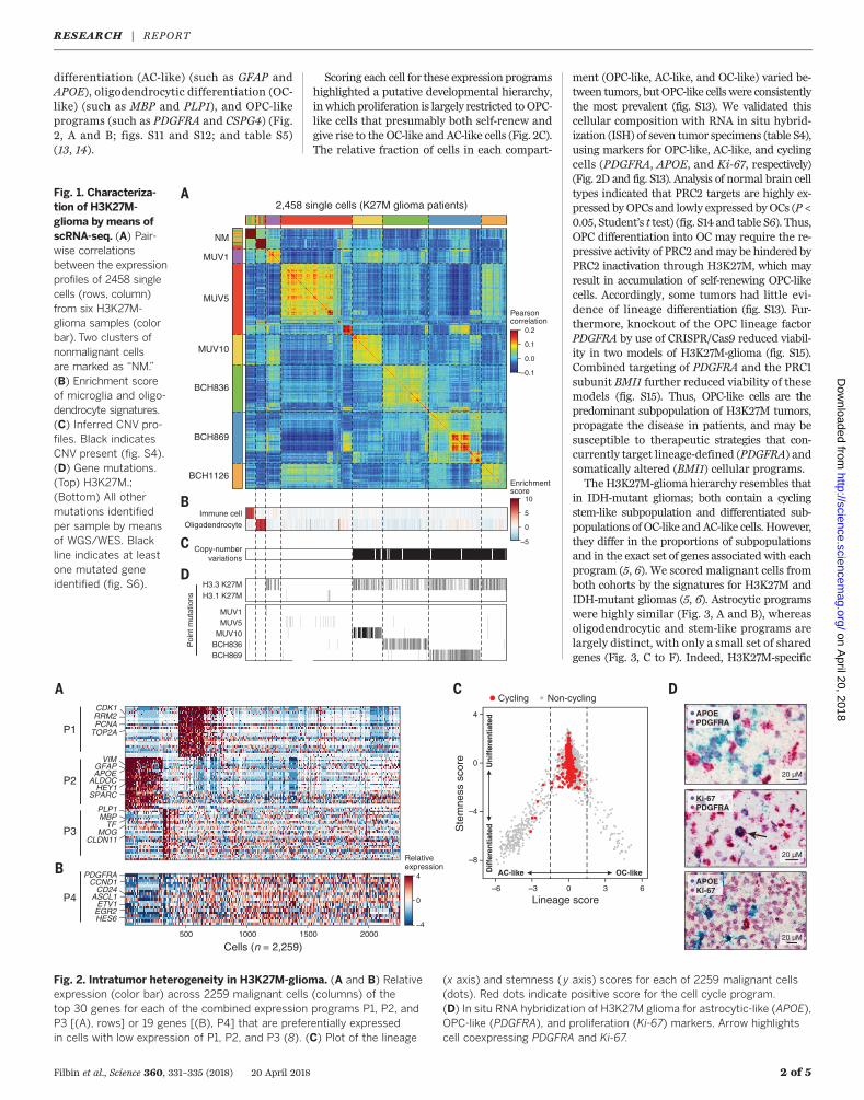
differentiation (AC-like) (such as GFAP andAPOE), oligodendrocytic differentiation (OC-like) (such as MBP and PLP1), and OPC-likeprograms (such as PDGFRA and CSPG4) (Fig.2, A and B; figs. S11 and S12; and table S5)(13, 14).
Scoring each cell for these expression programshighlighted a putative developmental hierarchy,inwhich proliferation is largely restricted to OPC-like cells that presumably both self-renew andgive rise to the OC-like and AC-like cells (Fig. 2C).The relative fraction of cells in each compart-
ment (OPC-like, AC-like, and OC-like) varied be-tween tumors, butOPC-like cells were consistentlythe most prevalent (fig. S13). We validated thiscellular composition with RNA in situ hybrid-ization (ISH) of seven tumor specimens (table S4),using markers for OPC-like, AC-like, and cyclingcells (PDGFRA, APOE, and Ki-67, respectively)(Fig. 2D and fig. S13). Analysis of normal brain celltypes indicated that PRC2 targets are highly ex-pressed by OPCs and lowly expressed byOCs (P <0.05, Student’s t test) (fig. S14 and table S6). Thus,OPC differentiation into OC may require the re-pressive activity of PRC2 andmay be hindered byPRC2 inactivation through H3K27M, which mayresult in accumulation of self-renewing OPC-likecells. Accordingly, some tumors had little evi-dence of lineage differentiation (fig. S13). Fur-thermore, knockout of the OPC lineage factorPDGFRA by use of CRISPR/Cas9 reduced viabil-ity in two models of H3K27M-glioma (fig. S15).Combined targeting of PDGFRA and the PRC1subunit BMI1 further reduced viability of thesemodels (fig. S15). Thus, OPC-like cells are thepredominant subpopulation of H3K27M tumors,propagate the disease in patients, and may besusceptible to therapeutic strategies that con-currently target lineage-defined (PDGFRA) andsomatically altered (BMI1) cellular programs.The H3K27M-glioma hierarchy resembles that
in IDH-mutant gliomas; both contain a cyclingstem-like subpopulation and differentiated sub-populations of OC-like andAC-like cells. However,they differ in the proportions of subpopulationsand in the exact set of genes associated with eachprogram (5, 6). We scored malignant cells fromboth cohorts by the signatures for H3K27M andIDH-mutant gliomas (5, 6). Astrocytic programswere highly similar (Fig. 3, A and B), whereasoligodendrocytic and stem-like programs arelargely distinct, with only a small set of sharedgenes (Fig. 3, C to F). Indeed, H3K27M-specific
Filbin et al., Science 360, 331–335 (2018) 20 April 2018 2 of 5
Fig. 1. Characteriza-tion of H3K27M-glioma by means ofscRNA-seq. (A) Pair-wise correlationsbetween the expressionprofiles of 2458 singlecells (rows, column)from six H3K27M-glioma samples (colorbar). Two clusters ofnonmalignant cellsare marked as “NM.”(B) Enrichment scoreof microglia and oligo-dendrocyte signatures.(C) Inferred CNV pro-files. Black indicatesCNV present (fig. S4).(D) Gene mutations.(Top) H3K27M.;(Bottom) All othermutations identifiedper sample by meansof WGS/WES. Blackline indicates at leastone mutated geneidentified (fig. S6). H3.3 K27M
H3.1 K27M
MUV1MUV5
MUV10BCH836BCH869
NM
BCH836
BCH869
BCH1126
MUV1
MUV5
MUV10
2,458 single cells (K27M glioma patients)
Immune cell
Oligodendrocyte
Copy-numbervariations
Poi
nt m
utat
ions
B
C
D
–0.1
0.0
0.1
0.2
Enrichmentscore
0
–5
5
10
Pearsoncorrelation
A
500 1000 1500 2000
Cells (n = 2,259)
B
CDK1RRM2
TOP2APCNA
VIMGFAPAPOE
ALDOCHEY1
SPARC
PLP1MBP
TFMOG
CLDN11
C
P1
P2
P3
Relativeexpression
–4
0
4PDGFRACCND1
CD24ASCL1
ETV1
HES6EGR2
–6 0 3–3 6
–8
–4
0
4
Lineage score
Ste
mne
ss s
core
Dif
fere
nti
ated
Un
iffe
ren
tiat
ed
AC-like OC-like
Cycling Non-cyclingA
P4
APOEPDGFRA
20 µM
20 µM
APOEKi-67
20 µM
Ki-67PDGFRA
D
Fig. 2. Intratumor heterogeneity in H3K27M-glioma. (A and B) Relativeexpression (color bar) across 2259 malignant cells (columns) of thetop 30 genes for each of the combined expression programs P1, P2, andP3 [(A), rows] or 19 genes [(B), P4] that are preferentially expressedin cells with low expression of P1, P2, and P3 (8). (C) Plot of the lineage
(x axis) and stemness (y axis) scores for each of 2259 malignant cells(dots). Red dots indicate positive score for the cell cycle program.(D) In situ RNA hybridization of H3K27M glioma for astrocytic-like (APOE),OPC-like (PDGFRA), and proliferation (Ki-67) markers. Arrow highlightscell coexpressing PDGFRA and Ki-67.
RESEARCH | REPORTon A
pril 20, 2018
http://science.sciencemag.org/
Dow
nloaded from
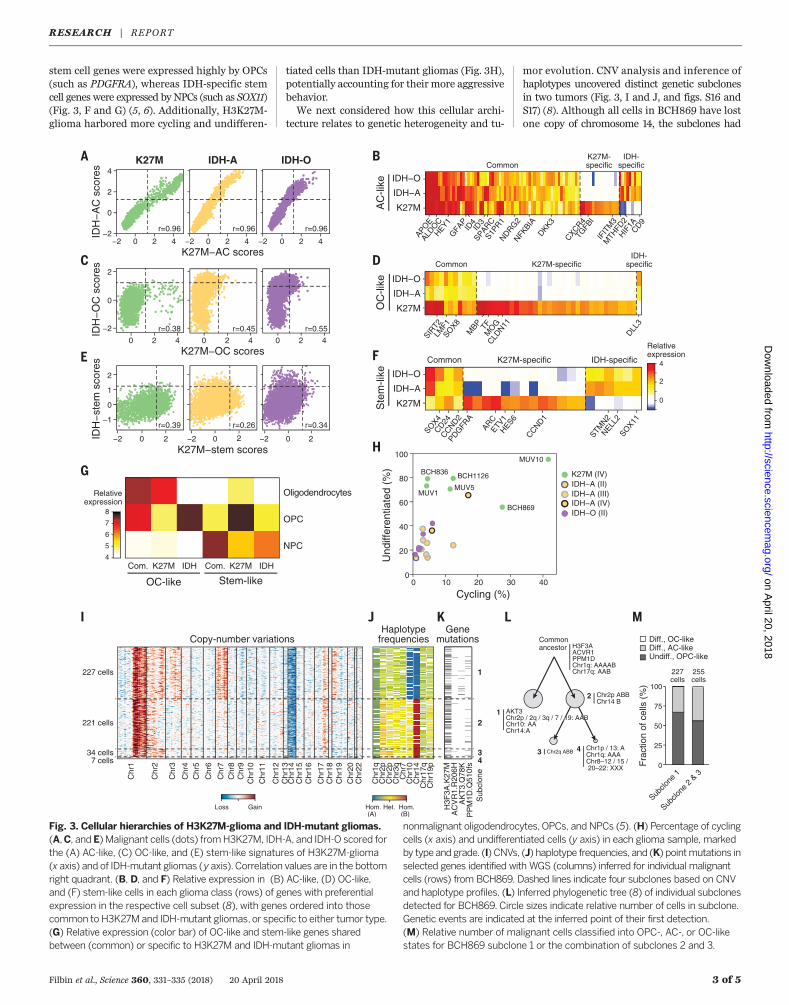
stem cell genes were expressed highly by OPCs(such as PDGFRA), whereas IDH-specific stemcell genes were expressed by NPCs (such as SOX11)(Fig. 3, F and G) (5, 6). Additionally, H3K27M-glioma harbored more cycling and undifferen-
tiated cells than IDH-mutant gliomas (Fig. 3H),potentially accounting for their more aggressivebehavior.We next considered how this cellular archi-
tecture relates to genetic heterogeneity and tu-
mor evolution. CNV analysis and inference ofhaplotypes uncovered distinct genetic subclonesin two tumors (Fig. 3, I and J, and figs. S16 andS17) (8). Although all cells in BCH869 have lostone copy of chromosome 14, the subclones had
Filbin et al., Science 360, 331–335 (2018) 20 April 2018 3 of 5
SOX4
CD24CCND2
PDGFR
A
ARCETV
1HES6
CCND1
STMN2
NELL2
SOX11
−2 0 2 4−2
0
2
4
IDH
−A
C s
core
sID
H−
OC
sco
res
IDH
−st
em s
core
s
−2 0 2 4
K27M−AC scores
K27M−OC scores
K27M−stem scores
−2 0 2 4
0 2 4
−2
0
2
0 2 4 0 2 4
K27M IDH-A IDH-OA
APOE
ALDO
CHEY1G
FAP
ID4
ID3
SPARCS1P
R1NDRG
2NFK
BIADKK3
CXCR4TG
FBI
IFIT
M3
MTH
FD2
HIF1A
CD9
SIRT2
LMF1
SOX8
MBP TF
MO
GCLD
N11
DLL3
K27M
IDH−O
IDH−A
CommonK27M-specific
K27M-specific
IDH-specific
IDH-specific
K27M
IDH−O
IDH−A
K27M
IDH−O
IDH−A
B
C D
E F
Common
K27M-specific IDH-specificCommon
0 10 20 30 400
20
40
60
80
100
Cycling (%)
Und
iffer
entia
ted
(%) BCH836
BCH869
MUV1MUV5
MUV10
BCH1126 K27M (IV)IDH−A (II)IDH−A (III)IDH−A (IV)IDH−O (II)
H
G
AC
-like
O
C-li
ke
Ste
m-li
ke
NPC
Oligodendrocytes
OPC
Relativeexpression
OC-like
K27M IDH
Stem-like
K27M IDH4
5
6
7
8
Com. Com.
−2 0 2
–1
0
1
2
−2 0 2 −2 0 2
Relativeexpression
0
2
4
Chr
1qC
hr2p
Chr
2pC
hr3q
Chr
7C
hr10
Chr
14C
hr17
qC
hr19
p
221 cells
7 cells
1
2
34
227 cells 227cells
255cells
34 cells
H3F
3A.K
27M
AC
VR
1.R
206H
AK
T3.
Q78
KP
PM
1D.Q
510f
sS
ubcl
one
Chr
1
Chr
2
Chr
3
Chr
4C
hr5
Chr
6
Chr
7C
hr8
Chr
9C
hr10
Chr
11
Chr
12C
hr13
Chr
14C
hr15
Chr
16
Chr
17C
hr18
Chr
19C
hr20
Chr
22
Copy-number variationsHaplotype
frequenciesGene
mutations
Subclo
ne 1
Subclo
ne 2
& 3
Fra
ctio
n of
cel
ls (
%) 100
75
50
25
0
Commonancestor H3F3A
ACVR1PPM1DChr1q: AAAABChr17q: AAB
3 Chr2q ABB
1
4 Chr1p / 13: AChr1q: AAAChr8–12 / 15 / 20–22: XXX
2 Chr2p ABBChr14 B
AKT3
Chr10: AAChr14:A
Undiff., OPC-likeDiff., AC-likeDiff., OC-like
I J K L M
Chr2p / 2q / 3q / 7 / 19: AAB
Loss Gain Hom.(A)
Het. Hom.(B)
r=0.96 r=0.96 r=0.96
r=0.38 r=0.45 r=0.55
r=0.39 r=0.26 r=0.34
Fig. 3. Cellular hierarchies of H3K27M-glioma and IDH-mutant gliomas.(A,C, and E) Malignant cells (dots) fromH3K27M, IDH-A, and IDH-O scored forthe (A) AC-like, (C) OC-like, and (E) stem-like signatures of H3K27M-glioma(x axis) and of IDH-mutant gliomas (y axis). Correlation values are in the bottomright quadrant. (B, D, and F) Relative expression in (B) AC-like, (D) OC-like,and (F) stem-like cells in each glioma class (rows) of genes with preferentialexpression in the respective cell subset (8), with genes ordered into thosecommon to H3K27M and IDH-mutant gliomas, or specific to either tumor type.(G) Relative expression (color bar) of OC-like and stem-like genes sharedbetween (common) or specific to H3K27M and IDH-mutant gliomas in
nonmalignant oligodendrocytes, OPCs, and NPCs (5). (H) Percentage of cyclingcells (x axis) and undifferentiated cells (y axis) in each glioma sample, markedby type and grade. (I) CNVs, (J) haplotype frequencies, and (K) pointmutations inselected genes identified with WGS (columns) inferred for individual malignantcells (rows) from BCH869. Dashed lines indicate four subclones based on CNVand haplotype profiles. (L) Inferred phylogenetic tree (8) of individual subclonesdetected for BCH869. Circle sizes indicate relative number of cells in subclone.Genetic events are indicated at the inferred point of their first detection.(M) Relative number of malignant cells classified into OPC-, AC-, or OC-likestates for BCH869 subclone 1 or the combination of subclones 2 and 3.
RESEARCH | REPORTon A
pril 20, 2018
http://science.sciencemag.org/
Dow
nloaded from
different haplotypes, indicating that two dis-tinct events led to loss of alternate chromosome14 alleles (Fig. 3J). Additionally, certain somaticgenemutations could be assigned to just one of thesubclones (Fig. 3K). Use of both CNVs and haplo-type frequencies enabled inference of phyloge-netic trees (Fig. 3L, fig. S17D, and table S7) (8).Each genetic subclone contained cells spanning asimilar diversity of cellular states, although withsome variation in their relative proportions (Fig.3M and fig. S17E). This suggests that distinct ge-netic subclones share similar developmental hier-archies in H3K27M-glioma.Next, we profiled 863 single cells from several
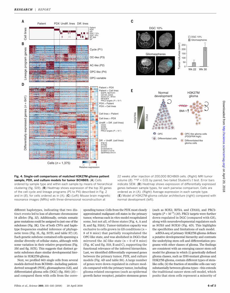
models derived from BCH869—including patient-derived xenograft (PDX), gliomaspheres (GS), anddifferentiated glioma cells (DGC) (fig. S18) (15)—and compared them with cells from the corre-
sponding tumor. Cells from the PDXmost closelyapproximated malignant cell states in the primarytumor, whereas each in vitromodel recapitulatedsome, but not all, of these states (Fig. 4, A andB, and fig. S19A). Tumor-initiation capacity wasexclusive to cells grown in GS conditions (n =8 of 8 mice) that partially recapitulated theOPC-like state, and was abolished in DGCs thatmirrored the AC-like state (n = 0 of 8 mice)(Fig. 4C and fig. S19, B and C), supporting thefunctional relevance of the inferred hierarchies.Last, we identified differentially expressed genesbetween the primary tumor, PDX, and culturemodels (Fig. 4D and table S8). A large numberof genes were down-regulated in culture mod-els comparedwith the primary tumor, includingglioma-related oncogenes (such as epidermalgrowth factor receptor), putative stemness genes
(such as SOX2, RFX4, and CD133), and PRC2-targets (P < 10−9) (10). PRC2 targets were furtherdown-regulated in DGC (compared with GS),along with neurodevelopmental regulators suchas SOX6 and SOX10 (Fig. 4D). This highlightsthe specificities and limitations of each model.scRNA-seq of primary H3K27M-glioma defines
a putative developmental hierarchy and contraststhe underlying stem cell and differentiation pro-grams with other classes of glioma. The findingsare consistent with an emerging cancer stem cellmodel for gliomas in which (i) genetically definedglioma classes, such as IDH-mutant gliomas andH3K27M-glioma, contain different types of stem-like cells; (ii) the fraction of stemlike cells can varysubstantially between glioma types—this extendsthe traditional cancer stem cell model, whichposits that stem cells represent a minority of
Filbin et al., Science 360, 331–335 (2018) 20 April 2018 4 of 5
OPC-like (P4)
OPC-variable
Patient PDXC
ell l
ines
A
B
Line
age
prog
ram
gen
esD
iffer
entia
lly e
xpre
ssed
gen
es (
n =
641
)
Cycle (P1)
OC-like (P3)
AC-like (P2)
Patient > PDX
PDX > Patient
Patient > Cell lines
PDX > Cell lines
Cell lines > Patient
Cell lines > PDX
Undiff. > Diff. (cell lines)
Diff. > Undiff. (cell lines)
EGFR, NOTCH1CD133, CD24SOX2-OT, RFX4NFIA/B/XPRC2 targets (P < 10–9)
CD24SOX6SOX10PRC2 targets (P < 10–7)
IFI6/16IFITM2STAT2S100A4/6/10S100A11/13/16SERPING1/H1/I1
D
Cells (n = 1,375)
Undiff. lines Diff. lines
Relative expression40–4
PatientPDXGSDGC 7.5%DGC 10%SF AdhGF–
C
E
50
0
150
250
Wk 22 Wk 30
DGC 10%Gliomaspheres
100
200
Tum
or v
olum
es (
mm
3 )
**
**
DGC 10%
Gliomaspheres
H3K27Mglioma
PR
C2
Normaldevelopment
PRC2
BMI-1
OPC-like glioma cells(PDGFRA-high)
PD
GF
RA
Differentiated-likeglioma cells
OPC
Differentiated cells
Fig. 4. Single-cell comparisons of matched H3K27M-glioma patientsample, PDX, and culture models for tumor BCH869. (A) Cellsordered by sample type and within each sample by means of hierarchicalclustering (fig. S19). (B) Heatmap shows expression of the top 30 genesof the cell cycle and lineage programs (P1 to P4) described in Fig. 2and in (8), for cells ordered as in (A). (C) (Left) Mouse brain magneticresonance images (MRIs) with three-dimensional reconstruction at
22 weeks after injection of 200,000 BCH869 cells. (Right) MRI tumorvolume (8). **P < 0.01 by paired, two-tailed Student’s t test. Error barsindicate SEM. (D) Heatmap shows expression of differentially expressedgenes between sample types, for each pairwise comparison. Cells areordered as in (A). (Right) Average expression in each sample type.(E) Model of H3K27M-glioma cellular architecture (right) compared withnormal development (left).
RESEARCH | REPORTon A
pril 20, 2018
http://science.sciencemag.org/
Dow
nloaded from
malignant cells; (iii) differentiation hierarchiesplay a critical role in the functional propertiesof glioma cells because self-renewal and tumor-propagating potential are exclusive to the mostprimitive cells; and (iv) genetic subclones in tumorstend to share similar cellular architecture. Ourstudy also highlights opportunities for therapeu-tic intervention.We show thatOPC-like cells driveH3K27M-glioma, suggesting that the OPCmarkerPDGFRA could be a lineage-defined therapeutictarget, relevant even in the absence of geneticamplification or mutation. H3K27M-glioma alsooverexpress the PRC1 subunit BMI1 and are sen-sitive to its inhibition, either alone or in combi-nation with PDGFRA inhibition, hinting at apotential compensatory mechanism for PRC2dysfunction (Fig. 4E). Thus, lineage-defined andsomatically altered cellular programs inH3K27M-glioma suggest complementary opportunitiesfor therapeutic intervention in these incurablemalignancies.
REFERENCES AND NOTES
1. D. Sturm et al., Nat. Rev. Cancer 14, 92–107 (2014).2. K. Funato, T. Major, P. W. Lewis, C. D. Allis, V. Tabar, Science
346, 1529–1533 (2014).3. P. W. Lewis et al., Science 340, 857–861 (2013).4. A. P. Patel et al., Science 344, 1396–1401 (2014).5. I. Tirosh et al., Nature 539, 309–313 (2016).6. A. S. Venteicher et al., Science 355, eaai8478 (2017).7. S. Picelli et al., Nat Protoc. 9, 171–181 (2014).8. Material and methods are available as supplementary
materials.9. B. B. Liau et al., Cell Stem Cell 20, 233–246.e7 (2017).10. I. Ben-Porath et al., Nat. Genet. 40, 499–507 (2008).11. S. Darmanis et al., Cell Reports 21, 1399–1410 (2017).12. A. Kreso et al., Nat. Med. 20, 29–36 (2014).13. G. La Manno et al., Cell 167, 566–580.e19 (2016).14. S. Marques et al., Science 352, 1326–1329 (2016).15. M. L. Suvà et al., Cell 157, 580–594 (2014).
ACKNOWLEDGMENTS
We thank L. Gaffney for assistance with figures. Funding: Thiswork was supported by grants from the Alex LemonadeStand (M.M. and M.L.Su.), the Wang Family Fund (M.L.Su.,
A.R., B.E.B., and M.M.), the Smith Family Foundation (M.L.Su.),the Broad Institute Broadnext10 (M.L.Su. and O.R.-R.), theV Foundation for Cancer Research (M.L.Su.), the HowardGoodman Fellowship at Massachussetts General Hospital (MGH)(M.L.Su.), the Merkin Institute Fellowship at the Broad Instituteof MIT and Harvard (M.L.Su.), the Rachel Molly MarkoffFoundation (M.L.Su. and B.E.B.), NIH–National Cancer Institute(NCI) brain cancer SPORE P50CA165962 (M.G.F., T.T.B.,and M.L.Su.), start-up funds from the MGH department ofPathology, The Cure Starts Now Foundation, Hope forCaroline Foundation, Julian Boivin Courage for CuresFoundation, Abbie’s Army, Michael Mosier Defeat DIPGFoundation, Reflections of Grace Foundation, The Cure StartsNow Australia, Brooke Healey Foundation, Soar With GraceFoundation, Jeffrey Thomas Hayden Foundation, Cure BrainCancer Foundation, The Jones Family Foundation, MusellaFoundation, Pray, Hope Believe Foundation, Smiles for SophieFoundation, Benny’s World, Love Chloe Foundation, Aiden’sAvengers, A Cure from Caleb Society, The Operation GraceWhite Foundation, Ryan’s Hope, Wayland Villars DIPGFoundation, American Childhood Cancer Organization, JulianaRose Donnelly Trust, Sheila Jones & Friends, The Ellie KavalierosDIPG Research Fund, Voices Against Brain Cancer, The DIPGCollaborative, Zach Carson Foundation, the Micky CzechFoundation, The Guglietti Family Trust, Prayers From MariaFoundation, Ryan Harvey Foundation, Markoff Art in Giving, andthe Brock Fleming Foundation. M.G.F. holds a Career Awardfor Medical Scientist from Burroughs Wellcome Fund and K12Paul Calabresi Career Award for Clinical Oncology–TrainingProgram in Nervous System Tumors (K12CA090354). I.T. wassupported by the Zuckerman STEM Leadership Programand the Benoziyo Endowment Fund for the Advancement ofScience. V.H. was supported by a European MolecularBiology Organization long-term fellowship and a Human FrontierScience Program fellowship. C.N. is supported by the PlacideNicod Foundation. K.L.L., L.G., and M.W.K. received supportfrom NCI grant P01 CA142536. K.E. is supported by theFondation Nuovo-Soldati. P.B. and R.B were supported by theSt. Baldrick’s Foundation, the Pediatric Brain Tumor Foundation,Gray Matters Brain Cancer Foundation Olivia CaldwellFoundation, and NCI (grants R01CA219943 and R01CA188228).A.R. was supported by funds from the Howard Hughes MedicalInstitute, the Klarman Cell Observatory, STARR cancerconsortium, NCI grant 1U24CA180922, NCI grant R33CA202820,by the Koch Institute Support (core) grant P30-CA14051 fromNCI, the Ludwig Center, and the Broad Institute. B.E.B. wassupported by the NIH Common Fund and National CancerInstitute (grant DP1CA216873), the American Cancer Society,the Ludwig Center at Harvard Medical School, and theBernard and Mildred Kayden MGH Research Institute Chair.Flow cytometry and sorting services were supportedby shared instrumentation grant 1S10RR023440-01A1. M.M.
was supported by the California Institute of RegenerativeMedicine (CIRM) grants RB4-06093 and RN3-06510,Lyla Nsouli Foundation, and the Virginia and D. K. Ludwig Fundfor Cancer Research. C.L., A.M.C., and J.M. are supported byThe Alicia Pueyo Foundation. Author contributions: M.G.F.,I.T., V.H., B.E.B., A.R., and M.L.Su. conceived the project anddesigned the study. M.G.F. collected and processed tumorsamples, performed all cell culture assays, and plannedmouse studies. I.T. and V.H. performed all computationalanalyses. M.L.Sh., L.E.E., N.D.M., C.N., N.F., K.P., C.M.H.,C.H., J.G., K.E., A.B., C.S., T.C., C.D., L.G., C.L., A.M.C., J.M.,A.P., M.P., A.A., T.T.B., M.W.K., G.F., and I.S. helped withcollecting tumors and generating single-cell sequencingdata. N.D.M. helped with CRISPR experiments. R.M. and C.C.L.provided flow cytometry expertise. M.G.F., V.H., and P.V.G.designed and performed targeted sequencing experiments.K.Y., G.G., P.B., and R.B. provided support for genomic andgenetic analyses. M.L.Sh. performed in situ hybridizationexperiments. M.P.F. and M.M.-L. provided samples for in situhybridization experiments. C.M. and M.M. developed normalhuman cell cultures used in the study. D.M.B. and Q.-D.N.performed in vivo mouse studies. K.W.W., O.R.R., D.N.L.,I.S., K.L.L., and T.R.G. provided experimental and analyticalsupport. M.G.F., I.T, V.H., A.R., B.E.B., and M.L.Su. interpretedresults and wrote and edited the manuscript with feedback fromall authors. Competing interests: B.E.B. is an advisor andequity holder for Fulcrum Therapeutics, 1CellBio, andHiFiBio. A.R. is a scientific advisory board member forThermoFisher Scientific, Syros Pharmaceuticals, and DriverGroup. M.G.F., I.T., V.H., A.R., B.B., and M.L.Su. areco-inventors on a U.S. provisional patent application(U.S. 62/585,468) relating to advances described in thismanuscript filed by the Broad Institute, MIT, and MGH.G.G. has patent applications on tools for cancer genomeanalysis. All other authors have no competing interests.Data and materials availability: Data generated for this studyare available through the Gene Expression Omnibus (GEO,accession no. GSE102130) or the Broad Institute Single-CellPortal (https://portals.broadinstitute.org/single_cell/study/single-cell-analysis-in-pediatric-midline-gliomas-with-histone-h3k27m-mutation). All other data are available in themanuscript or the supplementary materials.
SUPPLEMENTARY MATERIALS
www.sciencemag.org/content/360/6386/331/suppl/DC1Material and MethodsFigs. S1 to S19Tables S1 to S8References (16–27)
7 August 2017; accepted 26 February 201810.1126/science.aao4750
Filbin et al., Science 360, 331–335 (2018) 20 April 2018 5 of 5
RESEARCH | REPORTon A
pril 20, 2018
http://science.sciencemag.org/
Dow
nloaded from
Developmental and oncogenic programs in H3K27M gliomas dissected by single-cell RNA-seq
and Mario L. SuvàWucherpfennig, David N. Louis, Michelle Monje, Irene Slavc, Keith L. Ligon, Todd R. Golub, Aviv Regev, Bradley E. Bernstein Kieran, Pratiti Bandopadhayay, Rameen Beroukhim, Gerhard Fritsch, Gad Getz, Orit Rozenblatt-Rosen, Kai W.Luo, Andreas Peyrl, Mara Popovic, Amedeo Azizi, Tracy T. Batchelor, Matthew P. Frosch, Maria Martinez-Lage, Mark W.
C.Christian Dorfer, Liliana Goumnerova, Cinzia Lavarino, Angel M. Carcaboso, Jaume Mora, Ravindra Mylvaganam, Christina Christopher Mount, Peter van Galen, Dennis M. Bonal, Quang-De Nguyen, Alexander Beck, Claire Sinai, Thomas Czech,Nelli Frank, Kristine Pelton, Christine M. Hebert, Christine Haberler, Keren Yizhak, Johannes Gojo, Kristof Egervari, Mariella G. Filbin, Itay Tirosh, Volker Hovestadt, McKenzie L. Shaw, Leah E. Escalante, Nathan D. Mathewson, Cyril Neftel,
DOI: 10.1126/science.aao4750 (6386), 331-335.360Science
, this issue p. 331Sciencedeveloping therapies.stable tumor-propagating potential. The analysis also identified a lineage-specific marker that may be useful in
like profiles that contributed to their−with other gliomas, these cancers had distinct oncogenic programs and stem cellresembling oligodendrocyte precursor cells, whereas differentiated malignant cells were a smaller fraction. In comparison
oncogenic programs, genetics, and cellular hierarchies of H3K27M-glioma. Tumors were mainly composed of cells used a single-cell sequencing approach to study the et al.type of childhood cancer with few options for treatment. Filbin
Diffuse midline gliomas with histone H3 lysine27-to-methionine mutations (H3K27M-glioma) are an aggressiveThe cellular composition of H3K27M gliomas
ARTICLE TOOLS http://science.sciencemag.org/content/360/6386/331
MATERIALSSUPPLEMENTARY http://science.sciencemag.org/content/suppl/2018/04/18/360.6386.331.DC1
CONTENTRELATED
http://stm.sciencemag.org/content/scitransmed/9/375/eaah6510.fullhttp://stm.sciencemag.org/content/scitransmed/9/381/eaaf2968.fullhttp://stm.sciencemag.org/content/scitransmed/10/422/eaam7577.fullhttp://stm.sciencemag.org/content/scitransmed/10/430/eaao2731.full
REFERENCES
http://science.sciencemag.org/content/360/6386/331#BIBLThis article cites 25 articles, 8 of which you can access for free
PERMISSIONS http://www.sciencemag.org/help/reprints-and-permissions
Terms of ServiceUse of this article is subject to the
is a registered trademark of AAAS.Sciencelicensee American Association for the Advancement of Science. No claim to original U.S. Government Works. The title Science, 1200 New York Avenue NW, Washington, DC 20005. 2017 © The Authors, some rights reserved; exclusive
(print ISSN 0036-8075; online ISSN 1095-9203) is published by the American Association for the Advancement ofScience
on April 20, 2018
http://science.sciencem
ag.org/D
ownloaded from


![New PDF DocumentTMA Wire Round size: IPack/10pcs 1 Pack/ 1 Opcs Rect size. S.S Straight Wire Round size: IPack/20pcs Rect size. 1 Pack/ 1 Opcs Ligature S.S Wire [50g/1 roll] T.S.H.C0](https://img.pdfslide.net/doc/110x75/5f425229fdc979428d6c3778/new-pdf-document-tma-wire-round-size-ipack10pcs-1-pack-1-opcs-rect-size-ss.jpg)