Embed Size (px)
Citation preview
1
Cancer Prevention Research CAPR-15-0109 Revision
Clinical Trial of Acolbifene in Premenopausal Women at High Risk for Breast Cancer Carol J. Fabian1, Bruce F. Kimler*2, Carola M. Zalles3, Teresa A. Phillips1, Trina Metheny1, Brian K. Petroff1,4, Thomas C. Havighurst5, KyungMann Kim5, Howard H. Bailey6, Brandy M. Heckman-Stoddard7 * Corresponding Author: Bruce F. Kimler, Ph.D., Department of Radiation Oncology, University of Kansas Medical Center, 3901 Rainbow Boulevard, Kansas City, KS 66160 USA. Phone: 913-588-4523; fax: 913-588-3679; e-mail: [email protected] Affiliations: 1 C.J. Fabian, T.A. Phillips, T. Metheny, B.K. Petroff Department of Internal Medicine, University of Kansas Medical Center, 3901 Rainbow Boulevard, Kansas City, KS 66160 USA. 2 B.F. Kimler Department of Radiation Oncology, University of Kansas Medical Center, 3901 Rainbow Boulevard, Kansas City, KS 66160 USA. 3 C.M. Zalles Mercy Hospital, 3663 S Miami Ave, Miami, FL 33133 USA 4 B.K. Petroff Current affiliation: Diagnostic Center for Population and Animal Health, Pathobiology and Diagnostic Investigation, College of Veterinary Medicine, Michigan State University, 4125 Beaumont Road, 220L, Lansing, MI 48910 USA. 5T.C. Havighurst, K. Kim Department of Biostatistics and Medical Informatics, University of Wisconsin Madison, 600 Highland Ave, Box 4675, Madison, WI 53792 6 H. H. Bailey University of Wisconsin Carbone Cancer Center, 600 Highland Ave., K4/654, Madison, WI 53792-6164 7 B.M. Heckman-Stoddard Division of Cancer Prevention, National Cancer Institute, 9609 Medical Center Drive, Room 5E326 MSC 9783, Bethesda, MD 20892-9783 Running Title: Acolbifene for Breast Cancer Prevention Key Words: Breast Cancer Risk Biomarkers, SERM, RPFNA
Cancer Research. on July 14, 2019. © 2015 American Association forcancerpreventionresearch.aacrjournals.org Downloaded from
Author manuscripts have been peer reviewed and accepted for publication but have not yet been edited. Author Manuscript Published OnlineFirst on September 21, 2015; DOI: 10.1158/1940-6207.CAPR-15-0109
2
Financial Support: Supported by Subcontract 938N232 from the University of Wisconsin Cancer Consortium for “Phase I and Phase II Clinical Trials of Cancer Chemopreventive Agents” (PI: Howard H. Bailey, M.D.); NO1-CN-35153, National Cancer Institute, National Institutes of Health. Study agent was provided by Endorecherche, Inc. of Quebec, Canada. Conflicts of Interest: None Word Count: Abstract = 250 Text = 4,635 References: 59 Figures + Tables: 6
Cancer Research. on July 14, 2019. © 2015 American Association forcancerpreventionresearch.aacrjournals.org Downloaded from
Author manuscripts have been peer reviewed and accepted for publication but have not yet been edited. Author Manuscript Published OnlineFirst on September 21, 2015; DOI: 10.1158/1940-6207.CAPR-15-0109
3
Abstract
The purpose of this study was to assess the feasibility of using the selective estrogen
receptor modulator (SERM) acolbifene as a breast cancer prevention agent in
premenopausal women. In order to do so we assessed change in proliferation in
benign breast tissue sampled by random periareolar fine needle aspiration (RPFNA) as
a primary endpoint, along with changes in other risk biomarkers and objective and
subjective side effects as secondary endpoints. Twenty-five women with cytologic
hyperplasia +/- atypia and ≥2% of breast epithelial cells staining positive for Ki-67,
received 20 mg acolbifene daily for 6-8 months, and then had benign breast tissue and
blood risk biomarkers re-assessed. Ki-67 decreased from a median of 4.6%
(interquartile range, 3.1 – 8.5%) at baseline to 1.4% (IQR, 0.6 – 3.5%) after acolbifene
(P<0.001; Wilcoxon signed rank test), despite increases in bioavailable estradiol. There
were also significant decreases in expression (RT-qPCR) of estrogen inducible genes
that code for pS2, ER-α, and PgR (p≤0.026). There was no significant change in serum
IGF-1, IGFBP3, IGF-1:IGFBP3 ratio, or mammographic breast density. Subjective side
effects were minimal with no significant increase in hot flashes, muscle cramps,
arthralgias, or fatigue. Objective measures showed a clinically insignificant decrease in
lumbar spine bone density (DEXA) and an increase in ovarian cysts but no change in
endometrial thickness (sonography). In summary, acolbifene was associated with
favorable changes in benign breast epithelial cell proliferation and estrogen inducible
gene expression but minimal side effects, suggesting a Phase IIB placebo-controlled
trial evaluating it further for breast cancer prevention.
Cancer Research. on July 14, 2019. © 2015 American Association forcancerpreventionresearch.aacrjournals.org Downloaded from
Author manuscripts have been peer reviewed and accepted for publication but have not yet been edited. Author Manuscript Published OnlineFirst on September 21, 2015; DOI: 10.1158/1940-6207.CAPR-15-0109
4
Introduction
The selective estrogen receptor modulator (SERM) tamoxifen is the only FDA-
approved agent for breast cancer risk reduction in premenopausal women 35 years of
age or older with a Gail Model estimated 5-year risk of breast cancer of 1.66% or higher
and/or atypical hyperplasia, or in situ cancer [1]. A recent meta-analysis suggests that
use of tamoxifen in the chemopreventive setting reduced risk ~33% compared to
placebo among high risk women [2]. Current ASCO guidelines suggest including
tamoxifen in discussion of risk reduction strategies for high risk women [1] but tamoxifen
uptake for risk eligible women remains low [3] primarily due to concerns about side
effects and lack of demonstrated survival benefit [1, 3]. The benefit: risk ratio for
tamoxifen used as primary prevention in high risk premenopausal women is generally
seen as particularly favorable given the lack of significantly increased incidence of
serious side effects in women < 50 years old in the NSABP P-1 trial [4, 5]. Yet the rate
of tamoxifen uptake for premenopausal women attending high risk clinics has been
cited as only 10% [6]. Concerns about induction of menstrual abnormalities and
perimenopausal symptoms are the likely reasons that most younger women are
reluctant to take tamoxifen. Tamoxifen can also result in an increase in size or number
of ovarian cysts as well as bone density loss in premenopausal women [7, 8].
Currently, tamoxifen for primary prevention is regarded as a preference-sensitive
decision [3, 6, 9]. A dramatic increase in uptake of endocrine therapy for primary
prevention by premenopausal women is probably dependent on development of an
agent with fewer uterine side effects and perimenopausal symptoms.
Acolbifene (EM-652.HCl) is a fourth generation SERM of the benzopyran class
which has been found to have no estrogen agonist effects in either the breast or
Cancer Research. on July 14, 2019. © 2015 American Association forcancerpreventionresearch.aacrjournals.org Downloaded from
Author manuscripts have been peer reviewed and accepted for publication but have not yet been edited. Author Manuscript Published OnlineFirst on September 21, 2015; DOI: 10.1158/1940-6207.CAPR-15-0109
5
endometrium [10-13]. Acolbifene and its prodrug (EM-800) have been associated with
reduction of growth of tumor xenografts [14] as well as the incidence of DMBA-induced
rat mammary cancer [15]. The lack of estrogen agonist activity in the uterus of EM-800
as well as reported activity in tamoxifen-resistant metastatic disease [13] made it an
attractive agent for assessment for treatment and prevention. Although efficacy has
been reported in treatment trials of post-menopausal women, few pre-menopausal
women have been treated in this setting. .
The overall purpose of this pilot study was to assess the suitability of acolbifene
as a prevention drug for breast cancer in premenopausal women as assessed by
tolerability and favorable modulation of risk biomarkers for breast cancer. Tolerability
encompassed subjective side effects relating to vasomotor symptoms, and quality of
life, as well as objectively measured change in ovarian cysts, endometrial thickness,
and by pelvic ultrasonography and bone density by dual energy x-ray absorption
(DEXA). Primary biomarker assessment was to estimate the effect of acolbifene on
proliferation assessed by Ki-67 in benign breast tissue acquired by random periareolar
fine needle aspiration (RPFNA). Secondary objectives explored modulation of other
breast cancer risk biomarkers in blood and breast tissue, including serum insulin-like
growth factor 1 (IGF-1) and insulin-like growth factor binding protein 3 (IGFBP3) and
mammographic breast density. Exploratory markers included change in mRNA levels of
selected estrogen response genes which code for estrogen receptor alpha, pS2,
progesterone receptor, growth regulation by estrogen in breast cancer 1 (GREB-1), and
splice variants for C-X-C motif chemokine 12 which codes for stromal cell-derived factor
1 (SDF-1).
Cancer Research. on July 14, 2019. © 2015 American Association forcancerpreventionresearch.aacrjournals.org Downloaded from
Author manuscripts have been peer reviewed and accepted for publication but have not yet been edited. Author Manuscript Published OnlineFirst on September 21, 2015; DOI: 10.1158/1940-6207.CAPR-15-0109
6
Materials and Methods
Cohort and Trial Eligibility
Potential participants were required to be at increased risk for breast cancer as
defined by any one or more of the following criteria: 1) five-year Gail predicted
probability of breast cancer >1.67% or 5X that of an average woman of the same age
[16]; 2) known deleterious BRCA1/2 mutation carrier or a family history consistent with
hereditary breast cancer; 3) prior diagnosis of atypical hyperplasia, lobular or ductal
carcinoma in situ; or 4) a prior random periareolar fine needle aspiration (RPFNA)
showing hyperplasia with atypia [17]. A normal mammogram performed within three
months of the baseline RPFNA on days 1-10 of the menstrual cycle was required with at
least 5% estimated visual mammographic density. Participants were required to use
birth control (hormonal, intrauterine device, or double barrier) for the duration of the
study.
Protocols for screening RPFNA (HSC 4601; NCT00291096) and for the
acolbifene intervention (HSC 10588; NCT00853996) were approved by the University of
Kansas Medical Center Human Subjects Committee. Separate consents were utilized
for the screening and interventional protocols. For entry into study, the RPFNA
specimen must have exhibited cytologic evidence of hyperplasia with or without atypia
[18] with a Masood cytology index score of ≥14 [19]; and have ≥2% positive staining for
Ki-67.
RPFNA and Cytomorphology
RPFNA was performed (CJF) on two sites per breast under local anesthesia
during the follicular phase (day 1-10) of the menstrual cycle. The first aspiration pass
Cancer Research. on July 14, 2019. © 2015 American Association forcancerpreventionresearch.aacrjournals.org Downloaded from
Author manuscripts have been peer reviewed and accepted for publication but have not yet been edited. Author Manuscript Published OnlineFirst on September 21, 2015; DOI: 10.1158/1940-6207.CAPR-15-0109

7
per site (four sites total) was placed in a 2-ml cryovial containing 0.5 ml PBS,
immediately immersed in liquid nitrogen and transferred to a -80°C freezer within 12
hours for later use in gene expression assays. Remaining specimens were pooled from
both breasts in a single 15 cc tube containing 9 cc of CytoLyt™ and 1 cc of 10% neutral
buffered formalin. Cells were spun, washed and resuspended in PreservCyt™ after at
least 24 hours in CytoLyt™. Aliquots were processed to slides using a ThinPrep™
(Hologic LP, Malborough, MA) Non-Gyn standard protocol. Slides for cytomorphology
and Ki-67 were Papanicolaou-stained using an RNase-free technique.
All slides were assessed by a single cytopathologist (CMZ) who assigned a
categorical assessment of non-proliferative, hyperplasia, borderline hyperplasia with
atypia, or hyperplasia with atypia [18]; as well as a Masood semi-quantitative index
score [19].
Ki-67 Immunocytochemistry
Only slides having >500 epithelial cells visible by Papanicolaou-staining were
further processed for Ki-67. After de-staining, antigen retrieval was performed with a 10
mM citrate buffer (pH 6) in a Biocare (Walnut Creek, CA) decloaking chamber for two
minutes at 120°C. Slides were stained with a MIB-1 monoclonal antibody (M7240 Dako
Cytomation, Carpenteria, CA) at a 1:20 dilution using a Dako autostainer [20].
Hyperplastic clusters were preferentially assessed and the number of cells with
unequivocal nuclear staining out of a total of 500 cells were assessed manually by two
technicians and a consensus score recorded [20].
Gene Expression by RT-qPCR
Cancer Research. on July 14, 2019. © 2015 American Association forcancerpreventionresearch.aacrjournals.org Downloaded from
Author manuscripts have been peer reviewed and accepted for publication but have not yet been edited. Author Manuscript Published OnlineFirst on September 21, 2015; DOI: 10.1158/1940-6207.CAPR-15-0109
8
Total RNA was extracted from frozen RPFNA samples using Trizol® LS
according to the manufacturer’s instructions. The RNA collected was thus reflective of
adipocytes, stroma, and epithelial cells. RNA was amplified using MessageAmp™II
aRNA amplification kit (Life Technologies Grand Island NY) and reverse transcribed to
cDNA using SMARTScribe™ Reverse Transcriptase (Clontech Laboratories, Inc.,
Mountain View, CA) and random nonamer primers. Real-time PCR (qPCR) was
performed in the Breast Cancer Prevention Laboratory via 5’ nuclease assay using
hydrolysis probes as previously described [21].
Reference transcripts were β-glucuronidase (GUSB), β-actin (ACTB), cyclophilin
A (peptidylprolyl isomerase A, PPIA), hypoxanthine phosphoribosyltransferase 1
(HPRT1), cytokeratin 19 (KRT19), and E-cadherin (CDH1). Tested transcripts were
estrogen receptor 1 (ESR1) for estrogen receptor alpha (ERα), trefoil factor 1 (TFF1) for
pS2, two splice variants of C-X-C motif chemokine 12 (CXCL12) which code for stromal
cell derived factor 1 (SDF1α) and 2 (SDF-1β), growth regulation by estrogen in breast
cancer 1 (GREB1), steroid sulfatase (STS), progesterone receptor (PGR), cyclin D1
(CCND1), insulin-like growth factor 1 receptor (IGF1R), and keratin 5 (KRT5). Baseline
and post-intervention specimens were assessed together. PCR reactions were run on
an Applied Biosystems Prism 7000 Sequence Detection System. The quantity of each
biomarker transcript in a sample is expressed relative to the level of the reference
transcript HPRT1 which showed the least change between paired specimens. Further
normalization by epithelial cell markers (cytokeratin 19 and E-Cadherin) was not
indicated based on lack of significant directional change in these markers; but if done,
the results of statistical analysis for the tested transcripts were nor materially altered.
Cancer Research. on July 14, 2019. © 2015 American Association forcancerpreventionresearch.aacrjournals.org Downloaded from
Author manuscripts have been peer reviewed and accepted for publication but have not yet been edited. Author Manuscript Published OnlineFirst on September 21, 2015; DOI: 10.1158/1940-6207.CAPR-15-0109
9
Hormones, IGF-1, and IGFBP-3
Blood was obtained for analysis of estradiol and sex hormone binding globulin
(SHBG) during the follicular phase (day 1-10) of the menstrual cycle, at the time of
RPFNA. Fasting blood for progesterone, SHBG, testosterone, insulin-like growth factor-
1 (IGF-1) and its binding protein IGFBP-3 was obtained at days 20-24 of the menstrual
cycle. Samples were stored frozen at -80°C until analysis. Commercial kits from R&D
Systems, Inc. (Minneapolis MN) were used for enzyme-linked immunoabsorbent assay
(ELISA) of IGF-1 (DG100) and IGFBP-3 (DGB300). Commercial kits from Diagnostics
Biochem Canada (Dorchester ONT, Canada) were used for enzyme immunoassay of
estradiol (CAN-E-430), progesterone (CAN-P-305), testosterone (CAN-TE-250) and
ELISA of SHBG (CAN-SHBG-4010). Baseline and post-intervention specimens were
run together with pooled serum controls to assess batch variation. Bioavailable
estradiol and testosterone were calculated according to standard formulae [22, 23].
Mammographic Breast Density
Digital mammograms were converted to a common, de-identified format for
breast density assessments. The left cranial caudal view was generally used for
assessments by a single reviewer (CJF) using the Cumulus software program
developed by Boyd and Yaffee [24]. Breast density was calculated as percent dense
area compared to the entire breast area. Baseline and post-intervention
mammographic images were assessed together in a blinded fashion [25].
Adverse Events and Quality of Life
Subject-reported adverse events were recorded using NCI common toxicity
criteria (version 3.0). Subjects were contacted monthly for adverse events reporting.
Cancer Research. on July 14, 2019. © 2015 American Association forcancerpreventionresearch.aacrjournals.org Downloaded from
Author manuscripts have been peer reviewed and accepted for publication but have not yet been edited. Author Manuscript Published OnlineFirst on September 21, 2015; DOI: 10.1158/1940-6207.CAPR-15-0109

10
For quantitative assessment of quality of life aspects, specific information was collected
monthly regarding the frequency and severity of muscle cramps and hot flashes. The
Health Assessment Questionnaire II (HAQ-II) and the Brief Fatigue Inventory (BFI)
questionnaire were also completed at baseline and post-intervention.
Safety Assessments by Pelvic Sonography and DEXA
To monitor for possible side effects that might relate to administration of a SERM,
pelvic sonography and dual energy x-ray absorption (DEXA) bone density assessments
were performed at baseline and post-intervention on all subjects. Number and size of
ovarian cysts and endometrial thickness were recorded by the evaluating radiologist.
From DEXA, the T-score was used to evaluate bone mineral density for both the femur
and lumbar spine.
Study Agent
Acolbifene was provided by Endorecherche, Inc. (Quebec City, Quebec, Canada)
as 20 mg capsules. Subjects were instructed to take one capsule orally each day.
Sample Size and Statistical Analysis
Our planned accrual was 44 subjects, anticipating a 10% dropout rate. With 40
evaluable subjects, there would be at least 80% power to detect an effect size (defined
as the mean change divided by the standard deviation of change) of 0.45 or greater for
change in Ki-67 (as percent of cells staining positive) at a two-sided level of 0.05 in a
one-sample t-test. After 25 subjects had been accrued (and 9 completed study) a
technical problem with the Ki-67 immunocytochemistry staining was identified and
accrual was temporarily suspended. By the time the staining problem was resolved
(see Supplemental File 1), all 25 subjects had completed study (no dropouts) and had
Cancer Research. on July 14, 2019. © 2015 American Association forcancerpreventionresearch.aacrjournals.org Downloaded from
Author manuscripts have been peer reviewed and accepted for publication but have not yet been edited. Author Manuscript Published OnlineFirst on September 21, 2015; DOI: 10.1158/1940-6207.CAPR-15-0109
11
post-study RPFNA specimens evaluable for the primary endpoint. A decision was
made to not re-open the study for further accrual but to stop at 25 subjects. With this
number of evaluable subjects, there would still be 80% power to detect an effect size of
0.59.
For the primary endpoint of change in Ki-67, which did not appear normally
distributed, the non-parametric Wilcoxon signed-rank test was used to assess if
acolbifene had any effect on this marker. Similarly, for the secondary endpoints, serum
hormones, IGF-1 and IGFBP3 levels, IGF-1: IGFBP-3 ratio, breast density, and gene
expression, the Wilcoxon test was also used. For qualitative dichotomous outcomes,
McNemar’s test was used. Two-sample comparisons were made using non-parametric
Mann-Whitney test. All analyses were conducted by IBM, SPSS, version 20. As these
secondary analyses were all considered exploratory, no corrections were made for
multiple comparisons.
Cancer Research. on July 14, 2019. © 2015 American Association forcancerpreventionresearch.aacrjournals.org Downloaded from
Author manuscripts have been peer reviewed and accepted for publication but have not yet been edited. Author Manuscript Published OnlineFirst on September 21, 2015; DOI: 10.1158/1940-6207.CAPR-15-0109
12
Results
Screening and Enrollment
A total of 75 high risk women were screened by RPFNA, of whom three (4%)
were not medically eligible and 39 (52%) were not eligible on the basis of Ki-67 <2.0%
and/or cytomorphology. A total of eight elected not to participate in the intervention,
leaving 25 (33%) who enrolled and received acolbifene. The first subject started in
March 2009, the last started in December 2009, and the last subject completed study in
July 2010.
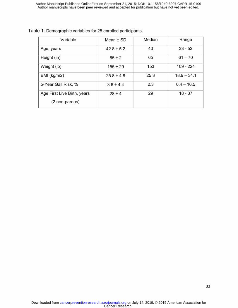
Demographic and Risk Information
Demographic and risk information for the 25 premenopausal women enrolled is
shown in Table 1. All subjects were Caucasian, with one self-identified as Hispanic.
Seven subjects (28%) were taking oral contraceptives.
Retention and Compliance
All 25 women enrolled completed the intervention, met the study definition of
compliance, had a repeat RPFNA, and provided paired biomarker data for assessment
of change over the study period. The minimum value for compliance was 81% of
prescribed agent, and the median compliance was 95%, based on subject-maintained
logs and returned pill counts. The median duration on study agent was 204 days (range
182 to 243). Per protocol, the nominal 6 months of study agent could be extended to 8
months for purposes of scheduling RPFNA with menstrual cycle.
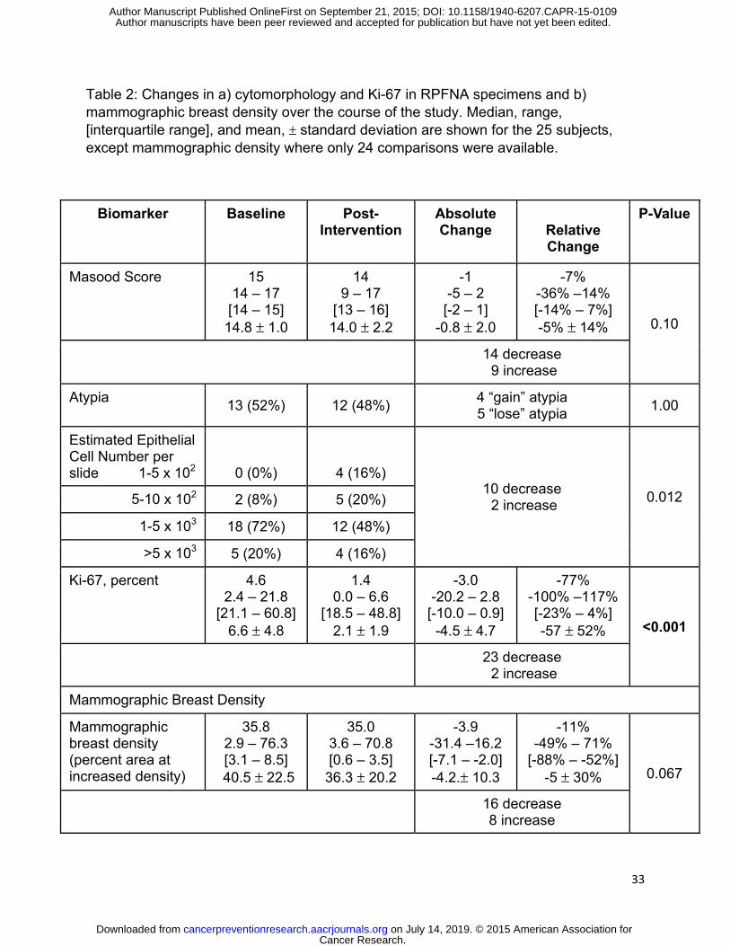
Changes in Ki-67 and Cytomorphology in Benign Breast Tissue
The median level of Ki-67 staining at baseline was 4.6% (range 2.4% to 21.8%;
(interquartile range (IQR), 3.1 – 8.5%); post-intervention, the median was 1.4% (range
Cancer Research. on July 14, 2019. © 2015 American Association forcancerpreventionresearch.aacrjournals.org Downloaded from
Author manuscripts have been peer reviewed and accepted for publication but have not yet been edited. Author Manuscript Published OnlineFirst on September 21, 2015; DOI: 10.1158/1940-6207.CAPR-15-0109
13
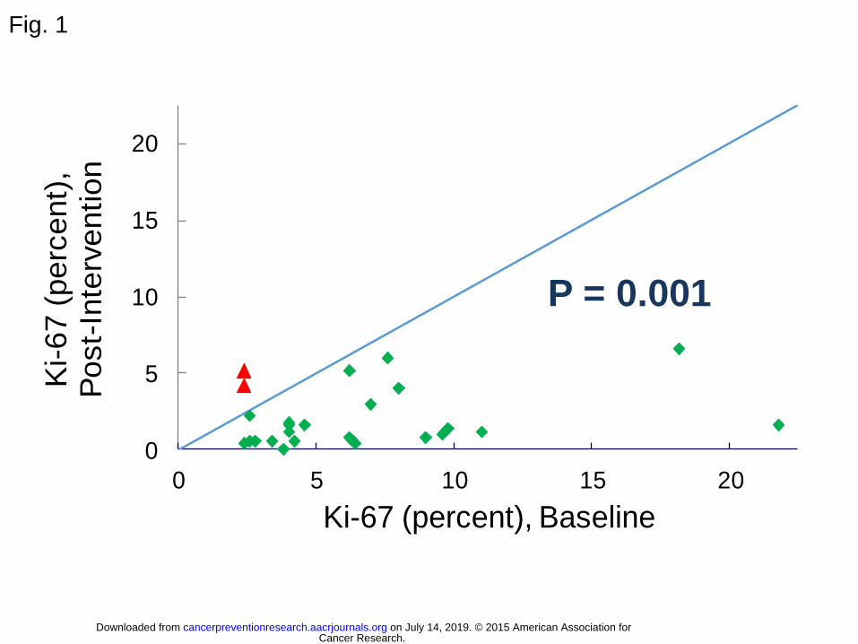
0% to 6.6%; IQR, 0.6 – 3.5%) (Table 2). The median change was -3.0% (range -20.2%
to 2.8%; IQR, -7.1 – -2.0%), which corresponded to a relative change of -77% with a
range of -100% (zero staining post-intervention) to 117% (IQR, -88% – -53%) (p<0.001,
Wilcoxon). Despite increases in serum bioavailable estradiol (see below), a decrease in
Ki-67 positive staining was noted in 23/25 (92%) subjects (Figure 1). There were no
differences between the seven oral contraceptive (OC) users and 18 non-users for
expression of Ki-67: baseline (p=0.93), post-intervention (p=1.0), change (p=0.88), or
relative change (p=0.69) (Mann-Whitney test). Both OC users and non-users showed
decreases (5/7, p=0.091; 18/18, p=0.001) with intervention.
There were no significant changes in cytomorphology over the course of the
intervention, either by a categorical descriptor or by Masood score (Table 2). Thirteen
of the 25 women exhibited hyperplasia with atypia at baseline versus 12 at study
conclusion. Similarly, for the semi-quantitative Masood cytomorphology score, median
score was 15 at baseline and 14 at second RPFNA, with a median 1 point decrease
(p=0.10, Wilcoxon).
Changes in Gene Expression in Benign Breast Tissue
Seventeen paired specimens (baseline and post-intervention) were available for
RT-qPCR analysis for levels of mRNA. Specimens from eight women were excluded
from analysis because either the baseline or post-intervention specimen was grossly
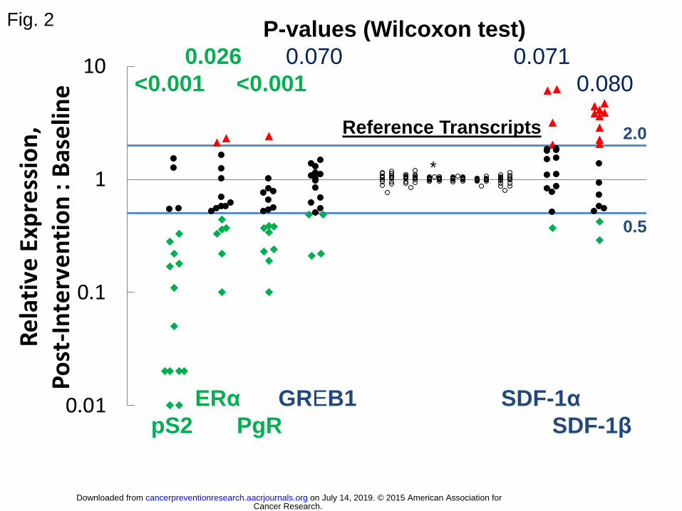
bloody. Significant decreases (p<0.05) were noted for transcripts for three estrogen
inducible genes that code for pS2, ERα, and PgR. There were also borderline
significant decreases for GREB1 and borderline significant increases for SDF-1α and
Cancer Research. on July 14, 2019. © 2015 American Association forcancerpreventionresearch.aacrjournals.org Downloaded from
Author manuscripts have been peer reviewed and accepted for publication but have not yet been edited. Author Manuscript Published OnlineFirst on September 21, 2015; DOI: 10.1158/1940-6207.CAPR-15-0109
14
SDF-1β (Figure 2). There were no changes noted for keratin 5, insulin-like growth factor
1 receptor, cyclin D1, or steroid sulfatase.
Changes in Mammographic Breast Density
There was no statistically significant change in mammographic breast density,
expressed as percent of breast area with increased density, from baseline (median
35.8%) to post-intervention (median 35.0%),with an average of 9 months between
mammograms (Table 2). Breast density was statistically significantly (p<0.001) lower at
baseline and post-intervention for women with higher BMI (dichotomized at the median
of 25 Kg/m2); but there was no difference for either absolute or relative change in
density.
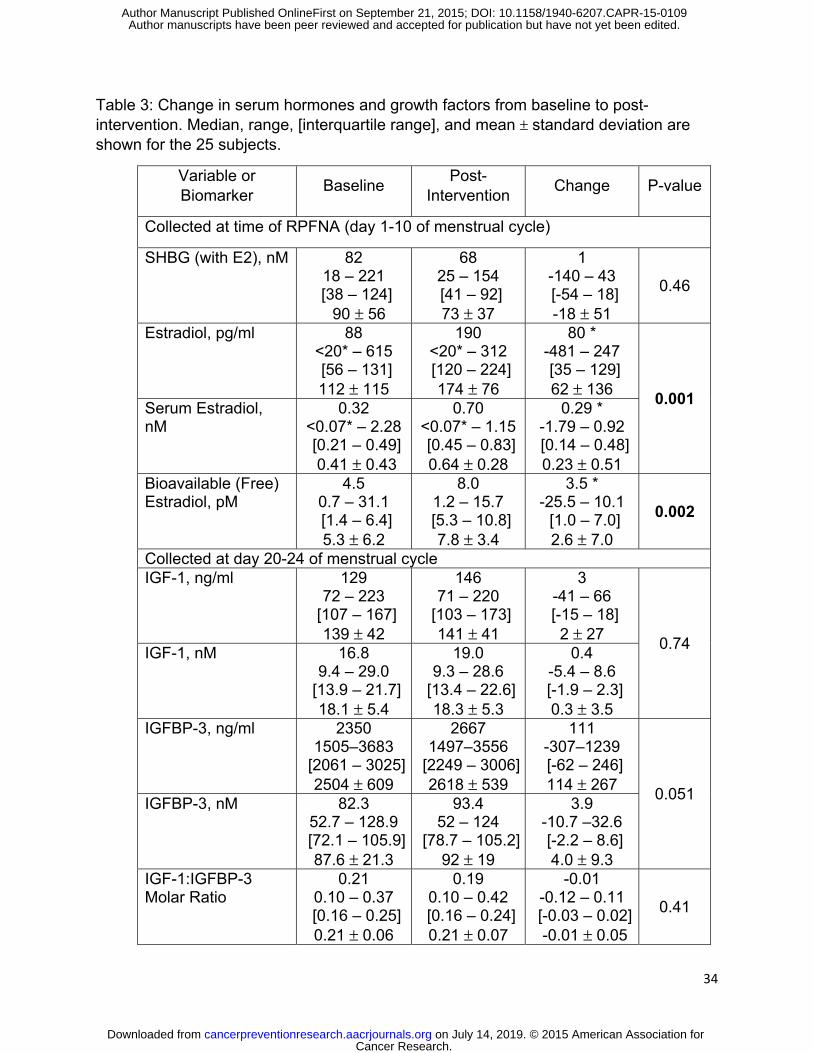
Change in Serum Hormones and IGF-1 and IGFBP3
Follicular phase (days 1-10 of cycle) estradiol and bioavailable estradiol
increased by medians of 78% and 110%, respectively, relative to baseline (p≤0.002)
(Table 3). Both OC users and non-users exhibited significant (p=0.018; p=0.044)
increases in bioavailable estradiol. For OC users this was due not only to an increase
in estradiol but also a significant decrease (7/7 subjects; p=0.018) in SHBG. Luteal
phase total testosterone increased by a median of ~30% relative to baseline (p=0.002).
Bioavailable testosterone did not change for the 18 OC non-users (p=0.91); but did
increase in each of the seven OC users (p=0.018), in part due to significant (7/7;
p=0.018) decreases in SHBG. OC users also had lower levels of bioavailable
testosterone at baseline than non-users (median 2.3 nM vs 17.2 nM; p=0.046). There
were no statistically significant changes for progesterone, IGF-1, IGFBP3, or the IGF-
1:IGFBP3 molar ratio (Table 3),
Cancer Research. on July 14, 2019. © 2015 American Association forcancerpreventionresearch.aacrjournals.org Downloaded from
Author manuscripts have been peer reviewed and accepted for publication but have not yet been edited. Author Manuscript Published OnlineFirst on September 21, 2015; DOI: 10.1158/1940-6207.CAPR-15-0109
15
Self-Reported Adverse Events
Five (20%) of 25 subjects reported no adverse events, 11 (44%) subjects
reported only grade 1 events, seven (28%) subjects reported a grade 2 event, and two
(8%) subjects reported a grade 3 event. One subject reported at the post-intervention
visit that grade 3 hot flashes had begun approximately two months earlier (after 4
months on study agent). One subject reported a grade 3 dizziness that began
approximately 6 weeks after starting study agent; this was considered to be unrelated to
study agent. A total of 71 adverse events (50 grade 1, 19 grade 2, two grade 3) were
self-reported, with only half being attributed by the protocol chair (CJF) as possibly or
probably related to study agent. Most common reported AEs (percent of subjects)
included irregular menses (32%), leg/muscle cramps (25%), diarrhea (16%), and hot
flashes (16%). No Serious Adverse Events were reported. Nor did any subject drop out
of the study due to AEs.
Quantitative Assessment of Hot Flashes, Menstrual Irregularities, Musculoskeletal
Symptoms, and General Quality of Life
Consistent with the low incidence of study-related moderate or severe adverse
events, no significant changes were observed for the quantitative assessments of
quality of life.
Problems with hot flashes were assessed for average number per day and
intensity. Only six women reported mild to moderate hot flashes prior to starting drug,
and only for two were these as frequent as daily. Five of the six with initial hot flashes
did not report hot flashes at their post-intervention visit; for the sixth, there was a slight
increase in number and intensity. Five other participants with no hot flashes at baseline
Cancer Research. on July 14, 2019. © 2015 American Association forcancerpreventionresearch.aacrjournals.org Downloaded from
Author manuscripts have been peer reviewed and accepted for publication but have not yet been edited. Author Manuscript Published OnlineFirst on September 21, 2015; DOI: 10.1158/1940-6207.CAPR-15-0109
16
reported infrequent hot flashes post-intervention. Overall, there was no effect of
acolbifene use on symptoms associated with hot flashes.
The Health Assessment Questionnaire II (HAQ-II) measures interference in daily
activities from arthralgias and joint pain. No woman reported a score above zero at
baseline and only one had a non-zero score (1.0) post-intervention. Thus, acolbifene
use was not associated with joint discomfort or disability. Similar results were obtained
for self-reported incidence, frequency, and severity of muscle cramps. Only three
women reported mild muscle cramps prior to starting drug. For two, no muscle cramps
were reported post-intervention; for the third, there was no change in any aspect of
muscle cramp symptoms. For three other women with no muscle cramps at baseline,
there were mild muscle cramps reported post-intervention. There was thus no adverse
effect of acolbifene use on symptoms associated with muscle cramps.
The Brief Fatigue Inventory (BFI) measures intensity of fatigue and interference
with daily activities. BFI scores at baseline and post-intervention were similar, reflecting
no change overall (p=0.82, Wilcoxon test). Medians were 9 and 10; ranges were 0 – 54
and 0 – 44; and means/standard deviations were 12.8 ± 13.2 and 12.9 ± 13.5,
respectively. For change over the study, there was a median of 1, range of -13 – 13,
and mean of -0.2 ± 7.6.
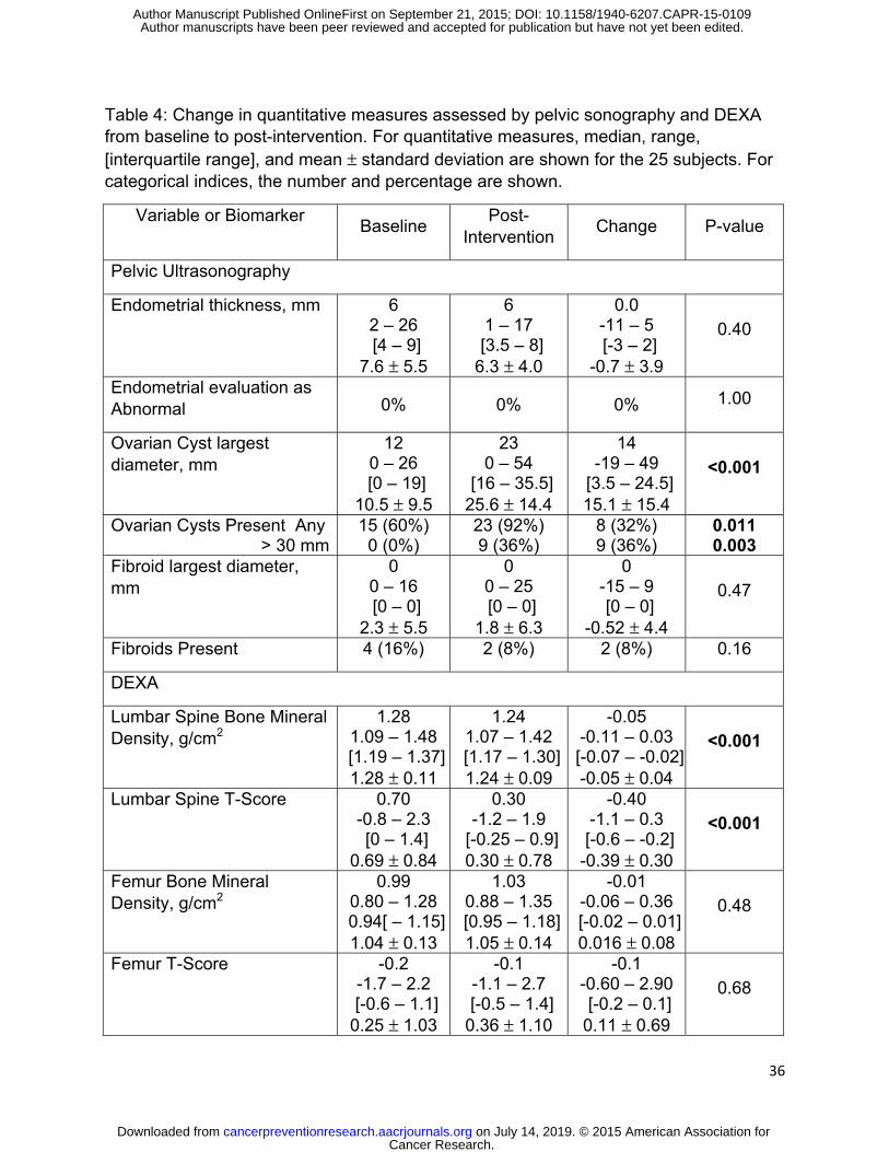
Gynecologic Parameters Assessed by Pelvic Ultrasonography
Endometrial thickness was unchanged over the course of the study (Table 4). In
contrast, the number of women in whom ovarian cysts could be visualized increased
from 15 (60%) to 23 (82%) (p=0.011, McNemar’s test). The largest diameter of ovarian
Cancer Research. on July 14, 2019. © 2015 American Association forcancerpreventionresearch.aacrjournals.org Downloaded from
Author manuscripts have been peer reviewed and accepted for publication but have not yet been edited. Author Manuscript Published OnlineFirst on September 21, 2015; DOI: 10.1158/1940-6207.CAPR-15-0109
17
cysts increased from a median of 12 mm at baseline to 23 mm post-intervention
(p<0.001).
Bone Density Assessed by DEXA
From DEXA assessments (Table 4), there was a statistically significant (p<0.001)
but minor decrease in lumbar spine bone density measurements. Median changes
were -0.04 g/cm2 (range -0.11 to +0.03) and -0.40 (range -1.10 to +0.30) for T-score.
Only one participant showed a clinically significant T-score decrease of at least one unit,
from -0.1 to -1.2. There was no observable effect on femur bone density or percent
body fat measured.
Cancer Research. on July 14, 2019. © 2015 American Association forcancerpreventionresearch.aacrjournals.org Downloaded from
Author manuscripts have been peer reviewed and accepted for publication but have not yet been edited. Author Manuscript Published OnlineFirst on September 21, 2015; DOI: 10.1158/1940-6207.CAPR-15-0109
18
Discussion
This is the first report of the effect of the SERM acolbifene on benign breast
tissue of healthy premenopausal women. There was favorable modulation of the risk
biomarker Ki-67 as well as expression of several estrogen responsive genes including
pS2 and PgR despite dramatic increases in serum estradiol levels. There were no
subjects who discontinued use because of side effects and no increase in endometrial
thickness. Clinically insignificant decreases in lumbar spine bone density were
observed following 6-8 months exposure, as well as an asymptomatic increase in
ovarian cysts. Overall, acolbifene appears to modulate tissue risk biomarkers in a
similar fashion as tamoxifen; but in this single arm study hot flashes and other
perimenopausal symptoms were not increased as would be expected with tamoxifen.
Nor was there an increase in endometrial thickness.
Ki-67 was selected as the primary risk biomarker endpoint of this study because
proliferation is permissive for cancer development. In observational studies, Ki-67 was
higher in foci of hyperplasia and atypical hyperplasia of women who subsequently
developed breast cancer than in those that did not [26, 27]. Women with ≥2% of cells in
atypical foci labeling for Ki-67 had a 4-fold increased risk for breast cancer [27]. By
eligibility criterion, a minimum baseline Ki-67 of 2% was required in clusters of cells
judged to be hyperplastic by cytologic criteria. The reduction in Ki-67 observed was
statistically significant and almost universal (23/25 paired specimens), consistent with
the well-known effects induced by tamoxifen in early breast cancer in short-term window
of opportunity trials and premenopausal benign breast tissue [28-30]. In neo-adjuvant
cancer treatment studies, reduction of or low post-tamoxifen Ki-67 in tumor tissue is
Cancer Research. on July 14, 2019. © 2015 American Association forcancerpreventionresearch.aacrjournals.org Downloaded from
Author manuscripts have been peer reviewed and accepted for publication but have not yet been edited. Author Manuscript Published OnlineFirst on September 21, 2015; DOI: 10.1158/1940-6207.CAPR-15-0109
19
associated with superior recurrence-free survival [31]. However, a serial biopsy study
reported by Moshin and Allred in 2005 did not show an effect of 1 year of tamoxifen vs
control on Ki-67 in a small number of women with benign hyperplastic foci generally
without atypia [32].
We did not see any reduction in cytologic evidence of atypia after 6 months of
acolbifene use. Cytologic evidence of atypia by RPFNA, like atypical hyperplasia in
diagnostic biopsies, is associated with increased risk [17], but there is no evidence that
short-term use of a SERM, including tamoxifen, will significantly change morphology in
benign breast tissue [32, 33].
Reduction observed at the transcript (mRNA) level of the estrogen inducible
genes for pS2 and PgR is qualitatively similar for acolbifene as that observed with
tamoxifen [34, 35]. Expression of the gene for ERα was reduced dramatically and
GREB1 which is an estrogen response gene associated with proliferation [34, 36] was
slightly reduced. There was no clear effect on the chemokine stromal cell-derived
factor 1 (SDF-1) which is important for viability of stem cells and has been implicated in
ligand-independent phosphorylation of the estrogen receptor and tamoxifen resistance
[37-39] Tamoxifen has been variably associated with increases in SDF-1 [34, 40, 41].
A recent report assessing the short term effects of several SERMs and fulvestrant on a
large number of genes in mammary cancer from ovariectomized mice suggests that
acolbifene reverses the effect of estradiol on more estrogen inducible genes than
tamoxifen, raloxifene, or fulvestrant [42].
The risk biomarkers of serum IGF-1 to IGFBP3 ratio [43] and mammographic
breast density [24] are known to be modulated by tamoxifen [33, 44, 45]. In IBIS-1
Cancer Research. on July 14, 2019. © 2015 American Association forcancerpreventionresearch.aacrjournals.org Downloaded from
Author manuscripts have been peer reviewed and accepted for publication but have not yet been edited. Author Manuscript Published OnlineFirst on September 21, 2015; DOI: 10.1158/1940-6207.CAPR-15-0109
20
tamoxifen reduced mammographic breast density in premenopausal women with
baseline density of >10% by a mean of 13% compared to placebo [45]. The median
absolute decrease in mammographic density of 3.9% after 6 months of acolbifene was
not statistically significant although a greater numerical effect is likely had the drug been
given longer [46] and might have reached significance had more subjects been entered
into the trial. Although acolbifene did not significantly modulate either IGF-1 or breast
density, this does not necessarily mean that acolbifene is a less effective anti-estrogen
than tamoxifen. Aromatase inhibitors modulate neither IGF-1 nor mammographic
breast density but have demonstrated efficacy in prevention and are generally viewed
as more effective than tamoxifen in a low estrogen environment [46, 47].
Acolbifene was associated with an increase in serum estradiol levels and ovarian
cysts. Increased mid-cycle and luteal levels of estrogen and increased ovarian cysts
have been observed for tamoxifen where the prevalence in asymptomatic
premenopausal women undergoing regular pelvic ultrasound monitoring has been
reported as up to 80% without regard to cyst size and approximately 30% for cysts of 30
mm or greater diameter [7, 48-51]. In the absence of symptoms, these cysts are
probably of little clinical significance and are likely due to prolonged elevation of FSH in
the follicular phase combined with elevated mid-cycle or luteal estradiol for both
tamoxifen and acolbifene [7]. Increase in ovarian cyst formation after acolbifene tended
to resolve shortly after the drug was stopped (data not shown). We found no change in
SHBG or free testosterone. Information on SHBG and free testosterone in
premenopausal women without breast cancer is limited but SHBG is generally
increased and free testosterone generally reduced in postmenopausal women after
Cancer Research. on July 14, 2019. © 2015 American Association forcancerpreventionresearch.aacrjournals.org Downloaded from
Author manuscripts have been peer reviewed and accepted for publication but have not yet been edited. Author Manuscript Published OnlineFirst on September 21, 2015; DOI: 10.1158/1940-6207.CAPR-15-0109
21
tamoxifen [52]. There was a minimal reduction in premenopausal bone density similar to
that observed with tamoxifen [53]. Finally, there was a statistically significant, but
clinically insignificant, decrease in white blood cells and platelets (data not shown),
similar to what has been observed with tamoxifen [33].
Importantly, from the standpoint of uptake of a prevention agent by
premenopausal women, there was no evidence of worsening of hot flashes, other
perimenopausal or musculoskeletal symptoms, or overall quality of life; nor was there
any evidence of endometrial thickening. In contrast in the NSABP P-1 trial, hot flashes
were reported by 81% of individuals randomized to 5 years of tamoxifen vs 65% of
those randomized to placebo [54] and endometrial thickening is commonly observed in
premenopausal women taking tamoxifen in the absence of concomitant Goserelin [55,
56].
The ability of a SERM to act as an agonist or antagonist depends on hormone
levels and the specific tissue as activator/repressor levels vary by tissue type.
Acolbifene differs from tamoxifen in that it blocks the co-activator SRC-1 expressed in
high amounts in uterine but not breast tissue [12], thus explaining acolbifene’s lack of
agonist effect on the uterus. Acolbifene has the potential to be more effective than
tamoxifen as it inhibits both the AF-1 and AF-2 functions of both ERα and ERβ, while
the inhibitory action of tamoxifen is limited to AF-2. SERMs which block only AF2 are
likely to have partial estrogen agonist activity [reviewed in 57]. A SERM’s relative
potency depends on many factors including its bioavailability, serum and tissue half-life,
affinity for the estrogen receptor, and rate of ubiquination of the ligand-ER complex [58].
Cancer Research. on July 14, 2019. © 2015 American Association forcancerpreventionresearch.aacrjournals.org Downloaded from
Author manuscripts have been peer reviewed and accepted for publication but have not yet been edited. Author Manuscript Published OnlineFirst on September 21, 2015; DOI: 10.1158/1940-6207.CAPR-15-0109
22
The plasma half-life of tamoxifen is ~7 days [59] whereas that of acolbifene is ~24 hours
[Personal communication, Fernand Labrie, Endorecherche, Inc., Quebec, Canada].
Limitations of this study include the small number of subjects and lack of a
control (placebo) arm. Because of the eligibility criterion of Ki-67 ≥2%, and the resulting
baseline mean Ki-67 of our cohort being higher than the population mean, there is a risk
that the apparent decrease in Ki-67 was the result of a regression to the mean artifact.
Without a parallel placebo arm one cannot conclusively distinguish between this
possibility and a true effect of acolbifene. In addition, the large number of variables
considered, without correction for multiple comparisons, increases the risk of type I error
for the exploratory biomarkers. None-the-less, a number of factors were identified as
potential pharmacodynamic effect markers or that might assist in elucidation of
mechanisms of action; these can be evaluated further in future trials.
In conclusion, acolbifene was associated with a favorable side effect profile, and
an apparent favorable modulation of risk biomarkers including proliferation as well as
the estrogen response genes for pS2, ERα, and PgR. Given the lack of demonstrated
increase in hot flashes and other subjective symptoms, acolbifene should be compared
to placebo (2 arms) or placebo and tamoxifen (3 arms) in a Phase IIB trial for
premenopausal women with modulation of benign breast tissue proliferation and
vasomotor symptoms as co-primary endpoints.
Cancer Research. on July 14, 2019. © 2015 American Association forcancerpreventionresearch.aacrjournals.org Downloaded from
Author manuscripts have been peer reviewed and accepted for publication but have not yet been edited. Author Manuscript Published OnlineFirst on September 21, 2015; DOI: 10.1158/1940-6207.CAPR-15-0109
23
References
1. Visvanathan K, Hurley P, Bantug E, Brown P, Col NF, Cuzick J, et al. Use of
pharmacologic interventions for breast cancer risk reduction: American Society of
Clinical Oncology clinical practice guideline. J Clin Oncol 2013; 31:2942-62
2. Cuzick J, Sestak I, Bonanni B, Costantino JP, Cummings S, DeCensi A, et al.
SERM Chemoprevention of Breast Cancer Overview Group. Selective oestrogen
receptor modulators in prevention of breast cancer: an updated meta-analysis of
individual participant data. Lancet 2013; 381:1827-34
3. Ropka ME, Keim J, Philbrick JT. Patient decisions about breast cancer
chemoprevention: A systematic review and meta-analysis J Clin Oncol 2010;
28:3090–5
4. Day R, Ganz PA, Costantino JP, Cronin WM, Wickerham DL, Fisher B. Health-
related quality of life and tamoxifen in breast cancer prevention: a report from the
National Surgical Adjuvant Brest and Bowel Project P-1 Study. J Clin Oncol 1999;
17:2659–69
5. Jordan VC. Estrogen, selective estrogen receptor modulation, and coronary heart
disease. Something or nothing. J Natl Cancer Inst 2001; 93:2-4
6. Donnelly LS, Evans DG, Wiseman J, Fox J, Greenhalgh R, Affen J, et al. Uptake of
tamoxifen in consecutive premenopausal women under surveillance in a high-risk
breast cancer clinic. Br J Cancer 2014; 110:1681-7
7. Cohen I, Figer A, Tepper R, Shapira J, Altaras MM, Yigael D, et al. Ovarian
overstimulation and cystic formation in premenopausal tamoxifen exposure:
comparison between tamoxifen-treated and nontreated breast cancer patients.
Gynecol Oncol 1999; 72:202-7
Cancer Research. on July 14, 2019. © 2015 American Association forcancerpreventionresearch.aacrjournals.org Downloaded from
Author manuscripts have been peer reviewed and accepted for publication but have not yet been edited. Author Manuscript Published OnlineFirst on September 21, 2015; DOI: 10.1158/1940-6207.CAPR-15-0109
24
8. Buijs C, Willemse PH, de Vries EG, Ten Hoor KA, Boezen HM, Hollema H, et al.
Effect of tamoxifen on the endometrium and the menstrual cycle of premenopausal
breast cancer patients. Int J Gynecol Cancer 1996; 19:677-81
9. Melnikow J, Paterniti D, Azari R, Kuenneth C, Birch S, Kuppermann M, et al.
Preferences of women evaluating risks of tamoxifen (POWER) study of preferences
for tamoxifen for breast cancer risk reduction. Cancer 2005; 103:1996-2005
10. Labrie F, Labrie C, Belanger A, Simard J, Gauthier S, Luu-The V, et al. EM-652
(SCH 57068), a third generation SERM acting as pure antiestrogen in the mammary
gland and endometrium. J Steroid Biochem Mol Biol 1999; 69:51-84
11. Labrie F, Simard J, Labrie C, Bélanger A. EM-652 (SCH 57068), a pure SERM in the
mammary gland and endometrium. Références en Gynécologie Obstétrique 2001;
8:331-6.
12. Labrie F, Labrie C, Belanger A, Simard J, Giguere V, Tremblay A, et al. EM-652
(SCH57068), a pure SERM having complete antiestrogenic activity in the mammary
gland and endometrium. J Steroid Biochem Mol Biol 2001; 79:213-25.
13. Labrie F, Champagne P, Labrie C, Roy J, Laverdière J, Provencher L, et al. Activity
and safety of the antiestrogen EM-800, the orally active precursor of acolbifene, in
tamoxifen-resistant breast cancer. J Clin Oncol 2004; 22:864-71.
14. Roy J, Couillard S, Gutman M, Labrie F. A novel pure SERM achieves complete
regression of the majority of human breast cancer tumors in nude mice. Breast
Cancer Res Treat 2003; 81:223-9
15. Luo S, Stojanovic M, Labrie C, Labrie F. Inhibitory effect of the novel anti-estrogen
EM-800 and medroxyprogesterone acetate on estrone-stimulated growth of
Cancer Research. on July 14, 2019. © 2015 American Association forcancerpreventionresearch.aacrjournals.org Downloaded from
Author manuscripts have been peer reviewed and accepted for publication but have not yet been edited. Author Manuscript Published OnlineFirst on September 21, 2015; DOI: 10.1158/1940-6207.CAPR-15-0109
25
dimethylbenz(a) anthracene-induced mammary carcinoma in rat. Int J Cancer 1997;
73:580-6
16. Gail MH, Costantino JP, Bryant J, Croyle R, Freedman L, Helzlsouer K, et al.
Weighing the risks and benefits of tamoxifen treatment for preventing breast cancer.
J Natl Cancer Inst 1999; 91:1829–46
17. Fabian CJ, Kimler BF, Zalles CM, Klemp JR, Kamel S, Zeiger S, et al. Short-term
breast cancer prediction by random periareolar fine-needle aspiration cytology and
the Gail risk model. J Natl Cancer Inst 2000; 92:1217-27
18. Zalles C, Kimler BF, Kamel S, McKittrick R, Fabian CJ. Cytologic patterns in random
aspirates from women at high and low risk for breast cancer. Breast J 1995; 1:343-9
19. Masood S, Frykberg ER, McLellan GL, Scalapino MC, Mitchum DG, Bullard JB.
Prospective evaluation of radiologically directed fine-needle aspiration biopsy of
nonpalpable breast lesions. Cancer 1990; 66:1480-7
20. Fabian CJ, Kimler BF, Zalles CM, Klemp JR, Petroff BK, Khan QJ, et al. Reduction
in Ki-67 in benign breast tissue of high risk women with the lignan
secoisolariciresinol diglycoside (SDG). Cancer Prev Res 2010; 3:1342-50
21. Phillips TA, Fabian CJ, Kimler BF, Petroff BK. Assessment of RNA in human breast
tissue sampled by random periareolar fine needle aspiration and ductal lavage and
processed as fixed or frozen specimens. Reprod Biol 2013; 13:75-81
22. Endogenous Hormones and Breast Cancer Collaborative Group. Free estradiol and
breast cancer risk in postmenopausal women: comparison of measured and
calculated values. Cancer Epidemiol Biomarkers Prev 2003; 12:1457–61.
Cancer Research. on July 14, 2019. © 2015 American Association forcancerpreventionresearch.aacrjournals.org Downloaded from
Author manuscripts have been peer reviewed and accepted for publication but have not yet been edited. Author Manuscript Published OnlineFirst on September 21, 2015; DOI: 10.1158/1940-6207.CAPR-15-0109
26
23. Vermeulen A, Verdonck G. Representativeness of a single point plasma
testosterone level for the long term hormonal milieu in men. J Clin Endocrinol Metab
1992; 74:939-42
24. Boyd NF, Byng JW, Jong RA, Fishell EK, Little LE, Miller AB, et al. Quantitative
classification of mammographic densities and breast cancer risk: results from the
Canadian National Breast Screening Study. J Natl Cancer Inst 1995; 87:670-5
25. Stone J, Gunasekara A, Martin LJ, Yaffe M, Minkin S, Boyd NF. The detection of
change in mammographic density. Cancer Epidemiol Biomarkers Prev 2003;
12:625-30
26. Shaaban AM, Sloane JP, West CR, Foster CS. Breast cancer risk in usual ductal
hyperplasia is defined by estrogen receptor-alpha and Ki-67 expression. Am J
Pathol 2002; 160:597-604
27. Santisteban M, Reynolds C, Barr Fritcher EG, Frost MH, Vierkant RA, Anderson SS,
et al. Ki67: a time-varying biomarker of risk of breast cancer in atypical hyperplasia.
Breast Cancer Res Treat 2010; 121:431-7
28. Decensi A, Robertson C, Viale G, Pigatto F, Johansson H, Kisanga ER, et al. A
randomized trial of low-dose tamoxifen on breast cancer proliferation and blood
estrogenic biomarkers. J Natl Cancer Inst 2003; 95:779-90
29. DeCensi A, Guerrieri-Gonzaga A, Gandini S, Serrano D, Cazzaniga M, Mora S, et
al. Prognostic significance of Ki-67 labeling index after short-term presurgical
tamoxifen in women with ER-positive breast cancer. Ann Oncol 2011; 22:582-7
Cancer Research. on July 14, 2019. © 2015 American Association forcancerpreventionresearch.aacrjournals.org Downloaded from
Author manuscripts have been peer reviewed and accepted for publication but have not yet been edited. Author Manuscript Published OnlineFirst on September 21, 2015; DOI: 10.1158/1940-6207.CAPR-15-0109
27
30. de Lima GR, Facina G, Shida JY, Chein MB, Tanaka P, Dardes RC, et al. Effects of
low dose tamoxifen on normal breast tissue from premenopausal women. Eur J
Cancer 2003; 39:891-8
31. Dowsett M, Smith IE, Ebbs SR, Dixon JM, Skene A, Griffith C, et al. Short-term
changes in Ki-67 during neoadjuvant treatment of primary breast cancer with
anastrozole or tamoxifen alone or combined correlate with recurrence-free survival.
Clin Cancer Res 2005; 11(2 Pt 2):951s-8s
32. Mohsin SK, Allred DC, Osborne CK, Cruz A, Otto P, Chew H, Clark GM, Elledge
RM. Morphologic and immunophenotypic markers as surrogate endpoints of
tamoxifen effect for prevention of breast cancer. Breast Cancer Res Treat. 2005;
94:205-11
33. Euhus D, Bu D, Xie XJ, Sarode V, Ashfaq R, Hunt K, et al. Tamoxifen
downregulates ets oncogene family members ETV4 and ETV5 in benign breast
tissue: implications for durable risk reduction. Cancer Prev Res 2011; 4:1852-62
34. Rae JM, Johnson MD, Scheys JO, Cordero KE, Larios JM, Lippman ME. GREB 1 is
a critical regulator of hormone dependent breast cancer growth. Breast Cancer Res
Treat 2005; 92:141-9
35. Frasor J, Stossi F, Danes JM, Komm B, Lyttle CR, Katzenellenbogen BS. Selective
estrogen receptor modulators: discrimination of agonistic versus antagonistic
activities by gene expression profiling in breast cancer cells. Cancer Res 2004;
64:1522-33
Cancer Research. on July 14, 2019. © 2015 American Association forcancerpreventionresearch.aacrjournals.org Downloaded from
Author manuscripts have been peer reviewed and accepted for publication but have not yet been edited. Author Manuscript Published OnlineFirst on September 21, 2015; DOI: 10.1158/1940-6207.CAPR-15-0109
28
36. Ghosh MG, Thompson DA, Weigel RJ. PDZK1 and GREB1 are estrogen-regulated
genes expressed in hormone-responsive breast cancer. Cancer Res 2000; 60:6367-
75
37. Rhodes LV, Short SP, Neel NF, Salvo VA, Zhu Y, Elliott S, et al. . Cytokine receptor
CXCR4 mediates estrogen-independent tumorigenesis, metastasis, and resistance
to endocrine therapy in human breast cancer. Cancer Res 2011; 71:603-13
38. Dubrovska A, Hartung A, Bouchez LC, Walker JR, Reddy VA, Cho CY, et al.
CXCR4 activation maintains a stem cell population in tamoxifen-resistant breast
cancer cells through AhR signalling. Br J Cancer 2012; 107:43-52
39. Kucia M, Jankowski K, Reca R, Wysoczynski M, Bandura L, Allendorf DJ, et al.
CXCR4-SDF-1 signalling, locomotion, chemotaxis and adhesion. J Mol Histol 2004;
35:233-45
40. Kubarek Ł, Jagodzinski PP. Epigenetic up-regulation of CXCR4 and CXCL12
expression by 17 beta-estradiol and tamoxifen is associated with formation of DNA
methyltransferase 3B4 splice variant in Ishikawa endometrial adenocarcinoma cells.
FEBS Lett 2007; 581:1441-8
41. Pietkiewicz PP, Lutkowska A, Lianeri M, Jagodzinski PP. Tamoxifen epigenetically
modulates CXCL12 expression in MCF-7 breast cancer cells. Biomed Pharmacother
2010; 64:54-7
42. Calvo E, Luu-The V, Belleau P, Martel C, Labrie F. Specific transcriptional response
of four blockers of estrogen receptors on estradiol-modulated genes in the mouse
mammary gland. Breast Cancer Res Treat 2012; 134:625-47
Cancer Research. on July 14, 2019. © 2015 American Association forcancerpreventionresearch.aacrjournals.org Downloaded from
Author manuscripts have been peer reviewed and accepted for publication but have not yet been edited. Author Manuscript Published OnlineFirst on September 21, 2015; DOI: 10.1158/1940-6207.CAPR-15-0109
29
43. Hankinson SE, Willett WC, Colditz GA, Hunter DJ, Michaud DS, Deroo B, et al.
Circulating concentrations of insulin-like growth factor-I and risk of breast cancer.
Lancet 1998; 351:1393-6
44. Bonanni B, Johansson H, Gandini S, Guerrieri-Gonzaga A, Torrisi R, Sandri MT, et
al. Effect of low dose tamoxifen on the insulin-like growth factor system in healthy
women. Breast Cancer Res Treat 2001; 69:21-7
45. Cuzick J, Warwick J, Pinney E, Warren RM, Duffy SW. Tamoxifen and breast
density in women at increased risk of breast cancer. J Natl Cancer Inst 2004;
96:621-8
46. Cigler T, Richardson H, Yaffe MJ, Fabian CJ, Johnston D, Ingle JN, et al. A
randomized, placebo-controlled trial (NCIC CTG MAP.2) examining the effects of
exemestane on mammographic breast density, bone density, markers of bone
metabolism and serum lipid levels in postmenopausal women. Breast Cancer Res
Treat 2011; 126:453-61
47. Goss PE, Ingle JN, Alés-Martínez JE, Cheung AM, Chlebowski RT, Wactawski-
Wende J, et al. Exemestane for breast-cancer prevention in postmenopausal
women. N Engl J Med 2011; 364:2381-2391
48. Premkumar A, Venzon DJ, Avila N, Johnson DV, Remaley AT, Forman MR, Eng-
Wong J, Zujewski J, Stratton P. Gynecologic and hormonal effects of raloxifene in
premenopausal women. Fertil Steril. 2007; 88:1637-44.
49. Shushan A, Peretz T, Uziely B, Lewin A, Mor-Yosef S. Ovarian cysts in
premenopausal and postmenopausal tamoxifen-treated women with breast cancer.
Am J Obstet Gynecol. 1996; 174(1 Pt 1):141-4
Cancer Research. on July 14, 2019. © 2015 American Association forcancerpreventionresearch.aacrjournals.org Downloaded from
Author manuscripts have been peer reviewed and accepted for publication but have not yet been edited. Author Manuscript Published OnlineFirst on September 21, 2015; DOI: 10.1158/1940-6207.CAPR-15-0109
30
50. Inal MM, Incebiyik A, Sanci M, Yildirim Y, Polat M, Pilanci B, Nayki C, Camuzcuoğlu
H. Ovarian cysts in tamoxifen-treated women with breast cancer. Eur J Obstet
Gynecol Reprod Biol. 2005; 120:104-6
51. Berliere M, Duhoux FP, Dalenc F, Baurain JF, Dellevigne L, Galant C, Van Maanen
A, Piette P, Machiels JP. Tamoxifen and ovarian function. PLoS One. 2013;
8:e66616
52. Kostoglou-Athanassiou I, Ntalles K, Gogas J, Markopoulos C, Alevizou-Terzaki V,
Athanassiou P, Georgiou E, Proukakis C. Sex hormones in postmenopausal women
with breast cancer on tamoxifen. Horm Res. 1997; 47:116-20.
53. Powles TJ, Hickish T, Kanis JA, Tidy A, Ashley S. Effect of tamoxifen on bone
mineral density measured by dual energy x-ray absorptionmetry in health
premenopausal and postmenopausal women. J Clin Oncol 1996; 14:78-84
54. Day R. Quality of life and tamoxifen in a breast cancer prevention trial: a summary
of findings from the NSABP P-1 study. National Surgical Adjuvant Breast and Bowel
Project. Ann N Y Acad Sci. 2001; 949:143-50.
55. Chang J, Powles TJ, Ashley SE, Iveson T, Gregory RK, Dowsett M Variation in
endometrial thickening in women with amenorrhea on tamoxifen. Breast Cancer Res
Treat. 1998; 48:81-5.
56. Yang H, Zong X, Yu Y, Shao G, Zhang L, Qian C, Bian Y, Xu X, Sun W, Meng X,
Ding X, Chen D, Zou D, Xie S, Zheng Y, Zhang J, He X, Sun C, Yu X, Ni J.
Combined effects of goserelin and tamoxifen on estradiol level, breast density, and
endometrial thickness in premenopausal and perimenopausal women with early-
Cancer Research. on July 14, 2019. © 2015 American Association forcancerpreventionresearch.aacrjournals.org Downloaded from
Author manuscripts have been peer reviewed and accepted for publication but have not yet been edited. Author Manuscript Published OnlineFirst on September 21, 2015; DOI: 10.1158/1940-6207.CAPR-15-0109
31
stage hormone receptor-positive breast cancer: a randomised controlled clinical trial.
Br J Cancer. 2013; 109:582-8
57. Fabian CJ, Kimler BF: Selective estrogen receptor modulators for primary prevention
of breast cancer. J Clin Oncol 2005; 23:1644-55
58. Martinkovich S, Shah D, Planey SL, Arnott JA. Selective estrogen receptor
modulators: tissue specificity and clinical utility. Clin Interv Aging 2014; 9:1437-52
59. Jordan VC, New insights into the metabolism of tamoxifen and its role in the
treatment and prevention of breast cancer. Steroids 2007; 72:829–842.
Cancer Research. on July 14, 2019. © 2015 American Association forcancerpreventionresearch.aacrjournals.org Downloaded from
Author manuscripts have been peer reviewed and accepted for publication but have not yet been edited. Author Manuscript Published OnlineFirst on September 21, 2015; DOI: 10.1158/1940-6207.CAPR-15-0109
32
Table 1: Demographic variables for 25 enrolled participants.
Variable Mean ± SD Median Range
Age, years 42.8 ± 5.2 43 33 - 52
Height (in) 65 ± 2 65 61 – 70
Weight (lb) 155 ± 29 153 109 - 224
BMI (kg/m2) 25.8 ± 4.8 25.3 18.9 – 34.1
5-Year Gail Risk, % 3.6 ± 4.4 2.3 0.4 – 16.5
Age First Live Birth, years
(2 non-parous)
28 ± 4 29 18 - 37
Cancer Research. on July 14, 2019. © 2015 American Association forcancerpreventionresearch.aacrjournals.org Downloaded from
Author manuscripts have been peer reviewed and accepted for publication but have not yet been edited. Author Manuscript Published OnlineFirst on September 21, 2015; DOI: 10.1158/1940-6207.CAPR-15-0109
33
Table 2: Changes in a) cytomorphology and Ki-67 in RPFNA specimens and b) mammographic breast density over the course of the study. Median, range, [interquartile range], and mean, ± standard deviation are shown for the 25 subjects, except mammographic density where only 24 comparisons were available.
Biomarker Baseline Post- Intervention
Absolute Change
Relative Change
P-Value
Masood Score 15 14 – 17
[14 – 15] 14.8 ± 1.0
14 9 – 17
[13 – 16] 14.0 ± 2.2
-1 -5 – 2
[-2 – 1] -0.8 ± 2.0
-7% -36% –14% [-14% – 7%] -5% ± 14% 0.10
14 decrease 9 increase
Atypia 13 (52%) 12 (48%) 4 “gain” atypia 5 “lose” atypia 1.00
Estimated Epithelial Cell Number per slide 1-5 x 10² 0 (0%) 4 (16%)
10 decrease 2 increase 0.012 5-10 x 102 2 (8%) 5 (20%)
1-5 x 10³ 18 (72%) 12 (48%)
>5 x 10³ 5 (20%) 4 (16%)
Ki-67, percent 4.6 2.4 – 21.8
[21.1 – 60.8] 6.6 ± 4.8
1.4 0.0 – 6.6
[18.5 – 48.8] 2.1 ± 1.9
-3.0 -20.2 – 2.8
[-10.0 – 0.9] -4.5 ± 4.7
-77% -100% –117% [-23% – 4%] -57 ± 52% <0.001
23 decrease 2 increase
Mammographic Breast Density
Mammographic breast density (percent area at increased density)
35.8 2.9 – 76.3 [3.1 – 8.5] 40.5 ± 22.5
35.0 3.6 – 70.8 [0.6 – 3.5] 36.3 ± 20.2
-3.9 -31.4 –16.2 [-7.1 – -2.0] -4.2.± 10.3
-11% -49% – 71%
[-88% – -52%] -5 ± 30% 0.067
16 decrease 8 increase
Cancer Research. on July 14, 2019. © 2015 American Association forcancerpreventionresearch.aacrjournals.org Downloaded from
Author manuscripts have been peer reviewed and accepted for publication but have not yet been edited. Author Manuscript Published OnlineFirst on September 21, 2015; DOI: 10.1158/1940-6207.CAPR-15-0109
34
Table 3: Change in serum hormones and growth factors from baseline to post-intervention. Median, range, [interquartile range], and mean ± standard deviation are shown for the 25 subjects.
Variable or Biomarker Baseline Post-
Intervention Change P-value
Collected at time of RPFNA (day 1-10 of menstrual cycle)
SHBG (with E2), nM 82 18 – 221 [38 – 124] 90 ± 56
68 25 – 154 [41 – 92] 73 ± 37
1 -140 – 43 [-54 – 18] -18 ± 51
0.46
Estradiol, pg/ml 88 <20* – 615 [56 – 131] 112 ± 115
190 <20* – 312 [120 – 224] 174 ± 76
80 * -481 – 247 [35 – 129] 62 ± 136 0.001 Serum Estradiol,
nM 0.32
<0.07* – 2.28 [0.21 – 0.49] 0.41 ± 0.43
0.70 <0.07* – 1.15 [0.45 – 0.83] 0.64 ± 0.28
0.29 * -1.79 – 0.92 [0.14 – 0.48] 0.23 ± 0.51
Bioavailable (Free) Estradiol, pM
4.5 0.7 – 31.1 [1.4 – 6.4] 5.3 ± 6.2
8.0 1.2 – 15.7 [5.3 – 10.8] 7.8 ± 3.4
3.5 * -25.5 – 10.1
[1.0 – 7.0] 2.6 ± 7.0
0.002
Collected at day 20-24 of menstrual cycle IGF-1, ng/ml 129
72 – 223 [107 – 167] 139 ± 42
146 71 – 220
[103 – 173] 141 ± 41
3 -41 – 66 [-15 – 18] 2 ± 27 0.74 IGF-1, nM 16.8
9.4 – 29.0 [13.9 – 21.7] 18.1 ± 5.4
19.0 9.3 – 28.6
[13.4 – 22.6] 18.3 ± 5.3
0.4 -5.4 – 8.6 [-1.9 – 2.3] 0.3 ± 3.5
IGFBP-3, ng/ml 2350 1505–3683
[2061 – 3025]2504 ± 609
2667 1497–3556
[2249 – 3006]2618 ± 539
111 -307–1239 [-62 – 246] 114 ± 267 0.051 IGFBP-3, nM 82.3
52.7 – 128.9 [72.1 – 105.9]87.6 ± 21.3
93.4 52 – 124
[78.7 – 105.2]92 ± 19
3.9 -10.7 –32.6 [-2.2 – 8.6] 4.0 ± 9.3
IGF-1:IGFBP-3 Molar Ratio
0.21 0.10 – 0.37 [0.16 – 0.25] 0.21 ± 0.06
0.19 0.10 – 0.42 [0.16 – 0.24] 0.21 ± 0.07
-0.01 -0.12 – 0.11 [-0.03 – 0.02] -0.01 ± 0.05
0.41
Cancer Research. on July 14, 2019. © 2015 American Association forcancerpreventionresearch.aacrjournals.org Downloaded from
Author manuscripts have been peer reviewed and accepted for publication but have not yet been edited. Author Manuscript Published OnlineFirst on September 21, 2015; DOI: 10.1158/1940-6207.CAPR-15-0109

35
Variable or Biomarker Baseline Post-
Intervention Change P-value
Progesterone, ng/ml 3.5 0.4 – 26.2 [0.9 – 3.5] 4.4 ± 5.5
2.8 0.5 – 36.3 [0.7 –9.7 ] 7.1 ± 10.0
0.0 -10.7 – 30.1 [-2.6 – 1.9] 2.7 ± 9.7 0.78 Progesterone, nM 11.0
1.1 – 83.3 [2.7 – 18.8] 14.0 ± 17.3
8.9 1.5 – 115.3 [2.1 – 30.7] 22.6 ± 31.7
-0.1 -34.1 – 95.8 [-8.3 – 6.1] 8.6 ± 30.7
SHBG, nM 87 22 – 276 [59 – 193] 117 ± 80
85 34 – 179 [48 – 135] 93 ± 46
6 -227 – 60 [-45 – 18] -24 ± 73
0.53
Testosterone, ng/ml 0.40 <0.08* – 2.59 [0.18 – 0.83] 0.63 ± 0.64
0.58 0.19 – 3.52 [0.29 – 0.83] 0.85 ± 0.91
0.16 -0.21 – 2.33 [0.02 – 0.24] 0.22 ± 0.48 0.002 Testosterone, nM 1.37
<0.28* – 9.00 [0.63 – 2.87] 2.18 ± 2.21
2.0 0.64 – 12.21 [1.00 – 2.86] 2.93 ± 3.15
0.53 -0.74 – 8.09 [0.06 – 0.80] 0.75 ± 1.66
Bioavailable (Free) Testosterone, pM
13.4 1.1 – 120.2 [4.6 – 35.1] 25.1 ± 30.7
17.3 3.8 – 194.4 [9.5 – 38.3] 32.4 ± 41.6
4.1 -26.9 – 118.2
[-2.1 – 8.5] 7.3 ± 26.8
0.13
* One woman had estradiol levels below limit of detection at both times and was imputed to have no change. Another women had testosterone levels below limit of detection only at baseline and was considered to have exhibited an increase.
Cancer Research. on July 14, 2019. © 2015 American Association forcancerpreventionresearch.aacrjournals.org Downloaded from
Author manuscripts have been peer reviewed and accepted for publication but have not yet been edited. Author Manuscript Published OnlineFirst on September 21, 2015; DOI: 10.1158/1940-6207.CAPR-15-0109
36
Table 4: Change in quantitative measures assessed by pelvic sonography and DEXA from baseline to post-intervention. For quantitative measures, median, range, [interquartile range], and mean ± standard deviation are shown for the 25 subjects. For categorical indices, the number and percentage are shown.
Variable or Biomarker Baseline Post-Intervention Change P-value
Pelvic Ultrasonography
Endometrial thickness, mm 6 2 – 26 [4 – 9]
7.6 ± 5.5
6 1 – 17 [3.5 – 8]
6.3 ± 4.0
0.0 -11 – 5 [-3 – 2]
-0.7 ± 3.9
0.40
Endometrial evaluation as Abnormal 0% 0% 0% 1.00
Ovarian Cyst largest diameter, mm
12 0 – 26 [0 – 19]
10.5 ± 9.5
23 0 – 54
[16 – 35.5] 25.6 ± 14.4
14 -19 – 49
[3.5 – 24.5] 15.1 ± 15.4
<0.001
Ovarian Cysts Present Any > 30 mm
15 (60%) 0 (0%)
23 (92%) 9 (36%)
8 (32%) 9 (36%)
0.011 0.003
Fibroid largest diameter, mm
0 0 – 16 [0 – 0]
2.3 ± 5.5
0 0 – 25 [0 – 0]
1.8 ± 6.3
0 -15 – 9 [0 – 0]
-0.52 ± 4.4
0.47
Fibroids Present 4 (16%) 2 (8%) 2 (8%) 0.16
DEXA
Lumbar Spine Bone Mineral Density, g/cm2
1.28 1.09 – 1.48 [1.19 – 1.37]1.28 ± 0.11
1.24 1.07 – 1.42 [1.17 – 1.30]1.24 ± 0.09
-0.05 -0.11 – 0.03 [-0.07 – -0.02] -0.05 ± 0.04
<0.001
Lumbar Spine T-Score 0.70 -0.8 – 2.3 [0 – 1.4]
0.69 ± 0.84
0.30 -1.2 – 1.9
[-0.25 – 0.9]0.30 ± 0.78
-0.40 -1.1 – 0.3 [-0.6 – -0.2]
-0.39 ± 0.30
<0.001
Femur Bone Mineral Density, g/cm2
0.99 0.80 – 1.28 0.94[ – 1.15]1.04 ± 0.13
1.03 0.88 – 1.35 [0.95 – 1.18]1.05 ± 0.14
-0.01 -0.06 – 0.36 [-0.02 – 0.01] 0.016 ± 0.08
0.48
Femur T-Score -0.2 -1.7 – 2.2 [-0.6 – 1.1]
0.25 ± 1.03
-0.1 -1.1 – 2.7 [-0.5 – 1.4]
0.36 ± 1.10
-0.1 -0.60 – 2.90 [-0.2 – 0.1]
0.11 ± 0.69
0.68
Cancer Research. on July 14, 2019. © 2015 American Association forcancerpreventionresearch.aacrjournals.org Downloaded from
Author manuscripts have been peer reviewed and accepted for publication but have not yet been edited. Author Manuscript Published OnlineFirst on September 21, 2015; DOI: 10.1158/1940-6207.CAPR-15-0109
37
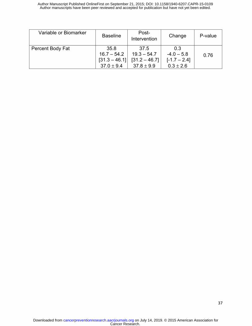
Variable or Biomarker Baseline Post-Intervention Change P-value
Percent Body Fat 35.8 16.7 – 54.2 [31.3 – 46.1]37.0 ± 9.4
37.5 19.3 – 54.7 [31.2 – 46.7]37.8 ± 9.9
0.3 -4.0 – 5.8 [-1.7 – 2.4] 0.3 ± 2.6
0.76
Cancer Research. on July 14, 2019. © 2015 American Association forcancerpreventionresearch.aacrjournals.org Downloaded from
Author manuscripts have been peer reviewed and accepted for publication but have not yet been edited. Author Manuscript Published OnlineFirst on September 21, 2015; DOI: 10.1158/1940-6207.CAPR-15-0109
38
Figure Legends Figure 1: Ki-67 expression (percent of cells staining positive) post-intervention as
a function of baseline value. Baseline aspiration values are shown on the x-axis;
repeat aspiration on the y-axis. The line represents no change in value; triangles
above the line denote an increase and diamonds below the line a decrease.
Figure 2: Effects of acolbifene on relative change (post-intervention:baseline) in
expression (by RT-qPCR) of genes that code for relevant proteins. There was
minimal change for the six reference transcripts assessed (Cytokeratin 19, E-
cadherin, HPRT1, Cyclophilin A, β-actin, β-glucuronidase). HPRT1 (*) was used
for normalization purposes.
Cancer Research. on July 14, 2019. © 2015 American Association forcancerpreventionresearch.aacrjournals.org Downloaded from
Author manuscripts have been peer reviewed and accepted for publication but have not yet been edited. Author Manuscript Published OnlineFirst on September 21, 2015; DOI: 10.1158/1940-6207.CAPR-15-0109
0
5
10
15
20
0 5 10 15 20
Ki-
67 (
pe
rcent)
, P
ost-
Inte
rve
ntio
n
Ki-67 (percent), Baseline
P = 0.001
Fig. 1
Cancer Research. on July 14, 2019. © 2015 American Association forcancerpreventionresearch.aacrjournals.org Downloaded from
Author manuscripts have been peer reviewed and accepted for publication but have not yet been edited. Author Manuscript Published OnlineFirst on September 21, 2015; DOI: 10.1158/1940-6207.CAPR-15-0109
0.01
0.1
1
10
Re
lati
ve E
xpre
ssio
n,
Po
st-I
nte
rve
nti
on
: B
ase
line
2.0
0.5
ERα GREB1 SDF-1α
pS2 PgR SDF-1β
Reference Transcripts
P-values (Wilcoxon test)
0.026 0.070 0.071
<0.001 <0.001 0.080
*
Fig. 2
Cancer Research. on July 14, 2019. © 2015 American Association forcancerpreventionresearch.aacrjournals.org Downloaded from
Author manuscripts have been peer reviewed and accepted for publication but have not yet been edited. Author Manuscript Published OnlineFirst on September 21, 2015; DOI: 10.1158/1940-6207.CAPR-15-0109
Published OnlineFirst September 21, 2015.Cancer Prev Res Carol J. Fabian, Bruce F. Kimler, Carola M. Zalles, et al. Risk for Breast CancerClinical Trial of Acolbifene in Premenopausal Women at High
Updated version
10.1158/1940-6207.CAPR-15-0109doi:
Access the most recent version of this article at:
Material
Supplementary
09.DC1
http://cancerpreventionresearch.aacrjournals.org/content/suppl/2015/09/19/1940-6207.CAPR-15-01Access the most recent supplemental material at:
Manuscript
Authoredited. Author manuscripts have been peer reviewed and accepted for publication but have not yet been
E-mail alerts related to this article or journal.Sign up to receive free email-alerts
Subscriptions
Reprints and
To order reprints of this article or to subscribe to the journal, contact the AACR Publications
Permissions
Rightslink site. Click on "Request Permissions" which will take you to the Copyright Clearance Center's (CCC)
.09http://cancerpreventionresearch.aacrjournals.org/content/early/2015/09/19/1940-6207.CAPR-15-01To request permission to re-use all or part of this article, use this link
Cancer Research. on July 14, 2019. © 2015 American Association forcancerpreventionresearch.aacrjournals.org Downloaded from
Author manuscripts have been peer reviewed and accepted for publication but have not yet been edited. Author Manuscript Published OnlineFirst on September 21, 2015; DOI: 10.1158/1940-6207.CAPR-15-0109


![Cancer Prevention Research CAPR-15-0109 Revision · endometrium [10-13]. Acolbifene and its prodrug (EM-800) have been associated with ... (Hologic LP, Malborough, MA) Non-Gyn standard](https://img.pdfslide.net/doc/110x75/5e37ed1f584c240891632fb3/cancer-prevention-research-capr-15-0109-revision-endometrium-10-13-acolbifene.jpg)