Embed Size (px)
Citation preview
Retrospective Theses and Dissertations Iowa State University Capstones, Theses andDissertations
1972
Canine hyperthermia with cerebral protectionRobert William CarithersIowa State University
Follow this and additional works at: https://lib.dr.iastate.edu/rtd
Part of the Biomedical Engineering and Bioengineering Commons
This Dissertation is brought to you for free and open access by the Iowa State University Capstones, Theses and Dissertations at Iowa State UniversityDigital Repository. It has been accepted for inclusion in Retrospective Theses and Dissertations by an authorized administrator of Iowa State UniversityDigital Repository. For more information, please contact [email protected].
Recommended CitationCarithers, Robert William, "Canine hyperthermia with cerebral protection " (1972). Retrospective Theses and Dissertations. 5891.https://lib.dr.iastate.edu/rtd/5891
INFORMATION TO USERS
This dissertation was produced from a microfilm copy of the original document. While the most advanced technological means to photograph and reproduce this document have been used, the quality is heavily dependent upon the quality of the original submitted.
The following explanation of techniques is provided to help you understand markings or patterns which may appear on this reproduction.
1. The sign or "target" for pages apparently lacking from the document
photographed is "Missing Page(s)". If it was possible to obtain the missing page(s) or section, they are spliced into the film along with adjacent pages. This may have necessitated cutting thru an image and duplicating adjacent pages to insure you complete continuity.
2. When an image on the film is obliterated with a large round black mark, it is an indication that the photographer suspected that the
copy may have moved during exposure and thus cause a blurred image. You will find a good image of the page in the adjacent frame.
3. When a map, drawing or chart, etc., was part of the material being
photographed the photographer followed a definite method in "sectioning" the material. It is customary to begin photoing at the upper left hand corner of a large sheet and to continue photoing from left to right in equal sections with a small overlap. If necessary, sectioning is continued again — beginning below the first row and continuing on until complete.
4. The majority of users indicate that the textual content is of greatest value, however, a somewhat higher quality reproduction could be made from "photographs" if essential to the understanding of the dissertation. Silver prints of "photographs" may be ordered at
additional charge by writing the Order Department, giving the catalog number, title, author and specific pages you wish reproduced.
University Microfilms 300 North Zeeb Road Ann Arbor, Michigan 48106
A Xerox Education Company
I I
73-3864
CARITHERS, D.V.M., Robert William, 1932-
CANINE HYPERTHERMIA WITH CEREBRAL PROTECTION.
Iowa State University, Ph.D., 1972
Engineering, biomedical
University Microfilms, A XEROX Company, Ann Arbor, Michigan
Canine hyperthermia with cerebral protection
by
Robert William Carithers
A Dissertation Submitted to the
Graduate Faculty in Partial Fulfillment of
The Requirements for the Degree of
DOCTOR OF PHILOSOPHY
Majors: Biomedical Engineering Veterinary Clinical Sciences
Approved:
In Charge of Major Work
For the Major Department
For the Graduate College
Iowa Stats University Ames, Iowa
1972
Signature was redacted for privacy.
Signature was redacted for privacy.
Signature was redacted for privacy.
PLEASE NOTE:
Some pages may have
ind is t inct pr in t .
F i lmed as received.
Univers i ty Microf i lms, A Xerox Educat ion Company
11
TABLE OF CONTENTS
Page
LIST OF SYMBOLS iii
INTRODUCTION 1
LITERATURE REVIEW 3
Thermoregulation 3
Influence of Temperature on Metabolism 6
Fever 11
Cerebral Heat Exchange Mechanism 13
Anatony 13
MATERIALS AND METHODS 17
RESULTS AND DISCUSSION 27
Temperature Responses 27
Clinical Observations 37
Pathology 42
Short Term Clinical Pathology 43
MATHEMATICAL MODEL 45
Mathematical Solution 45
Solution Utilizing the Analog Computer 50
Comparison of Model with Experimental Data 51
CONCLUSIONS AND RECOMMENDATIONS 54
LITERATURE CITED 57
ACKNOWLEDGEMENTS 64
APPaNUiA: bUWMilKÏ or ut\xe\
iii
LIST OF SYMBOLS
D=t Died=time after treatment (hrs.)
E Euthanized in one week
f Respiratory rate (min
t Time (min)
T Temperature, °C
Subscripts :
B Bath
B^ Brain
c Core
f Respiratory rate (min ^)
i Initial
N Irrigation (Nose)
sc Subcutaneous
1
INTRODUCTION
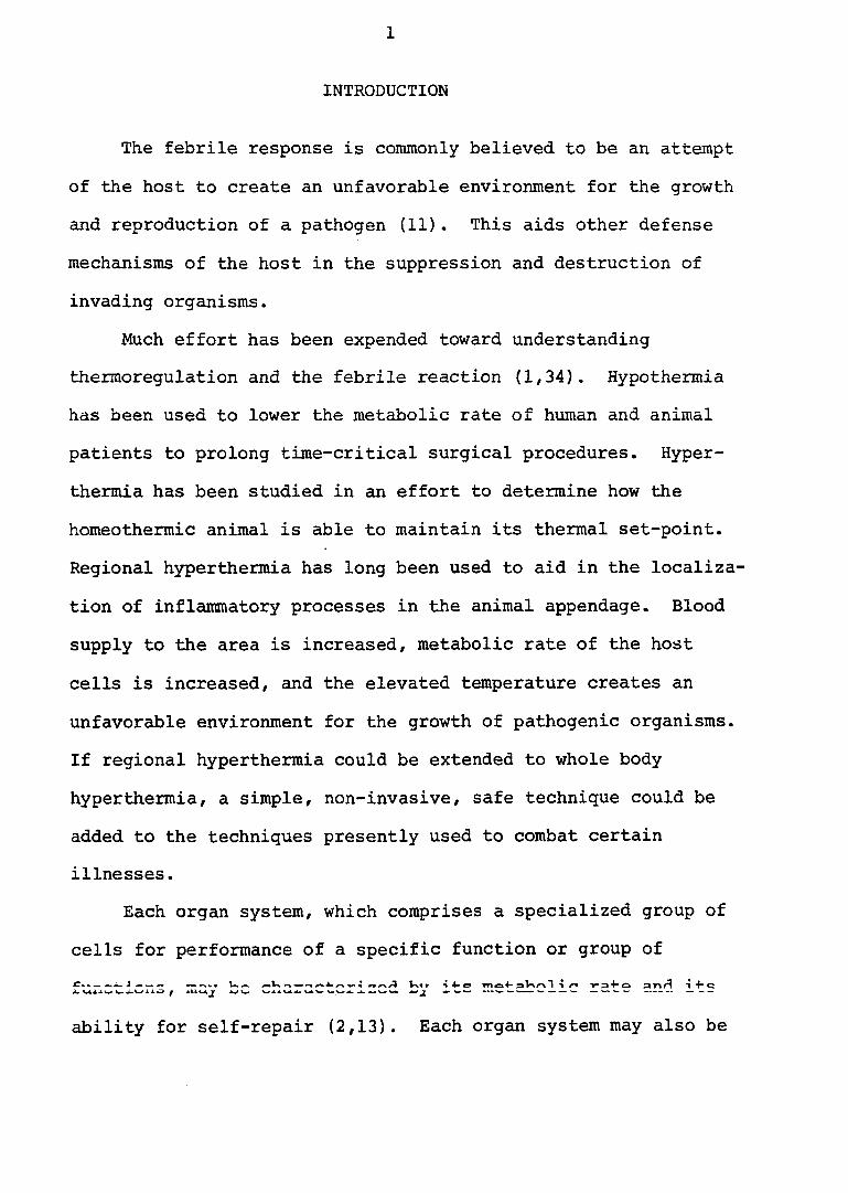
The febrile response is commonly believed to be an attempt
of the host to create an unfavorable environment for the growth
and reproduction of a pathogen (11). This aids other defense
mechanisms of the host in the suppression and destruction of
invading organisms.
Much effort has been expended toward understanding
thermoregulation and the febrile reaction (1,34). Hypothermia
has been used to lower the metabolic rate of human and animal
patients to prolong time-critical surgical procedures. Hyper
thermia has been studied in an effort to determine how the
homeothermic animal is able to maintain its thermal set-point.
Regional hyperthermia has long been used to aid in the localiza
tion of inflammatory processes in the animal appendage. Blood
supply to the area is increased, metabolic rate of the host
cells is increased, and the elevated temperature creates an
unfavorable environment for the growth of pathogenic organisms.
If regional hyperthermia could be extended to whole body
hyperthermia, a simple, non-invasive, safe technique could be
added to the techniques presently used to combat certain
illnesses.
Each organ system, which comprises a specialized group of
cells for performance of a specific function or group of
^ Vn* r n 4- cj sa T 1 ̂ ^ A VA A X ^ ^ A i ^ ^ M W* Jt ^ w » V — — — — —
ability for self-repair (2,13). Each organ system may also be
2
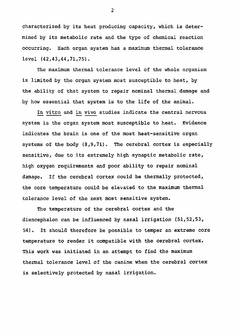
characterized by its heat producing capacity, which is deter
mined by its metabolic rate and the type of chemical reaction
occurring. Each organ system has a maximum thermal tolerance
level (42,43,44,71,75).
The maximum thermal tolerance level of the whole organism
is limited by the organ system most susceptible to heat, by
the ability of that system to repair nominal thermal damage and
by how essential that system is to the life of the animal.
In vitro and in vivo studies indicate the central nervous
system is the organ system most susceptible to heat. Evidence
indicates the brain is one of the most heat-sensitive organ
systems of the body (8,9,71). The cerebral cortex is especially
sensitive, due to its extremely high synaptic metabolic rate,
high oxygen requirements and poor ability to repair nominal
damage. If the cerebral cortex could be thermally protected,
the core temperature could be elevated to the maximum thermal
tolerance level of the next most sensitive system.
The temperature of the cerebral cortex and the
diencephalon can be influenced by nasal irrigation (51,52,53,
54). It should therefore be possible to temper an extreme core
temperature to render it compatible with the cerebral cortex.
This work was initiated in an attempt to find the maximum
thermal tolerance level of the canine when the cerebral cortex
is selectively protected by nasal irrigation.
3
LITERATURE REVIEW
Thermoregulation
Two thermoregulatory centers have been identified within
the central nervous system (12,57). Located in the medial
preoptic area is the paired heat loss center. The other
center is called the heat production and conservation center.
This paired center is located in the caudal hypothalamus
dorsolateral to the mammillary bodies (6,7,12). Stimulation
of the heat loss center in the canine induces cutaneous vaso-
dilitation, panting, salivation, and decreased muscle tone.
The heat loss center also has an influence on the supra-optic
and paraventricular nuclei so that during excessive salivation
or sweating the antidiuretic hormone is released, conserving
body fluids by renal water retention (4,5,6,7,12). Stimulation
of the heat production and conservation center causes vaso
constriction of cutaneous vessels, pilierection, increased
secretion of epinephrine, and shivering (5,12,30,37). Inhibi
tory information passes between the heat loss center and the
heat production center. This functions as a servomechanism to
aid in regulation of the thermal set point (12,38,49,50).
These centers receive information about the thermal state of
the external environment. This information is sensed by thermo
receptors located in the skin (49) , transmitted by afferent
nerve fibers to the central nervous system, and passed up the
4
lateral funiculus of the spinal cord to the hypothalamus.
Thermoreceptors are also located around the thermoregulatory
centers in the brain. They function to sense the blood
temperature of the hypothalamus (12,59).
Thermoreceptors are also located somewhere in the core
tissues of the body (32). Thermal information from all sources
is received by the thalamus, and is relayed to the thermo
regulatory centers, as well as to other parts of the brain.
The thermoregulatory centers integrate all the sensory inputs
with the other body functions to maintain the thermal set-
point.
Information calling for conservation of heat or heat dis
sipation is distributed to various parts of the brain from the
thermoregulatory centers. The thermoregulatory center thus
has been considered a body thermostat (34). The thermal
operating point or set-point has been studied quite extensively.
Hardy (34) summarized the results of many investigations by
stating that temperature is the regulated variable, and that
the regulator has the properties of proportional and rate
control, but not integral control. Guieu and Hardy (30) found
that the preoptic area functioned as an area of integration as
well as initiation of temperature information. Transient and
long-term thermal stresses affect thermal receptors differently.
Thermal stress originating from inside the body also affects
the thermal receptors differently from those originating from
5
outside (34).
The regulation of body temperature in the cold or neutral
zones is largely affected by the peripheral receptors, whereas
regulation of body temperature in the hot zone is more under
the control of the central receptors (34).
The three important effector elements of thermoregulatory
control are the metabolic response to cold, the sweating or
panting response to heat and control of the vasomotor exchange.
Below environmental temperatures near neutrality the vasomotor
system is in a full state of constriction. This results in a
minimum of heat flow from the blood to the surface. Above the
environmental temperature zone near neutrality (28-30®C) there
is reduction of vasomotor tone and active vasodilator activity
(17,18,28), causing a marked increase in peripheral conduction.
In the zone of vasomotor regulation, minor increases and
decreases in tissue conductance maintain thermal balance.
The tongue of the dog is an important blood-air heat
exchanger during hyperthermia, as was pointed out by Ederstrom
(16). He observed large increases in flow to the tongue, with
little increase to the foot, ear or intestine. The skin is
not an important mechanism of heat dissipation in the dog, as
evidenced by the sparsity of sweat glands (58).
Murakami et al. (59) found a diminished response to temper
ature change following the administration of anesthesia. Of
the anesthetics he studied, chlorolose-urethan was the least
6
depressive. Ingram and Smith (41) found that some anesthetics
produced persistent vasodilation, but with methoxyflurane and
urethan their results indicated a curvilinear relationship
between brain temperature and peripheral blood flow over the
entire range of brain temperatures. They concluded the effects
of direction and rate of change of brain temperature were
equivocal. Type of anesthetic and the level of its use also
influences the heart rate and electrical activity, and the
respiratory rate and depth. Methoxyflurane, used at light
anesthetic levels, has little influence on these activities but
will put the anrjnal in a mild state of metabolic acidosis (67).
Influence of Temperature on Metabolism
Chemical reaction rates of body metabolism roughly follow
van't Hoff's rule, which states that the velocity of chemical
reactions is increased two-fold or more for each rise of 10®
centigrade in temperature. Dukes (72) restricts this to more
physiological limits by stating that a rise of 1®C causes an
increase of 10 to 20% in metabolic rate. In man, a 13% eleva
tion in metabolic rate for each degree centigrade is seen.
Hubbard et al. (40) describes the Q^q as the ratio between
the metabolic rate at a given temperature and the rate at a
temperature 10®C higher. Therefore, when the is measured,
the temperature range must also be given. Comparisons are
relevant only when temperature ranges are exactly the same.
7
They are, thus, not really linear over a wide biological range.
Whole body hypothermia was used in 1938, when body
temperature was reduced to 32.2°C. Later, temperatures were
reduced to 27.5®C and 21.1'C. Hypothermia aided in reducing
the metabolic rate of the patient, thus affording an increase
in time available for surgery (20,48). It was also used in
attempts to reduce edema in cases of severe human cerebral
trauma (21). Local hypothermia was accomplished by circulating
ice water through a capsule inserted into the skull, but was
accompanied by brain abscesses and cerebritis. There was
bradycardia, depressed respiration and increase in pulse
pressure. Cerebral hypoxia and ventricular fibrillation are
complications of whole body hypothermia (20).
When the environmental temperature is increased such that
cooling by convection and radiation cannot eliminate metabolic
heat, panting or sweating is initiated and the excess heat is
released by evaporation (72). The threshold for increased
evaporative heat loss is influenced by time and temperature of
exposure, acclimatization, health of subject and level of basal
metabolic rate (33,46,55,56). Heart rate is also increased,
and urine production is diminished (26,36,43). In the dog,
Hammel e^ a^. (33), have noted a linear relationship between
rectal temperature and panting on exposure to heat. With an
increase in rectal temperature from 38 to 38.8®C, there is a
twenty—fold increase xn evaporative heat loss from the lungs.
8
Findley (24,25) noted that as respiration rate increased,
actual pulmonary ventilation remained almost constant. Panting
results in evaporative cooling in the upper respiratory pas
sages and does not cause cooling in the lungs (24,65). After
a period of panting, pCOg decreases, pH increases and blood
lactate is elevated (17,26,42,68). Urinary output drops, due
to a decrease in renal plasma flow (42,43,52). Plasma Cortisol
levels increase (14) and cardiac output is increased (63).
Durotoye and Grayson (15) studied production of heat in
the gastrointestinal tract of the dog. They found the duodenum
was 0.6°C hotter, and the ileum, stomach and large intestine
were 0.5°C hotter than the aorta. The portal vein was 0.35®C
hotter than the aorta, while the rectum had the same tempera
ture as the aorta. They concluded that the G. I. tract pro
duced 60% more heat than the liver, and that splanchnic heat
accounted for about 33% of the total body heat production.
In animals that pant (dog, sheep, swine), the thermal
stimuli for panting arise primarily from core receptors and
peripheral receptors (32,74) and these stimuli will override
the chemoreceptor center for respiration if the animal becomes
alkalotic (31).
Alterations in tissue electroyte concentrations indicate
changes in membrane permeability. Spurr and Barlow (68) noted
that if the canine rectal temperature were elevated to 42.5®C
for one hour, electrolyte and fluid shifts occurred in various
9
body tissues. He noted an increase in intracellular sodium in
the liver, jejunum and brain, and a decrease in intra-cellular
potassium in the liver and jejunum. He concluded these organs
contributed heavily to extracellular potassium concentration.
Other dyscrasias become evident during the extreme hyper
thermia. Field et al. (23) found a disappearance of reflexes
and death at 42-45®C in the rat. He concluded that this was
the temperature range where a progressive decrease in ©2 con
sumption occurred. In the pig, maximum respiratory rate was
recorded at rectal temperatures of 41 to 41.5*C. If rectal
temperature exceeded 41.5®C, respiratory rate declined and
death was imminent (69). In a study of human patients that
suffered heat stroke from a mining accident, 90% survived.
These patients suffered centrolobular degeneration or necrosis
of hepatocytes and congestion. These changes were completely
reversible and were believed to be due to hypoxia and thermal
injury (44).
Frankel et al. (26,27) intensively studied blood chemistry
during progressive hyperthermia. Changes in blood chemistry
began to develop at the critical temperature of 42°C. Studies
of blood gases and lactates demonstrated that tissue hypoxia
developed. He concluded that failure of external respiration
was not the primary physiological derangement during progres
sive hvoerthermia. In studvinq liver lactates, pyruvates and
10
pyridine nucleotides, Frascella and Frankel (28) concluded that
the site of initial failure during progressive hyperthermia
was not the mammalian liver.
Burger and Fuhrman (13), utilizing ̂ vitro studies, found
the cerebral cortex more sensitive to heat damage than the
liver or the renal cortex. His technique utilized decrease in
Og consumption as the biochemical criterion. He found damage
to the cerebral cortex after sixty to ninety minutes, with the
temperature held at 40-41*C, and also after thirty to forty
minutes at a temperature of 43®C. The liver was damaged by
temperatures of 45°C for sixty minutes, the demonstrable
kidney damage required 44®C for sixty minutes.
Nemoto and Frankel (61) felt that cerebral failure was not
due to cerebral hypoxia, but to limitation of the nucleotide
supply required for cerebral glucose transport. During his
series of investigations of progressive hyperthermia, elevation
of cerebral Og consumption and glucose consumption was reversed
between 42®C and 43*C. Upon examination of Purkinji cells
from rats which were stressed at 40 to 42®C for three hours,
Kucherenko (46) noted reactive and destructive changes of
cellular membrane systems. Swelling of mitochondria, clearing
of the mitochondrial matrix, and partial destruction of the
cristae were observed. Fragmentation of membranes of the
endocytoplasmic reticulum and Golgi hypertrophy were also seen.
Lysis of the degenerative products occurred in six hours, with
restoration of normal ultrastructure in twenty-four hours.
11
studying independently, Wang et al. (75) and Rozanora
et al. (64) felt that uncoupling of oxidative phosphorylation
may be a mechanism of cellular breakdown during acute hyper
thermia. Denaturation of proteins, especially of enzyme
systems, was also observed in cerebral tissue during hyper
thermia (2,70,71,73).
In examining the above information, the CNS is implicated
as the first organ system to succumb to extreme hyperthermia.
Evidence also incriminates the denaturation of enzymes and/or
an uncoupling of oxidative phosphorylation as the mechanism of
action.
Fever
Fever is an elevation of body temperature in response to
some diseases. It is a mechanism whereby the body creates an
unfavorable environment for a disease condition (11). Per
fusion of the cerebral ventricles with sodium ions markedly
increases the body temperature, while perfusion of calcium
ions decreases body temperature (22,60). Possibly, the thermal
set-point may be raised by altering the balance between these
two actions in the cerebrospinal fluid (60). However, most
investigators agree with Atkins (11) that the mechanism of
action involves the release of bacterial endotoxins. Bacteria
release an endotoxin which, in turn, causes granulocytes to
release an endopyrogen. This endopyrogen acts on the thermo
regulatory center to cause an elevation in body temperature.
12
Atkins also stated that a disease process becomes less severe
if the body temperature is raised artificially, thereby adding
credence to the concept that fever is a defense mechanism.
Von Ardenne et al. (8,9,10) was able to arrest growth of
several types of carcinogenic tissues in rats and mice by
exposing them to hot water baths of varying temperatures for
appropriate durations. After treatment at 40°C for one hour,
two hours, or four hours, the experimental animals, as well as
the control animals, had mitotic figures in the cancerous
tissue. At 41°C for sixty minutes, animal death occurred
because of the excessive heat. But at 40.5°C for sixty minutes,
tumor growth in heat-treated rodents was arrested for a period
of two to six months, while untreated rodents died within a
two-week period. Von Ardenne improved his results by combining
hyperthermia with the use of cyclophosphamide, a chemical used
as an aid in arresting certain types of carcinogenic prolifera
tion. He postulated that hyperthermia caused denaturation of
the enzyme systems in cancerous tissue before it affected the
normal tissue. It also increased membrane permeability, there
by permitting an increased amount of cyclophosphamide to enter
the cells. Kirsch et al. (45) confirmed the latter statement
with Pg2 labeled cyclophosphamide.
13
Cerebral Heat Exchange Mechanism
Magilton and Swift described a mechanism by which nasally
respired air could influence the temperature of the brain (51,
54). This involved essentially two heat exchangers. One was
the air-venous blood interface which was located at the alar
fold of the nasal maxilloturbinate. This effectively cooled
blood which drained to the second heat exchanger, which was a
venous blood-arterial blood heat exchanger. The cooled venous
blood received thermal energy from arterial blood that supplied
the brain (52). Subsequent study revealed that this system was
not passive. It was under dynamic vascular and neural control
and through changes in temperatures exposed to the nasal mucosa
a definite, repeatable alteration could be noted in temperature
of brain parenchyma, cerebrospinal fluid pressure, blood
pressure and heart rate (51,53).
Magilton and Swift (52,54) demonstrated by cold water
irrigation that brain temperature could be decreased to 12°C
below body temperature, although hot water irrigation could not
elevate brain temperature much above normal body temperature.
Anatomy
The alar fold is a bilateral bulbous rostral extension
of the maxilloturbinate in the dog. It is richly supplied with
blood and contains large venous sinuses (58). The alar fold
functions with the turbinates as a heat exchanger between the
14
blood and respired air (52).
The cooled venous blood draining the alar fold returns to
the body by two separate routes after it is collected by the
dorsal and lateral nasal vein. One is directly to the heart by
the facial vein and the other is an indirect route which
involves another heat exchanger. The latter route courses the
blood through the angularis oculi vein over the ventro-medial
angle of the bony orbit into the orbital canal, and continues
through the ophthalmic vein, multifurcating into a venous
plexus. The majority of the blood then enters the cavernous
sinus, while a minor portion flows to the internal maxillary
vein to return to the central venous pool (58).
The cavernous sinus is a cavity formed by the separation
of the durai sheath of the brain, which is filled with venous
blood. This sinus is constantly located on the floor of the
middle cranial fossa. It is paired and connected anteriorly
with the orbital plexus through the orbital foramen. The
paired cavernous sinuses are connected medially by inter
cavernous sinuses rostral to and caudal to the stalk of the
dorsum sellae. The hypophysis is located rostral to the
rostral intercavernous sinus. A third intercavernous sinus may
inconstantly be located rostral to the hypophysis (58). The
intercavernous sinuses are small, which restricts the venous
communication between right and left cavernous sinuses (51).
15
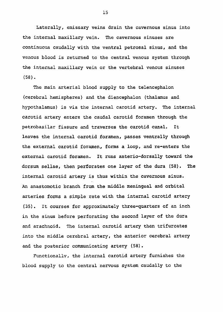
Laterally, emissary veins drain the cavernous sinus into
the internal maxillary vein. The cavernous sinuses are
continuous caudally with the ventral petrosal sinus, and the
venous blood is returned to the central venous system through
the internal maxillary vein or the vertebral venous sinuses
(58).
The main arterial blood supply to the telencephalon
(cerebral hemispheres) and the diencephalon (thalamus and
hypothalamus) is via the internal carotid artery. The internal
carotid artery enters the caudal carotid foramen through the
petrobasilar fissure and traverses the carotid canal. It
leaves the internal carotid foramen, passes ventrally through
the external carotid foramen, forms a loop, and re-enters the
external carotid foramen. It runs anterio-dorsally toward the
dorsum sellae, then perforates one layer of the dura (58). The
internal carotid artery is thus within the cavernous sinus.
An anastomotic branch from the middle meningeal and orbital
arteries forms a simple rete with the internal carotid artery
(35). It courses for approximately three-quarters of an inch
in the sinus before perforating the second layer of the dura
and arachnoid. The internal carotid artery then trifurcates
into the middle cerebral artery, the anterior cerebral artery
and the posterior communicating artery (58).
Functionally, the internal carotid artery furnishes the
blood supply to the central nervous system caudally to the
16
region of the rostral colliculus. This blood is then drained
into the various durai sinuses of the cranial vault, and is
returned to the central venous pool (58).
If the venous blood of the alar fold were cooled, the
base of the brain would be cooled two ways, directly and in
directly. The first way is by direct extension through the
durai surface from the cavernous sinus to the ventral portions
of the brain. The second is by cooling the arterial blood
supply to the cerebral hemispheres (52).
17
MATERIALS AND METHODS
In order to determine the maximum thermal tolerance level
of a body system, core temperature must be controlled. Core
temperature is a function of the thermal "set point" of the
hypothalamus, temperature of the environment and temperature
of respired air. It is readily apparent that only an indirect
control of core temperature can be maintained.
Environmental temperature can be controlled most constantly
by a circulating water bath. Length of hair, although main
tained short in this series of experiments, does not interfere
significantly with heat transfer into the skin from the water
bath (39). This is because virtually all the heat transfer is
through convection.
The warm water bath consisted of a hundred-gallon water
tank mounted on wheels for maneuverability. Heat loss from the
tank walls was minimized by attaching a one-inch-thick expanded
foam plastic sheet to the outside.
The time required to cool the bath from 42®C to 41®C at
room temperature was one hour. Water circulation was
adequately maintained with a large submersible impeller pump.^
No temperature gradients were observed in various locations in
the tank when the temperature was held constant. Upon heating
or cooling, the water transient gradients were noted, but no
^Model no 7114, Cole-Parmer Instrument Co., Chicago, 111.
18
thermal oscillations occurred because whirlpool effects were
minimized.
Temperature of the water was increased or decreased by
adding hot water (55®C) or cold water (10°C). Excess water
was removed with another impeller pump.^ The desired water
temperature was quite easily achieved and maintained within
narrow tolerance limits.
A restraining device made of rods was suspended in one
end of the tank. The lower jaw was anchored to this device to
immobilize the canine head. Care was maintained to not inter
fere with the venous drainage of the tributaries of the maxil
lary vein. This was the only restraint required to stabilize
the partially submerged animal with the exception of anchoring
the tail to prevent lateral deviation. Cold water nasal irri
gating tubes were fabricated from a blood transfusion set. A
hypodermic thermistor was placed in the irrigating line within
two inches of the outflow. Irrigating water temperature was
held at approximately 8°C, +^2®C/ by a refrigerator coil. This
water was circulated through the irrigation tubing to the nose
by a roller pump^. Approximately symmetrical water pressure
^Model no. 7111, Cole-Parmer Instrument Co., Chicago, 111.
2 Yellow Springs Instrument Co., Yellow Springs, Ohio.
^Randolph Pump model 500, The Randolph Co., Houston, Tex.
19
was maintained to irrigate only the alar fold of each nostril
with the water returning cranially out of the nose. The water
then fell into a pan and returned to the refrigerator reservoir.
Endotracheal intubation, which was utilized for gas anesthesia^,
prevented any entrance of water into the trachea. It also
prevented the tongue from functioning as a cooling organ.
Low resistance wire, with a steel needle electrode, was
utilized for each of the three electrocardiographic electrodes.
Modification of these leads was necessary to record heart
electrical activity of the submerged dog. An epoxy film was
placed over the lead to within three-eights inch of the tip.
It was thus possible to place the electrode well under the skin
of the animal and avoid any direct electrical contact between
the water bath and the electrode. The high skin and epoxy
resistance formed an effective shield from the water bath.
2 Three wheatstone bridge thermometers with accompanying
rectal probes were utilized to monitor two rectal temperatures
2 and one bath temperature. Four thermistor needle electrodes
were each utilized in a resistive bridge circuit to monitor
subcutaneous temperature. All thermistors were calibrated
utilizing the same glass mercury thermometer as a reference.
Equivalent resistances at 2®C increments were determined for
^Metofane, Pitman-Moore, Inc., Ft. Wash., Pa.
2 Yellow Springs Instrument Co., Yellow Springs, Ohio.
20
the needle electrodes. Calibrations were made before and after
each experimental procedure.
Respiratory rate was measured by a thermistor placed in
the endotracheal tube. Respiratory and heart rate, four sub
cutaneous temperatures, bath, nasal irrigation and rectal
temperatures were continuously monitored on a twelve-channel
pen recorder^.
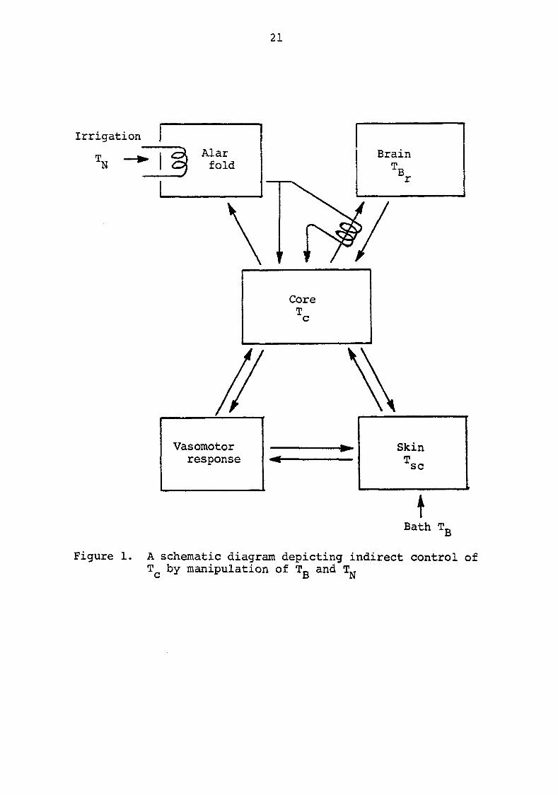
A schematic diagram (Figure 1) depicts the indirect
control of core temperature by the manipulation of bath water
and irrigating water temperatures.
Healthy mature pound dogs of either sex, that weighed
between five and fourteen Kgms., were utilized for the experi
ment. Dog size was restricted by the water bath dimensions.
During the two-week acclimatization period, baseline data were
obtained. Baseline data consisted of a fecal examination for
2 endoparasites, blood analysis, electroencephalogram , electro-
2 cardiogram , complete physical and neurological examination.
Blood analysis included determinations of packed cell
volume, hemoglobin, total red cell count^, total white cell
count^, differential white count, platelets, cell morphology,
^Polygraph Model 7, Grass Instrument Co., Quincy, Mass.
2 Dymograph Model R-411, Beckman Instruments, Inc.,
Schiller Park,, 111.
^coulter counter Model r , Coulter Electronics, Inc., Hialeah, Fla.
21
Core
Vasomotor
Bath T B
Figure 1. A schematic diagram depicting indirect control of by manipulation of Tg and
22
11 2 total protein , fibrinogen , blood urea nitrogen , serum
3 4 4 glutamic pyruvic transaminase , sodium and potassium content.
Animals that did not have acceptable "normal" values were
eliminated from the experiment.
Feed and water were withheld eighteen hours prior to the
experiment. Baseline data were obtained after animals were
anesthetized prior to the experimental procedure. These data
included another blood analysis, clotting time, urinalysis^
and blood-gas^ analysis. The urinalysis included qualitative
determinations for blood, protein, sugar, pH and a microscopic
examination. The blood-gas determinations involved partial
pressure of oxygen (PvOg)/ partial pressure of carbon dioxide
(PVCO2) and concentration of hydrogen ions (pH). Blood-gas
measurements were again determined at a point two-thirds
through the experiment. Immediately after the experiment,
blood analysis, clotting time, blood-gas analysis, urinalysis
(including specific gravity) and urine volume were again
determined on the surviving dogs.
^T. S. meter, American Optical.
2 Unimeter model D-250, Biodynamics Inc., Indianapolis, Ind.
3 Reitman-Frankel Procedure, Dade, Miami, Fla.
4 Flame Photometer, Instrumentation Lab., Inc., Lexington,
Mass.
s "Hemacombistix Ames Co,, Elkhart, Ind.
^Corning Model 16 blood gas analyzer. Corning Glass works. Corning, N.Y,
23
Surviving dogs were maintained without therapy other than
nursing care for one week. At the end of that time, a blood
analysis, ECG, EEC, neurological and physical examination were
made prior to euthanasia. A necropsy was performed with tissue
samples obtained for histopathological studies from the liver,
spleen, kidney, pancreas, thyroid, thymus, parathyroid, lymph
node, left ventricle, aortic valve, aorta, lung, urinary
bladder, stomach fundus, descending duodenum, gall bladder,
terminal ileum, diaphragm, skeletal muscle and sometimes the
skin. Sections of the central nervous system included cerebral
and cerebellar cortex, basal ganglia of the cerebrum and
cerebellum, thalamus, septal area, hypothalamus, regions
through the oculomotor and trochlear nucleus, brachium pontis,
caudal medulla, and high cervical region.
Anesthesia was induced on two dogs with thiamylal Na.^
After baseline samples were obtained, the dogs were placed in
the 37® water bath side by side, but were spaced for maximum
water circulation over the body surface.
Anesthesia was maintained in a light plane with methoxy-
flurane gas . Heart rate and condition were monitored via lead
II electrocardiogram.
Subcutaneous temperatures were obtained from the right and
left mid-thoracic regions of both dogs. The electrode for
^Surital, Parke Davis & Co., Detroit, Mich.
2 Metofane, Pitman Moore, Inc., Ft. Wash., Pa.
24
recording rectal temperature was placed high into the colon, as
close to the left colic flexure as possible.
Bath water was heated at the rate of one-half degree
centigrade per minute until the desired bath temperature was
reached. The two dogs were treated identically with the
exception that when the core temperature of the dog to be
irrigated reached 41*C, irrigation of the alar fold was
initiated, with a water temperature of approximately 8®C. When
the bath water reached the predetermined temperature, this
temperature was maintained for a certain predefined period,
the bath was then cooled back to 36®C at a rate of 1 to 2®C per
minute. Fast recordings were obtained prior to bath heating
and at ten-minute intervals throughout the procedure.
Bath temperatures of 41*C, 43®C, and 45®C were selected
with each temperature held for durations of thirty, sixty and
ninety minutes. Three pairs of dogs were included at each
component of the time-temperature matrix, with the exception
of each extreme, namely 41°C for thirty minutes and sixty
minutes and the control component at 45®C for sixty and ninety
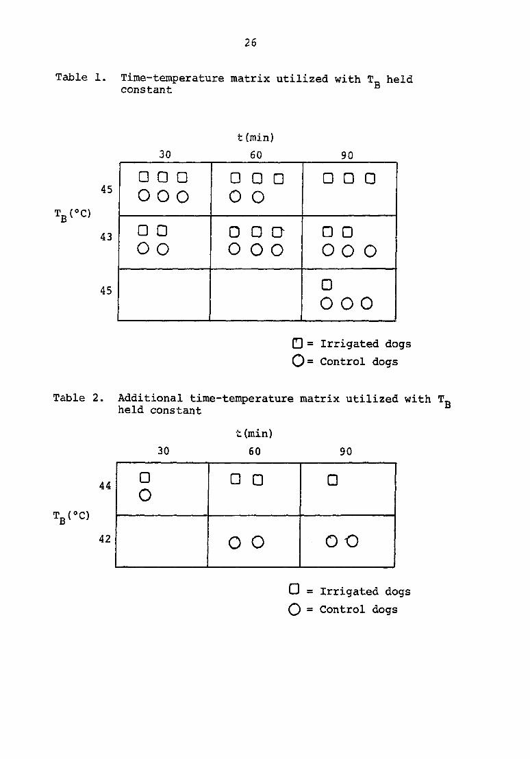
minutes (Table 1).
Parameters to formulate the matrix were set to include
time and temperature extremes of in vivo and in vitro studies
(8,23,69,70). After initial trials the thirty and sixty minute
hTonks at 43®C were eliminated because a response of rapid
respirations could not be elicited in the control dogs. At
25
45°C the control animals could not survive past fifteen to
twenty-five minutes. The irrigated dogs exposed to 45°C for
longer than one hour suffered such marked respiratory, cardiac
and cutaneous alterations that this extreme was also eliminated.
Additional information was obtained by adding an irrigated
group at 44®C and a control group at 42*C (Table 2). The core
temperatures of these two groups were approximately the same.
Core temperature along with bath temperature could therefore
be held constant by comparing the various time-temperature
groups with each other. Data were accumulated to formulate a
descriptive mathematical model (66).
These additional experiments were conducted on only one
dog at a time. Rectal and stomach temperatures were simul
taneously recorded with subcutaneous temperatures of the right
and left lateral aspect of the foreleg and the right and left
lateral aspect of the rib cage next to the twelfth rib.
Respiratory and heart rates were also monitored.
B:ain temperatures of seven irrigated and three control
dogs were compared to their own core temperatures. The brain
thermistor probe was positioned in the right corpus striatum
in a plane with the caudal aspect of the thalamus. The loca
tion was verified on post mortem examination.
The stomach and rectal temperatures of fifteen dogs were
comnared.
26
Table 1. Time-temperature matrix utilized with T_ held constant
30
t (min)
60 90
45 0 0 0
0 0 o 0 0 0 0 o
O 0 0
43
o o
• o
ODD O 0 0 o
• o
• o
45 0
0 0 0
0 = Irrigated dogs 0= Control dogs
Table 2. Additional time-temperature matrix utilized with T„ held constant
t (min)
30 60 90
44 0 0 0 0 44 0
42 0 0 o o
O = Irrigated dogs
O = Control dogs
27
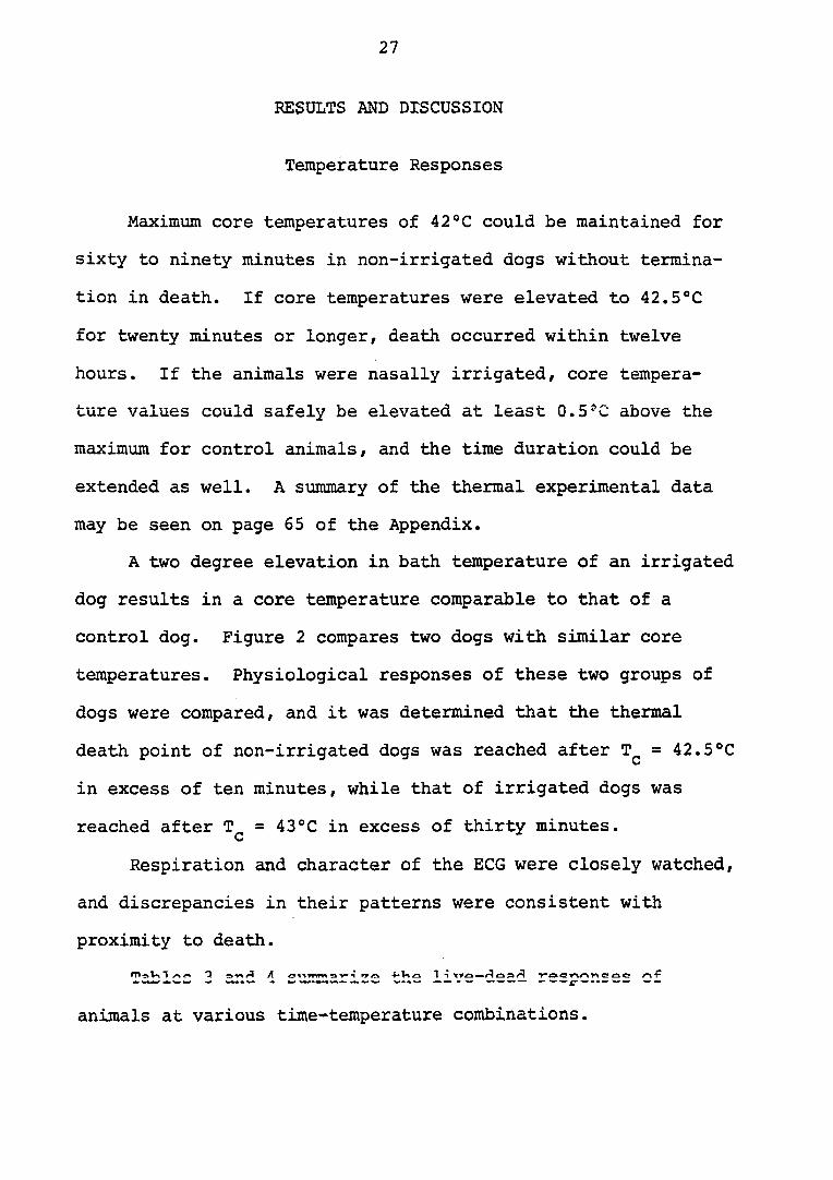
RESULTS AND DISCUSSION
Temperature Responses
Maximxam core temperatures of 42®C could be maintained for
sixty to ninety minutes in non-irrigated dogs without termina
tion in death. If core temperatures were elevated to 42.5®C
for twenty minutes or longer, death occurred within twelve
hours. If the animals were nasally irrigated, core tempera
ture values could safely be elevated at least 0.5'C above the
maximum for control animals, and the time duration could be
extended as well. A summary of the thermal experimental data
may be seen on page 65 of the Appendix.
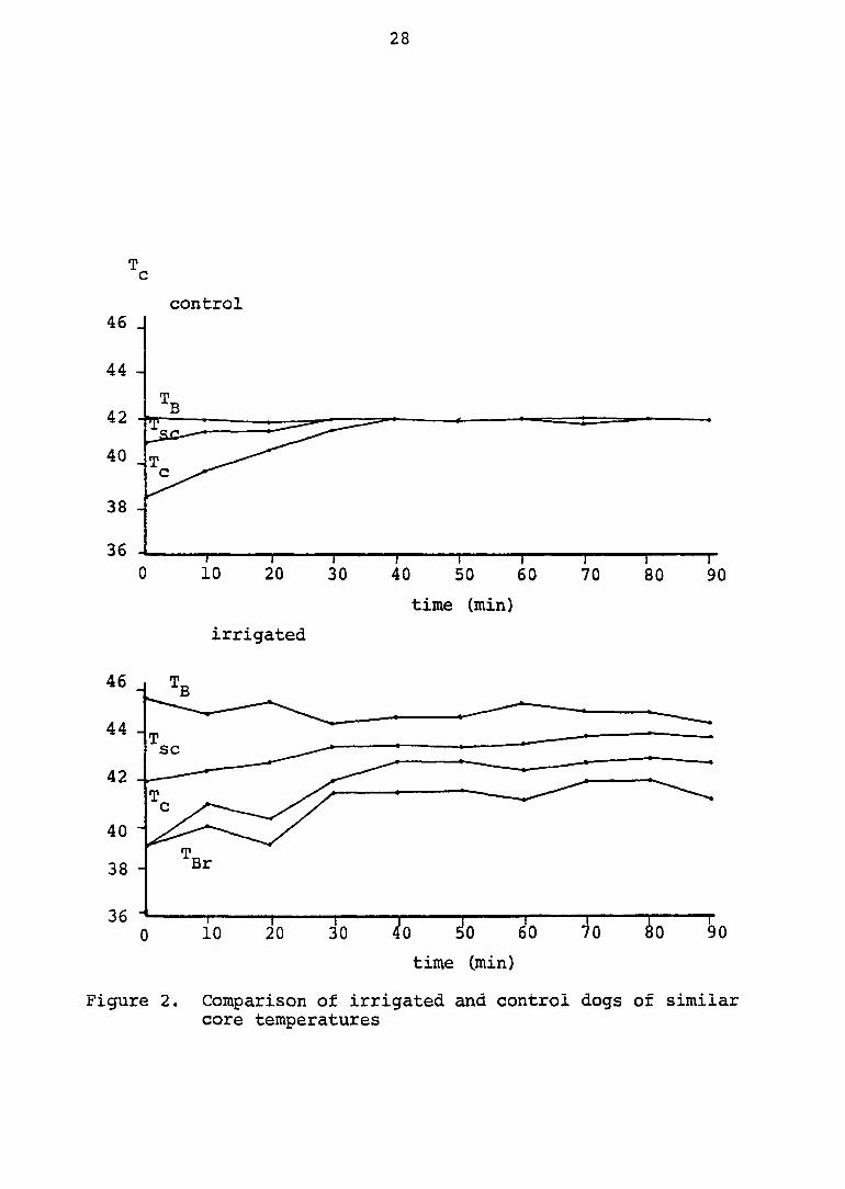
A two degree elevation in bath temperature of an irrigated
dog results in a core temperature comparable to that of a
control dog. Figure 2 compares two dogs with similar core
temperatures. Physiological responses of these two groups of
dogs were compared, and it was determined that the thermal
death point of non-irrigated dogs was reached after T^ = 42.5®C
in excess of ten minutes, while that of irrigated dogs was
reached after T^ = 43°C in excess of thirty minutes.
Respiration and character of the ECG were closely watched,
and discrepancies in their patterns were consistent with
proximity to death.
animals at various time-temperature combinations.
28
T c
control 46 J
44 -
42
40
38 -
36
0 10 20 30 40 50 60 70 80 90
time (min)
irrigated
46
44 -sc
42
Br 38 -
time (.min)
Figure 2. Comparison of irrigated and control dogs of similar core temperatures
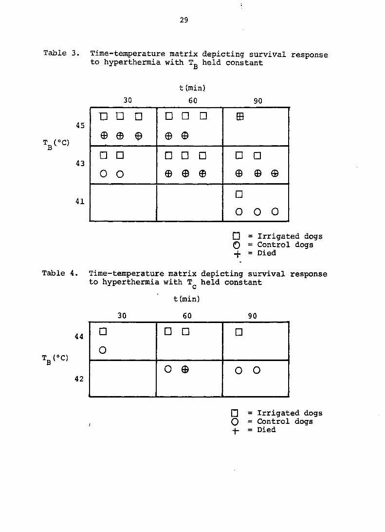
29
Table 3. Time-temperature matrix depicting survival response to hyperthermia with T„ held constant
45
TgC'C)
43
41
• = Irrigated dogs O = Control dogs
= Died
Table 4. Time-temperature matrix depicting survival response to hyperthermia with held constant
t (min)
30 60 90
44
Tg(»C)
42
• = Irrigated dogs O = Control dogs
= Died
t (min)
30 60 90
•
p
p
•
•
p m
© © ^ e e
•
•
•
•
•
•
•
o
o © 0 © ® © ©
•
0 0 0
• • • •
o
0 © 0 o
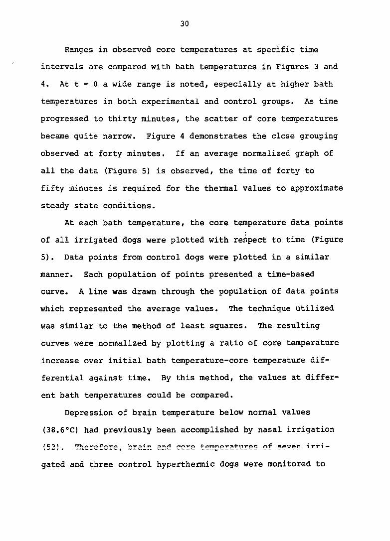
30
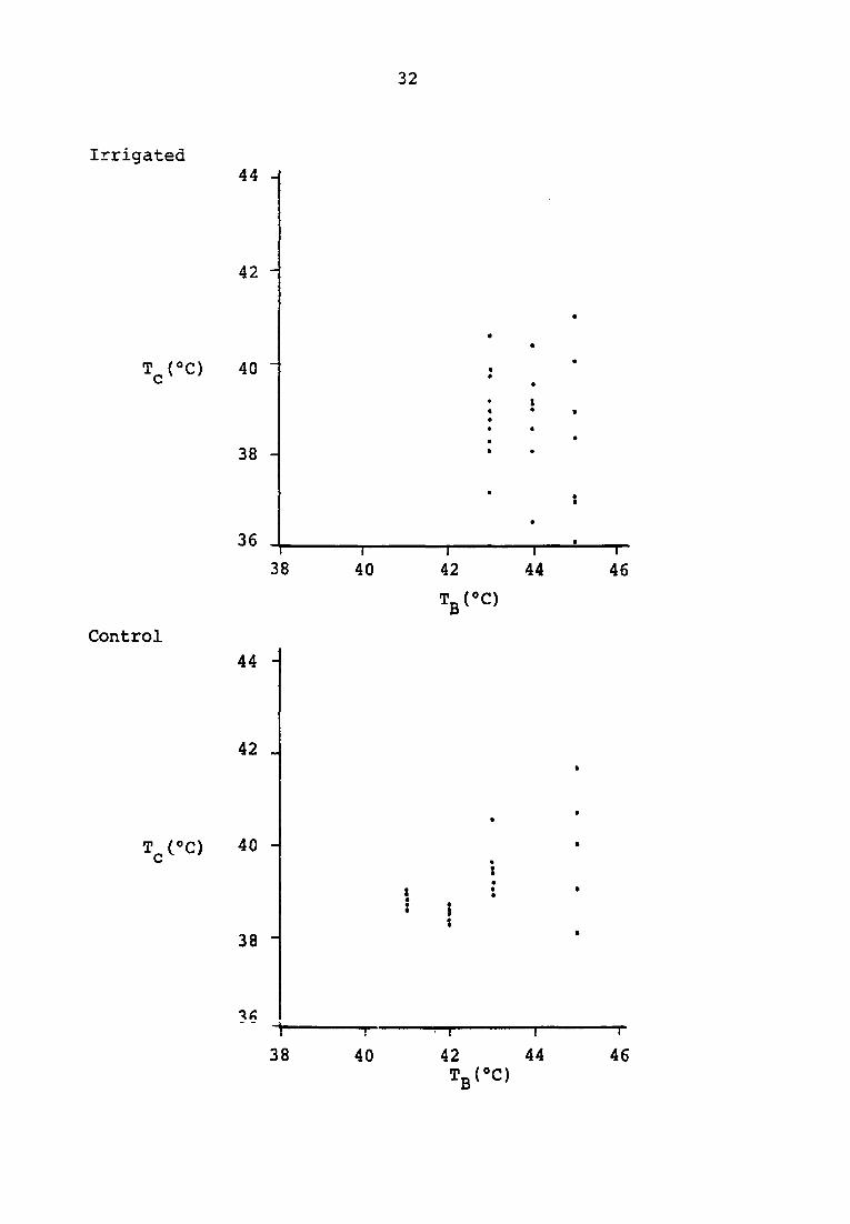
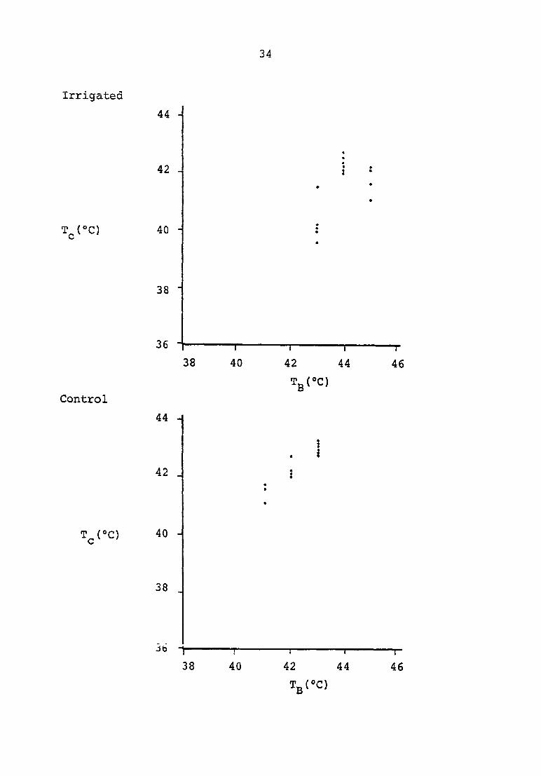
Ranges in observed core temperatures at specific time
intervals are compared with bath temperatures in Figures 3 and
4. At t = 0 a wide range is noted, especially at higher bath
temperatures in both experimental and control groups. As time
progressed to thirty minutes, the scatter of core temperatures
became quite narrow. Figure 4 demonstrates the close grouping
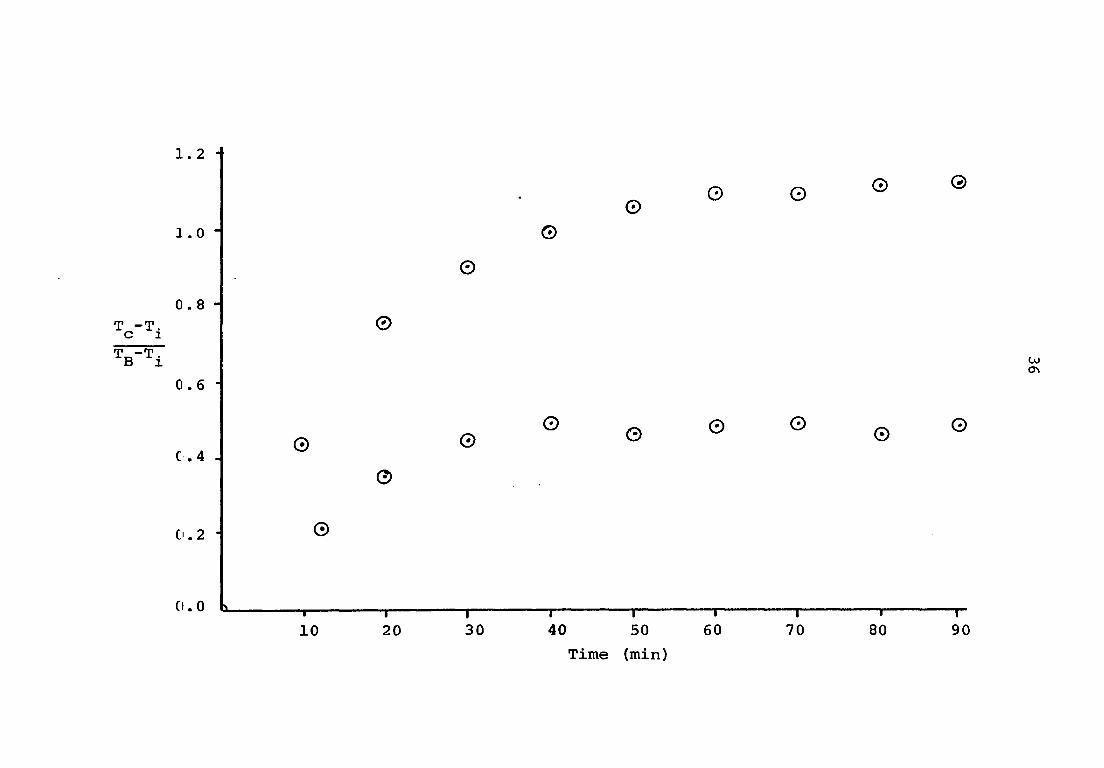
observed at forty minutes. If an average normalized graph of
all the data (Figure 5) is observed, the time of forty to
fifty minutes is required for the thermal values to approximate
steady state conditions.
At each bath temperature, the core temperature data points
of all irrigated dogs were plotted with respect to time (Figure
5). Data points from control dogs were plotted in a similar
manner. Each population of points presented a time-based
curve. A line was drawn through the population of data points
which represented the average values. The technique utilized
was similar to the method of least squares. The resulting
curves were normalized by plotting a ratio of core temperature
increase over initial bath temperature-core temperature dif
ferential against time. By this method, the values at differ
ent bath temperatures could be compared.
Depression of brain temperature below normal values
(38.6®C) had previously been accomplished by nasal irrigation
gated and three control hyperthermic dogs were monitored to
Figure 3. Collated comparison of core temperature and bath temperature
t = 0.0 minutes
32
Irrigated
T^(°C)
Control
T^CC)
4 4 -J
4 2 -
4 0
3 8 -
3 6
3 8
4 4 -
4 2 _
4 0 -
3 8 -
3 8
"T— 4 0
-T-
4 0
n 4 2
TgCO
—T" 4 4
—I
4 2 T g ( * C )
4 4
-T 4 6
—r
4 6
Figure 4. Collated comparison of core temperature and bath temperature
time = 40 min
34
Irrigated
Control
T^CC)
44 -
4 2 -
T ^ ( ° C ) 4 0
3 8 -
3 6
3 8
4 4 H
4 2
4 0 -
3 8
Jb
38
40
!
4 0
4 2
T g ( ® C )
I
4 4
4 2
—r-
4 4
Tg(®C)
T
4 6
1-
4 6
Figure 5. Normalized experimental data upper curve = control lower curve = experimental
1.2
0. 0 L -1— 10
©
T 20
©
© ©
©
©
30
© © © © ©
©
U) m
© © © © © ©
1 1 1 1 1 r 40 50 60 70 80 90
Time (min)
37
determine whether nasal irrigation could maintain a lower,
safer temperature in the brain than the core temperature of
these animals. It was shown that a differential between brain
and core temperatures of 1° to 2®C could be maintained for the
duration of the timed procedure.
Removal of the irrigating water resulted in an elevation
of brain temperature to the existing hyperthermic core tempera
ture. Replacement of the irrigator resulted in depression of
brain temperature (Figure 6). Brain temperature corresponded
to core temperature in the non-irrigated dogs.
Recordings shown in Figure 6 were made at the terminus of
the experiment, when water was being cooled. The control dog
was dying, as is evidenced by the ECG tracing.
Core temperatures, as determined by stomach temperatures,
rectal temperatures, and peritoneal temperatures, all agree
consistently within 0.1°C, unless the animal was being heated
or cooled. Stomach or rectal contents seemed responsible for
a delay in response time for those sites.
Clinical Observations
Several clinical observations were made on both experi
mental and control animals which had core temperatures
approaching 42.5®C. During the procedure, the anesthetic plane
band width narrowed. The animals moved from one anesthetic
stage to another very rapidly, due to the rapid metabolic rate
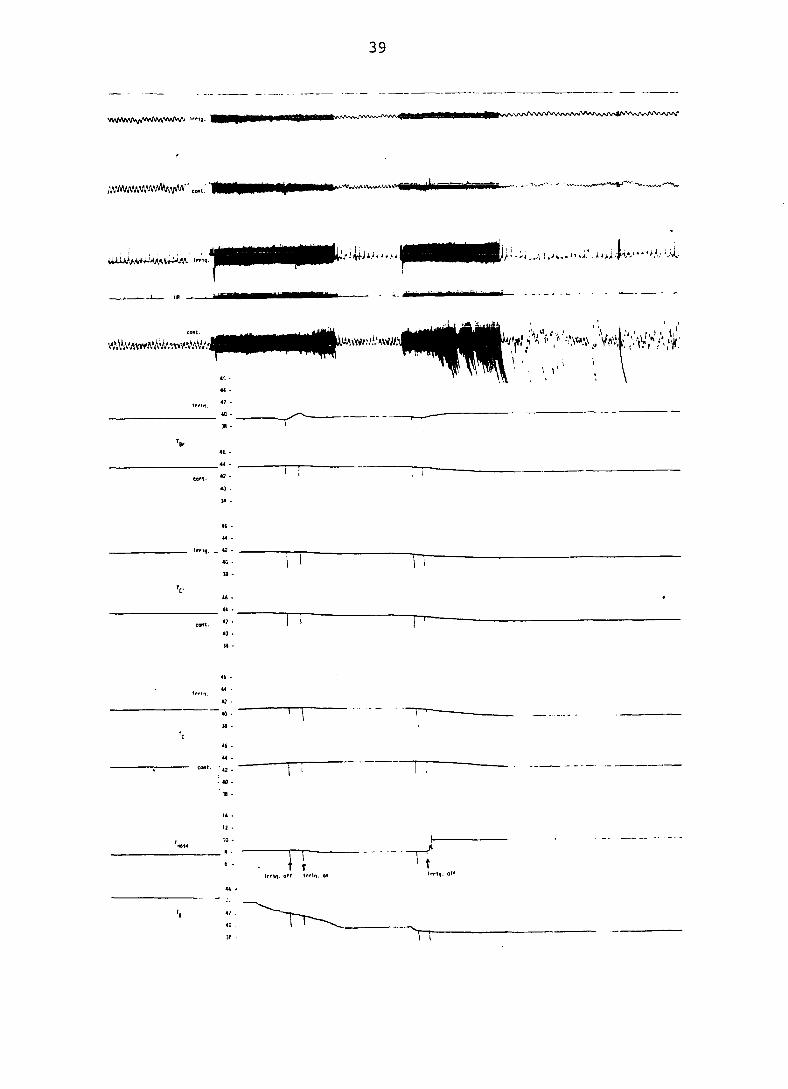
Figure 6. Photograph of recording depicting influence of nasal irrigation on independent of
39
MM9H ^S/»/»,\ArU\l\A/V\r/V>V\AWVv\Aw%f\rv>4/VV\,VVV\Aw\y\^^^
;,VAVWW.'VWJVVVI;jiAV'' ,
• ! • • ' ' 1/ • '"i, ,,>\,i« ̂ . i 4 ^̂ ,•' ;jwA'"**''A-v'X-
\
TT , I
Tf Irrki. off 1'rto. @*
40
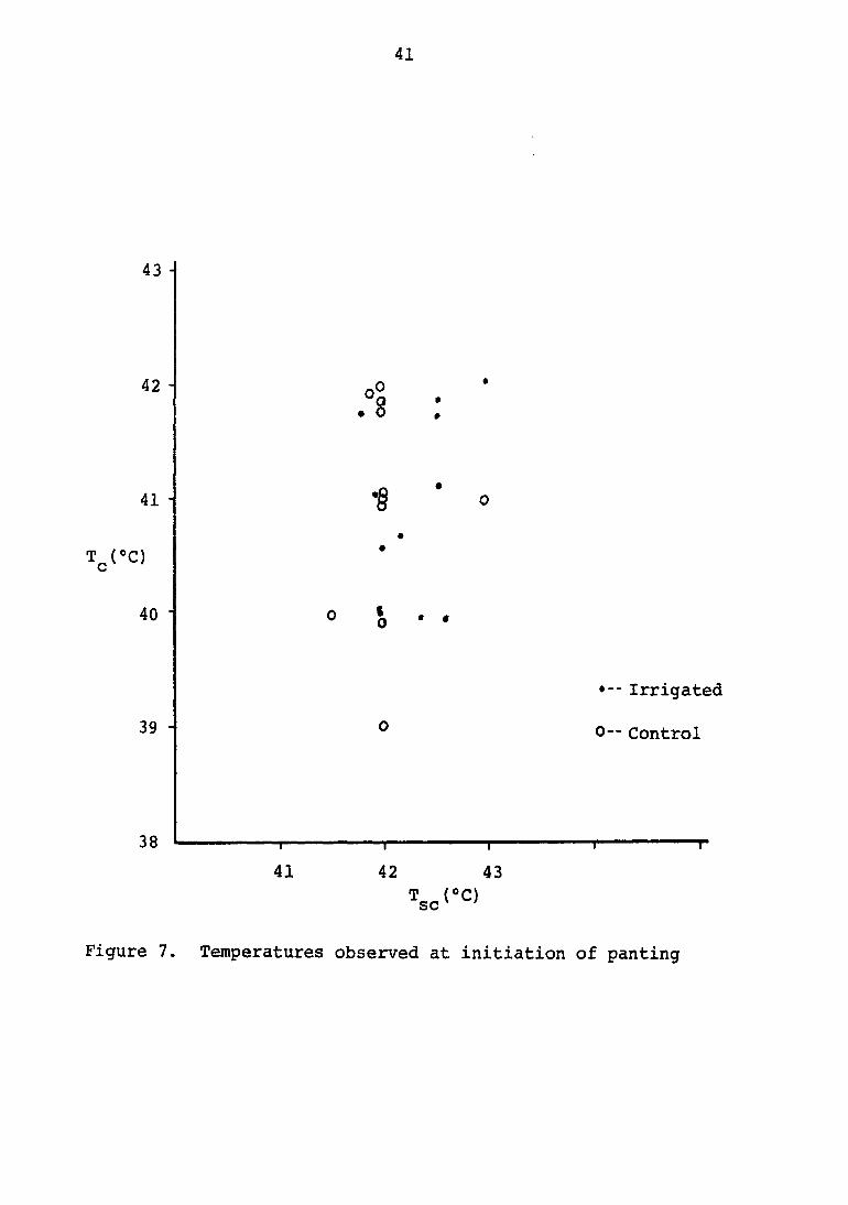
induced in the tissue cells. Panting was initiated when core
temperatures reached 40°C to 42°C, but initiation of panting
was more closely correlated with subcutaneous temperature.
Non-irrigated animals began panting when the subcutaneous
temperature reached 42®C. More spread was noted in the irri
gated animals, which showed initiation of panting at sub
cutaneous temperatures of 41.5 to 43®C (Figure 7). Heart rate
did not show any consistent relationship to either core or
subcutaneous temperature. An increase in heart rate was noted
with increased bath temperature, but no linearity between these
parameters could be maintained.
Death during the procedure was uneventful. Respiratory
rate diminished, while the amplitude was exaggerated. Ventri
cular tachycardia was noted; then, as heart rate slowed, the
R-S segment increased in amplitude and duration. No other
portions of the ECG tracing could be identified. All vital
signs subsequently faded. This trend was reversed if animals
were cooled rapidly when the abnormal respiratory and cardiac
signs appeared.
After the procedure, the recovery period was prolonged.
Often, animals were in a state of exhaustion for one to two
hours. Once recovery developed, animals rapidly regained normal
appetites, and no physical or neurological abnormalities were
noted. If the animals did not become conscious within two
hours after the procedure, death occurred within twelve hours.
41
43
42 -
41 -
T^(«C)
40
39 -
38
8 :
0 {, . •
41 42
-I—
43
Tsc(°CI
• Irrigated
0-- Control
Figure 7. Temperatures observed at initiation of panting
42
These clinical signs are consistent with cited information
referring to regenerative capacities of thermally sensitive
organs. If hyperthermia follows van't Hoff's rule, metabolic
rates of the animals with core temperatures of 42°C are 50 to
75% higher than in the normal state.
Examination of electrocardiographic and electroencephalo-
graphic data showed no observable alteration when terminal
recordings were compared to initial recordings in either
irrigated or surviving control dogs. Clinicopathological data
showed no alteration when compared to the initial values in
either irrigated or control dogs later than twenty-four hours
after the experimental procedure.
Pathology
No remarkable lesions were evidenced in either the experi
mental or control animals which survived one week after
treatment. Dog 115 suffered from chronic interstitial
pneumonia that flared to an acute form. Dog number 152 suf
fered from chronic hepatitis. Both dogs were eliminated from
the experiment.
Gross pathological changes observed in those animals which
died acutely, revealed few petechia on the serosal surface of
the stomach and intestine. Occasionally, the mesenteric lymph
nr«/^oe waro onlavrrorl anfl orloma+-nnc . Tn animale f r» Kafh
temperatures of 45® to 46®C, ecchymotic and suffusion hemor
rhages were seen on the inside thigh, scrotum and inguinal
43
region. Microscopically, the gross lesions were supported, but
no lesions were demonstrated in the central nervous system.
Pathology was apparently restricted mostly to the ultra-
structural and biochemical level, because death was so rapid.
This observation bears out cited information in the literature
review.
Short Term Clinical Pathology
Blood gases demonstrated a shift to metabolic acidosis
prior to the treatment after the animal was anesthetized.
During the procedure, after the dog had been panting for a
period of time, there consistently was a shift to respiratory
alkalosis with partial compensatory metabolic acidosis. After
the procedure, surviving animals reverted to slight metabolic
acidosis, with partial compensatory respiratory alkalosis.
Dogs will develop slight dehydration during cage confine
ment. This, along with induction of anesthesia, is sufficient
to render the animal in a mild state of metabolic acidosis.
The second blood-gas sample was taken after the animal had been
panting for a period of time, which accounts for the moderate
state of respiratory alkalosis. Renal shut-down and reduction
of the panting state reverted the animal back to a mild state
of metabolic acidosis.
Althouah extreme hvoerthermia would be expectpd fn inrreaee
clotting time, no consistent pattern was noted. Urine volume
44
diminished markedly when the core temperature was maintained
close to 42.5°C. This was more striking in non-irrigated than
in irrigated dogs. Urine production volumes were not taken in
animals prior to treatment, so quantitative comparisons with
normal temperatures were not made. However, urine volume in
the non-irrigated animals was much less than that of irrigated
dogs in the same bath temperature. If core temperatures were
held identical, urine volume did not differ.
Increases in protein and red blood cells were observed
in the urine, but these were not marked. These were probably
due to catheterization, and not to hyperthermia, as increases
were seen in all animals, including those that did not receive
sufficient heat to initiate panting.
Twenty-four hours after the treatment, animals that had
achieved core temperatures in excess of 41.5*C had neutrophilia
and lymphopenia. Occasionally, measured fibrinogen levels were
elevated. The hemograms described no other abnormalities.
Hemograms were similar to those seen in any stress syndrome of
the canine.
Blood samples collected close to animal expiration revealed
excessive hemolysis and cell dyscrasias, and no conclusions
could be drawn other than that the blood cells were very fragile.
45
MATHEMATICAL MODEL
Bath temperature (Tg), duration of immersion (t), irriga
tion temperature (T^) and irrigation flow rate are the control
ling parameters or driving functions. In order to obtain pre
dictable core temperature values the experimental design was
presented to develop a family of curves at various temperature
ranges and durations. These values were then normalized so
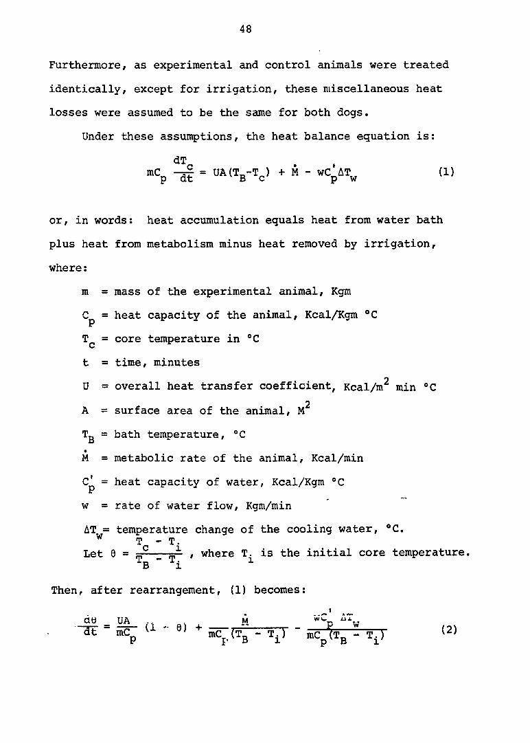
they could be compared with each other (Figure 8). A mathe
matical model was thus developed utilizing a ratio of core
temperature increase over initial bath temperature-core
temperature differential, holding irrigation temperature and
flow rate constant. Assuming metabolic rate is approximately
linear with small temperature increases, the time required to
achieve a desired core temperature can be determined by
selecting an appropriate bath temperature.
Mathematical Solution
The first law of thermodynamics requires that energy be
conserved. In the absence of work, the heat accumulation of a
system equals heat input plus heat generated minus heat lost.
The system here was defined as including the water bath, dog and
cooled irrigating water. It was assumed that negligible heat
was gained or lost with respect to the body in the air passage
ways. Although the head wa> not submerged, it was assumed that
significant amounts of heat were not lost through its surface.

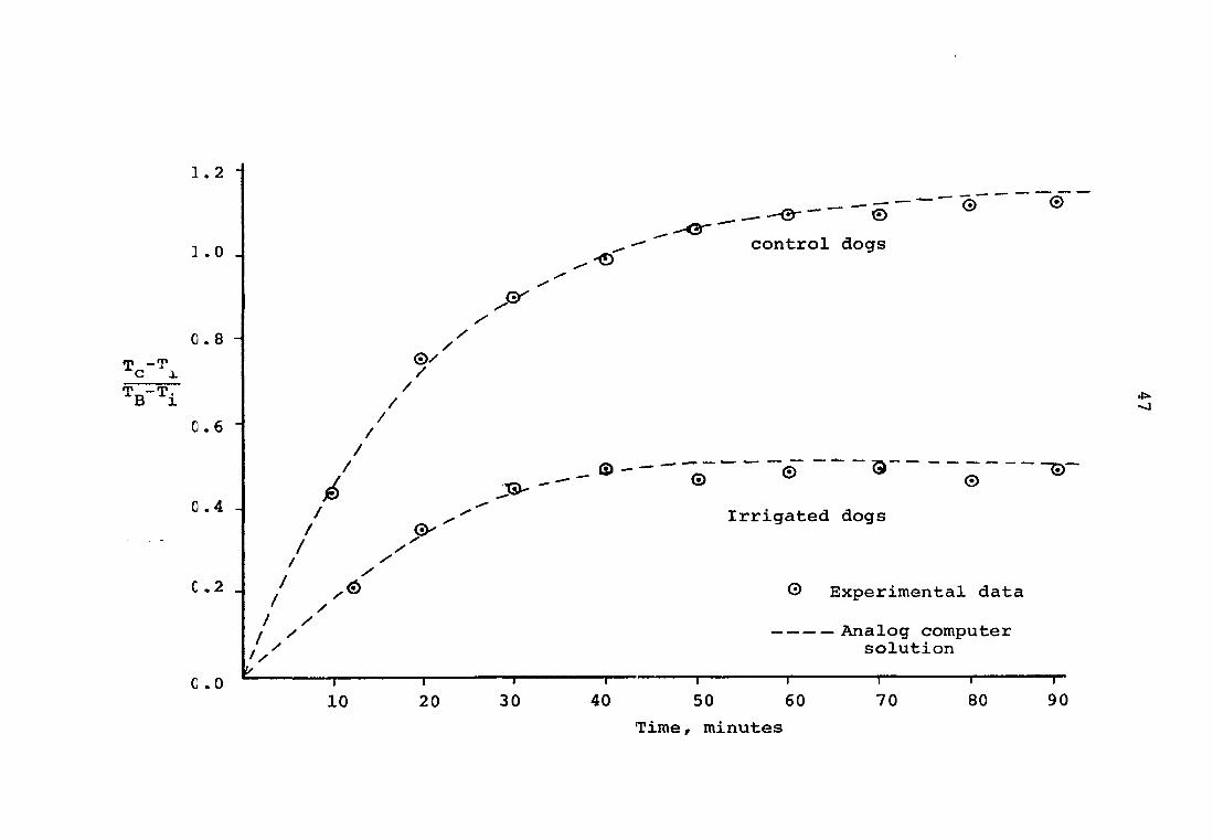
Figure 8. Comparison of experimental data with a mathematical model
1 . 2 -
0 . 8
0.6
0.4
G .2
©/ /
/ /
/ /
/ /
JSX-
/ / /
/ / y u I
10 -i— 20
—r-30
<3 ^ control dogs
-<D
® - ' ~ ® ® g , " ~ ® '
Irrigated dogs
G Experimental data
Analog computer solution
—I 1 1 1 1 r-40 50 60 70 80 90
Time, minutes
48
Furthermore, as experimental and control animals were treated
identically, except for irrigation, these miscellaneous heat
losses were assumed to be the same for both dogs.
Under these assumptions, the heat balance equation is:
dT mCp = DA(Tg-T^) + M - wCpiT^ (1)
or, in words: heat accumulation equals heat from water bath
plus heat from metabolism minus heat removed by irrigation,
where:
m = mass of the experimental animal, Kgm
Cp = heat capacity of the animal, Kcal/Kgm ®C
= core temperature in ®C
t = time, minutes
U = overall heat transfer coefficient, Kcal/m^ min °C
2 A = surface area of the animal, M
Tg = bath temperature, ®C
M = metabolic rate of the animal, Kcal/min
Cp = heat capacity of water, Kcal/Kgm "C
w = rate of water flow, Kgm/min
AT = temperature change of the cooling water, ®C. w T - T.
Let 0 = — , where T. is the initial core temperature. " ̂ i 1
Then, after rearrangement, (1) becomes:
do UA M wC AT
"ïït " mCp " 9) + - T^) - mCp^Tg - T.) ^2)
49
Now let a = UA mC
and b = M
and c =
" ''i'
then gp = a(l - 8) + b - c, which can be solved to produce:
e = = + b - c ^ _ g-atj (3)
This fits the initial condition:
6(0) = 3_jLA_=_£(I _ e°) = * + b - _ 1) = 0,
and can also be used to get the correct steady-state (long
time) result:
6C") = a + b - c 1 -a + b - c
In the control dogs, where c = 0 (no irrigation), the data
points at ninety minutes approached a value of;
ew = ̂ = oH = 1-150 ( 4 )
where the values of a, b, (and c) are determined either by
Equation 2 on an analog computer, as will be described below.
50
In the experimental dog, as can be seen in Figure 8,
eW = a + b - c ̂ 0.444^-^0.236 ^ ̂ . 0.540 (5)
These results agree very well with the collated experimental
data, as shown in Figure 8. The dashed line through the
experimental data points is the result of plotting the solution
(Equation 3) using the parameters a, b and c as determined
above.
Solution Utilizing the Analog Computer
Equation 2 was solved on an EAI Tr-48 analog computer,
using
1 volt = 0.1 units of 9, 0' = 100
and 1 second = 10 minutes of real time, T = ̂
the equation becomes:
^ = a'(10 - 0') + b' - c' (6)
and when scaled (min
§ =iM2^ao-e., p p 1 p 1
The following results were obtained, directly from the
computer, by fitting the data in Figure 8;
a' = = 0.386 min'^, p
51
lô b' - rô lOOM
mC AT. P 1
-1 = 0.058 min ,
and
lô c' - I?
lOOwC AT, P V
mC AT. P 1
= 0.236 min ^
Comparison of Model with Experimental Data
The computer results for the coefficients a, b and c were
then compared to the estimated values of the experimental
parameters.
Water bath parameters
a' = = 0.386 min'l (8) mC P
Substituting known values for the average dog (3),
UA = (—251) (il^) = 16 Kcal/hr°C, (9)
which is reasonable, since the dog's surface area (A) ̂ 0.5 to
0.7M^, and the heat transfer coefficient (U) 20 to 25 Kcal/M^
hr°C for this situation.
Metabolic parameters
10 M 0.058 ^ mC .T. " ïïiin '
P 1
or again substituting known parameters,
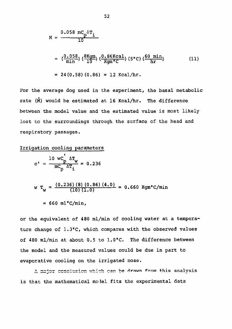
52
M =
,0.058\ /8Kgm\ ,0.86Kcal\ /60 min I min 'I 10 'I Kgm'C ' ^ hr mm
(11)
= 24(0.58)(0.86) = 12 Kcal/hr.
For the average dog used in the experiment, the basal metabolic
rate (M) would be estimated at 16 Kcal/hr. The difference
between the model value and the estimated value is most likely
lost to the surroundings through the surface of the head and
respiratory passages.
Irrigation cooling parameters
or the equivalent of 480 ml/min of cooling water at a tempera
ture change of 1.3*C, which compares with the observed values
of 480 ml/min at about 0.5 to 1.0*C. The difference between
the model and the measured values could be due in part to
evaporative cooling on the irrigated nose.
A r.ajcr conclusion vrhich can be drawn from this analysis
is that the mathematical moïel fits the experimental data
w T. w
(0.236)(8) (0.86) (4.0) (10)(1.0)
= 0.660 Kgm°C/min
= 660 ml*C/min
53
extremely well, in that it:
(1) gives the proper transient and steady-state results,
(2) describes the proper relative behavior between the
experimental dogs and the control dogs, with only the term
representing irrigative cooling (c) being added, and
(3) estimates proper and reasonable values for both the
known and unknown experimental parameters.
54
CONCLUSIONS AND RECOMMENDATIONS
Extreme whole body hyperthermia was achieved by elevating
core temperatures of dogs to 42®C, using a warm water bath.
Irrigation of the alar fold of the nose with cold water (10 to
15°C) provided a means of elevating core temperature 0.5 to
1.5°C without lasting side effects. A brain-core temperature
differential could be maintained by this technique for a long
period of time (more than two hours). Maximum tolerable core
temperature for the non-irrigated dog was 42®C for one to one
and one-half hours, whereas that for the irrigated dog was in
excess of 42.5®C. Time required for core temperature to
reach steady-state was approximately forty minutes after
correct bath temperature was achieved. Best results required
a treatment duration of one to one and one-half hours. Panting
was usually initiated at a subcutaneous temperature of 42®C.
Dyspnea and ventricular tachycardia were observed when the
animal approached death. These parameters, when monitored
closely, revealed an "on-line" index with respect to viability.
Death appeared to be due to heat exhaustion or hypovolemic
shock.
An effective mathematical model of the system, that
described the dynamic temperature changes, was developed. The
analog solution matched the normalized experimental data points.
The solution to the mathematical model also appeared to predict
proper and reasonable values for known and unknown experimental
55
parameters.
Although available evidence indicates that maximum thermal
tolerance levels are dictated by thermal denaturation of
enzymes involved in cellular respiration, availability of
metabolites may play an important role. Increases in metabolic
rate required increases in glucose and oxygen utilization. If
excessive quantities of glucose and oxygen were made available
to the cells, the metabolic limitation might be reduced,
thereby permitting greater elevation of core temperature (19).
Blood glucose determinations, oxygen consumption measure
ments, improved thermal monitoring, ultrastructural pathological
studies, and automation of the physical system are major areas
for further investigation. The present design was adequate to
demonstrate that cerebral protection could be maintained under
conditions of hyperthermia, and it was also adequate to permit
a reasonable determination of the parameters required.
This technique provides a potential means by which to
elevate the metabolic rate of cells to the point at which
intracellular enzymes will be denatured and the cells destroyed.
Cells with a high metabolic rate at normal body temperature
should be the first to reach this limiting state during hyper
thermia. Rapidly growing cancer cells have a very high
metabolic rate, as do cells of cerebral tissue. Cerebral
4-i->^v-ms 1 TA TninimuiTi of 0.5®C below W** V V w- — ^ — - - -

56
core temperature, as shown in this investigation. Therefore,
core temperature can conceivably be elevated sufficiently to
jeopardize the viability of cancer cells without cerebral
damage.
57
LITERATURE CITED
1. Adams, T., Morgan, M., Hunter, W. and Holmes, K. Temperature regulation of the unanesthetized cat during mild cold and severe heat stress. J. Aopl. Physiol. 29 (6): 852. 1970.
2. Alenina, T. V. The change in the dry residue of some tissues with overheating of the body (English translation). Tr. Smolensk Med. Inst. 26: 435. 1968.
3. Altman, P. and Dittmer, D., ed. Biology data book. Washington, D.C., Fed. of Amer. Soc. for Exp. Biol. 1964.
4. Andersson, B. Cold defense reactions elicited by electrical stimulation within the septal area of the brain in goats. Acta Physiol. Scand. 41: 90. 1951.
5. Andersson, B., Grant, R. and Larsson, S. Central control of heat loss mechanisms in the goat. Acta Physiol. Scand. 37: 261. 1956.
6. Andersson, B. and Persson, N. Pronounced hypothermia elicited by prolonged stimulation of the heat loss centre in conscious goats. Acta Physiol. Scand. 41: 10. 1957.
7. Andersson, B. and Persson, N. Pronounced hypothermia elicited by prolonged stimulation of the heat loss centre in unanesthetized goats. Acta Physiol, Scand. 41: 277. 1957.
8. Ardenne, M. von. The selective amplification of primary cancer cell damage as a basic mechanism of the multistep cancer therapy (English translation). Ill Simposio internazionale sul trattamento loco regionale dei tumori. Lecture. Dec. 10, 1969.
9. Ardenne, M. Von. Uber die methode der intensivierten O2 - mehrschritt-prophylaxe gegen krebs bzw, krebs-metastasen. Dtsch. Ges. Wesen 27: in press. 1972.
10. Ardenne, M. von and Rieber, F. On the present status of whole body extreme hyperthermia as an element of multistep carcinoma therapy (English translation). Z. Krebsforsch 69 (4): 341. 1967.
58
11. Atkins, E. Pathogenesis of fever. Physiol, Rev. 40: 580. 1960.
12. Breazile, J. Textbook of veterinary physiology. Philadelphia, Lea and Febiger. 1971.
13. Burger, F. J. and Fuhrman, F. Evidence of injury by heat in mammalian tissues. Am. J. Physiol. 206 (5): 1057. 1964.
14. Collins, K. J., Few, J. D., Forward, T. J, and Giec, L. A. Stimulation of adrenal glucocorticoid secretion in man by raising the body temperature. J. Physiol. 202 (3): 645. 1969.
15. Durotoye, A. 0. and Grayson, J. Heat production in the G. I. tract of the dog. J. Physiol. (London) 214 (3); 417. 1971.
16. Ederstrom, H. E. Blood flow changes in the dog during hyperthermia. Am. J. Physiol. 176: 347. 1954.
17. Edholm, 0. G., Fox, R. H. and Macpherson, R. K. The effect of cutaneous anesthesia on skin blood flow. J. Physiol. 132: 15. 1956.
18. Edholm, 0. G., Fox, R. H. and Macpherson, R. K. Vasomotor control of the cutaneous blood vessels in the human forearm. J. Physiol. 139: 455. 1957.
19. Farkas, M., Mozsa, S. and Donhoffer, S. The effect of hypoxic hypoxia and environmental temperature on body temperature and oxygen consumption in the course of pyrogen-induced fever. Acta Physiol. Acad. Sci. (Hung.) 30: 155. 1966.
20. Fay, T. Early experiences with local and generalized refrigeration of the human brain. Jour. Neurosurg. 16: 239. 1959.
21. Fay, T. Observations on generalized refrigeration in cases of severe cerebral trauma. Res. Publ. Ass. Nerv. Ment. Dis. 24: 611. 1945.
22. Feldberg, W. and Saxena, P. Mechanism of action of pyrogen. J. Physiol. (London) 211 (1): 245. 1970.
23. Field, J., Fuhrman, F. A. and Martin, A. W. Effect of temperature on the oxygen consumption of brain tissue. J. Neurophysiol. 7: 117,. 1944.
59
24. Findlay, J. D. The respiratory activity of calves subjected to thermal stress. J. Physiol. 136: 300. 1957.
25. Findlay, J. D. The respiratory behaviour of calves exposed to increasing thermal stress. J. Physiol. 130: 16. 1955.
26. Frankel, H. M., Ellis, J. P., Jr. and Cain, S. M. Development of tissue hypoxia during progressive hyperthermia in dogs. Am. J. Physiol. 205 (4); 733. 1963.
27. Frankel, H. M. and Ferrante, F. L. Effect of arterial pC02 on the appearance of increased lactate during hyperthermia. Am. J. Physiol. 210 (6): 1269. 1966.
28. Frascella, D. and Frankel, H. M. Liver pyridine nucleotides, lactate and pyruvate, in hyperthermic rats. Am. J. Physiol. 217 (1): 207. 1969.
29. Grant, R. T. and Boiling, H. E. Further observations of the vascular responses of the human limb to body warming: Evidence for sympathetic vasodilator nerves in the normal subject. Clin. Sci. 3: 273. 1938.
30. Guieu, J. D. and Hardy, J. D. Effects of heating and cooling of the spinal cord on preoptic unit activity. J. Appl. Physiol. 29 (5); 675. 1970.
31. Hales, J. R. S., Bligh, J. and Maskrey, M. Cerebrospinal fluid acid-base balance during respiratory alkalosis in the panting animal. Am. J. Physiol. 219 (2): 469. 1970.
32. Hales, J. R. S., Kao, F. F., Mei, S. S., Wang, C. and Grelenstein, M. Panting in heated cross circulated dogs. Am. J. Physiol. 218 (5): 1389. 1970.
33. Hammel, H. T., Wyndham, C. H. and Hardy, J. D. Heat production and heat loss in the dog at 8-36®C. environmental temperature. Am. J. Physiol. 194: 99. 1958.
34. Hardy, J. D. Physiology of temperature regulation. Physiol. Rev. 41: 521. 1961.
60
35. Hayward, J. and Baker, M. A comparative study of the role of cerebral arterial blood in the regulation of brain temperature in 5 mammals. Brain Res. 16 (2): 417. 1970.
36. Hellman, K. and Weiner, J. S. Antidiuretic substance in urine following exposure to high temperatures. J. Appl. Physiol. 6: 194. 1953.
37. Hemingway, A., Forgrave, P. and Birzis, L. Nervous control of shivering. Ladd Air Force Base, Alaska, Arctic Aeromed. Lab. Rept. No. 1, Project No. 22; 1301. June, 1954.
38. Hensel, H. Physiologie der thermoreception. Ergeb. Physiol. 47; 166. 1952.
39. Hildwein, G. and Malan, A. Effect of hair clipping on thermal reoulation in the angora guinea pig. Arch. Sci. Physiol. 24 (1): 347. 1971.
40. Hubbard, J., Llinas, R. and Quastel, D. Electrophysiological analysis of synaptic transmission. Baltimore, The William and Wilkins Co. 1969.
41. Ingram, D. L. and Smith, R. Brain temperature and cutaneous blood flow in the anesthetized pig. J. Appl. Physiol. 29 (5): 698. 1970.
42. Kanter, G. S. Effect of heat on regulation of body fluids and electrolytes in dogs. Am. J. Physiol. 178: 259. 1954.
43. Kanter, G. S. Heat and excretion in man. J. Appl. Physiol. 7; 533. 1955.
44. Kew, M., Bersohn, I., Steftel, H. and Kent, G. Liver damage in heat stroke. Amer. J. Med. 49 (2): 192. 1970.
45. Kirsch, R., Schmidt, D., Chr, H., Gabsch and Hohaus, B. On the effect of hyperthermia of the whole body on concentration of cyclophosphamide in normal tissue and tumors (English translation). Z. Gesamte Exp. Med. 145 (1): 41. 1968.
46. Kucherenko, R. P. Submicroscopic changes in the purkinji cells of the cerebellum under the effect of hyperthermia (English translation). Tsitologiya 12 (3); 312. 1970.
61
47. Lichton, I. J. Competition between sweat glands and kidneys for salt and water in man. J. Appl. Physiol. 11: 223. 1957.
48. Lundberg, N., Nielsen, K. and Nilsson, E. Deep hypothermia in intracranial surgery. J. Neurosurg. 13: 235. 1956.
49. MacDonald, D. K. C. and Wyndham, C. H. Heat transfer in man. J. Appl, Physiol. 3: 342. 1950.
50. Machle, W. and Hatch, T. F. Heat: man's exchanges and physiological responses. Physiol. Rev. 27: 200. 1947.
51. Magilton, J. and Swift, C. Cardiovascular responses to induced brain temperature changes. J. Physiologie, Tome 63 (3): 328. 1971.
52. Magilton, J. and Swift, C. Description of two physiological heat exchange systems for the control of brain temperature. Inst. Elect. Electron. Engrs. Ann. Rocky Mountain Bioeng. Symp. 5th: 24. 1968.
53. Magilton, J. and Swift, Ci Responses of veins draining the nose to alar fold temperature changes in the dog. J. Appl. Physiol. 27 (1): 18. 1969.
54. Magilton, J. and Swift, C. Thermoregulation of the canine brain by alar fold and intervening arteriovenous heat exchange systems. Physiologist 10: 241., 1967.
55. Matsui, N. Ability to perspire in relation to sexuality. Bull. Res. Inst. Diath. Med. (Japan) 6: 60. 1955.
56. Matsui, N. Study on the difference of the ability to perspire according to age, with particular observation in childhood cind old age. Bull. Res. Inst. Diath. Med. (Japan) 6: 70. 1955.
57. Meyer, H. H. Theorie des fiebers und seiner behand-lung. Verhandl. deut. Bes inn Med. 30: 15. 1913.
58. Miller, M. E., Christensen, G. and Evans, H. E. Anatony of the dog. Philadelphia, W. B. Saunders. 1964. Reprinted, 1968.
59. Murakami, N., Stolwijk, J. A. J. and Hardy, J. D. Responses ot preoptic neurons to anesthetics and peripheral stimulation. Am. J. Physiol. 213: 1015. 1967.
62
60. Ityers, R. D., Veale, W. and Yaksh, T. Changes in body temperature of the anesthetized monkey produced by sodium and calcium ions perfused through the cerebral ventricles. J. Physiol. (London) 217 (2): 381. 1971.
61. Nemoto, E. M. and Frankel, H. Cerebral oxygenation and metabolism during progressive hyperthermia. Am. J. Physiol. 219 (6): 1784. 1970.
62. Radigan, L. R. and Robinson, S. Effects of environmental heat stress and exercise on renal blood flow and filtration rate. J. Appl. Physiol. 2: 185. 1949.
63. Rowel1, L. B./ Brengelmann, G. L. and Murray, J. Cardiovascular responses to sustained high skin temperature in resting man. J. Appl. Physiol. 27 (5) : 673. 1969.
64. Rozanora, E. S., Medvedkova, A. and Mikhailenko, M. Effect of hyperthermia on the phosphorylase activity in different tissues (English translation). Smolensk Med. Inst. 26: 426. 1968.
65. Schulke, L. and Kading, 0. Uber den einfluss exogener hyperthermie und thermischer hyperventilation auf den partialdruck des COn im blut des schweines. Arch. Exp. Veterinarmed. 22 (3): 633. 1968.
66. Seagrave, R. C. Biomedical applications of heat and mass transfer. Ames, Iowa, I.S.U. Press. 1971.
67. Somas, L., ed. Textbook of veterinary anesthesia. Baltimore, Md., The Williams and Wilkins Co. 1971.
68. Spurr, G. B. and Barlow, G. Tissue electrolytes in hyperthermic dogs. J. Appl. Physiol. 28 (1); 13. 1970.
69. Steinhardt, M., Studzinski, T., Lyhs, L. and Borchardt, D. Thermoreguiatoriche hyperventilation, speichel-und harnabgabe beim hausschwein in exogener hyperthermie. Arch. Exp. Veterinarmed. 24 (3): 793. 1970.
70. Stunzhas, P. M. Spectrometric characterization of the proteins of the brain of overheated rats (English , translation). Tr. Smolensk Med. Inst. 26: 431. 1968.
63
71. Stunzhas/ P. M. Condition of some functional groups of soluable proteins of the brain of overheated rats (English translation). Tr. Smolensk Med. Inst. 26: 431. 1968.
72. Swenson, M. J., ed. Dukes' physiology of domestic animals. 8th ed. Ithaca, N.Y., Cornell Univ. Press. 1970.
73. Vladimirov, S. V. and Polyakova, G. Hypothalamo-hypophyseal neurosecretory system of dogs under conditions of solar thermal overheating (English translation). Arkh. Anat. Gistol. Embriol. 57 (10): 74. 1969.
74. Waites, G. M. H. Effect of heating the scrotum of the ram on respiration and body temperature. Quart. J. Exptl. Physiol. 46: 314. 1962.
75. Wang, J. K., Moffitt, E. A. and Rosevear, J. W. Oxidative phosphorylation in acute hyperthermia. Anesthesiology 30 (4): 439. 1969.
64
ACKNOWLEDGEMENTS
The author wishes to express his sincere thanks to Dr. R.
C. Seagrave for his assistance and enthusiasm in this project.
Mrs. Joyce Feavel and Dr. Mike Brown lended valuable
assistance and support when it was needed. Dr. Curran Swift
availed himself to help with the complicated monitoring
problems. Dr. Jim Magilton offered encouragement and his
laboratory facilities.
Thanks to Dr. P. T. Pearson and Dr. N. R. Cholvin for
their staunch support during the author's training program.
Appreciation is extended to the department of veterinary
pathology for their assistance in this project. This study
was funded in part by ISU Grant No. 405-23-007.
Finally appreciation is expressed to Drs. Klaus Rowedder
and Jeanine Carithers without whose support this program could
not have been completed.
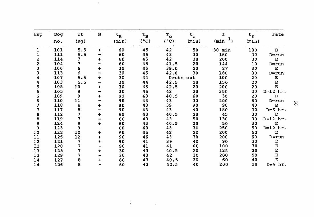
APPENDIX: SUMMARY OF EXPERIMENTAL DATA
Exp Dog Wt N tg Tg
no. (Kg) (min) (®C)
1 101 5.5 + 60 45 1 111 5.5 — 60 45 2 114 7 + 60 45 2 104 7 - 60 45 3 106 6 + 30 45 3 113 6 - 30 45 4 107 5.5 + 30 44 4 103 5.5 - 30 44 5 108 10 + 30 45 5 105 9 — 30 45 6 109 9 + 90 43 6 110 11 — 90 43 7 118 8 + 90 43 7 117 8 - 90 43 8 112 7 + 60 43 8 119 7 - 60 43 9 124 9 + 60 43 9 123 9 - 60 43 10 122 10 + 60 45 11 125 12 + 90 46 12 121 7 + 90 41 12 120 7 — 90 41 13 128 7 + 30 43 13 129 7 — 30 43 14 127 8 + 60 43 14 126 8 — 60 43
t f tm Fate c c _ 1 f Fate
°C) (min) (min (min)
42 50 30 min 180 E 43 30 160 30 D=run 42 30 200 30 E 41.5 20 144 10 D=run 39.0 20 27 30 E 42.0 30 180 30 D=run Probe out 100 20 E 42.5 20 150 20 E 42.5 20 200 20 E 42 20 250 30 D=12 hr 40.5 60 30 20 E 43 30 200 80 D=run 39 90 90 40 E 43 40 180 30 D=6 hr 40.5 20 45 30 E 43 50 130 30 D=12 hr 40.5 20 50 30 E 43 30 250 50 D=12 hr 42 20 200 50 E 43 30 200 60 D=run 39 40 90 30 E 41 60 100 70 E 40.5 20 125 30 E 42 30 200 50 E 40.5 30 60 40 E 42.5 40 200 30 D=4 hr
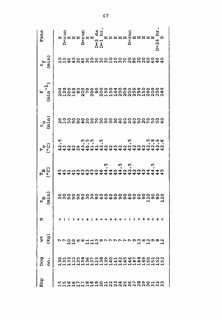
67
Q) -P
h H
fi fi nJ 3 3 >0
r
E
E E r E E
II Il m iH Û Q II II Q û
C 0
H H M H H W H M H M H &
x:
CN II Q
C m -H
•P Ê
o o o o o o o o o o o o o o o o o o o o o o o
iH I
«H C •H E
o o o o o o o o o o o o o o o o o o o o o o o ocNovoœor-oooommoooomininoomo M H M r4 (M (S M iH H rH CJ M CM M CN iH <N CM (N C>4
C O-H
•P e
o o o o o o o o o o o o o o o o o o o o o o o cNHmino\TrcNr>jr>infomnTry5CNvomcNr^mm*i'
m m m m m m in œ vo vo
O U b* o
(NmCN(NO\mOr4r4(NCN(NmCNCN(NCN(NCN(NMCN(N
m m m m
m U E4 o
inir>HHronnroinvoo*a'inor4CNP«icNnoTj«*j'in
f i o o o o o o o o o o o o o o o o o o o o o o o m-H mmmo\(T\o\mmo\vovovo(T)mo\vomvomcNO\o\(N
+J g H H
+ I I + I + I + + + + + + I I I + + + + +
•p s W r^r^oovovor4r4mo\r~t~»r^r~r^r-o\mvo(Nmo\N
H r—t iHHH rHiH r4
tj> . oHfocNinTrvor^moocnOfHtNinvop»c»(y>oHcNro 0 O mmmmmmmmHmmooTfTi'Ti'Ti'Ti'Trifiinuiifi O fi HHHHiHHHrHrH'HrHrHrHrHiHrHrHrHrHrHHrHH
I ininvovor^r>>oooocftoHfNco«ti<invD[^oo<T*oHfNro HHHHHHHiHHfMCNCNCNCMCNCNJCNCNCNrnmoon