Embed Size (px)
Citation preview

Canine neoplasia
Carol Naranjo, LV, DACVP, DECVP, PhDIDEXX Laboratories

IRIDOCILIARY EPITHELIAL TUMORS

Iridociliary epithelial tumors – dogs
• 2nd most common primary intraocular neoplasm
• Middle-aged dogs
• Retrievers
• Origin:
– Pigmented or non-pigmented CB epithelium
– Posterior iris epithelium (pigmented)

Iridociliary epithelial neoplasia
• Adenoma
– Non uveo-invasive
– Uveo-invasive
• Adenocarcinoma (scleral invasion)
• Pleomorphic adenocarcinoma

Iridociliary adenoma/carcinoma – Gross
• Well-delineated
• PC >>> AC
• Cradling lens
• 90% non-pigmented (light tan – pink)
– Pigmented (DDX. melanocytic neoplasia)


Iridociliary epithelial neoplasms – Micro
• Ribbons, trabeculae, cords, papillae, acini
• Asteroid hyalosis
• 60% thick PAS-positive membranes
• 50% hyaluronic acid (Alcian blue +ive)
– DDx w/ metastastic neoplasia


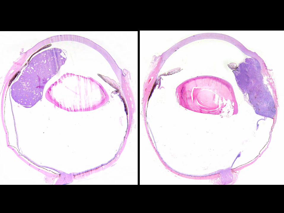
Iridociliary epithelial neoplasms – IHC
• Vimentin, NSE +ive
• S100 variable
• Cytokeratin:
– Benign tumors –ive
– Increasing staining w/ invasiveness

Courtesy R. Dubielzig (COPLOW)
Vimentin Cytokeratin
Iridociliary epithelial neoplasms – IHC
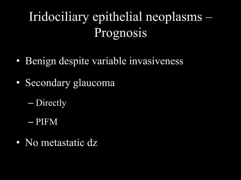
Iridociliary epithelial neoplasms –Prognosis
• Benign despite variable invasiveness
• Secondary glaucoma
– Directly
– PIFM
• No metastatic dz
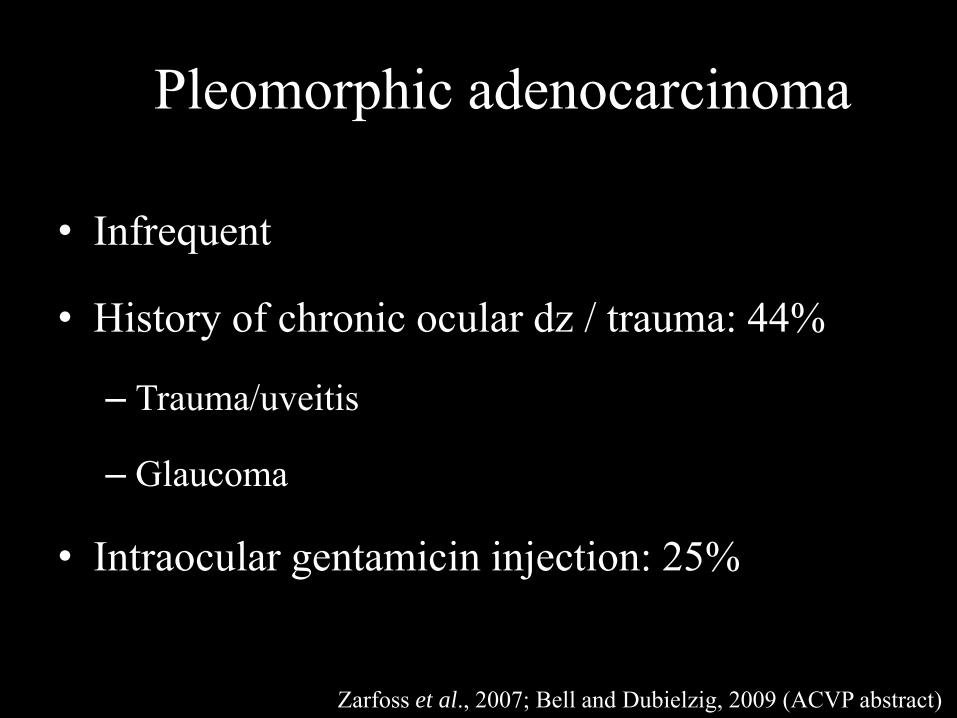
Pleomorphic adenocarcinoma
• Infrequent
• History of chronic ocular dz / trauma: 44%
– Trauma/uveitis
– Glaucoma
• Intraocular gentamicin injection: 25%
Zarfoss et al., 2007; Bell and Dubielzig, 2009 (ACVP abstract)

Pleomorphic adenocarcinoma
• Gross:
– Poorly defined mass diffuse
– Filling the globe
• Histo: irregular cords, nests, anaplastic cells
• Poor prognosis (local recurrence, mets)

Pleomorphic adenocarcinoma – Gross

Pleomorphicadenocarcinoma – Histo

Pleomorphic adenocarcinoma – IHCVimentin
100% positive (n = 16)
Pancytokeratin
75% positive (n = 16)
Bell & Dubielzig, ACVP abstracts, 2009

PNET

Neoplasia w/ neural differentiation
• PNET:
– Primitive neuroectodermal tumors
• Neuroblastoma, ependimoblastoma, retinoblastomas,
medulloepithelioma
• Eye:
– PNET: peripheral retina / CB
– Medulloepiteliomas: CB (dog) or ON (horse) >>> retina

Medulloepithelioma
• PNET
• Shared features with iridociliary tumors
• Rare in dogs, horses >>> others
• Not always young dogs
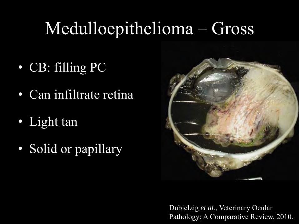
Medulloepithelioma – Gross
• CB: filling PC
• Can infiltrate retina
• Light tan
• Solid or papillary
Dubielzig et al., Veterinary Ocular Pathology; A Comparative Review, 2010.

Medulloepithelioma – Micro
• Necrosis w/ survival around blood vessels
• Rosettes
• Teratoid: muscle, bone, cartilage, neuropil
• IHC:
– TERT +ive
– Vimentin and cytokeratin: limited +ivity

Dubielzig et al., Veterinary Ocular Pathology; A Comparative Review, 2010.

Medulloepithelioma – Prognosis
• Locally invasive
– Enucleation
• Mets very infrequent

¿Retinoblastoma?
• Sparse reports, some arguable
• Tumors with neural differentiation (PNET) but
do not meet all the criteria

Flexner-Wintersteiner rosettes Homer-Wright rosettes
Fleurettes Yanoff and Sassani’s Ocular Pathology, 2015

Courtesy of Dr. Dubielzig, COPLOW

Retinoblastoma in dogs?
• Retinoblastoma-like:– Fleurettes / rosettes (1 layer)
• Medulloepithelioma– Pseudorosettes

Regan et al., 2013IHC IRBP
Retinoblastoma – like Medulloepithelioma

UVEAL SCHWANNOMA OF BLUE EYED DOGS

Uveal schwannoma of blue-eyed dogs
• SCTBED
• Blue-eyed / partially blue-eyed
– Siberian huskies, Border Collies, Catahoula hound
• Recent report in a brown-eyed dog (Marlo et al., 2018)
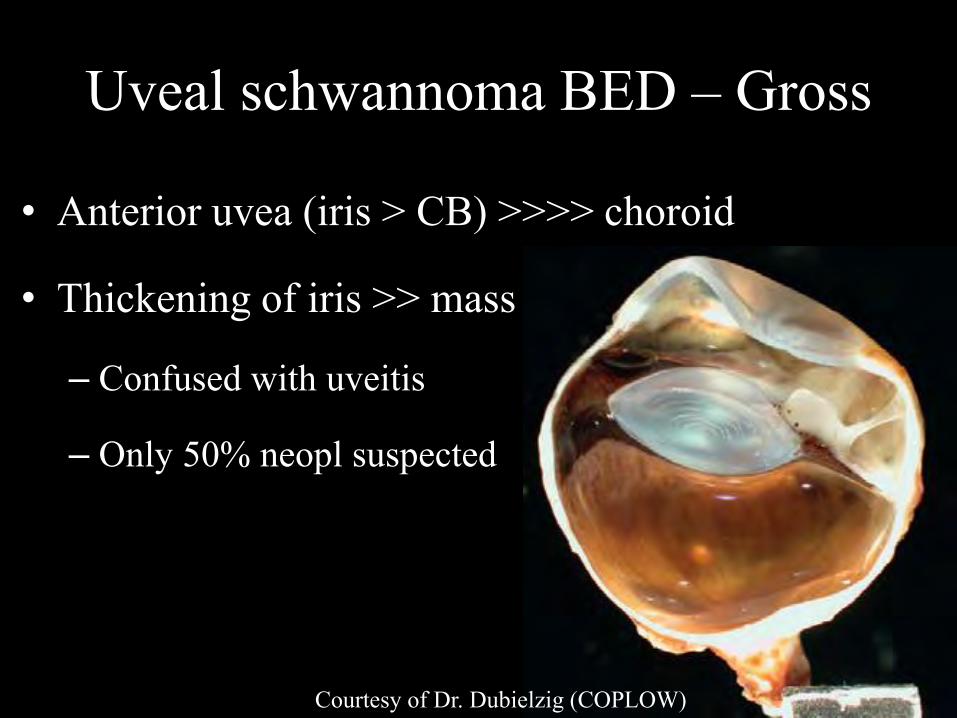
Uveal schwannoma BED – Gross
• Anterior uvea (iris > CB) >>>> choroid
• Thickening of iris >> mass
– Confused with uveitis
– Only 50% neopl suspected
Courtesy of Dr. Dubielzig (COPLOW)


Case material from COPLOW

Uveal schwannoma BED – Histo
• Poorly demarcated
• Slender to plump spindle cells
• Bland to markedly atypical cells
• Loose fascicles to tight bundles and whorls
• May invade sclera


Uveal schwannoma BED – IHC
• Vimentin +
• S100 +
• GFAP +
– Schwann cells of nonmyelinated nerves of iris stroma
Courtesy of Dr. Dubielzig (COPLOW)

Uveal schwannoma BED – EM
• Long interdigitating cytoplasmic processes
• Intermittent basal laminae at plasma mb
• Peripheral nerve sheath origin
Zarfoss et al., 2007

Uveal schwannoma BED – Prognosis
• Invasive: recurrence w/in orbit / scleral shell
• Mets rarely reported:
– Lungs, liver, mesenteric LN
– Path features linked to malignancy? (scleral invasion?)

METASTATIC TUMORS

Metastatic tumors to the globe (uvea)
• Dogs
– Lymphoma > histiocytic sarcoma
– Carcinoma, melanoma, hemangiosarcoma, OSA,…
– Carcinoma: mammary
– Extraskeletal OSA or chondrosarcoma
• Search for primary tumor!

Uveal lymphoma
• Most common metastatic neoplasm
• 37% dogs w/ lymphoma have uveal involvement
– Preceding systemic signs, presenting complaint
• 2nd clinical sign (after lymphadenopathy)
• Bilat (not always)

Uveal lymphoma – Gross
• Anterior > posterior uvea
• Mass-like lesion or diffuse (DDx. uveitis)
• Light tan
• Hematological abnormalities (hemorrhage)


Uveal lymphoma – Micro
• Monomorphic round cells
– Iris, CB > retina, choroid, limbus, ON, peripheral n.
• DDx. melanoma / histiocytic sarcoma


Uveal lymphoma – IHC
• B cell: CD20, CD79a, PAX5
• T cell: CD3
• Both described in the eye

Uveal lymphoma – Prognosis
• Guarded
• Systemic: stage V if associated w/ hemorrhage
– Survival 60-70% of those w/o ocular involvement
• ¿Primary?


Primary uveal lymphoma?• Wiggans et al., 2014
– Neurologic signs indep of ON involvement
– ¿Staging?
• Lanza et al., 2017:
– 61% w/o systemic signs (@ Dx)
– No progression
– Median survival: 769 d vs 103 d

Histiocytic sarcoma
• Metastatic
• Not infreq presented for ocular dz first
• Rottweiler > Retrievers > BMD
• Adult to senile
• Unilat (typically)

Histiocytic sarcoma – Gross• Light tan • Anterior uvea

Histiocytic sarcoma – Micro
• Round cells w/ abundant cytoplasm
– DDx. amelanotic melanoma
• Multinucleation, karyomegaly


Histiocytic sarcoma – IHC
• Vimentin +
• CD18 +
• Melan A –
• CD3/CD20 –

Histiocytic sarcoma – Prognosis • Grave, survival 0-6 mo post-enucleation

• Unilat bilat
• Anterior > posterior uvea
• Neoplastic cells w/in blood vessels
• 2 patterns:
– Discrete mass
– Diffuse infiltrates lining the inner aspect of uvea
Metastatic tumors to the globe


Primary vs. metastatic carcinomas
Primary
benign
Primary
malignant
Metastatic
Cytokeratin - +/- + Vimentin + + -

Metastatic malignant melanoma