Embed Size (px)
Citation preview
Revised Scientific Review Panel Draft April 2011
1
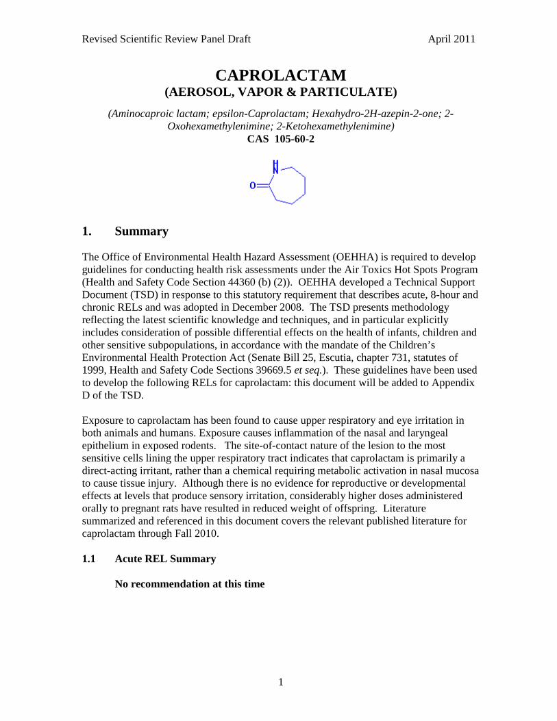
CAPROLACTAM (AEROSOL, VAPOR & PARTICULATE)
(Aminocaproic lactam; epsilon-Caprolactam; Hexahydro-2H-azepin-2-one; 2-
Oxohexamethylenimine; 2-Ketohexamethylenimine) CAS 105-60-2
1. Summary The Office of Environmental Health Hazard Assessment (OEHHA) is required to develop guidelines for conducting health risk assessments under the Air Toxics Hot Spots Program (Health and Safety Code Section 44360 (b) (2)). OEHHA developed a Technical Support Document (TSD) in response to this statutory requirement that describes acute, 8-hour and chronic RELs and was adopted in December 2008. The TSD presents methodology reflecting the latest scientific knowledge and techniques, and in particular explicitly includes consideration of possible differential effects on the health of infants, children and other sensitive subpopulations, in accordance with the mandate of the Children’s Environmental Health Protection Act (Senate Bill 25, Escutia, chapter 731, statutes of 1999, Health and Safety Code Sections 39669.5 et seq.). These guidelines have been used to develop the following RELs for caprolactam: this document will be added to Appendix D of the TSD. Exposure to caprolactam has been found to cause upper respiratory and eye irritation in both animals and humans. Exposure causes inflammation of the nasal and laryngeal epithelium in exposed rodents. The site-of-contact nature of the lesion to the most sensitive cells lining the upper respiratory tract indicates that caprolactam is primarily a direct-acting irritant, rather than a chemical requiring metabolic activation in nasal mucosa to cause tissue injury. Although there is no evidence for reproductive or developmental effects at levels that produce sensory irritation, considerably higher doses administered orally to pregnant rats have resulted in reduced weight of offspring. Literature summarized and referenced in this document covers the relevant published literature for caprolactam through Fall 2010. 1.1 Acute REL Summary No recommendation at this time
Revised Scientific Review Panel Draft April 2011
2
1.2 8-Hour REL Summary
8-Hour inhalation reference exposure level
7 µg/m3 (1 ppb)
Critical effect(s) Inflammatory changes of nasal and laryngeal epithelium in rodents
Hazard index target(s) Upper respiratory system 1.3 Chronic REL Summary
Chronic inhalation reference exposure level
2 µg/m3 (0.5 ppb)
Critical effect(s) Inflammatory changes of nasal and laryngeal epithelium in rodents
Hazard index target(s) Upper respiratory system 2. Physical and Chemical Properties [from HSDB (2006), unless noted
otherwise]
Description A semi-volatile white, hygroscopic, crystalline solid or flakes with unpleasant odor
Molecular formula C6H11NO Molecular weight 133.16 g/mol Density 1.05 g/cm3 @ 25 °C Boiling point 270 °C Melting point 69.3 °C Vapor pressure 0.001 mm Hg @ 20ºC (68ºF)
0.0021 mm Hg @ 25°C (77ºF), saturated vapor concentration = 13 mg/m3
Odor threshold 0.3 mg/m3 (Gross, 1984) Solubility Very soluble in water, benzene, diethyl ether,
and ethanol. Soluble in chlorinated solvents, petroleum distillates, and cyclohexene
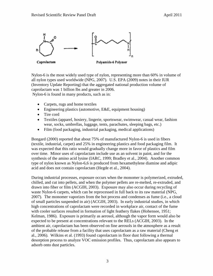
Conversion factor 1 ppm = 4.63 mg/m3 (as vapor) @ 25° C 3. Occurrence and Major Uses Caprolactam is the monomer used in the polymerization process to manufacture synthetic fibers and resins, known as Nylon-6 (Cooper et al., 1993; NPG, 2007). Nylon-6 was first developed for commercial use in the 1930’s. This type of nylon has also been called polyamide-6, which refers to the type of polymer produced by the interaction of an amino group of one caprolactam molecule and a carboxylic group of another caprolactam molecule to give a protein-like structure. The reaction, shown in Figure 1, entails a ring-opening polymerization: Figure 1. Ring opening polymerization of caprolactam (Lander, 2002)
Revised Scientific Review Panel Draft April 2011
3
Nylon-6 is the most widely used type of nylon, representing more than 60% in volume of all nylon types used worldwide (NPG, 2007). U.S. EPA (2009) notes in their IUR (Inventory Update Reporting) that the aggregated national production volume of caprolactam was 1 billion lbs and greater in 2006. Nylon-6 is found in many products, such as in:
• Carpets, rugs and home textiles • Engineering plastics (automotive, E&E, equipment housing) • Tire cord • Textiles (apparel, hosiery, lingerie, sportswear, swimwear, casual wear, fashion
wear, socks, umbrellas, luggage, tents, parachutes, sleeping bags, etc.) • Film (food packaging, industrial packaging, medical applications)
Bongard (2000) reported that about 75% of manufactured Nylon-6 is used in fibers (textile, industrial, carpet) and 25% in engineering plastics and food packaging film. It was expected that this ratio would gradually change more in favor of plastics and film over time. Minor uses of caprolactam include use as an solvent in paint, and for the synthesis of the amino acid lysine (IARC, 1999; Bradley et al., 2004). Another common type of nylon known as Nylon-6,6 is produced from hexamethylene diamine and adipic acid and does not contain caprolactam (Hegde et al., 2004). During industrial processes, exposure occurs when the monomer is polymerized, extruded, chilled, and cut into pellets, and when the polymer pellets are re-melted, re-extruded, and drawn into fiber or film (ACGIH, 2003). Exposure may also occur during recycling of waste Nylon-6 carpets, which can be reprocessed in full back to its raw material (NPG, 2007). The monomer vaporizes from the hot process and condenses as fume (i.e., a cloud of small particles suspended in air) (ACGIH, 2003). In early industrial studies, in which high concentrations of caprolactam were recorded in workplace air, contact of the fume with cooler surfaces resulted in formation of light feathery flakes (Hohensee, 1951; Kelman, 1986). Exposure is primarily as aerosol, although the vapor form would also be expected to be present at concentrations relevant to the RELs (ACGIH, 2003). In the ambient air, caprolactam has been observed on fine aerosols in the atmosphere as a result of the probable release from a facility that uses caprolactam as a raw material (Cheng et al., 2006). Wilkins et al. (1993) found caprolactam in floor dust following a thermal desorption process to analyze VOC emission profiles. Thus, caprolactam also appears to adsorb onto dust particles.
Revised Scientific Review Panel Draft April 2011
4
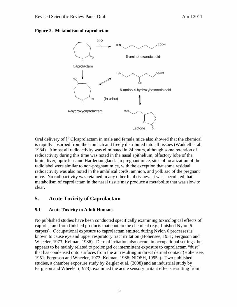
Measurable levels of caprolactam have been found primarily in indoor air as a result of release of the vapor or particulate from carpeting containing Nylon-6 (IWMB, 2003). The polymerization process of caprolactam to nylon polymer may not be 100 percent efficient, thus allowing some of the un-polymerized caprolactam into the final product. Goldblatt et al. (1954) noted that the polymerized fiber contains approximately one percent of the unreacted caprolactam monomer. A more recent study suggests that total caprolactam contaminants, plus lesser amounts of its various oligomers, are present at 1% or less in some Nylon-6 products (Venema et al., 1993). These oligomers found following polymerization include cyclic oligomers (i.e., the cyclic dimer, trimer and tetramer, etc.) as well as some linear oligomers (Krajnik et al., 1982; Ballistreri et al., 1987; Bonifaci et al., 1991; Venema et al., 1993). Based on the measured emission rate of caprolactam from carpet samples, modeled air concentrations for office and classroom scenarios ranged from 39 to 450 µg/m3 (IWMB, 2003). A chamber study found caprolactam emissions from some polyamide carpets resulted in chamber concentrations ranging from 6 to 97 µg/m3 on the 28th day of chamber testing (Wilke et al., 2004). In indoor monitoring studies, caprolactam was detected in all floor dust samples collected during an indoor air study in nine public buildings (Wilkins et al., 1993). In another study, the average caprolactam concentration in a new California relocatable classroom during school hours over 8 weeks following installation of a Nylon-6 broadloom carpet was 22.2 µg/m3 (range: 10.6 - 30.1 µg/m3) (Hodgson et al., 2004). The emission rate of caprolactam following installation of the carpet was about 5 mg/h prior to occupancy, and dropped to 3 mg/h 27 weeks after first occupancy. Similar relocatable classrooms that installed upgraded carpets containing Nylon-6,6 emitted low to non-detectable concentrations of caprolactam (maximum: 1.4 µg/m3). 4. Metabolism In rats, Kirk et al. (1987) observed that approximately 16% of ingested caprolactam in diet was excreted in urine as 4-hydroxycaprolactam and a small amount as the non-hydroxylated acid, 6-aminohexanoic acid (Figure 2). Following a single oral dose of [14C]caprolactam in male rats, 77.6% of the radioactivity was excreted in urine, 3.5% in the feces, and 1.5% in the expired air in 24 hrs (Unger et al., 1981). The half-life of disappearance of radioactivity from the blood was 2.98 hr. Similar to the findings by Kirk et al., Unger et al. (1981) observed two metabolites, which comprised 79.3 and 17.7% of the total urinary radioactivity. However, Unger et al. made no attempt to identify these urinary metabolites. Unchanged caprolactam represented only 2.3% of the total urinary radioactivity. The radiolabeled caprolactam was widely distributed among the tissues of the rat with concentrations mostly similar to that in the blood. The radioactivity was consistently lower in fat relative to the blood in the first 24 hrs, indicating a low affinity of caprolactam and its metabolites for adipose tissue.
Revised Scientific Review Panel Draft April 2011
5
Figure 2. Metabolism of caprolactam
Oral delivery of [14C]caprolactam in male and female mice also showed that the chemical is rapidly absorbed from the stomach and freely distributed into all tissues (Waddell et al., 1984). Almost all radioactivity was eliminated in 24 hours, although some retention of radioactivity during this time was noted in the nasal epithelium, olfactory lobe of the brain, liver, optic lens and Harderian gland. In pregnant mice, sites of localization of the radiolabel were similar to non-pregnant mice, with the exception that some residual radioactivity was also noted in the umbilical cords, amnion, and yolk sac of the pregnant mice. No radioactivity was retained in any other fetal tissues. It was speculated that metabolism of caprolactam in the nasal tissue may produce a metabolite that was slow to clear. 5. Acute Toxicity of Caprolactam 5.1 Acute Toxicity to Adult Humans No published studies have been conducted specifically examining toxicological effects of caprolactam from finished products that contain the chemical (e.g., finished Nylon 6 carpets). Occupational exposure to caprolactam emitted during Nylon 6 processes is known to cause eye and upper respiratory tract irritation (Hohensee, 1951; Ferguson and Wheeler, 1973; Kelman, 1986). Dermal irritation also occurs in occupational settings, but appears to be mainly related to prolonged or intermittent exposure to caprolactam “dust” that has condensed onto surfaces from the air resulting in direct dermal contact (Hohensee, 1951; Ferguson and Wheeler, 1973; Kelman, 1986; NIOSH, 1995a). Two published studies, a chamber exposure study by Zeigler et al. (2008) and an industrial study by Ferguson and Wheeler (1973), examined the acute sensory irritant effects resulting from
Revised Scientific Review Panel Draft April 2011
6
directly measured concentrations of caprolactam. These two studies are summarized and assessed for REL development below. The caprolactam occupational exposure standards are also summarized in this section. Because caprolactam is a respiratory irritant, the available data for the potential of caprolactam acting as a sensitizing agent for allergic asthma is also discussed. Ferguson and Wheeler (1973) Ferguson and Wheeler (1973) exposed 5 healthy unacclimated male workers at a caprolactam polymer plant to caprolactam vapor concentrations of 10, 14, 25, and 104 ppm (46, 65, 116, and 482 mg/m3, respectively) while they were standing or conversing for several minutes downwind from a known emission source. The smoking status of the workers was not reported. ‘Unacclimated’ was defined as workers who were experienced in the work environment, but were not continuously exposed in their ordinary duties. Although the authors report exposure was to the “vapor” form of caprolactam, the concentrations were above the saturated vapor concentration of 13 mg/m3 indicating that exposure was probably mostly to caprolactam aerosol. Air sampling for the exposures was set at a height of about 60 inches from the floor and was averaged over a minimum of 30 minutes (Ferguson and Wheeler, 1973). The instrumentation for these exposures was presumably the same used for estimating 8-hour time weighted average concentrations in the second part of the study: a liquid absorption train that consisted of three flasks in series was set up to collect the caprolactam in measured volumes of air. The flasks were half-filled with water and connected with one another by fritted glass delivery tubes. The train was connected to a fixed vacuum supply through a wet test meter. A similar arrangement was also employed that used gas traps rather than flasks, and a portable vacuum pump was used as a vacuum source. Two-mm glass beads were used in the first trap as a sparger. Both techniques were considered effective, as no caprolactam was found in the last trap in either train. This air sampling technique was a commonly used method in the 1970’s, but some particulates could have been missed due to the use of fritted glass (Gill, 2011). Most or all of the workers in the Ferguson and Wheeler study reported transient nasal and throat irritation at all concentrations, including 4 out of 5 exposed individuals exposed to 10 ppm (46 mg/m3). Eye irritation was noted only in one volunteer at the highest concentration. The authors did not indicate if throat irritation was a result of mouth-breathing (due to the unpleasant odor and/or nasal irritation) by the volunteers. The degree of discomfort felt by the workers was considered dose-responsive, but was not quantified due to wide differences in the degree of discomfort between individual subjects. The scalar functions used in an attempt to evaluate degrees of discomfort were not presented. Some of the volunteers were exposed to similar concentrations for up to 30 min, but the sensory effects were not clearly stated or quantified. Brief exposure to 400-1200 ppm caprolactam was described as extremely irritating, resulting in a “choking” response.
Revised Scientific Review Panel Draft April 2011
7
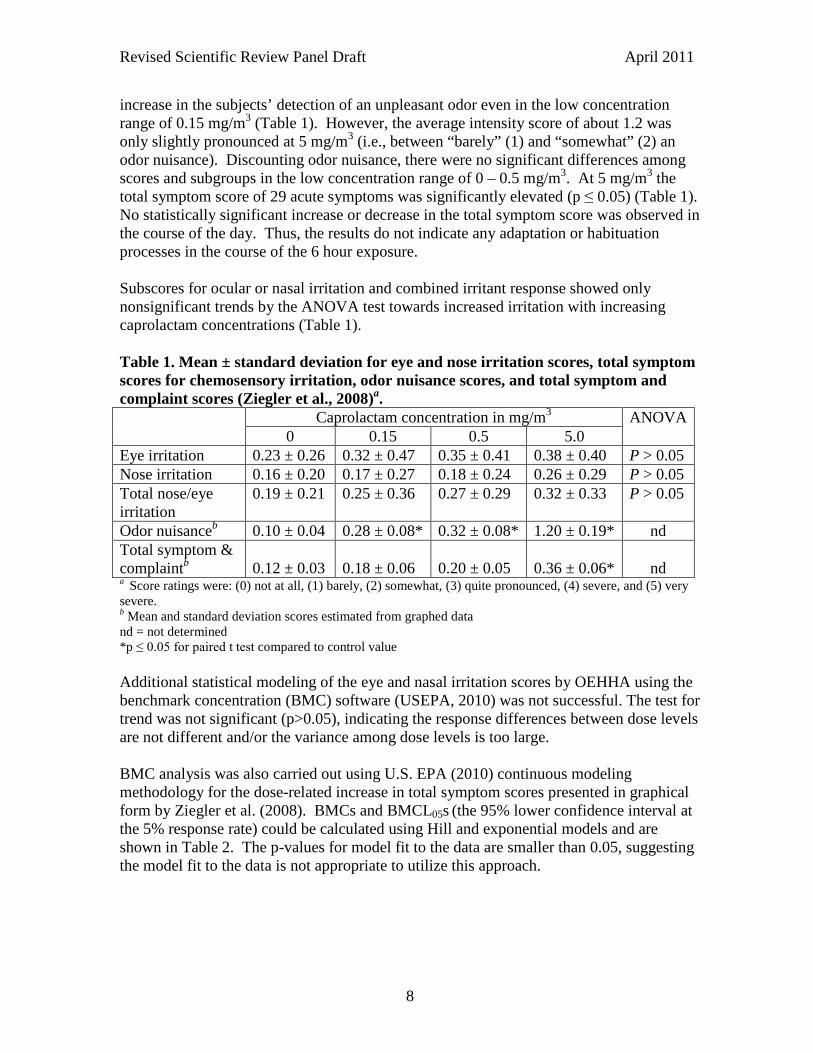
A similar acute exposure study was arranged at a caprolactam monomer plant although specific conditions of the exposure were not presented by the authors (Ferguson and Wheeler, 1973). In this part of the study, 14 ppm (65 mg/m3) did not result in distress or discomfort. The authors speculated that the conditions of 100% humidity at the monomer plant (the polymer plant was described as having near normal humidity) may have been a factor in reducing sensory irritation. They also noted that concentrations at the monomer plant appeared to be more uniform, suggesting that the greater variability in the concentration at the polymer plant resulted in brief high exposures leading to sensory irritation. However, the authors did not present quantitative data to show the variation in caprolactam concentration during the acute exposures. The authors concluded that the irritant response threshold for the workers is at or near 10 ppm (46 mg/m3) caprolactam, and that 5 ppm (23 mg/m3) is 50% of the "discomfort" threshold and "somewhat below the no-effect level". The authors state that additional support for their worker threshold value of 5 ppm is based on no reported distress in active and semi-active areas at concentrations up to about 7 ppm (discussed in Section 6.1). Ziegler et al. (2008) To address possible chemosensory effects of caprolactam at low concentrations reflecting the indoor environment, Ziegler et al. (2008) conducted chamber exposures of 20 adult subjects (10 men and 10 women) to 0, 0.15, 0.5, and 5 mg/m3 caprolactam vapor for 6 hours on 4 successive days. Chemosensory effects were assessed by questionnaire, by measures of blinking frequency and assessment of conjunctival hyperemia based on digital slit lamp photographs taken during exposure, and by measures of nasal resistance using active anterior rhinometry before and after exposures. They observed nonsignificant trends towards higher blink frequencies and increased nasal resistance with increasing caprolactam concentrations, but no evidence of change in eye redness. Questionnaires were used to evaluate 29 symptoms and to generate a total daily score. Six adjectives were presented for rating the intensity of symptoms from zero (not at all) to 5 (very severe). The 29 symptoms were also placed into seven subscores and evaluated. The subscores and symptoms include: 1) non-specific symptoms: feeling of weakness, headache, dizziness, felling of being unwell; 2) not classified: blurred vision, irritation to the throat, skin irritation; 3) sensations of bad taste: very unpleasant taste in the mouth, unpleasant taste, foul taste; 4) respiratory symptoms: pressure on the chest, coughing, dyspnea; 5) olfactory symptoms: perception of bad air, foul smell, unpleasant smell, stink; 6) nasal irritation: nasal irritation, itching nose, dry nose, runny nose, burning nose; 7) irritation to the eyes: tiredness of the eyes, itchy eyes, burning eyes, eye irritation, dry eyes, watery eyes, redness of the eyes. A second section evaluating well-being (i.e., tension, tiredness, annoyance and general well-being) was rated on a visual analog scale from one (no symptoms) to seven (severe symptoms). The questionnaires were administered before exposure and after 1, 3 and 6 hours of exposure. Caprolactam exposure was also associated with a statistically significant
Revised Scientific Review Panel Draft April 2011
8
increase in the subjects’ detection of an unpleasant odor even in the low concentration range of 0.15 mg/m3 (Table 1). However, the average intensity score of about 1.2 was only slightly pronounced at 5 mg/m3 (i.e., between “barely” (1) and “somewhat” (2) an odor nuisance). Discounting odor nuisance, there were no significant differences among scores and subgroups in the low concentration range of 0 – 0.5 mg/m3. At 5 mg/m3 the total symptom score of 29 acute symptoms was significantly elevated (p ≤ 0.05) (Table 1). No statistically significant increase or decrease in the total symptom score was observed in the course of the day. Thus, the results do not indicate any adaptation or habituation processes in the course of the 6 hour exposure. Subscores for ocular or nasal irritation and combined irritant response showed only nonsignificant trends by the ANOVA test towards increased irritation with increasing caprolactam concentrations (Table 1). Table 1. Mean ± standard deviation for eye and nose irritation scores, total symptom scores for chemosensory irritation, odor nuisance scores, and total symptom and complaint scores (Ziegler et al., 2008)a. Caprolactam concentration in mg/m3 ANOVA
0 0.15 0.5 5.0 Eye irritation 0.23 ± 0.26 0.32 ± 0.47 0.35 ± 0.41 0.38 ± 0.40 P > 0.05 Nose irritation 0.16 ± 0.20 0.17 ± 0.27 0.18 ± 0.24 0.26 ± 0.29 P > 0.05 Total nose/eye irritation
0.19 ± 0.21 0.25 ± 0.36 0.27 ± 0.29 0.32 ± 0.33 P > 0.05
Odor nuisanceb 0.10 ± 0.04 0.28 ± 0.08* 0.32 ± 0.08* 1.20 ± 0.19* nd Total symptom & complaintb
0.12 ± 0.03
0.18 ± 0.06
0.20 ± 0.05
0.36 ± 0.06*
nd
a Score ratings were: (0) not at all, (1) barely, (2) somewhat, (3) quite pronounced, (4) severe, and (5) very severe. b Mean and standard deviation scores estimated from graphed data nd = not determined *p ≤ 0.05 for paired t test compared to control value Additional statistical modeling of the eye and nasal irritation scores by OEHHA using the benchmark concentration (BMC) software (USEPA, 2010) was not successful. The test for trend was not significant (p>0.05), indicating the response differences between dose levels are not different and/or the variance among dose levels is too large. BMC analysis was also carried out using U.S. EPA (2010) continuous modeling methodology for the dose-related increase in total symptom scores presented in graphical form by Ziegler et al. (2008). BMCs and BMCL05s (the 95% lower confidence interval at the 5% response rate) could be calculated using Hill and exponential models and are shown in Table 2. The p-values for model fit to the data are smaller than 0.05, suggesting the model fit to the data is not appropriate to utilize this approach.

Revised Scientific Review Panel Draft April 2011
9
Table 2. BMC continuous model results for total symptom and complaint score at 1 hour of exposure to caprolactam. Model
BMC (mg/m3)
BMCL05 (mg/m3)
P-value
Hill 0.31 0.18 0.037 Exponential 0.35 0.24 0.025 Other statistical analyses were attempted on the trend data using a ranking procedure using the Friedman test. Based on the information provided in the Ziegler et al. study, five relevant outcomes were chosen from the study: blink frequency, eye redness, nasal resistance, reported irritation to the eyes, and reported irritation to the nose. From tables and figures in the study, median average blink frequency values, mean eye redness at the highest exposure duration, mean difference in total nasal resistance before and after exposure, and median eye and nose irritation scores were selected. For each outcome, the responses at each exposure level were ranked from 1 to 4 (Table 3). Ties were assigned the average of the two possible ranks. To this matrix of rankings, a Friedman test was applied to ascertain whether non-random differences in rankings existed over the concentration gradient. Significant differences in rankings by concentration were found (p = .02), and a subsequent application of Page’s Trend Test found an association between increasing exposure and the measure rankings (L=144.5, p < .01). Table 3. Rank order of response by dose among five outcome measures Study Measure Exposure Concentration (mg/m3) 0 0.15 0.5 5.0 Blink frequency median
2 3 1 4
Redness at max time (360 min)
1.5 3 1.5 4
Nasal resistance 1 2 3 4 Eye sx score median
1 2 3.5 3.5
Nasal sx score median
1 2 3 4
There are some concerns about using the Friedman test in this context. The test is a non-parametric test that typically uses individual data, not summary statistics, as was done here, to assign rankings. Applying rankings to summary statistics ignores the distribution and variance. Because the rankings were not created using the individual values, parametric assumptions are still needed because a single summary statistic is being used to represent a distribution for a specific exposure-outcome combination. This would be more problematic when using the means supplied in the paper, since many of these measures generate skewed distributions for which a mean would be a poor representative. Indeed, the Ziegler paper admits needing to use non-parametric tests to correct for the non-normal distributions that were present in these data.
Revised Scientific Review Panel Draft April 2011
10
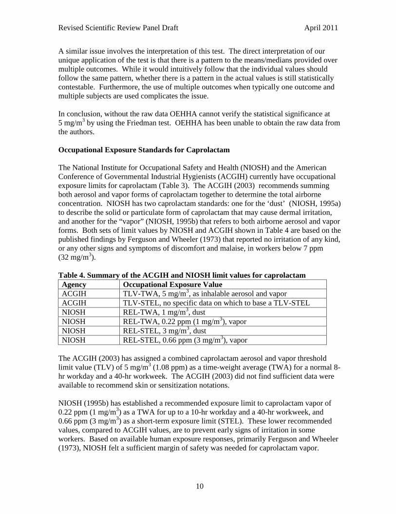
A similar issue involves the interpretation of this test. The direct interpretation of our unique application of the test is that there is a pattern to the means/medians provided over multiple outcomes. While it would intuitively follow that the individual values should follow the same pattern, whether there is a pattern in the actual values is still statistically contestable. Furthermore, the use of multiple outcomes when typically one outcome and multiple subjects are used complicates the issue. In conclusion, without the raw data OEHHA cannot verify the statistical significance at 5 mg/m3 by using the Friedman test. OEHHA has been unable to obtain the raw data from the authors. Occupational Exposure Standards for Caprolactam The National Institute for Occupational Safety and Health (NIOSH) and the American Conference of Governmental Industrial Hygienists (ACGIH) currently have occupational exposure limits for caprolactam (Table 3). The ACGIH (2003) recommends summing both aerosol and vapor forms of caprolactam together to determine the total airborne concentration. NIOSH has two caprolactam standards: one for the ‘dust’ (NIOSH, 1995a) to describe the solid or particulate form of caprolactam that may cause dermal irritation, and another for the “vapor” (NIOSH, 1995b) that refers to both airborne aerosol and vapor forms. Both sets of limit values by NIOSH and ACGIH shown in Table 4 are based on the published findings by Ferguson and Wheeler (1973) that reported no irritation of any kind, or any other signs and symptoms of discomfort and malaise, in workers below 7 ppm (32 mg/m3). Table 4. Summary of the ACGIH and NIOSH limit values for caprolactam Agency Occupational Exposure Value ACGIH TLV-TWA, 5 mg/m3, as inhalable aerosol and vapor ACGIH TLV-STEL, no specific data on which to base a TLV-STEL NIOSH REL-TWA, 1 mg/m3, dust NIOSH REL-TWA, 0.22 ppm (1 mg/m3), vapor NIOSH REL-STEL, 3 mg/m3, dust NIOSH REL-STEL, 0.66 ppm (3 mg/m3), vapor
The ACGIH (2003) has assigned a combined caprolactam aerosol and vapor threshold limit value (TLV) of 5 mg/m3 (1.08 ppm) as a time-weight average (TWA) for a normal 8-hr workday and a 40-hr workweek. The ACGIH (2003) did not find sufficient data were available to recommend skin or sensitization notations. NIOSH (1995b) has established a recommended exposure limit to caprolactam vapor of 0.22 ppm (1 mg/m3) as a TWA for up to a 10-hr workday and a 40-hr workweek, and 0.66 ppm (3 mg/m3) as a short-term exposure limit (STEL). These lower recommended values, compared to ACGIH values, are to prevent early signs of irritation in some workers. Based on available human exposure responses, primarily Ferguson and Wheeler (1973), NIOSH felt a sufficient margin of safety was needed for caprolactam vapor.
Revised Scientific Review Panel Draft April 2011
11
For caprolactam dust, NIOSH (1995a) assigned a TWA value of 1 mg/m3, and a STEL of 3 mg/m3. These exposure levels appear to be largely based on an unpublished letter to the TLV committee in 1972 when occupational limits were being determined for caprolactam (Ferguson and Wheeler, 1973; ACGIH, 2001). In this letter, airborne caprolactam dust was reported to be irritating to the skin of some individuals at 5 mg/m3, but provide adequate protection at 1 mg/m3 particularly when combined with respirator use. From this description, it can probably be inferred that the caprolactam dust limits also lower the deposition of the dust onto surfaces that workers come in direct contact, perhaps not unlike occupational exposure conditions described by Kelman (1986) in Section 6.1 below. The Occupational Safety and Health Administration (OSHA) has not promulgated a permissible exposure limit (PEL) for either caprolactam vapor or caprolactam dust. However, Cal/OSHA lists a PEL of 1 mg/m3 and STEL of 3 mg/m3 for caprolactam dust (Cal/OSHA, 2011). For the vapor form, Cal/OSHA lists a PEL of 5 ppm (20 mg/m3) and a STEL of 10 ppm (40 mg/m3). These occupational standards were likely adopted from earlier ACGIH (2001) standards for caprolactam before they were revised by the ACGIH in 2003. The ACGIH (2001) originally had higher exposure limits that mirrored Ferguson and Wheeler’s conclusion that a worker threshold value of 5 ppm (23 mg/m3) is recommended because no distress was reported in active and semi-active areas at concentrations up to about 7 ppm. However, subsequent reduction of the ACGIH (2003) exposure limit to well below this worker threshold value suggests a lack of confidence in the reported threshold findings by Ferguson and Wheeler (1973). Human Sensitization Studies and Case Reports A review by Gross (1984) of the eastern European occupational studies conducted in the 1960s and 1970s suggests a significant number of workers may develop hypersensitivity to caprolactam. However, the methodology was not adequately described in these studies and there was co-exposure to other chemicals. Acute exposure studies conducted in the West, some of which were unpublished, indicated caprolactam did not cause skin irritation or was a mild irritant, although there is one case report described below of neurological effects and dermatitis resulting from short-term exposure. Short-term studies also suggest that it does not act as a sensitizing agent. A few case reports of apparent hypersensitivity resulting from prolonged exposure to caprolactam are described in Section 6.1. Goldblatt et al. (1954) applied a 5% aqueous caprolactam solution to the skin of the inner forearms of six normal persons (4 men, 2 women) as a patch test left in contact for 48 hours. Goldblatt et al. also applied a 5% caprolactam solution in either alcohol or olive oil to the same area on volunteers and allowed to dry. The process was repeated daily for four days. In all cases, no irritant effects were produced. The authors concluded with these short-term exposures that caprolactam is not a skin irritant and no evidence was found that it could act as a sensitizing or dermatitis agent.
Revised Scientific Review Panel Draft April 2011
12
In an unpublished study carried out in Haskell Laboratory in 1941, three human volunteers had a 1% aqueous solution of caprolactam applied to the skin (Haskell Laboratory, 1950). No skin irritation was produced. No other methods or descriptive information was provided. In an unpublished study conducted in 1952-53 and recently reported to the U.S. EPA (2009), a patch test was conducted in 204 human subjects to determine whether or not Nylon-6 containing 3-5% water-extractable caprolactam and dimers would produce primary skin irritation and/or sensitization in occupational exposures. No primary irritation or allergic sensitization was observed in the tested subjects. No other methods or descriptive information was provided. In a case report, a 22-year-old man developed dermatitis of the hands and feet, fever and grand mal seizures three days after being transferred to a section of a plastics plant that involved caprolactam use (Tuma et al., 1981). The caprolactam dust coated his clothing and exposed areas of skin when he arrived at the hospital. The symptoms cleared after a few days of observation while the skin lesions showed desquamation (peeling) and erythema (redness and swelling). A comprehensive neurological investigation showed no organic CNS abnormalities, suggesting the seizures were a result of caprolactam exposure. The authors did not indicate any respiratory distress was present, and a chest roentgenogram was normal. This case report by Tuma et al. (1981) indicates some serious implications for acute exposure to caprolactam. However, this is the only report that could be located indicating seizures in humans due to caprolactam exposure. In addition, exposure may have occurred by routes other than inhalation. This patient appears to have been heavily exposed to caprolactam, given that his clothes and skin were covered with caprolactam dust. Studies in rabbits indicate concentrated caprolactam (50% aqueous caprolactam solution) placed on the skin can be absorbed (Haskell Laboratory, 1950). However, it appears likely that in both the rabbit report and in the Tuma et al. case report that the concentrated caprolactam damaged the skin, which then facilitated dermal absorption of caprolactam. Considering that large, often fatal, bolus doses by gavage were needed in animal studies to induce convulsions (see Section 5.3 below) OEHHA speculates that the patient may have consumed caprolactam, perhaps resulting in his illness and seizures. The potential for this patient being hypersensitive to chemical exposure was also not explored. Finally, co-exposure with other chemicals during, or just before, the man was transferred to the area of the plant using caprolactam was not investigated. 5.2 Acute Toxicity to Infants and Children No studies were located regarding acute toxicity to infants and children exposed to caprolactam. We found no studies of inhalation exposure to young or pregnant animals that could shed insight into acute toxicity in infants and children.
Revised Scientific Review Panel Draft April 2011
13
5.3 Acute Toxicity to Experimental Animals Relatively few peer-reviewed studies of acute caprolactam exposure in experimental animals have been conducted. Acute inhalation, oral and parental exposure studies are summarized below, including some non-peer-reviewed studies, to provide the full spectrum of effects resulting from acute intoxication from caprolactam exposure. Due to caprolactam’s respiratory irritant action, dermal and inhalation sensitization studies are also reviewed. All sensitization studies were not peer-reviewed. The BASF Chemical Company conducted unpublished acute exposure studies in the 1960s and 70s that were reported by Ritz et al. (2002). In the rat, an oral LD50 of 1155 mg/kg is reported. Symptoms of acute intoxication were tonoclonic convulsions. Rabbits and cats are said to be more sensitive to caprolactam, but no data was provided. In an acute toxicity study on rats and mice, the NTP (1982) administered caprolactam in corn oil by gavage to groups of five males and five females. The LD50 for male and female mice were 2070 and 2490 mg/kg, respectively. The LD50 for male and female rats were 1650 and 1210 mg/kg, respectively. No signs or symptoms of toxicity were discussed. Goldblatt et al. (1954) observed 66% mortality in rats injected intraperitoneally with 800 mg/kg caprolactam with the appearance of delayed spasms. Lower non-fatal doses (500-600 mg/kg) resulted in tremors, apprehension, depression of temperature, and occasional chromodacryorrhea. Concentrations of 900 mg/kg and above proved fatal and resulted in epileptiform convulsions, salivation, and bleeding from the nares. Goldblatt et al. (1954) also injected rabbits intravenously with non-fatal doses of caprolactam ranging from 100 to 300 mg/kg. The effects were intense, but transient, and included apprehension, salivation, accelerated respiration, tremors, convulsions, opisthotonic-type muscle contractions, and mydriasis (dilatation of the pupil of the eye). Similar results were observed in the foreign toxicology literature (published mainly in Russian and German) of the 1950s and 1960s and reported in a review by Gross (1984). Caprolactam LD50 studies in experimental animals and exposure to high doses of caprolactam by intravenous and intraperitoneal injection produced tremors, epileptiform convulsions, salivation and bleeding from the nostrils. In an unpublished study by Haskell Laboratory (1950), an approximate lethal dose of 3375 mg/kg was observed in rats administered by gavage. Rats receiving 1500 mg/kg developed convulsions and some showed slight bleeding from the nose and mouth. In an unpublished industrial study, four-hour exposure of rats to 5,250, 8,350, or 10,120 mg/m3 caprolactam aerosol via a head-nose inhalation system resulted in eyelid closure, shallow to spasmodic breathing, and mild to strong defense reactions (BASF, 1985). After exposure, steppage gait, bloody nasal secretions, spasmodic breathing, marked tremor, and bloody lacrimation were observed. LC50s of 9,600 and 7,080 mg/m3 were recorded for male and female rats, respectively. In rats that died, general circulatory congestion, elevated hyperemia of the lung, moderate to severe fatty degeneration of the liver, and ischemic tubular nephrosis in the kidney cortex were found. No additional
Revised Scientific Review Panel Draft April 2011
14
deaths occurred after one day post-exposure and all surviving animals appeared normal 3 days post-exposure. Histopathological examination of the organs in surviving rats 14 days post-exposure was described as “unremarkable”. In another unpublished study, two rats exposed to a nominal concentration of 14,000 ppm caprolactam for 17 min showed signs of general discomfort and inflammation around the eyes and nose (Haskell Laboratory, 1950). No gross pathology or micropathology was detected at sacrifice following a nine-day observation period. The U. S. Consumer Product Safety Commission contracted a study of sensory and pulmonary irritation in Swiss-Webster mice exposed to compounds emitted from carpet and carpet-related products, including caprolactam (CPSC, 1996). The animals were placed in a head-only glass plethysmograph and exposed to 13.5 mg/m3 caprolactam vapor, the highest attainable exposure concentration. The methodology called for a one hour exposure, followed by a recovery period of 15 minutes in clean air, then exposure to the same concentration of caprolactam for another hour. Sensory irritation is indicated when the group of mice showed a 12% or greater decrease in the mean respiratory frequency, the minimum level of respiratory depression needed to classify an exposure as having a positive sensory irritation response (CPSC, 1996). By this approach, no measurable sensory irritation or reduction in respiratory rate was observed in the mice during the caprolactam exposure. However, the CPSC (1996) notes that measurable respiratory irritation in mice using this method usually occurs at levels 10 to 100 times higher than levels which would result in irritation in humans. Inhalation and Dermal Sensitization/Irritation Studies In a skin absorption study, a 50% aqueous solution of caprolactam was applied to a shaved area between the shoulder blades of rabbits (Haskell Laboratory, 1950). The approximate lethal dose was 3375 mg/kg producing pathology similar to shock. Clinical observations included tremors, convulsions, and bleeding from the mouth and nose analogous to those observed in rats receiving oral doses. Edema and congestion of the skin at the site of application was noted, which may have increased dermal absorption as a result of skin damage. Gross (1984) reviewed the eastern European literature conducted in the 1970s concerning dermal sensitization studies in animals. It was claimed in these studies that both intracutaneous and dermal application of caprolactam in guinea pigs resulted in sensitization. In one of two cases, it was claimed guinea pigs became sensitized to caprolactam by inhalation. However, other studies described below could not reproduce assertions of inhalation sensitization, and dermal studies characterized caprolactam, at best, to be a mild sensitizer. In a non-peer-reviewed study, groups of four male albino guinea pigs were exposed for 30 min on 5 consecutive days to 3, 10, or 30 mg/m3 aerosols (1.5 micron) generated from a 15% aqueous caprolactam solution (Rinehart et al., 1997; USEPA, 1998). On day 19, 26,
Revised Scientific Review Panel Draft April 2011
15
33 and 40, animals were challenged for 30 min with 30 mg/m3 caprolactam. Animals were monitored with whole-body plethysmography for indications of irritation and coughing, and pulmonary hypersensitivity was monitored using respiratory frequency, tidal volume, and airway constriction as criteria for effect. Caprolactam failed to induce immediate or delayed pulmonary hypersensitivity with this protocol, which has been positive for ovalbumin and trimellitic anhydride. In addition, there was no evidence of respiratory tract irritation at any exposure concentration. In unpublished results carried out by the BASF Chemical Company, guinea pigs were exposed to repeated epicutaneous application (50% ether solution; 10 times) or intracutaneous injection (0.1% in physiological NaCl solution) (Ritz et al., 2002). Neither treatment caused local irritation or sensitization to the skin. In an unpublished study carried out in 1941, a skin irritation test with a 66% aqueous solution of caprolactam was carried out in 10 albino guinea pigs (Haskell Laboratory, 1950). Initial application of the aqueous caprolactam solution to unbroken shaved skin resulted in erythema in one animal, faint erythema in two other animals, and negative results in the remaining 7 animals. The researchers concluded caprolactam produced only mild dermal irritation in the guinea pigs. To further test for sensitization, a maximization test was conducted that consisted of a series of 6 treatments of a 66% aqueous solution of caprolactam to broken skin, or 6 intradermal injections of 0.1 ml of a 0.1% aqueous solution (Haskell Laboratory, 1950). This was followed by a rest period of two weeks, and then the 66% aqueous caprolactam solution was again applied to the unbroken skin at the same site as the original application. Seven of ten animals now had dermal reactions indicating that sensitization had occurred. A final intradermal injection and application to broken skin likewise showed an increase in intensity of the reaction consistent with sensitization. Although the sensitization potential is limited by using an irritant concentration for challenge treatment, the researchers considered caprolactam should be considered a mild sensitizer on the basis of the strength of the reaction. In a similar (unpublished) maximization test protocol, 20 female guinea pigs received intradermal application of caprolactam (3.0% w/v) in water, or topical application of caprolactam (75% w/v) in water (Springborn Laboratories Inc., 1991). Challenge responses in the induced animals were compared to those of the controls. Blood samples were obtained prior to study initiation and following the challenge for evaluation of standard hematology parameters. Additionally, plasma histamine was determined for selected test and control animals following challenge. Based on the concurrent mild dermal reaction in the control group animals and the fading of reactions from 24 to 48 hours, caprolactam was not considered to be a contact sensitizer. In a Buehler test in rabbits, 10 animals each were used in the challenge control and the rechallenge control groups (Springborn Laboratories Inc., 1991). Induction test animals were patched with 25% w/v caprolactam in water 3 times within 3 weeks. In the challenge phase, the test group animals received 25% w/v caprolactam in water for
Revised Scientific Review Panel Draft April 2011
16
injection in a patch. Dermal reaction was scored 24 and 48 hours after removal of the patch. Minimal dermal reaction was observed in both the test animals and negative control animals after the challenge as well as after the rechallenge. Mean dermal scores were also comparable between both groups. The skin sensitization potential of caprolactam was limited by using an irritant concentration for the challenge treatment. Therefore, caprolactam was not considered to be a contact sensitizer under the test conditions chosen. In an non-peer-reviewed dermal sensitization test by Rinehart et al. (1997), groups of 20 female albino guinea pigs were tested with 25% aqueous caprolactam solution using either the traditional modified Buehler or maximization test designs. Groups of 5 guinea pigs were treated with 5% DNCB (probably 1-Chloro-2,4-Dinitrobenzene) as a positive control. After the second challenge dose had been evaluated, blood samples were obtained for measurements of leukocytes, differential counts and plasma histamine levels. Neither test regimen showed positive results for animals treated with caprolactam. There were no body weight changes or any effects on hematologic components or plasma histamine levels caused by treatment with caprolactam. In summary, the acute animal data shows that large bolus doses of caprolactam given orally by gavage (hundreds of mg/kg), or injected intravenously or intraperitoneally, can result in convulsions. However, this severe neurological finding was not found in LC50 studies, in which lethal or near-lethal airborne concentrations of caprolactam result in severe tremors, but not convulsions. In addition, feeding studies in which caprolactam is mixed with food at maximum levels (see Section 6.2 below) does not appear to attain a high enough absorbed dose to lead to convulsions or tremors. Dermal and inhalation sensitization test were generally negative. A mild dermal sensitization may occur, but at concentrations that result in dermal damage in control animals. A concern with the overall acute data is that most of these reports were never peer-reviewed or published. 6. Chronic Toxicity of Caprolactam 6.1 Chronic Toxicity to Adult Humans Occupational exposure to caprolactam is known to cause dermal, eye and upper respiratory tract irritation with acute or recurrent acute exposure, but data on chronic toxicity endpoints resulting from prolonged caprolactam exposure in workers were considered by OEHHA to be inadequate for use as the basis of a chronic REL. Gross (1984) summarized the early foreign literature regarding industrial exposure to caprolactam. With a few exceptions, the pertinent publications were Russian. These reports describe diverse complaints and abnormalities of the various organ systems in people exposed in factories producing nylon. The exposures in no instance were only to caprolactam. Exposure to caprolactam was commonly associated with exposure to dinyl oxides, such as diphenyl oxide. Other chemicals often associated with caprolactam exposures were cyclohexane, cyclohexanol, cyclohexanone, benzene, acetone, and trichloroethylene.
Revised Scientific Review Panel Draft April 2011
17
End of shift complaints by workers exposed to caprolactam at a factory included irritability, nervousness, heartburn, bloating, nose bleeds, upper airway inflammation, and dry and chapped lips and noses (Hohensee, 1951). Exposure included both the vapor and crystal, or dust, forms of caprolactam. Headache in response to the odor and unpleasant taste of the caprolactam vapor was also reported. All these symptoms subsided after a short (but unspecified duration) stay in fresh air. Factory inspection of the caprolactam concentration in the spinning room revealed a concentration of 61 mg/m3, while the concentration in the laboratory room was 16-17 mg/m3. Although Ferguson and Wheeler (1973) were primarily focused on acute effects of airborne caprolactam exposure, the researchers also took 8-hr time-weighted average (TWA) measurements at two facilities and reviewed medical records. Other than dermal injuries resulting from direct contact to concentrated caprolactam solutions, no general health problems requiring medical follow-up were found in a review of medical records collected during the 18 years of plant operation. In addition, no worker had been removed or asked to be removed from exposure to caprolactam vapor for health reasons during plant operation. At the caprolactam polymer plant, approximate 8-hr time-weighted average (TWA) air samples were collected from various locations in a work area over five days (Ferguson and Wheeler, 1973). The 8-hr TWA air concentrations of caprolactam vapor during working hours were 3.2 ppm (14.8 mg/m3) with a range of 1.3 to 6.9 ppm (6.0 to 31.9 mg/m3) at location 1, and 1.1 ppm with a range of <0.5 to 4.5 ppm (<2.3 to 20.8 mg/m3) at location 2. Based on the percent time worked in specific locations of the caprolactam-contaminated rooms, the worker exposure durations were estimated to be about 15 to 45 min at location 1, and 1 to 4 hrs at location 2. At the caprolactam monomer plant, 8-hr TWA caprolactam vapor concentrations at various sites over a 3-week period were collected. The concentration of caprolactam sampled at various worksite locations ranged from 0.2 to 17.6 ppm (0.9 to 81.5 mg/m3). Worker exposure durations in the caprolactam-contaminated areas ranged from 10 min to almost 3 hrs. From the 8-hr TWA data collected, Ferguson and Wheeler (1973) concluded that working atmospheric concentrations up to about 7 ppm (32 mg/m3) at the caprolactam polymer plant generally resulted in no reported distress of interviewed workers in active and semi-active areas. This data supported their estimate of a worker irritant response threshold of 5 ppm (23 mg/m3) based on the acute exposure portion of their study. There are significant deficiencies in the Ferguson and Wheeler report that prevent it from use as the basis of an OEHHA chronic REL. As also noted by the U.S. EPA RfD/RfC Work Group, significant deficiencies included lack of information on the number of workers and the average duration and distribution of exposure (USEPA, 1998). Also, no historical air levels are given, all exposures are determined from area rather than personal samplers, and no attempt was made to reconstruct individual exposure histories.
Revised Scientific Review Panel Draft April 2011
18
Kelman (1986) conducted a clinical and occupational history of eight workers, seven of which were smokers, at a Nylon-6 manufacturing plant. Exposure was described as caprolactam vapor from heat-curing ovens, which subsequently condensed into a fume in the workplace air. Contact of the fume with cooler surfaces resulted in the formation of light feathery flakes. Average worker exposure was 4.8 years (range 9 months to 13 years) and mean atmospheric caprolactam dust concentrations at the time of the study were 84 mg/m3 (range: 22-168 mg/m3) for static samplers and 68 mg/m3 (range: 6-131 mg/m3) for personal samplers. The caprolactam dose and exposure durations for individual workers were not provided. Recovery of caprolactam vapor from distilled-water bubblers was considered negligible, which the authors interpreted as indicating exposure was limited to caprolactam dust. Kelman (1986) reported that several of the workers (number not given) complained of “some degree” of eye, nose, and throat irritation. It was unclear from the study if the irritation was chronic in nature. All but one reported peeling of the skin on the hands. Five workers showed abnormal maximal expiratory flow volumes. However, the author considered the lung function tests unremarkable when the smoking history of the workers was taken into account. Blood and urine samples were collected for assessment of hematological, hepatic and renal functions. No evidence of systemic toxicity was found. Billmaier et al. (1992) conducted an industrial exposure study of selected workers in two caprolactam plants, Chesterfield and Hopewell. Forty-nine workers were selected (27 smokers/ex-smokers) with 63 controls (workers not working in caprolactam areas, 42 smokers). The controls were matched to the exposed workers (all males) for age, race and smoking status. The workers selected had an average work exposure of 18.7 years (range 8.2-31.7 years) against matched controls. The level of caprolactam in the work areas was determined by industrial monitoring. The monitoring method detected total caprolactam and did not differentiate between various states of the material. The average concentrations from occasional monitoring over the previous 10 years at the Chesterfield plant averaged 4.5 mg/m3 in the Polymer 25 area and 9.9 mg/m3 (Spinning 26 area). Short term measurements of 15-59 minutes during specific plant operations that represented maximum short-term exposures to caprolactam ranged up to 34.8 mg/m3. For the Hopewell plant, the levels were 4.2-7.8 mg/m3 from occasional monitoring over 10 years, and an average of 17 mg/m3 with a range of 2.3-30.8 mg/m3 from short term measurements. Pulmonary function tests were obtained by Billmaier et al. (1992) from all exposed and control workers. Pulmonary function tests began in 1978. "Nurses notes" used were from Chesterfield workers. These notes were obtained from workers who were ill, injured, had a physical examination or a return to work examination, or others over a period of 11 years. Only a few episodes of injury or illness were noted in the medical records that were specifically related to caprolactam exposure. One employee reported dermatitis on two separate occasions, and another employee reported dermal irritation following direct exposure to a lactam-containing solution. A third employee complained of eye irritation on one occasion and reportedly inhaled partially polymerized nylon flakes on another occasion, leading to nausea. No specific caprolactam exposure-related nose or throat
Revised Scientific Review Panel Draft April 2011
19
symptomatology was reported. However, "symptoms" recorded in the notes may not have been assessed as this was optional. There were no significant differences between exposed workers and their controls in the pulmonary function tests or lung function over the years (Billmaier et al., 1992). Wide differences were shown in the initial (using a Collins Eagle spirometer from 1980 to 1988) and last (using a Puritan Bennet spirometer which replaced the Collins Eagle spirometer) FEV1/FVC ratios between smokers (n=21), ex-smokers (n=12) and non-smokers (n=7) but not between smokers and controls. The authors concluded that there would be differences in the FEV1/FVC ratios between the exposed workers and their controls if they were present. OEHHA notes several uncertainties with Billmaier et al. (1992) that preclude it from use as the basis of a chronic REL. Difference in the FEV1/FVC ratios in smokers, ex-smokers and non-smokers may be due to the fact that tobacco smoke is inhaled deeply whereas caprolactam may not be. Smokers could be heavy smokers, and they could smoke at work and during non-working hours; exposure to caprolactam occurs primarily at work. Other toxicological studies summarized in this document indicate the endpoint for caprolactam exposure is the upper respiratory tract. Thus, FEV1/FVC ratios may not be an effective measure of caprolactam effects. U.S. EPA (1998) also notes that the spirometry performed was not in accordance with current guidelines and quality assurance procedures. Another weakness is that individual worker exposure histories could not be clearly determined due to high variability in caprolactam levels and changes in job responsibilities throughout the workday. As noted earlier, the irritation data from "nurses notes" are probably unreliable and were apparently not collected systematically for all workers. Finally, the authors did not conduct a survey of the workers regarding sensory irritation symptoms or examine the upper respiratory tract for signs of inflammation. In an oral exposure study, groups of obese patients received either placebo (n = 26), 3 g (n = 62) or 6 g (n = 28) of caprolactam daily as wafers or as tablets for 18 months to investigate the chemical’s weight reduction properties (Riedl et al., 1963). In all instances, the patients were instructed to eat a 1000-calorie reducing diet. The subjects receiving the placebo showed no reduction in weight, while subjects treated with 3 and 6 gm caprolactam per day showed weight reductions averaging about 0.025 and 0.05 kg/day, respectively. The patients that were administered caprolactam showed essentially no toxic effects; thirst was reported by one patient and a rash was observed in another patient. Factoring in body weights at the beginning of the study, average daily caprolactam intake of patients administered 3 g caprolactam daily was approximately 26 and 28 mg/kg body weight for males and females, respectively. The average daily intake of patients administered 6 g caprolactam was approximately 52 and 56 mg/kg body weight for males and females, respectively.
Revised Scientific Review Panel Draft April 2011
20
Riedl et al. (1963) also investigated the effects of caprolactam on intermediary metabolism when obese patients were administered 1 g glucose per kg body weight. Caprolactam treatment was not clearly specified, but appeared to also consist of 3 or 6 g doses per day for at least 2 months prior to glucose loading. Blood lactic acid levels were reduced in those patients receiving caprolactam. Blood sugar and levels of citric acid and non-esterified fatty acids in blood were unaffected by caprolactam treatment. A few case reports of dermal hypersensitivity resulting from long-term exposure to caprolactam have been published (Aguirre et al., 1995; Hausen, 2003). Considering the widespread occupational and consumer use of Nylon-6 materials, the few reports of individuals becoming hypersensitive to caprolactam exposure appear to indicate that hypersensitivity is an unusual outcome of caprolactam exposure. No evidence for respiratory hypersensitivity was found in the literature. 6.2 Chronic Toxicity to Infants and Children No toxicity studies were located regarding prolonged animal inhalation exposure to caprolactam beginning at a young age. In an animal three-generation developmental study, reductions in body weight and food consumption were not found in first-generation (P1) rats exposed to caprolactam in feed, but were observed in the second- (P2) and third-generation (P3) rats treated with caprolactam (Serota et al., 1988). The P1 rats were young adults (approximately 6 weeks old) upon initiation of treatment. Since the P2 and P3 animals were exposed both in utero and through the early growth phase, the decreased body weights noted in the P2 and P3 animals were most likely due to the time of life in which treatment began. 6.3 Chronic Toxicity to Experimental Animals Only a few peer-reviewed, multi-day inhalation studies were found in the literature, and no chronic inhalation studies have been performed. Only one comprehensive subchronic inhalation study (Reinhold et al., 1998) has been conducted and is summarized and assessed below. Multi-day inhalation and long-term oral studies are also reviewed, many of which were unpublished industry studies, in order to provide a more complete picture of toxic effects resulting from long-term exposure to caprolactam. Reinhold et al. (1998) subchronic inhalation study In a 13-week study, Sprague-Dawley CD rats were exposed to caprolactam aerosol (mass median aerodynamic diameter = 3 µm; average geometric standard deviation = 1.7) at a concentration of 0, 24, 70, and 243 mg/m3 for 6 hours/day, 5 days/week (Reinhold et al., 1998). A second group of rats was similarly exposed but euthanized following a 4-week clean air recovery period. Treatment-related increases in respiratory (labored breathing) and secretory (nasal discharge) signs were noted in all groups during the caprolactam exposures, starting the second week and continuing through cessation of exposure at 13 weeks. Weekly physical exams noted an exposure-related trend toward increased
Revised Scientific Review Panel Draft April 2011
21
incidence of red staining (facial area), clear nasal discharge, and moist rales. A neurotoxicity evaluation was conducted just prior to sacrifice based on a functional observational battery including tests for neuromuscular function and coordination, central nervous system activity and excitability, sensorimotor responses, and autonomic function. No evidence of neurotoxicity was observed. At the 13-week sacrifice, no evidence of ophthalmoscopic lesions, clinical pathology, organ weight changes, or macroscopic pathology was observed. Microscopic evaluation observed treatment-related changes only in the nasoturbinal tissues and the larynx and are presented in Table 5 (Reinhold et al., 1998). No apparent treatment-related changes were observed in other regions of the respiratory system including the trachea, mainstem bronchi and lungs. Table 4 shows the regions of the nasal and laryngeal tissue where treatment-related lesions were observed, and the pathologist grading of the severity of those lesions. The graded responses in males and females were similar, so the data were combined. In the nasal region, respiratory epithelium showed a treatment-related increase in goblet cell hypertrophy/hyperplasia, and olfactory epithelium showed a treatment-related increase in incidence of intracytoplasmic eosinophilic material. In most control animals minimal changes were observed in the respiratory mucosa (19 of 20 rats), and minimal or slight changes were observed in the olfactory mucosa (17 of 20 rats). Thus, the increased severity of the nasal responses with increasing caprolactam concentration represents an exacerbation of the low-level changes that are presumably present in all rat groups. In the larynx, no lesions were apparent in the control animals (Table 5). With caprolactam exposure, laryngeal tissues showed a dose-related trend for increased incidence and severity of squamous or squamoid metaplasia or hyperplasia of the pseudostratified columnar epithelium covering the ventral seromucous gland. In five rats exposed to the highest caprolactam concentration of 243 mg/m3, minimal laryngeal keratinization of the metaplastic epithelium was observed.
Revised Scientific Review Panel Draft April 2011
22
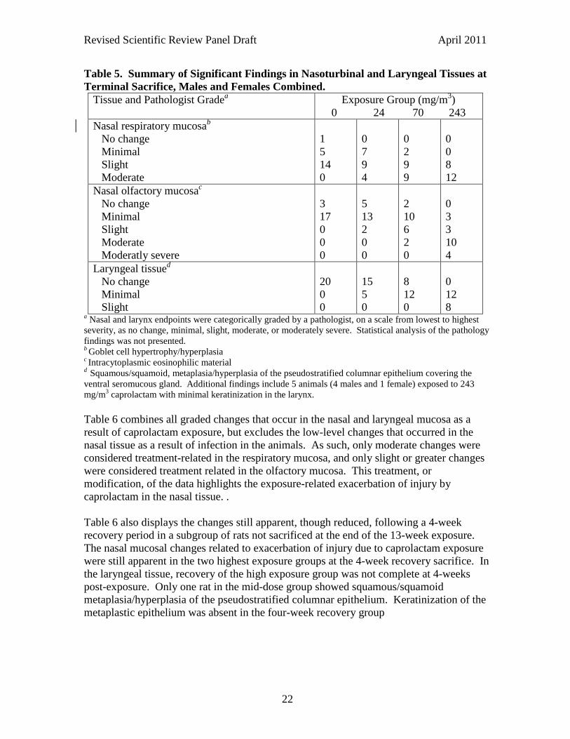
Table 5. Summary of Significant Findings in Nasoturbinal and Laryngeal Tissues at Terminal Sacrifice, Males and Females Combined.
Tissue and Pathologist Gradea Exposure Group (mg/m3) 0 24 70 243
Nasal respiratory mucosab No change Minimal Slight Moderate
1 5 14 0
0 7 9 4
0 2 9 9
0 0 8 12
Nasal olfactory mucosac No change Minimal Slight Moderate Moderatly severe
3 17 0 0 0
5 13 2 0 0
2 10 6 2 0
0 3 3 10 4
Laryngeal tissued No change Minimal Slight
20 0 0
15 5 0
8 12 0
0 12 8
a Nasal and larynx endpoints were categorically graded by a pathologist, on a scale from lowest to highest severity, as no change, minimal, slight, moderate, or moderately severe. Statistical analysis of the pathology findings was not presented.
b Goblet cell hypertrophy/hyperplasia c Intracytoplasmic eosinophilic material d Squamous/squamoid, metaplasia/hyperplasia of the pseudostratified columnar epithelium covering the ventral seromucous gland. Additional findings include 5 animals (4 males and 1 female) exposed to 243 mg/m3 caprolactam with minimal keratinization in the larynx. Table 6 combines all graded changes that occur in the nasal and laryngeal mucosa as a result of caprolactam exposure, but excludes the low-level changes that occurred in the nasal tissue as a result of infection in the animals. As such, only moderate changes were considered treatment-related in the respiratory mucosa, and only slight or greater changes were considered treatment related in the olfactory mucosa. This treatment, or modification, of the data highlights the exposure-related exacerbation of injury by caprolactam in the nasal tissue. . Table 6 also displays the changes still apparent, though reduced, following a 4-week recovery period in a subgroup of rats not sacrificed at the end of the 13-week exposure. The nasal mucosal changes related to exacerbation of injury due to caprolactam exposure were still apparent in the two highest exposure groups at the 4-week recovery sacrifice. In the laryngeal tissue, recovery of the high exposure group was not complete at 4-weeks post-exposure. Only one rat in the mid-dose group showed squamous/squamoid metaplasia/hyperplasia of the pseudostratified columnar epithelium. Keratinization of the metaplastic epithelium was absent in the four-week recovery group
Revised Scientific Review Panel Draft April 2011
23
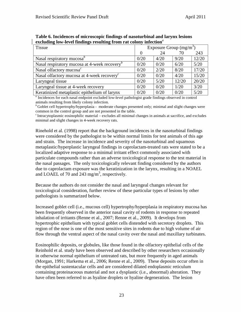
Table 6. Incidences of microscopic findings of nasoturbinal and larynx lesions excluding low-level findings resulting from rat colony infectiona Tissue Exposure Group (mg/m3)
0 24 70 243 Nasal respiratory mucosab 0/20 4/20 9/20 12/20 Nasal respiratory mucosa at 4-week recoveryb 0/20 0/20 6/20 5/20 Nasal olfactory mucosac 0/20 2/20 8/20 17/20 Nasal olfactory mucosa at 4-week recoveryc 0/20 0/20 4/20 15/20 Laryngeal tissue 0/20 5/20 12/20 20/20 Laryngeal tissue at 4-week recovery 0/20 0/20 1/20 3/20 Keratinized metaplastic epithelium of larynx 0/20 0/20 0/20 5/20 a Incidences for each nasal endpoint excluded low-level pathologist grade findings observed in control animals resulting from likely colony infection. b Goblet cell hypertrophy/hyperplasia - moderate changes presented only; minimal and slight changes were common in the control group and are not presented in the table. c Intracytoplasmic eosinophilic material – excludes all minimal changes in animals at sacrifice, and excludes minimal and slight changes in 4-week recovery rats. Rinehold et al. (1998) report that the background incidences in the nasoturbinal findings were considered by the pathologist to be within normal limits for test animals of this age and strain. The increase in incidence and severity of the nasoturbinal and squamous metaplastic/hyperplastic laryngeal findings in caprolactam-treated rats were stated to be a localized adaptive response to a minimal irritant effect commonly associated with particulate compounds rather than an adverse toxicological response to the test material in the nasal passages. The only toxicologically relevant finding considered by the authors due to caprolactam exposure was the keratinization in the larynx, resulting in a NOAEL and LOAEL of 70 and 243 mg/m3, respectively. Because the authors do not consider the nasal and laryngeal changes relevant for toxicological consideration, further review of these particular types of lesions by other pathologists is summarized below. Increased goblet cell (i.e., mucous cell) hypertrophy/hyperplasia in respiratory mucosa has been frequently observed in the anterior nasal cavity of rodents in response to repeated inhalation of irritants (Renne et al., 2007; Renne et al., 2009). It develops from hypertrophic epithelium with typical goblet cells distended with secretory droplets. This region of the nose is one of the most sensitive sites in rodents due to high volume of air flow through the ventral aspect of the nasal cavity over the nasal and maxillary turbinates. Eosinophilic deposits, or globules, like those found in the olfactory epithelial cells of the Reinhold et al. study have been observed and described by other researchers occasionally in otherwise normal epithelium of untreated rats, but more frequently in aged animals (Morgan, 1991; Harkema et al., 2006; Renne et al., 2009). These deposits occur often in the epithelial sustentacular cells and are considered dilated endoplasmic reticulum containing proteinaceous material and not a dysplastic (i.e., abnormal) alteration. They have often been referred to as hyaline droplets or hyaline degeneration. The lesion
Revised Scientific Review Panel Draft April 2011
24
increases in severity and extent with age and exposure to specific irritants, such as dimethylamine and cigarette smoke. The mechanism by which this lesion appears in aging rats, and the nature of the response to irritants, is not understood. Intracellular eosinophilic deposits have been observed in other studies in nasal respiratory mucosa and in other respiratory tract epithelium (Morgan, 1991; Renne et al., 2009), but either was not found in the Reinhold et al. animals, or was found in comparable incidence and severity in rats from both control and exposure groups. The region of the larynx investigated by Reinhold et al.(1998), the pseudostratified columnar epithelium on the ventral floor of the larynx at the base of the epiglottis, is especially sensitive to inhaled materials (Renne and Gideon, 2006; Renne et al., 2009). Squamous metaplasia as noted by Renne et al. (2009) may occur in association with acute and/or chronic inflammation or in the process of regeneration. Laryngeal squamous metaplasia has been characterized as a classic example of indirect metaplasia (Osimitz et al., 2007). Inhalation of an irritant damages sensitive respiratory or transitional epithelium, so that cells that proliferate to replace the lost cells produce a replacement epithelium that is better adapted to the new environment. In an expert workshop to evaluate larynx squamous metaplasia, a similar conclusion was made. This type of epithelial change is a result of transformation of the pre-existing epithelium to a squamous epithelium, with or without keritinization (Kaufmann et al., 2009). The lesion was classified as the morphologic correlate of an adaptive process from a more sensitive to a more resistant type of epithelium, which is indicative of local irritation. Focal, minor metaplastic changes that may also occasionally occur in control animals were considered “non-adverse”, while moderate to severe squamous metaplasia should be considered adverse as it may be associated with dysfunction. In humans, this dysfunction may result in hoarseness and an altered coughing reflex. In the rats exposed to caprolactam, exposure to the low and mid-level concentrations resulting in only a “minimal” grading for larynx metaplasia (Table 4). For an assessment of adversity (equivalent term to “toxicity”), the Panel (Kaufmann et al., 2009) felt it was more relevant to observe dysfunction of an organ or tissue (e.g., by test designed to measure muciliary clearance). For the Reinhold et al. (1998) rats, adversity was apparent due to the treatment-related increases in labored breathing, nasal discharge, red staining of the facial area, clear nasal discharge, and moist rales that began after approximately 1-2 weeks of exposure. An earlier paper by Osimitz et al. (2007) suggested that laryngeal squamous metaplasia should not be used as an endpoint for quantitative risk assessment, as it is well-differentiated, reversible and generally lacking signs of progression. This, in the opinion of the workshop Panel (Kaufmann et al., 2009), was not a responsible approach. All available information should be carefully considered by the pathologist, including other related health effects that are evaluated as “adverse”. The nasal lesions observed in control animals of the caprolactam study could also represent an upper respiratory infection present in the rat colony of the facility. Reinhold
Revised Scientific Review Panel Draft April 2011
25
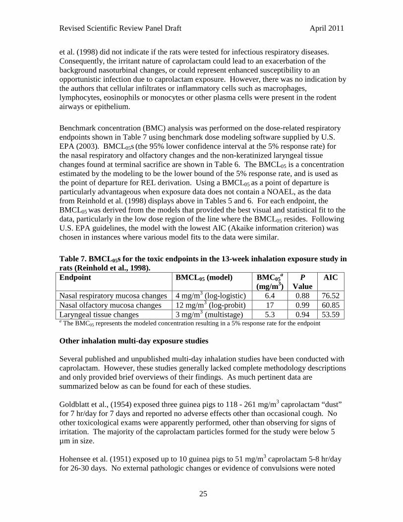
et al. (1998) did not indicate if the rats were tested for infectious respiratory diseases. Consequently, the irritant nature of caprolactam could lead to an exacerbation of the background nasoturbinal changes, or could represent enhanced susceptibility to an opportunistic infection due to caprolactam exposure. However, there was no indication by the authors that cellular infiltrates or inflammatory cells such as macrophages, lymphocytes, eosinophils or monocytes or other plasma cells were present in the rodent airways or epithelium. Benchmark concentration (BMC) analysis was performed on the dose-related respiratory endpoints shown in Table 7 using benchmark dose modeling software supplied by U.S. EPA (2003). BMCL05s (the 95% lower confidence interval at the 5% response rate) for the nasal respiratory and olfactory changes and the non-keratinized laryngeal tissue changes found at terminal sacrifice are shown in Table 6. The BMCL05 is a concentration estimated by the modeling to be the lower bound of the 5% response rate, and is used as the point of departure for REL derivation. Using a BMCL05 as a point of departure is particularly advantageous when exposure data does not contain a NOAEL, as the data from Reinhold et al. (1998) displays above in Tables 5 and 6. For each endpoint, the BMCL05 was derived from the models that provided the best visual and statistical fit to the data, particularly in the low dose region of the line where the BMCL05 resides. Following U.S. EPA guidelines, the model with the lowest AIC (Akaike information criterion) was chosen in instances where various model fits to the data were similar. Table 7. BMCL05s for the toxic endpoints in the 13-week inhalation exposure study in rats (Reinhold et al., 1998). Endpoint
BMCL05 (model) BMC05a
(mg/m3) P
Value AIC
Nasal respiratory mucosa changes 4 mg/m3 (log-logistic) 6.4 0.88 76.52 Nasal olfactory mucosa changes 12 mg/m3 (log-probit) 17 0.99 60.85 Laryngeal tissue changes 3 mg/m3 (multistage) 5.3 0.94 53.59 a The BMC05 represents the modeled concentration resulting in a 5% response rate for the endpoint Other inhalation multi-day exposure studies Several published and unpublished multi-day inhalation studies have been conducted with caprolactam. However, these studies generally lacked complete methodology descriptions and only provided brief overviews of their findings. As much pertinent data are summarized below as can be found for each of these studies. Goldblatt et al., (1954) exposed three guinea pigs to 118 - 261 mg/m3 caprolactam “dust” for 7 hr/day for 7 days and reported no adverse effects other than occasional cough. No other toxicological exams were apparently performed, other than observing for signs of irritation. The majority of the caprolactam particles formed for the study were below 5 µm in size. Hohensee et al. (1951) exposed up to 10 guinea pigs to 51 mg/m3 caprolactam 5-8 hr/day for 26-30 days. No external pathologic changes or evidence of convulsions were noted
Revised Scientific Review Panel Draft April 2011
26
during the exposures. Pathological and histological examination of a few of the animals revealed compound-related slight inflammation of the nasal mucosa and tracheal mucosa. However, no information was provided on the nature or extent of the inflammation or whether controls were free of this involvement. In a multi-day unpublished industry study, two rats were exposed to a nominal concentration of 3000 ppm, followed five days later by a series of five nominal 1 to 2 hr exposures ranging from 2700 to 6800 ppm on successive days (Haskell Laboratory, 1950). Nominal exposure entails calculating the loss of material to the gassing chamber when heated and the rate of air flow. No direct measurement of airborne caprolactam concentration is performed. General discomfort during the exposures, and inflammation around the eyes and nose were observed. Gross and microscopic pathological examination three days following the last exposure showed slight lung edema and congestion of the spleen, but no pathology in any other organs. In another unpublished study, 4 dogs, 6 guinea pigs, 6 rats and 2 rabbits were exposed subchronically to caprolactam fumes generated by heating the chemical in air. This study was conducted in 1952-53 and only recently reported to the U.S. EPA (2009). The composition of the fume was not evaluated and it was unclear from the report if the exposures were nominal or dynamic exposures. The authors and laboratory conducting the experiment are not identified and the document was labeled ‘company sanitized’. All animals were exposed 6 hrs/day for 43 exposures to 444 mg/m3. Half of the guinea pigs, rat and rabbits, and 3 of the dogs, were then exposed to 1020 mg/m3 on exposure 44 through 67 or 73 total daily exposures. In dogs, the fumes seemed to aggravate open sores and especially infections and soreness of the eyes (USEPA, 2009). Two of the four dogs displayed occasional muscle tremors during exposure to the low concentration of caprolactam. One of these dogs displayed severe muscle tremors, weakness, coughing with a dense white froth around the mouth when exposed to the high concentration during exposures 46 through 67. In all cases, the dogs were normal the next morning after exposure. One dog had a significant lowering of systolic pressure and pulse pressure but otherwise no other significant changes in weight, blood sugar, cholesterol, BUN, thymol turbidity or hematology. Gross pathology showed an indication of either acute duodenitis or gastroenteritis in two dogs, but it was suggested this was an aggravation of an existing gastro-intestinal disorder. Microscopic examination revealed no changes that were attributable to caprolactam. The study reported that one rabbit of the two exposed rabbits showed slight corneal damage and both rabbits showed mild irritation of the conjunctiva in both eyes (USEPA, 2009). No gross or microscopic pathology was observed in the rats or rabbits. In guinea pigs, one of the six had a lung reaction to a foreign body and a kidney showed evidence of regeneration of tubules. Another guinea pig had nephritis. A third guinea pig displayed consolidation of the apex of the right lung. No other gross or microscopic changes were detected in the remaining 3 guinea pigs.
Revised Scientific Review Panel Draft April 2011
27
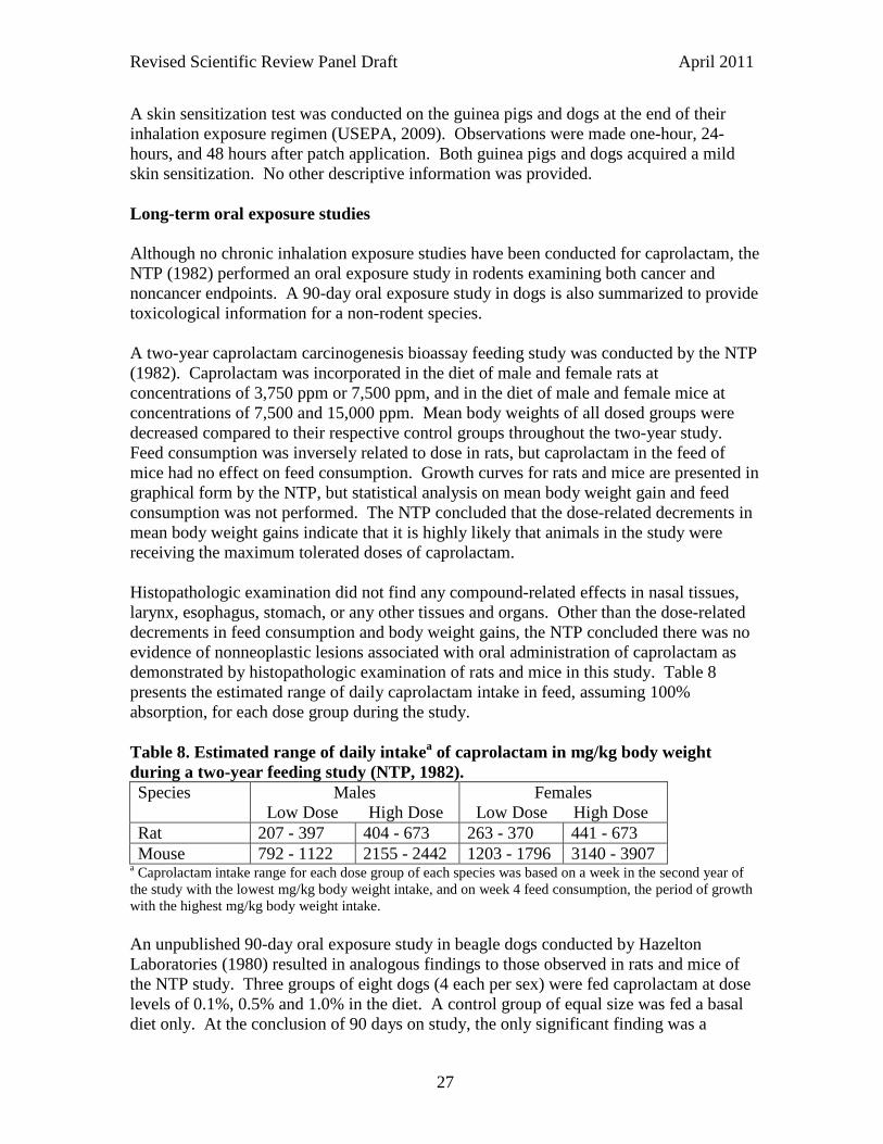
A skin sensitization test was conducted on the guinea pigs and dogs at the end of their inhalation exposure regimen (USEPA, 2009). Observations were made one-hour, 24-hours, and 48 hours after patch application. Both guinea pigs and dogs acquired a mild skin sensitization. No other descriptive information was provided. Long-term oral exposure studies Although no chronic inhalation exposure studies have been conducted for caprolactam, the NTP (1982) performed an oral exposure study in rodents examining both cancer and noncancer endpoints. A 90-day oral exposure study in dogs is also summarized to provide toxicological information for a non-rodent species. A two-year caprolactam carcinogenesis bioassay feeding study was conducted by the NTP (1982). Caprolactam was incorporated in the diet of male and female rats at concentrations of 3,750 ppm or 7,500 ppm, and in the diet of male and female mice at concentrations of 7,500 and 15,000 ppm. Mean body weights of all dosed groups were decreased compared to their respective control groups throughout the two-year study. Feed consumption was inversely related to dose in rats, but caprolactam in the feed of mice had no effect on feed consumption. Growth curves for rats and mice are presented in graphical form by the NTP, but statistical analysis on mean body weight gain and feed consumption was not performed. The NTP concluded that the dose-related decrements in mean body weight gains indicate that it is highly likely that animals in the study were receiving the maximum tolerated doses of caprolactam. Histopathologic examination did not find any compound-related effects in nasal tissues, larynx, esophagus, stomach, or any other tissues and organs. Other than the dose-related decrements in feed consumption and body weight gains, the NTP concluded there was no evidence of nonneoplastic lesions associated with oral administration of caprolactam as demonstrated by histopathologic examination of rats and mice in this study. Table 8 presents the estimated range of daily caprolactam intake in feed, assuming 100% absorption, for each dose group during the study. Table 8. Estimated range of daily intakea of caprolactam in mg/kg body weight during a two-year feeding study (NTP, 1982). Species Males
Low Dose High Dose Females
Low Dose High Dose Rat 207 - 397 404 - 673 263 - 370 441 - 673 Mouse 792 - 1122 2155 - 2442 1203 - 1796 3140 - 3907
a Caprolactam intake range for each dose group of each species was based on a week in the second year of the study with the lowest mg/kg body weight intake, and on week 4 feed consumption, the period of growth with the highest mg/kg body weight intake. An unpublished 90-day oral exposure study in beagle dogs conducted by Hazelton Laboratories (1980) resulted in analogous findings to those observed in rats and mice of the NTP study. Three groups of eight dogs (4 each per sex) were fed caprolactam at dose levels of 0.1%, 0.5% and 1.0% in the diet. A control group of equal size was fed a basal diet only. At the conclusion of 90 days on study, the only significant finding was a
Revised Scientific Review Panel Draft April 2011
28
slightly lower mean body weight of the 1.0% females compared to controls. A similar change was not observed for the males, and total food consumption was comparable between groups of both sexes. No difference from controls was observed in absolute or relative organ weights for any group. Gross and microscopic examination of tissues revealed no remarkable findings. Ophthalmologic findings were negative. No dose-related changes were observed for hematologic and serum chemistry samples. 7. Developmental and Reproductive Toxicity No studies were located investigating the developmental and reproductive toxicity of caprolactam by the inhalation route. In oral exposure studies, (Gad et al., 1987) dosed pregnant rats by gavage with caprolactam at 100, 500, or 1,000 mg/kg/day on gestation days 6-15. Increased maternal mortality was observed at the highest dose. A dose-related decrease in mean maternal body weight was observed with a statistically significant reduction (p≤0.05) in total body weight at the highest dose level (a 10 and 11% reduction on gestational days 15 and 20, respectively). A statistically significant reduction (p≤0.05) in mean weight change was observed during the treatment period at the two highest doses (5.2 and 2.3% mean weight gain at the mid- and high-dose, respectively, compared to a 13.4% weight gain for the control group). Food consumption was reduced in the two highest dose groups. The mean incidence of resorptions was increased at the highest dose. No dose-related skeletal anomalies or major malformations were noted among the offspring of any exposure group. An apparent dose-related increase in the mean number of skeletal variants per litter was observed, including incomplete ossification of the skull or vertebral column and the presence of extra ribs. However, no statistically significant difference in skeletal variation values between treated groups and the control group were noted. Gad et al. (1987) also dosed pregnant rabbits by gavage with caprolactam at 50, 150, or 250 mg/kg/day on gestational days 6-28. Sixteen percent mortality and statistically significantly decreased overall maternal body weight gain were observed at the highest dose. Corrected weight gain (i.e., weight gain minus weight of gravid uterus) was statistically significantly lower (p<0.05) from day 6 to 29 of gestation in the 150 mg/kg group. Absolute maternal body weights were unaffected in this mid-dose group. Mean fetal weights were statistically significantly reduced (p<0.05) by 12% in each of the two highest dose groups compared to controls. The incidence of major malformations was unaffected by caprolactam treatment. Minor skeletal anomalies included an increased incidence of unilateral or bilateral thirteenth ribs in the highest dose group. Gad et al. (1987) concluded that caprolactam given by gavage to two species up to levels that caused severe maternal toxicity did not support a finding of the compound causing either embyotoxicity or teratogenicity. Fetotoxicity was evidenced in rabbits by lower fetal weights at the two highest doses, and an increased incidence of 13th ribs at the highest dose level.
Revised Scientific Review Panel Draft April 2011
29
In a multi-generation study, rats were given a 1,000, 5,000, or 10,000 mg caprolactam/kg diet (ppm) over three generations (Serota et al., 1988). Each generation was treated over a 10-week period. Consistently lower mean body weights and food consumption were observed in both P2 and P3 parental generations at 5,000 and 10,000 ppm, but body weights were unaffected in P1 animals at all dose levels. The mean body weight changes were statistically significant (p≤0.05) in all high dose groups at all time points with weight reductions in both males and females ranging from 10 to 21% compared to controls. For mid-dose animals, a statistically significant change in mean body weight occurred only in P2 males, a 13% reduction compared to controls, during week four of exposure. Dose-related reductions of fetal body weights were observed in all filial generations. For example, statistically significant differences (p≤0.05) noted in F1a and F1b high dose groups (17 to 23% reductions compared to controls) and occasionally in mid-dose groups (11 to 14% reductions in F1b offspring only compared to controls). Based on mean body weight and mean food consumption values at week 10 in P1 females, caprolactam in the diet at 1000, 5000 and 10,000 ppm was equivalent to a daily dose of 697, 3542 and 5622 mg/kg caprolactam, respectively. No treatment-related effect on gross appearance, gross pathology, survival rate or number of pups was observed. A slight increase in the severity of spontaneous nephropathy was observed on histopathologic examination of males in the high-dose group of the first parental generation. Serota et al. (1988) concluded that caprolactam in the diet at the two highest exposures resulted in decreases in body weight in both pups and parental animals in utero through weaning. Similar effects on food consumption were also noted. Body weights were unaffected in P1 animals at all dose levels, and reduced food consumption was observed only at week 10 in P1 females. No effects were evident on reproductive performance or offspring survival, and only minimal kidney toxicity was observed in males at the highest dose level. In other oral exposure studies, (Salamone, 1989) reported no sperm abnormality in male mice treated with 222, 333, 500, 750, or 1,125 mg/kg caprolactam by gavage daily for five days, although mortality was evident at the highest dose. Following the fifth gavage with 1,125 mg/kg caprolactam, the mice immediately became motionless. In four of the nine mice treated, this inactivity was followed 10 min later by racing around the cage and death within seconds. These deaths are probably related to the method of oral treatment because exposure of mice up to 2155 to 2442 mg/kg caprolactam in feed for two years by the NTP (1982) did not result in an increase in mortality. A similar study in male rats did not observe DNA damage to spermatocytes following an oral dose of 750 mg/kg caprolactam (Working, 1989). The primary finding of the two developmental/reproductive toxicity oral exposure studies was that caprolactam may be fetotoxic due to reduced fetal body weight. Reductions in fetal weight in the gavage study occurred at the same dose levels that reductions in
Revised Scientific Review Panel Draft April 2011
30
maternal food consumption and body weight occurred. Based on this gavage study, the concomitant reduction in both maternal body weight and fetal weight make it difficult for OEHHA to conclude that caprolactam is exclusively fetotoxic. However, body weights of P1 rats in the multi-generation study were not reduced by caprolactam exposure yet resulted in reduced fetal weights in F1a and F1b offspring. This finding indicates a fetotoxic LOAEL of 5000 ppm caprolactam in feed, which is equivalent to a maternal daily dose of 3542 mg/kg. The calculated NOAEL is 697 mg/kg. Assuming 100% pulmonary absorption, the NOAEL is equivalent to an air concentration of 2440 mg/m3 (697 mg/kg x 70 kg body wt. / 20 m3/day) in a route-to-route extrapolation. Brief human exposures to lower caprolactam concentrations in the range of 1850 to 5560 mg/m3 (400 to 1200 ppm) have been characterized as extremely irritating, and subchronic exposures of rats to air concentrations as low as 24 mg/m3 have resulted in labored breathing and nasal secretory discharge. Applying a 100-fold uncertainty factor (10-fold UF each for interspecies and intraspecies extrapolation) for extrapolation from an animal developmental study to human exposure would produce a proposed REL of 24 mg/m3. The acute and chronic RELs of 770 µg/m3 and 2 µg/m3, respectively, are considerably lower than that derived from the oral multi-generation animal study. These findings show that the oral dose at which fetotoxicity occurs is likely not relevant to air concentrations of caprolactam for REL derivation due to upper respiratory tract injury occurring at lower concentrations. The acute, 8-hour and chronic RELs developed in this document based on caprolactam air exposures would be protective for reproductive/developmental effects. Therefore, OEHHA is using pulmonary and sensory irritation endpoints for the caprolactam inhalation RELs.
Revised Scientific Review Panel Draft April 2011
31
8. REL Derivations 8.1 Derivation of the Acute Inhalation Reference Exposure Level (1-hour
exposure) As noted above, only two studies exist that examined the acute sensory irritant effects resulting from directly measured concentrations of caprolactam. Because of limitations in the occupational study (Ferguson and Wheeler, 1973), and lack of a LOAEL in the chamber exposure study (Ziegler et al., 2008), an acute REL cannot be derived by OEHHA at this time. The only quantitative evidence for a caprolactam concentration resulting in sensory irritation in humans is from Ferguson and Wheeler (1973), in which brief exposure to 10 ppm (46 mg/m3) resulted in nasal and throat irritation. Although the exposures were reported to be to caprolactam “vapor”, all the exposures were above the saturated vapor pressure of 13 mg/m3 at 25°C. The form of caprolactam was not determined by the authors, but it was likely exposure was to the aerosol form with some smaller amount in the vapor form. In particular, the exposures were uncontrolled and the variability in concentration during exposure was not reported. The authors themselves observed that the caprolactam concentration varied enough over a 30 minute recording period to result in an average concentration that may have been below the actual threshold level for irritation. Also, the method used for measuring the caprolactam concentration is antiquated and probably resulted in loss of the chemical before reaching the analytical equipment. Combined with data from only five individuals exposed and no concentration examined at which sensory irritation was absent in all individuals (a NOAEL), OEHHA cannot reliably base an acute REL derivation using this study. In contrast to the occupational study in which only a LOAEL is reported, the chamber study by Ziegler et al. (2008) only reports a NOAEL. OEHHA normally does not use a study with a free-standing NOAEL as the basis of an acute REL. The human chamber exposure study by Ziegler et al. (2008) found a statistically significantly (p<0.05) increased subjective total symptom and complaint score at the highest concentration of 5 mg/m3. Increasing the power of the analysis by combining all 29 subjective symptom scores resulted in this finding. Individual subjective scores and subscore groups did not reach statistical significance on their own, except for odor annoyance, which showed that odor was the main driver for the increased total symptom score at 5 mg/m3. However, average odor intensity at the high concentration was only 1.2 (i.e., falling between “barely” (1) and “somewhat” (2) an odor nuisance). Severe physiological responses to odor can be the basis of acute RELs, such as the intense nausea and headache that can be induced by exposure to hydrogen sulfide. Ziegler et al. (2008) noted that three of the 20 volunteers perceived the odor nuisance as “severe” (a score rating of 4) at 5 mg/m3, while others did “not” feel annoyed “at all” (a score rating of 0) at this concentration. A wide variation in response to chemical odor is not unusual among human volunteers. However, the six hour exposures to caprolactam did not result in any volunteers leaving the chamber early, or appear to produce a physiological response
Revised Scientific Review Panel Draft April 2011
32
of the magnitude of hydrogen sulfide. The ambiguity in using the increased subjective total symptom and complaint score, in which odor annoyance appears to dominate and did not result in any severe physiological responses, precludes using this endpoint as the basis of the acute REL. Individual objective and subjective sensory irritation scores, including eye blink, nasal resistance, and eye and nose irritation, showed an increasing trend towards irritation, but did not reach statistical significance by the methods used by the authors. Without the raw data, OEHHA cannot verify these findings by using other statistical means. OEHHA has been unable to obtain the raw data from the authors. Further evidence that a sensory irritant level was probably not reached even though the odor was detected can be observed from the subjective mean eye and nose irritation scores shown in Table 1. The mean response difference between the control and 5 mg/m3 exposure level for nose irritation was 0.16 and 0.26, respectively, corresponding to a score rating between “not at all” (zero) and “barely” (1). Eye irritation showed a similar shallow response difference between control and high dose. Typically, when the sensory irritant threshold is reached the graded response should rise steeply. Over a range of hardly more than one order of magnitude of concentration, sensory irritation may increase from “barely detectatble” to “painful irritation” (Cain et al., 2006; Nielsen et al., 2007). Such a steep rise in the graded response was not apparent for caprolactam in this study, suggesting the sensory irritant threshold was not reached.
Revised Scientific Review Panel Draft April 2011
33
8.2 Derivation of the 8-Hour Inhalation Reference Exposure Level Study Reinhold et al. 1998 Study population Sprague-Dawley CD rats (10 animals/sex/group) Exposure method Discontinuous whole-body inhalation exposure to
0, 24, 70, and 243 mg/m3 caprolactam aerosol Critical effects Upper airway lesions of nasal and laryngeal
epithelium LOAEL 24 mg/m3 NOAEL Not observed BMCL05 3 mg/m3 Exposure continuity 6 hours per day, 5 days/week Exposure duration 13 weeks Average experimental exposure 1.607 mg/m3 (3 mg/m3 x 6/8 x 5/7) Human equivalent concentration 0.402 mg/m3 (for extrathoracic respiratory
effects, RGDR = 0.25 LOAEL uncertainty factor 1 (BMCL05 used as point of departure) Subchronic uncertainty factor 2 (for 13 wk exposure in rodents) (see below) Interspecies uncertainty factor
Toxicokinetic (UFA-k) 1 Toxicodynamic (UFA-d) √10 (default: no interspecies toxicodynamic data)
Intraspecies uncertainty factor Toxicokinetic (UFH-k) 1 (site of contact; no systemic effects)
Toxicodynamic (UFH-d) 10 (potential asthma exacerbation in children) Cumulative uncertainty factor 60 8-Hour air reference exposure level 7 µg/m3 (1 ppb)
The comprehensive subchronic exposure study in rats by Reinhold et al. (1998) is the basis of the 8-hour REL, resulting in a level of 7 µg/m3 (rounded up to one significant figure from 6.70 µg/m3), or 1 ppb (rounded down to one significant figure from 1.45 ppb). The occupational studies of long-term exposure to caprolactam were considered by OEHHA to be inadequate for use as a point of departure for 8-hour and chronic RELs (see Section 6.1 for reviews of the occupational studies). In the Reinhold et al. study, the authors aerosolized an aqueous caprolactam solution for the exposures, which had an MMAD of 3.0 µm and a geometric standard deviation of 1.7 µm. Particles of this size have been shown to deposit primarily in the upper respiratory airway (Stuart, 1984; Yeh et al., 1996). So it is not surprising that this was the region of the respiratory system affected by caprolactam exposure. Although little other evidence could be found in the literature regarding the MMAD of caprolactam found in the environment, it can be expected that a significant fraction of caprolactam at the level of the REL would be in a gas phase. Very water-soluble gases, including caprolactam, also deposit in the upper respiratory tract (OEHHA, 2008). This evidence, plus human industrial exposure data that shows nasal irritation as the primary acute response, indicates the primary respiratory endpoint is the upper respiratory region.
Revised Scientific Review Panel Draft April 2011
34
The 8-hr REL derivation is based on a BMCL05 = 3 mg/m3 for a pathology grading of minimal and slight increases in squamous/squamoid metaplasia/hyperplasia in the larynx of male and female rats exposed to caprolactam aerosol for 13 weeks (Reinhold et al., 1998). The BMCL05 for exacerbation of changes to the respiratory and olfactory nasal mucosa resulted in essentially the same value (respiratory BMCL05 = 4 mg/m3) or was slightly greater (olfactory BMCL05 = 12 mg/m3). Reinhold et al. (1998) regarded laryngeal keratinization of the metaplastic epithelium to be the primary adverse effect, resulting in a NOAEL of 70 mg/m3. The other effects in the upper respiratory system were considered by the researchers to be normal adaptive responses to an irritant, which they did not consider a toxicological endpoint. However, OEHHA RELs include health protection against mild irritant/inflammatory effects. These types of mild inflammatory changes are primary endpoints of toxicity as indicated in the Revised Air Toxics Hot Spots Program Technical Support Document for the Derivation of Noncancer Reference Exposure Levels and RELs for Six Chemicals (OEHHA, 2008). The irritant-related microscopic changes in the upper respiratory tract, combined with the observations of respiratory irritation/inflammation (nasal discharge, moist rales, red staining around the face) and labored breathing in all caprolactam-treated groups, support the lack of an observed NOAEL in the principal study. The BMCL05 = 3 mg/m3 was adjusted to an average experimental exposure of 1.6 mg/m3 for eight-hour exposures, seven days/week. The concentration at the BMCL05 is below the saturated vapor concentration of caprolactam of 13 mg/m3 at 25ºC. Thus, a greater proportion of caprolactam may be in gaseous form rather than the aerosol form used in the study. Although no studies have been conducted comparing the potency of gaseous or aerosol forms of caprolactam, the evidence in this document indicates both forms are expected to have the same toxicological endpoints. Given that the predominant form humans will be exposed to at the level of the RELs will likely be the gaseous form, a regional gas dose ratio (RGDR) approach will be used for the human equivalent concentration (HEC) adjustment. The RGDR of 0.25 was calculated using US EPA methodology (OEHHA, 2008) for extrapolation from rat and human exposure. The equation for gases with respiratory effects is:
RGDR = (MVa/MVh) / (SAa/SAh) Eq. 8-1 Where: MVa = animal minute volume MVh = human minute volume SAa = animal surface area for lung region of concern’ SAh = human surface area for lung region of concern
Surface areas for the region of concern, the extrathoracic region, for rat (15 cm2) and human (200 cm2) were obtained from Table F.1.1 in OEHHA (2008). Minute volume for rats were calculated using Eq. 8-2 below, using intercept (b0) and slope (b1) values from Table F.1.2 in OEHHA (2008). Body weight (BW) for both male and female rats
Revised Scientific Review Panel Draft April 2011
35
combined (0.323 kg) was averaged over the 13-week exposure duration from body weight tables in Reinhold et al. (1998) and the authors’ unpublished industrial data (Hoffman, 1997).
loge(MV) = b0 + b1 loge(BW) Eq. 8-2
Where
BW = 0.323 kg; b0 = -0.578; b1 = 0.821 loge(MV) = -0.578 + 0.821 loge(0.323 kg) MV = 0.222 L/min or 222 ml/min
For humans, an average adult male and female combined MV of 11,944 ml/min was estimated using breathing rate data from US EPA (2009). Thus:
RGDR = (222 ml/min/11,944 ml/min) / (15cm2/200 cm2) RGDR = 0.0186 / 0.075 RGDR = 0.25
A subchronic UF = 2 was incorporated into the REL derivation for extrapolation from 13-week exposure in the rats to chronic exposure. Although 13 weeks of exposure is 12.5% of the 2-year life expectancy of rats, which would entail use of a subchronic UF = 1 for >12% of lifetime exposure, U.S. EPA (1994) recommends using a subchronic adjustment factor for all 13-week studies regardless of species. OEHHA has typically used a subchronic UF = √10 for 13-week exposure studies in rats and mice. However, for rodent studies of this exposure duration a subchronic UF = 2 for upper respiratory irritants, when the resulting injury is considered mild, is appropriate. The basis for using a subchronic UF = 2 was derived from the numerous rodent studies with formaldehyde (OEHHA, 2008). Comparison of 13-week exposure studies with studies of longer duration up to 2 years shows that the NOAELs and LOAELs for upper airway injury are often the same, with only a 2-fold difference between chronic and 13-week study NOAELs and LOAELs in some cases. The 2-fold lower NOAELs and LOAELs were often a result of the choice of the formaldehyde exposure concentration used in the studies. The severity of the upper respiratory tract injury also supports a subchronic UF = 2. The pathology grading of the upper respiratory tract resulting from caprolactam exposure indicates only a mild increase in injury. The exacerbation by caprolactam exposure of normal nasal olfactory tissue degeneration was small, increasing from minimal to slight at the lowest dose of 24 mg/m3. The laryngeal tissue damage caused by caprolactam was minimal, at best, at the low dose. Overall, only a few cases of moderately severe tissue injury were observed, occurring in the high concentration exposure group in olfactory tissue. In addition, all animals survived to the end of the study and the treatment-related
Revised Scientific Review Panel Draft April 2011
36
labored breathing and nasal discharge generally decreased in incidence during the second half of the study. We did not apply an interspecies toxicokinetic UFA-k . Hybrid computational fluid dynamics and PBPK modeling for predicting nasal tissue dose metrics show that the predicted dose to the epithelium of the total nasal cavity following inhalation of an organic gas is similar, or slightly greater, in humans compared to rats (Frederick et al., 2001). Also, injury occurred to regions of the upper respiratory tract that are most sensitive to both chemical and mechanical irritants (primarily due to airflow characteristics) which indicates that caprolactam is primarily a direct-acting irritant, rather than a chemical requiring metabolic activation in nasal mucosa to cause tissue injury (Kilgour et al., 2000; Harkema et al., 2006; Renne et al., 2007; Kaufmann et al., 2009). In particular, Kaufmann et al. (2009) indicated the rat larynx may be more sensitive to inhaled irritant gases due to air flow characteristics than other species such as humans, monkeys and dogs. Therefore, the human equivalency concentration (HEC) adjustment for upper respiratory tract injury should also be sufficient for any residual interspecies toxicokinetics differences. We applied a default interspecies UFA-d of √10 to compensate for the absence of data on pharmacodynamic differences between species. Specifically, only one comprehensive animal inhalation study in rats has been performed with caprolactam. The toxicokinetic data for inspired upper respiratory irritants in humans suggest low interindividual variation and no dosimetry differences between adults and children (OEHHA, 2008). Thus, no UFH-k is applied for intraspecies toxicokinetic variation among individuals. While caprolactam is irritating to the upper respiratory tract, initiation or exacerbation of asthma by caprolactam has not been characterized. However, data summarized by OEHHA (2008) show asthmatics may be more sensitive to the effects of respiratory irritants than non-asthmatic individuals. Thus, an intraspecies toxicodynamic UF of 10 is applied to address the diversity in the human population, including children with asthma. Application of the cumulative UF = 60 to the human equivalent concentration of 0.402 mg/m3 resulted in an 8-hour REL of 7 µg/m3 (1 ppb) for caprolactam.
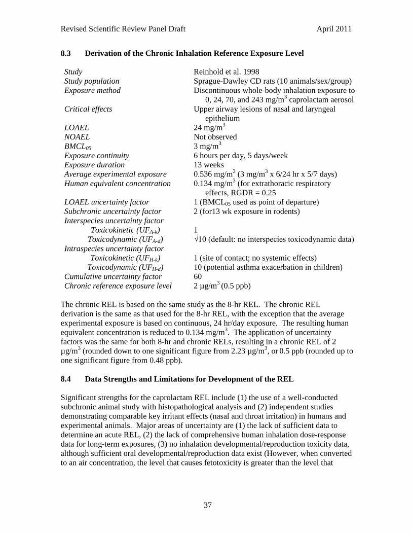
Revised Scientific Review Panel Draft April 2011
37
8.3 Derivation of the Chronic Inhalation Reference Exposure Level Study Reinhold et al. 1998 Study population Sprague-Dawley CD rats (10 animals/sex/group) Exposure method Discontinuous whole-body inhalation exposure to
0, 24, 70, and 243 mg/m3 caprolactam aerosol Critical effects Upper airway lesions of nasal and laryngeal
epithelium LOAEL 24 mg/m3 NOAEL Not observed BMCL05 3 mg/m3 Exposure continuity 6 hours per day, 5 days/week Exposure duration 13 weeks Average experimental exposure 0.536 mg/m3 (3 mg/m3 x 6/24 hr x 5/7 days) Human equivalent concentration 0.134 mg/m3 (for extrathoracic respiratory
effects, RGDR = 0.25 LOAEL uncertainty factor 1 (BMCL05 used as point of departure) Subchronic uncertainty factor 2 (for13 wk exposure in rodents) Interspecies uncertainty factor
Toxicokinetic (UFA-k) 1 Toxicodynamic (UFA-d) √10 (default: no interspecies toxicodynamic data)
Intraspecies uncertainty factor Toxicokinetic (UFH-k) 1 (site of contact; no systemic effects)
Toxicodynamic (UFH-d) 10 (potential asthma exacerbation in children) Cumulative uncertainty factor 60 Chronic reference exposure level 2 µg/m3 (0.5 ppb)
The chronic REL is based on the same study as the 8-hr REL. The chronic REL derivation is the same as that used for the 8-hr REL, with the exception that the average experimental exposure is based on continuous, 24 hr/day exposure. The resulting human equivalent concentration is reduced to 0.134 mg/m3. The application of uncertainty factors was the same for both 8-hr and chronic RELs, resulting in a chronic REL of 2 µg/m3 (rounded down to one significant figure from 2.23 µg/m3, or 0.5 ppb (rounded up to one significant figure from 0.48 ppb). 8.4 Data Strengths and Limitations for Development of the REL Significant strengths for the caprolactam REL include (1) the use of a well-conducted subchronic animal study with histopathological analysis and (2) independent studies demonstrating comparable key irritant effects (nasal and throat irritation) in humans and experimental animals. Major areas of uncertainty are (1) the lack of sufficient data to determine an acute REL, (2) the lack of comprehensive human inhalation dose-response data for long-term exposures, (3) no inhalation developmental/reproduction toxicity data, although sufficient oral developmental/reproduction data exist (However, when converted to an air concentration, the level that causes fetotoxicity is greater than the level that
Revised Scientific Review Panel Draft April 2011
38
results in severe pulmonary injury), (4) the absence of a NOAEL in the subchronic study, and (5) no chronic animal inhalation exposure studies.
9. References ACGIH. (2001). Caprolactam. American Conference of Governmental Industrial Hygienists, Cincinnati, Ohio. Online at: www.acgih.org. ACGIH. (2003). Caprolactam. American Conference of Governmental Industrial Hygienists, Cincinnati, Ohio. Online at: www.acgih.org. Aguirre A, Gonzalez Perez R, Zubizarreta J, Landa N, Sanz de Galdeano C and Diaz Perez JL (1995). Allergic contact dermatitis from epsilon-caprolactam. Contact Dermatitis 32(3): 174-5. Ballistreri A, Garazzo D, Giuffrida M, Montaudo G, Filippi A, Guaita C, Manaresi P and Pilati F (1987). Fast atom bombardment mass spectrometry identification of oligomers contained in poly(epsilon-caprolactam) and poly(butylene isophthalate). Macromolecules 20: 1029-32. BASF (1985). Test report: Acute inhalation toxicity LC50, 4 hours (rat): Evaluation of caprolactam (compound no. 84/217) as an aerosol liquid. pp. 1-21. Billmaier DJ, Knowlden NF and Stidham DW (1992). Caprolactam: A study of current workers. Allied-Signal Inc., Morristown, NJ. EPA/OTS Doc #86-920001041. 1-16, A-1-A-9. Bongard W (2000). Competitiveness of Nylon 6. Man-Made Fiber Year Book DSM Fibre Intermediates, Geleen, Netherlands: 14-15. Bonifaci L, Frezzotti D, Cavalca G, Malauti E and Ravanetti GP (1991). Analysis of epsilon-caprolactam and its oligomers by high-performance liquid chromatography. J Chromatog 585: 333-36. Bradley EL, Speck DR, Read WA and Castle L (2004). Method of test and survey of caprolactam migration into foods packaged in nylon-6. Food Addit Contam 21(12): 1179-1185. Cain WS, Lee NS, Wise PM, Schmidt R, Ahn BH, Cometto-Muniz JE and Abraham MH (2006). Chemesthesis from volatile organic compounds: Psychophysical and neural responses. Physiol Behav 88(4-5): 317-24. Cal/OSHA. (2011). Permissable Exposure Limits for Chemical Contaminants. California Occupational Safety and Health Administration, Online at: www.dir.ca.gov/title8/5155table_ac1.html.
Revised Scientific Review Panel Draft April 2011
39
Cheng Y, Li SM and Leithead A (2006). Chemical characteristics and origins of nitrogen-containing organic compounds in PM2.5 aerosols in the Lower Fraser Valley. Environ Sci Technol 40(19): 5846-52. Cooper JE, Cooke DP, Dickson WA, Noell LT, Beal BM and Pirtle LS (1993). Petition to Delist Caprolactam Under Section 112(b) of the Clean Air Act. AlliedSignal Inc. Properties, Standards, and Use of Caprolactam. BASF Corporation, DSM Chemicals North America Inc. pp. 2-1 to 2-7. CPSC. (1996). Sensory and Pulmonary Irritation Studies of Carpet System Materials and their Constituent Chemicals U.S. Consumer Product Safety Commission. Contract CPSC-C-94-1122. AQS Report No. 01890-06. Chapters 1 to 5 available at Internet address:http://www.cpsc.gov/LIBRARY/FOIA/FOIA98/os3519926D.pdf. Chapters 6 through Appendix B available at http://www.cpsc.gov/LIBRARY/FOIA/FOIA98/os/3512B0C.pdf. Ferguson WS and Wheeler DD (1973). Caprolactam vapor exposures. Am Ind Hyg Assoc J 34(9): 384-9. Frederick CB, Gentry PR, Bush ML, Lomax LG, Black KA, Finch L, Kimbell JS, Morgan KT, Subramaniam RP, Morris JB and Ultman JS (2001). A hybrid computational fluid dynamics and physiologically based pharmacokinetic model for comparison of predicted tissue concentrations of acrylic acid and other vapors in the rat and human nasal cavities following inhalation exposure. Inhal Toxicol 13(5): 359-76. Gad SC, Robinson K, Serota DG and Colpean BR (1987). Developmental toxicity studies of caprolactam in the rat and rabbit. J Appl Toxicol 7(5): 317-26. Gill K (2011). Written communication. Air Resources Board, California Environmental Protection Agency, Sacramento, CA. Goldblatt MW, Farquharson ME, Bennett G and Askew BM (1954). Epsilon-Caprolactam. Br J Ind Med 11(1): 1-10. Gross P (1984). Biologic activity of epsilon-caprolactam. Crit Rev Toxicol 13(3): 205-16. Harkema JR, Carey SA and Wagner JG (2006). The nose revisited: a brief review of the comparative structure, function, and toxicologic pathology of the nasal epithelium. Toxicol Pathol 34(3): 252-69. Haskell Laboratory (1950). Medical Research Project No. MR-173: Toxicity of Epsilon-Caprolactam. Report # HL-0022-50, Haskell Laboratory of Industrial Toxicology, Wilmington, Delaware.
Revised Scientific Review Panel Draft April 2011
40
Hausen BM (2003). Allergic contact dermatitis from colored surgical suture material: contact allergy to epsilon-caprolactam and acid blue 158. Am J Contact Dermat 14(3): 174-5. Hazelton Laboratories America Inc (1980). 90-Dat Toxicity Study in Dogs: Caprolactam, Final Report. Hazelton Laboratories America. Hegde RR, Dahiya A and Kamath MG (2004). Nylon Fibers. Online at: www.engr.utk.edu/mse/Textiles/Nylon%20fibers.htm. Hodgson AT, Shendell DG, Fisk WJ and Apte MG (2004). Comparison of predicted and derived measures of volatile organic compounds inside four new relocatable classrooms. Indoor Air 14 Suppl 8: 135-44. Hoffman GM. (1997). A 13-week inhalation toxicity study (with a 4-week recovery) of caprolactam (494-95A) in the rat via whole-body exposures. Huntingdon Life Sciences, East Millstone, NJ, Volume I, Study no.: 95-6095, IHF 3-95PA. Hohensee F (1951). On the pharmacological and physiological effects of e-caprolactam. Faserforsch Textultech 2: 299-303. HSDB (2006). Hazardous Substances Data Bank. Chemical/Physical Properties; Emergency Medical Treatment. National Library of Medicine, Bethesda, MD. Available at: http://toxnet.nlm.nih.gov. IARC (1999). Caprolactam. IARC Monographs on the Evaluation of Carcinogenic Risks to Humans. Re-evalualtion of Some Organic Chemicals, Hydrazine and Hydrogen Peroxide. World Health Organization, Lyon, France. 71 (part 2): 383-400. IWMB (2003). Integrated Waste Management Board. Building Material Emissions Study. Sacramento, CA. Available at: www.ciwmb.ca.gov/Publications. pp. 1-15, 34-37, 62-63, 90-92, 164-184 (App. C), 305-308 (App. O). Kaufmann W, Bader R, Ernst H, Harada T, Hardisty J, Kittel B, Kolling A, Pino M, Renne R, Rittinghausen S, Schulte A, Wohrmann T and Rosenbruch M (2009). 1st international ESTP expert workshop: "Larynx squamous metaplasia". A re-consideration of morphology and diagnostic approaches in rodent studies and its relevance for human risk assessment. Exp Toxicol Pathol 61(6): 591-603. Kelman GR (1986). Effects of human exposure to atmospheric epsilon-caprolactam. Hum Toxicol 5(1): 57-9. Kilgour JD, Simpson SA, Alexander DJ and Reed CJ (2000). A rat nasal epithelial model for predicting upper respiratory tract toxicity: in vivo-in vitro correlations. Toxicology 145(1): 39-49.
Revised Scientific Review Panel Draft April 2011
41
Kirk LK, Lewis BA, Ross DA and Morrison MA (1987). Identification of ninhydrin-positive caprolactam metabolites in the rat. Food Chem Toxicol 25(3): 233-239. Krajnik V, Bozek P, Kondelikova J and Kralicek J (1982). High-performance liquid chromatography of 6-caprolactam and its cyclic oligomers present in polyamide 6. J Chromatog 240: 539-42. Lander J-A. (2002). Structure Development in Silicate-Layered Polymer Nanocomposites. A Thesis Submitted for the Degree of Doctor of Philosophy. Uxbridge, Middlesex: Wolfson Centre for Materials Processing, Brunel University. Morgan KT (1991). Approaches to the identification and recording of nasal lesions in toxicology studies. Toxicol Pathol 19(4 Pt 1): 337-51. Nielsen GD, Wolkoff P and Alarie Y (2007). Sensory irritation: risk assessment approaches. Regul Toxicol Pharmacol 48(1): 6-18. NIOSH. (1995a). Occupational Safety and Health Guideline for Caprolactam Dust. National Institute for Occupational Safety and Health, U.S. Department of Health and Human Services. NIOSH. (1995b). Occupational Safety and Health Guideline for Caprolactam Vapor. National Institute for Occupational Safety and Health, U.S. Department of Health and Human Services. NPG (2007). Nylon-6. Nylon-6 Promotional Group, Online at: www.npg-6.com. NTP (1982). Carcinogenesis bioassay of caprolactam (CAS No. 105-60-2) in F344 rats and B6C3F1 mice (feed study). National Toxicology Program, Technical Report Series No. 214. Online at: http://ntp-server.niehs.nih.gov. OEHHA. (2008). Air Toxics Hot Spots Program Risk Assessment Guidelines. Technical Support Document for the Derivation of Noncancer Reference Exposure Levels. California Environmental Protection Agency, Office of Environmental Health Hazard Assessment, Oakland, CA. Online at: http://www.oehha.ca.gov/air/hot_spots/rels_dec2008.html. Osimitz TG, Droege W and Finch JM (2007). Toxicologic significance of histologic change in the larynx of the rat following inhalation exposure: a critical review. Toxicol Appl Pharmacol 225(3): 229-37. Reinhold RW, Hoffman GM, Bolte HF, Rinehart WE, Rusch GM, Parod RJ and Kayser M (1998). Subchronic inhalation toxicity study of caprolactam (with a 4-week recovery) in the rat via whole-body exposures. Toxicol Sci 44(2): 197-205.
Revised Scientific Review Panel Draft April 2011
42
Renne R, Brix A, Harkema J, Herbert R, Kittel B, Lewis D, March T, Nagano K, Pino M, Rittinghausen S, Rosenbruch M, Tellier P and Wohrmann T (2009). Proliferative and nonproliferative lesions of the rat and mouse respiratory tract. Toxicol Pathol 37(7 Suppl): 5S-73S. Renne RA and Gideon KM (2006). Types and patterns of response in the larynx following inhalation. Toxicol Pathol 34(3): 281-5. Renne RA, Gideon KM, Harbo SJ, Staska LM and Grumbein SL (2007). Upper respiratory tract lesions in inhalation toxicology. Toxicol Pathol 35(1): 163-9. Riedl O, Hruskova J, Stuchlikova E, Komarkova A, Novotna Blazkova B, Tenorova M, Renner J, Spala M and Tvaroh F (1963). Treatment of obesity with caprolactam. Rev Czech Med 24: 167-82. Rinehart WE, Alarie Y, Stock MF and Rush RE (1997). The sensitization potential of caprolactam. Int J Toxicol 16 (suppl 2): 22. Ritz J, Fuchs H, Kieczka H and Moran WC (2002). Caprolactam. In: Ullmann's Encyclopedia of Industrial Chemistry. Wiley-VCH Verlag GmbH & Co. KGaA, Weinheim. New York: 1-21. Salamone MF (1989). Abnormal sperm assay tests on benzoin and caprolactam. Mutat Res 224(3): 385-9. Serota DG, Hoberman AM, Friedman MA and Gad SC (1988). Three-generation reproduction study with caprolactam in rats. J Appl Toxicol 8(4): 285-93. Springborn Laboratories Inc. (1991). Life Sciences Division, SLS Study No. 3224.7, April 25, 1991. Stuart BO (1984). Deposition and clearance of inhaled particles. Environ Health Perspect 55: 369-90. Tuma SN, Orson F, Fossella FV and Waidhofer W (1981). Seizures and dermatitis after exposure to caprolactam. Arch Intern Med 141(11): 1544-5. U.S. EPA. (2009). Metabolically derived human ventilation rates: A revised approach based upon oxygen consumption rates. U.S. Environmental Protection Agency, National Center for Environmental Assessment, Washington, DC; EPA/600/R-06/129F. Unger PD, Salerno AJ and Friedman MA (1981). Disposition of 14C-caprolactam in the rat. Food Cosmet Toxicol 19: 457-462.
Revised Scientific Review Panel Draft April 2011
43
USEPA. (1994). Methods for derivation of inhalation reference concentrations and application of inhalation dosimetry. U.S. Environmental Protection Agency, EPA/600/8-90/066F. USEPA (1998). Caprolactam (CASRN 105-60-2). U. S. Envoronmental Protection Agency. Integrated Risk Information System. Available at: http://www.epa.gov/iris. pp.1-13. USEPA (2003). National Center for Environmental Assessment. Benchmark Dose Software, Version 1.3.2. Available online at: www.epa.gov/ncea/bmds.htm. USEPA. (2009). Epsilon-Caprolactam (CAS # 105-60-2). U.S. Environmental Protection Agency, Washington, DC, 8EHQ-09-17699, dated October 28, 2009. USEPA. (2010). Benchmark Dose Software. National Center for Environmental Assessment. Online at: http://www.epa.gov/ncea/bmds. Venema A, Henk van de Ven JFM, David F and Sandra P (1993). Supercritical fluid extraction of Nylon 6: An investigation into the factors affecting the efficiency of extraction of caprolactam and its oligomers. J High Resolution Chromatography 16: 522-24. Waddell WJ, Marlowe C and Friedman MA (1984). The distribution of [14C]caprolactam in male, female, and pregnant mice. Food Chem Toxicol 22(4): 293-303. Wilke O, Jann O and Brodner D (2004). VOC- and SVOC-emissions from adhesives, floor coverings and complete floor structures. Indoor Air 14 Suppl 8: 98-107. Wilkins CK, Wolkoff P, Gyntelberg F, Skov P and Valbjorn O (1993). Characterization of office dust by VOCs and TVOC release - Identification of potential irritant VOCs by partial least squares analysis. Indoor Air 3: 283-290. Working PK (1989). Assessment of unscheduled DNA synthesis in Fischer 344 rat pachytene spermatocytes exposed to caprolactam or benzoin in vivo. Mutat Res 224(3): 365-8. Yeh H-C, Cuddihy RG, Phalen RF and Chang I-Y (1996). Comparisons of calculated respiratory tract deposition of particles based on the proposed NCRP model and the new ICRP66 model. Aerosol Sci Technol 25: 134-40. Ziegler AE, Zimmer H and Triebig G (2008). Exposure study on chemosensory effects of epsilon-caprolactam in the low concentration range. Int Arch Occup Environ Health 81(6): 743-53.