Embed Size (px)
Citation preview
TTeell..:: 661133 886600--33111111 FFaaxx:: 661133 886600--33111122 331100--337777 DDaallhhoouussiiee SSttrreeeett,, OOttttaawwaa,, OOnnttaarriioo KK11NN 99NN88 CCAANNAADDAA wwwwww..ccaarr..ccaa iinnffoo@@ccaarr..ccaa
CAR CT Colonography Standards
Approved: January 2010
An International collaboration: Australasia; Canada; Europe; Japan; Korea.
The Canadian Association of Radiologists in conjunction with an International panel of experts has developed these
CAR CT Colonography Standards for the Canadian radiology profession.
The CAR reserves the right to publish, modify, update or otherwise amend this document to reflect the Canadian context as required.
The standards of the Canadian Association of Radiologists (CAR) are not rules, but are guidelines that attempt to define principles of practice that should generally produce radiological care. The physician and medical physicist may modify an existing standard as determined by the individual patient and available resources. Adherence to CAR standards will not assure a successful outcome in every situation. The standards should not be deemed inclusive of all proper methods of care or exclusive of other methods of care reasonably directed to obtaining the same results. The standards are not intended to establish a legal standard of care or conduct, and deviation from a standard does not, in and of itself, indicate or imply that such medical practice is below an acceptable level of care. The ultimate judgment regarding the propriety of any specific procedure or course of conduct must be made by the physician and medical physicist in light of all circumstances presented by the individual situation.
Canadian Association of Radiologists
CAR CT Colonography Standards 2
Canadian Association of Radiologists CAR CT Colonography Working Group
Dr. Lawrence Stein, Author and Consultant Radiologist Dr. Giles Stevenson, Consultant Radiologist Dr. Nasir Jaffer, Consultant Radiologist Dr. David Burling, Consultant Radiologist (UK) Dr. Paul O'Brien, Consultant Radiologist Dr. Sophie Laplante, Consultant Radiologist Dr. Andrew Peppin, Consultant Radiologist Dr. John Mathieson, Consultant Radiologist Dr. Richard Eddy, Consultant Radiologist Dr. Tanya Chawla, Consultant Radiologist Dr. Cindy Walsh, Consultant Radiologist Dr. Caroline Samson, Consultant Radiologist
UK Steering Group and Standards Committee Members Dr. David Burling, Corresponding Author and Consultant Radiologist Mrs. Christine Bloor, Radiographic Technologist Dr. Erika Denton, UK Department of Health Lead for Radiology Prof. Steve Halligan, Principal Investigator for SIGGAR 1 Trial Dr. Clive Kay, Chairman of BSGAR & Consultant Radiologist Dr. Andrew Lowe, Consultant Radiologist Mrs. Janice Muckian, Radiographic Technologist Mrs. Julie Nightingale Radiographic Technologist Dr. Giles Maskell, Registrar Royal College of Radiologists Mrs. Audrey Paterson, Society and College of Radiographers & Radiology Accreditation Program Prof. Julietta Patnick Director of Bowel Cancer Screening Program CBE Dr. Stuart Taylor, Consultant Radiologist Dr. Damian Tolan, Consultant Radiologist Dr. Roland Valori, UK Department of Health Lead for Endoscopy and Consultant Gastroenterologist Mr. Chris Wiltsher, Patient representative, Royal College Radiology
CONSULTATION GROUPS CANADA Dr. Lawrence Stein, Dr. Giles Stevenson and members of the Canadian Association of Radiologists CT Colonography Working Group
UK Independent sector British Society Gastroenterology & Joint Advisory group for Endoscopy The Association of Coloproctology
AUSTRALASIA Prof. Richard Mendelson of ARGANZ (RANZCR)
EUROPE Prof. Andrea Laghi of ESGAR (Via ESGAR CTC committee)
JAPAN Dr. Gen Iinuma, National Cancer Centre, Tokyo
KOREA Dr. Se Hyung Kim, Seoul National University Hospital
Canadian Association of Radiologists
CAR CT Colonography Standards 3
Table of Contents
EXECUTIVE SUMMARY ............................................................................................................................................................ 5 Methods .................................................................................................................................................................................. 6 A. PATIENT INFORMATION & CONSENT ................................................................................................................................. 7
Rationale ............................................................................................................................................................................ 7 Standards ........................................................................................................................................................................ 7
Physician Qualifications .......................................................................................................................................................... 9 Radiology technologists .......................................................................................................................................................... 9 B. BOWEL PREPARATION........................................................................................................................................................ 9
Rationale ............................................................................................................................................................................ 9 Standards ...................................................................................................................................................................... 10
C. SCANNER PARAMETERS & PROTOCOLS ........................................................................................................................... 11 Rationale .......................................................................................................................................................................... 11
Standards ...................................................................................................................................................................... 12 D. ON THE SCANNER TABLE .................................................................................................................................................. 13
Rationale .......................................................................................................................................................................... 13 Positioning .................................................................................................................................................................... 13 Colonic distension ......................................................................................................................................................... 13 Spasmolytics ................................................................................................................................................................. 14 Standards ...................................................................................................................................................................... 14
E. USE OF INTRAVENOUS CONTRAST ................................................................................................................................... 15 Rationale .......................................................................................................................................................................... 15
Standards ...................................................................................................................................................................... 16 F. ADDITIONAL POST CTC 'ONE STOP' TESTS ....................................................................................................................... 16
Standards ...................................................................................................................................................................... 17 G. PATIENT EXPERIENCE AND SAFETY .................................................................................................................................. 18
Rationale .......................................................................................................................................................................... 18 Standards ...................................................................................................................................................................... 18
H. INTERPRETATION METHODS AND CAD ............................................................................................................................ 20 Rationale .......................................................................................................................................................................... 20
Computer aided diagnosis (CAD) .................................................................................................................................. 20 Standards ...................................................................................................................................................................... 20
I. PATIENT MANAGEMENT & INTERVAL SURVEILLANCE ..................................................................................................... 21 Rationale .......................................................................................................................................................................... 21
Standards ...................................................................................................................................................................... 22 J. PLANNING VC TEAMS AND LISTS ..................................................................................................................................... 23
Rationale .......................................................................................................................................................................... 23 Standards ...................................................................................................................................................................... 24
K. MEASURING & MONITORING CTC ACTIVITY/OUTCOMES ............................................................................................... 25 Rationale .......................................................................................................................................................................... 25
Standards ...................................................................................................................................................................... 25 L. TRAINING AND ASSESSMENT ........................................................................................................................................... 27
Rationale .......................................................................................................................................................................... 27
Canadian Association of Radiologists
CAR CT Colonography Standards 4
Standards ...................................................................................................................................................................... 28 APPENDIX 1 ........................................................................................................................................................................... 30 CT COLONOGRAPHY PATIENT INFORMATION ...................................................................................................................... 30
What is CT colonography? ............................................................................................................................................... 30 Are there alternatives to CT colonography? .................................................................................................................... 30 What do I have to do before my CT colonography? ........................................................................................................ 30
Bowel preparation ........................................................................................................................................................ 30 Taking tablets and medicines........................................................................................................................................ 30
On the day of your test .................................................................................................................................................... 31 Where do I go when I arrive at the hospital? ............................................................................................................... 31 What happens during CT colonography?...................................................................................................................... 31 Are there any risks? ...................................................................................................................................................... 32 What happens after the test? ....................................................................................................................................... 32
(Optional) Same-day endoscopy ...................................................................................................................................... 32 Any further questions? .................................................................................................................................................... 33
APPENDIX 2 ........................................................................................................................................................................... 34 REPORTING AND DATA SYSTEM FOR ASYMPTOMATIC (A) AND SYMPTOMATIC (B) PATIENT POPULATIONS .................... 34
APPENDIX 2A – Excerpts from: ........................................................................................................................................ 34 CT colonography reporting and data system: a consensus proposal .............................................................................. 34 APPENDIX 2B – MANAGEMENT STRATEGY for symptomatic patients ............................................................................ 36
APPENDIX 3 ........................................................................................................................................................................... 37 BOWEL PREPARATION REGIMENS INCORPORATING DIETARY ADVICE ................................................................................ 37 CT Colonography Procedure Pamphlet ................................................................................................................................. 37
The test involves the following steps: .............................................................................................................................. 37 Risk and Benefits of the Study: ........................................................................................................................................ 37 Patient Instructions: ......................................................................................................................................................... 37
CT COLONOGRAPHY PATIENT INSTRUCTIONS FOR BOWEL AND FAECAL TAGGING ............................................................ 38 (DILUTE BARIUM AND TELEBRIX – TORONTO PREP) ....................................................................................................... 38
CT COLONOGRAPHY PATIENT INSTRUCTIONS FOR BOWEL AND FAECAL TAGGING ............................................................ 39 FOR SEVERELY CONSTIPATED – 3 DAY PREP .................................................................................................................... 39
IMPORTANT BOWEL PREPARATION INSTRUCTIONS: ................................................................................................... 39 CT COLONOGRAPHY DIET PREPARATION ............................................................................................................................. 40
PATIENT INSTRUCTIONS ................................................................................................................................................... 40 BARIUM ONLY FAECAL TAGGING AGENT ......................................................................................................................... 40
IMPORTANT BOWEL PREPARATION INSTRUCTIONS: ................................................................................................... 40 CT COLONOGRAPHY PREP DIET SHEET ................................................................................................................................. 41 LOW RESIDUE DIET SHEET..................................................................................................................................................... 42 ONE-DAY BOWEL PREP FOR CT COLONOSCOPY (VICTORIA) ................................................................................................ 43
(PATIENT INSTRUCTIONS) ................................................................................................................................................ 43 PICO-SALAX, DULCOLAX SUPPOSITORY & FAECAL TAGGING AGENTS (4.6 w/v %Barium & Gastrograffin/Telebrix) ..... 43
A) DAY BEFORE TEST ..................................................................................................................................................... 43 B) DAY OF TEST ............................................................................................................................................................. 44 C) AFTER THE TEST: ....................................................................................................................................................... 44
Canadian Association of Radiologists
CAR CT Colonography Standards 5
EXECUTIVE SUMMARY
CT colonography (CTC, Virtual colonoscopy) has been approved by several national groups for reimbursement for both diagnostic and screening applications in colorectal cancer and is widely available across the world in both public and independent clinical environments. However, performance is variable. In one UK study, accuracy of sub-specialist radiologists offering a CTC service in routine clinical practice, ranged from 53 to 93% for detection of large colonic pathology, mirroring results from other major European and US studies. The causes of variable performance are multi-factorial, occurring at different stages along the diagnostic pathway. Highest quality CTC is provided by centres which combine the best technical strategies in order to produce optimal patient experience and outcome.
Knowledge and understanding of CTC prior to examination is a patient's right and will likely enhance both compliance with bowel preparation regimens and co-operation with the subsequent examination. Patients should be provided with appropriate, high quality information. The decision on whether to undergo CTC will require provision of detail on what the examination involves and its potential benefits and risks. This document recommends what and how information should be provided to patients, and provides illustrative examples in Appendices 1 and 3.
CTC technique, including colonic preparation, distension and use of intravenous contrast, fundamentally influences subsequent interpretation accuracy. There is an extensive peer-reviewed literature providing evidence-based strategies for optimising technique. CTC examinations generally utilise multi-detector row CT platforms but protocols vary, potentially resulting in excessive radiation dose delivered to patients or inappropriate use of intravenous contrast. In addition, suboptimal workflow patterns can be inefficient and result in longer examination times with detrimental effects on patient experience. Standards for scan protocols and technique are provided in this document.
An excellent patient safety profile is a critical determinant of CTC success but there are documented risks of colonic perforation and other complications. Survey data suggests complications are associated with potentially avoidable causes for example, poor catheter insertion technique and colonic perforation. Whilst perforations are rare, awareness of complications and knowledge of techniques for avoiding them are recommended.
In the past, methods of data interpretation have been the subject of considerable debate but experts now agree CTC software must provide both 2D and 3D display for accurate interpretation. In addition, new developments such as computer aided detection (CAD) systems are available, but there is limited information regarding its implementation in routine clinical practice.
Key factors determining the outcome of the CTC examination pathway will be addressed in this document, which aims to provide standards of best practice for public and independent imaging providers offering (or who would like to provide) CTC to patients.
Canadian Association of Radiologists
CAR CT Colonography Standards 6
Methods
This Standards document was jointly authored by an experienced committee comprising four consultant radiologists and four senior radiographers with combined expertise; delivering high quality VC in Canada and the NHS, supervising and conducting VC research (>100 peer-reviewed articles), organising and delivering VC training and developing standards for VC.
The following evidence was appraised and discussed at a one day meeting at the Royal College of Radiologists held in July 2008 followed by regular email communication and wider consultation with colleagues offering VC in a variety of clinical environments.
♦ Currently published literature available on Medline ♦ Articles in press including preliminary results from SIGGAR 1 ♦ Expert consensus including ESGAR (European Society of Gastrointestinal and Abdominal Radiology) consensus
statement (1) ♦ Expert opinion (standards committee and steering group) ♦ Standards from Radiology Accreditation Programme ♦ Experience from Breast Cancer Screening Programme ♦ Standards for Endoscopy - symptomatic patients and Bowel Cancer Screening Program
The format for these standards includes a concise rationale followed by 'Minimum standards' and 'Standards for best practice' and up to five key references. Standards are split into those for the service (in black) and those for the individual (in red). Additional notes are (in blue).
A draft version of these recommendations was sent for consultation in January 2009 for review by the groups listed above, in the UK, Canada and abroad.
Canadian Association of Radiologists
CAR CT Colonography Standards 7
A. PATIENT INFORMATION & CONSENT
Rationale Proper information must be provided to all patients undergoing examinations and patient choice will require an explanation of what the examination will involve, its benefits and risks (including side-effects and complications), and the alternatives to the particular procedure proposed. The quality of information provided to patients will influence their experience which is an auditable outcome and key marker of service quality.
It is the 'health professional carrying out the procedure (who) is ultimately responsible for ensuring that the patient understands what is being done.' As a result, radiology technologists performing CTC will frequently be the primary team members providing verbal information and answering patient questions. Under CAMRT and CMRTO (Ontario) this is classified as an authorised act which is within their scope of practice.
Consent may be implied, or expressed (either expressed oral or expressed written consent). The need for rectal catheterisation, intravenous injection/catheterisation and published complications indicate CTC is an invasive examination that warrants expressed patient consent.
Standards
Minimum ♦ National or Provincial guidance for development of patient information is adopted
◊ For example, in Canada the Canadian Association of Radiologists website is a helpful resource for developing information based on several guiding principles relating to communication with different patient groups. It also provides templates for written information in booklet or poster format (3).
♦ Prior to distribution of information, a centre's local patient information group should be consulted
♦ The following information should be conveyed to patients prior to undergoing bowel preparation and ideally with time to fully appraise examination options ◊ Brief explanation of what the test is, and what it is for ◊ What alternative tests are available ◊ Bowel preparation and diet instructions and why necessary including risks of electrolyte imbalance ◊ Examples of foods or liquids that can and cannot be eaten and when prior to examination (relevant to
different age and ethnic groups) ◊ Patients to advise CTC team prior to exam if any relevant medical conditions/allergies, with examples. ◊ Where patient should arrive and at what time ◊ Who to meet and what to expect upon arrival ◊ How long the examination will take ◊ Need for intravenous cannulation where appropriate ◊ Description of rectal catheter insertion and insufflation including anticipated type and level of
discomfort that may be experienced ◊ Techniques for avoiding adverse effects for example maintaining adequate hydration or use of
petroleum jelly at anal verge to avoid soreness ◊ Patient positioning on scanner table and option for lateral decubitus ◊ Risks of perforation, radiation, pain, vasovagal reactions, allergy to contrast (where appropriate)
Canadian Association of Radiologists
CAR CT Colonography Standards 8
◊ Possible need for additional imaging for example, additional CT staging examinations or
endoscopy/biopsy where a cancer is found. ◊ Appendix 1 provides appropriate example text for written information
♦ Written information should be provided in clear, easily understandable language (appropriate for a reading age of 12 years) and available in translation for those 'minority' languages encountered most frequently in local patient populations
♦ There must be a record, implied or expressed, that consent was obtained which should include the name and designation of the individual to whom the consent was given
♦ Written information should be provided in clear, easily understandable language (appropriate for a reading age of 12 years) and available in translation for those 'minority' languages encountered most frequently in local patient populations
♦ There must be a note in the patient's record of the discussion/procedure with patient
Best practice ♦ Audiovisual formats should be considered particularly for those patients who have difficulty with reading
♦ Further resources should be made available to 'expert' patients including web based information and peer reviewed articles. However, patients should be made aware that web resources may provide incomplete or conflicting information (5)
♦ Patient information sheets are reviewed annually and amended as necessary
♦ Patients frequently asked questions are incorporated into the published patient information
♦ A telephone number providing access to an experienced member of the team should be available to deal with additional questions prior to the day of examination
♦ Centres might consider obtaining formal written consent to help ensure that sufficient information has been provided to patients, to provide evidence that patients understood this information and giving patients a further opportunity to discuss any questions with experienced staff members
References 1. http://www.dh.gov.uk/en/Publichealth/Scientificdevelopmentgeneticsandbioethics/Consent/index.htm
2. http://www.nhsidentity.nhs.uk/
3. www.car.ca
4. http://www.dh.gov.uk/en/Managingyourorganisation/PatientAndPublicinvolvement/dh_076366
5. Sheran J, Dachman AH. Quality of CT colonography-related web sites for consumers. J Am Coll Radiol. 2008 Apr; 5(4):593-7.
Canadian Association of Radiologists
CAR CT Colonography Standards 9
Physician Qualifications
Physicians involved in the performance, supervision and interpretation of CT colonography should be Diagnostic Radiologists with a fellowship or certification in diagnostic radiology with the Royal College of Physicians and Surgeons of Canada, and / or the Collège des médecins du Québec. Equivalent foreign radiologist qualifications are also acceptable if the radiologist is certified by a recognized certifying body and holds a valid provincial licence. If results are being interpreted by teleradiology, the radiologist must hold a valid licence in the province in which the examination is performed as well as the province in which the interpretation is being made. The reading should conform to Canadian Teleradiology standards.
Appropriate clinical training, under supervision and with proper documentation should be obtained before radiologists perform or interpret these examinations independently. Attendance at a course in CT colonography is desirable as is documentation of satisfactory examination of not less than 50 cases. Continuing professional development must meet with the requirements of the Maintenance of Certification Program of the Royal College of Physicians and Surgeons of Canada or the Collège des médecins du Québec
Radiology technologists
The medical radiation technologists must have Canadian Association of Medical Radiation Technologists certification, or be certified by an equivalent licensing body recognized by the CAMRT.
Under the overall supervision of the radiologist, the technologist will have responsibility for patient comfort and safety, for examination preparation and performance, and for image technical evaluation and quality and applicable quality assurance.
The training of technologists engaged in specialty activities, including CT colonography shall meet with applicable and valid national and provincial specialty qualifications. During CT colonography, technologists may have responsibility for rectal tube insertion, balloon insufflation, introduction of carbon dioxide from an automated insufflator, intravenous or intramuscular injection of Buscopan (TN), intravenous injection of Glucagon (TN), for ensuring adequacy of the supine and prone sets of images, and for alternative sets when required, and in some cases for the primary interpretation of the images. Training in each of these endeavours should be thorough, and documented, including documentation of transfer of function when required by provincial, or health authority regulations. Technologists performing CT colonography should be encouraged to attend a course on this specific type of examination and maintain CME.
B. BOWEL PREPARATION
Rationale There are several options available when preparing the patient bowel for CTC. Purgatives (such as Pico sulphate, Citromag and Dulcolax) which result in minimal or no fluid residue are generally preferred over "wet" laxatives (such as PEG, polyethylene glycol) to achieve full bowel preparation. However, a balance must be struck between the idealised clean dry colon (with no obscuration of colonic mucosa by retained colonic contents) and the inconvenience and possible risks to patients of excessive laxation for example, dehydration and electrolyte disturbance.
Information on Bowel Preparation regimes for Virtual Colonoscopy may be obtained from the Canadian Association of Radiologists' website.
Laxative burden may be decreased or completely eliminated to improve patient tolerance and decrease risks. Oral administration of positive contrast over the day(s) prior to CTC results in "tagging" of residual colonic contents. Tagged residue of higher attenuation facilitates its differentiation from 'soft tissue' density neoplasia. Use of faecal tagging potentially reduces the proportion of CTC examinations deemed technically inadequate due to retained faecal residue.
Canadian Association of Radiologists
CAR CT Colonography Standards 10
Faecal tagging can be combined with full or reduced laxatives and is essentially mandatory where purgation is reduced). The optimal tagging protocol is currently unknown, but the most successful trials to date have all used faecal tagging, suggesting it holds an advantage.
Iodinated compounds such as gastrografin have an osmotic laxative effect resulting in a 'wetter' colon, frequently containing small or moderate volumes of tagged fluid. Gastrografin may cause mild abdominal discomfort in some patients but this can be limited by reducing dose. Barium is usually well tolerated by patients but results in a small or moderate volume of tagged solid residue (which may sediment out in the presence of residual fluid). Oral iodinated contrast is often less palatable than barium but this can be improved by diluting with an equal volume of water and mixing with fruit cordial. In addition, iodinated contrast has a very small but documented risk of allergy.
Some experts recommend combining barium and iodinated contrast to tag both solid and liquid residue respectively although gastrografin alone will label both in most patients (See sample protocol APPENDIX 3A and 3C). Inevitably, the routine use of tagging adds to costs and complexity of VC, and requires additional interpretative skill.
Standards
Minimum ♦ Full laxation (using "dry" purgatives") without faecal tagging remain current standard practice Example
bowel preparation and diet sheets are provided in Appendix 3
♦ Consideration should be given to potential allergy when prescribing iodinated oral contrast for outpatient use
Best practice ♦ Use of faecal tagging (barium, iodinated contrast e.g. gastrografin or a mixture) combined with full laxation
is probably beneficial but requires ◊ additional interpretative experience compared to full laxative untagged examinations ◊ additional resource by adding to cost and complexity of patient preparation
♦ Where experience permits (see below), different options for bowel preparation should be available to achieve as low a dose of laxative as possible according to the vulnerability of the patient group examined and target lesion. For example a reduced laxative regimen may be appropriate for a frail patient in whom the target lesion is cancer, whereas full laxation combined with faecal tagging might be appropriate in a fit, high risk patient where detection of subtle advanced polyps assumes greater clinical significance.
♦ Reduced laxative regimens combined with faecal tagging or single agent tagging regimens may also work well in practice, particularly for frail patients, but remain under investigation. Implementation should therefore be fully guided by the developing evidence base and only then by experienced centres (experience with standard regimens prior to implementing reduced laxative options (for example >150 cases)
References 1. Taylor SA, et al. Optimizing Bowel Preparation for Multidetector Row CT Colonography: Effect of Citramag and
Picolax. Clin Radiol 2003; 58:723-732
2. Iannaccone R et al. Computed tomographic colonography without cathartic preparation for the detection of colorectal polyps. Gastroenterology 2004; 127:1300-1311
Canadian Association of Radiologists
CAR CT Colonography Standards 11
3. Pickhardt PJ et al. Computed tomographic virtual colonoscopy to screen for colorectal neoplasia in asymptomatic adults. N Engl J Med 2003; 349:2191-2200
4. Lefere PA et al. Dietary faecal tagging as a cleansing method before CT colonography: initial results polyp detection and patient acceptance. Radiology 2002; 224:393-403.
5. Johnson CD et al. Noncathartic CT colonography with stool tagging: performance with and without electronic stool subtraction. AJR Am J Roentgenol 2008; 190:361-366
C. SCANNER PARAMETERS & PROTOCOLS
Rationale There is a considerable body of evidence (phantom, clinical studies and meta-analyses) which has aimed to define the optimal scan parameters for CTC over the last decade but at the same time, scanner technology has rapidly evolved (from single to 256 slice). In addition, scan protocols and radiation dose varies considerably between manufacturers and installation sites. The majority of research has been undertaken by centres utilising single slice, or 4 slice CT platforms whereas many centres now use 16, 32 and 64 slice scanners. Nevertheless three meta-analyses [1-3] have contributed useful data to help inform the standards for CTC detailed below.
Multi-detector row CT (MDCT) scanning is faster than single row platforms and therefore patients do not have to hold their breath for as long (reduced respiratory artefact) and the scan can be better timed in relation to maximal distension (also limiting associated patient discomfort). In addition, the latest CT platforms (generally 64 slice+) utilise dose modulation software which varies the tube current depending on body region and projection angle, resulting in significant dose savings in most patients. However, caution should be taken with obese patients, as it may in some instances increase their dose.
Dual patient positioning is mandatory for standard CTC but the scan parameters can be varied between the two scan acquisitions to achieve a compromise between minimising dose and permitting adequate visualisation of colonic and extra-colonic pathology.
kVp is generally fixed at 120kV in most centres and the mA is varied according to scan protocol, whereby there is a linear relationship between mA and effective patient dose. The inherently high contrast between luminal gas and colonic wall permits use of ultra-low dose protocols with mA as low as 10mA with adequate visualisation of clinically significant polyps (measuring 6mm or larger) (5). However, at this ultra-low level (often coupled with an increase to 140 kV), contrast between soft tissues is poor and there is excessive scan noise limiting extra-colonic organ review, which can be a particular issue in the obese patient. As a result, differing protocols are utilised according to scan indication and between the two scan acquisitions, for example asymptomatic (screening) CTC should be targeted towards the colon only, comprising prone and supine scans without intravenous contrast and using low mA for both scans (10-80mA), whereas for symptomatic patients, the mA may be increased to improve visualisation of extra-colonic organs. Where the requirement is for full visualisation of extra-colonic organs, for example staging of colonic cancer (including intravenously administered contrast) then CT scan parameters are generally adjusted to standard abdomino-pelvic CT levels (approximately 220mA).
The patient should be imaged in the cranio-caudal direction, which reduces respiratory artifact should the patient begin to breathe at the end of the scan.
Slice thickness is much less of a concern as recent MDCT platforms utilise narrow collimation (2mm or less for standard abdomino-pelvic CT). Notwithstanding, a slice thickness of less than 3mm is recommended for CTC, enabling adequate visualisation of medium sized polyps (6mm or larger). Conversely sub millimetre collimation is generally not recommended due to the resulting large volume of data (unnecessarily burdening data storage and processor capability) and increased image noise (resulting in increased radiation dose if the noise is automatically compensated for).
Canadian Association of Radiologists
CAR CT Colonography Standards 12
The ALARP (ALARA) principle (dose kept as low as reasonably practicable) should be applied such that protocols are chosen depending upon scan indication, and effective dose should be monitored locally. Several studies have quoted mean CTC doses of 8.8mSv, which is similar to barium enema dose, and potentially confers a 0.02% risk for inducing cancer in patients aged over 50 years [5], but dose equivalent to background radiation (<2mSv) are achievable with low mAs techniques. Where CTC is routinely used in a screening population, the cumulative dose must be considered over a longer timeframe. Whilst no national Dose Reference Levels (DRLs) exist at the present time, departments should work with their Radiation Protection Advisor (RPA) to set and monitor local DRLs. An excellent review of the dose encountered by patients undergoing virtual colonoscopy can be found at the International Atomic Energy Agency website RISKS http://rpop.iaea.org/RPOP/RPoP/Content/InformationFor/HealthProfessionals/1_Radiology/ComputedTomography/CTColonography.htm
Standards
Minimum ♦ Multi-detector CT (MDCT) should be used
◊ When older scanners are used, pitch / table feed per rotation should be sufficient to achieve anatomical coverage within a single breath hold to minimise movement artefact
◊ There is no published data supporting use of electron beam CT scanners for CTC
♦ An initial "scout" view is essential to assess bowel distension
♦ Dose should be kept as low as reasonably practicable (ALARP (ALARA)) ◊ 120 kVp is generally recommended ◊ mA according to scan indication, use of intravenous contrast ◊ Should reduce dose to minimum parameters (tailored to colon only) for at least one of scan
acquisitions, irrespective of clinical indication
♦ The patient should be imaged in the cranio-caudal direction
♦ Collimation / slice thickness should be </= 3mm and >/=1mm
♦ Effective doses should be monitored locally and dose reference levels set
Best practice ♦ When available, dose modulation should be utilised
◊ Caution should be taken with obese patients, as it may in some instances increase their dose
Key references 1. Sosna J, Morrin M, Kruskal J et al (2003) CT Colonography of colorectal polyps: A metaanalysis. AJR
2003;181:1593-1598
2. Mulhall B, Veerappan G, Jackson J (2005). Meta-analysis: Computed Tomographic Colonography. Ann Intern Med 2005;142:635-650
3. Halligan S, Altman D, Taylor S et al (2005). CT colonography in the detection of colorectal polyps and cancer: systematic review, meta-analysis, and proposed minimum data set for study level reporting. Radiology 2005;237:893-904
Canadian Association of Radiologists
CAR CT Colonography Standards 13
4. Tolan D, Armstrong E, Burling D et al (2007). Optimization of CT Colonography technique: a practical guide. Clin Rad (2007) 62, 819-827
5. Vogt C, Cohnen M, Beck A et al (2004). Detection of colorectal polyps by multislice CT colonography with ultra-low-dose technique: comparison with high-resolution videocolonoscopy. Gastrointestinal Endoscopy (2004) 60(2):201-9.
D. ON THE SCANNER TABLE
Rationale Positioning CTC is a dual position examination. This is required to optimise distension of all colonic segments so that solid or fluid residue will be redistributed by gravity, revealing previously obscured colonic mucosa and maximising detection of significant colonic pathology (1). Typically scans are performed in a supine and prone position. The prone position can be substituted with a lateral decubitus scan in patients who are immobile or who have respiratory problems. The order of scans can be determined locally. There is no evidence this order affects diagnostic performance.
In obese patients, prone positioning may result in collapse of the transverse colon as body fat is forcibly redistributed which in turn, compresses the colon. Use of a folded pillow to prop up the chest, or substituting the prone scan with a lateral decubitus acquisition will help improve distension.
In a small proportion of patients, a third scan acquisition is required to help distend a collapsed segment, for example adding a lateral decubitus where supine and prone positions have failed to adequately distend the sigmoid colon.
Colonic distension Good colonic distension is essential as poorly distended or collapsed segments both reduce polyp detection and mimic pathology.
A thin, flexible rectal catheter should be used rather than standard barium enema catheters (rigid plastic) as they are just as effective for distending the colon and better tolerated by patients. Carbon dioxide insufflation is preferred by patients to room air, with reduced post-procedure discomfort (2). Any prior history of colonic surgery must be obtained prior to catheterisation (3).
Colonic distension may be achieved manually, with an enema bulb, using either room air or carbon dioxide (via a reservoir), or with carbon dioxide via an automated insufflator. Carbon dioxide is better tolerated than air for insufflation, and more rapidly reabsorbed at the end of the procedure, which improves patient experience. Automated carbon dioxide insufflation provides better distension compared with manual techniques, but both methods are equally well tolerated (2). Automated insufflation carries further possible advantages for patient throughput and safety (with automatic cut out and venting facility if rectal pressure becomes excessive). Complications are uncommon (18 patients in 56,000 examinations in one series, but all 18 had manual insufflation rather than the safer automated pressure controlled insufflation) (3), and mild vasovagal symptoms are multifactorial (and associated with significant gas reflux into the small bowel). Regardless of the method of insufflation, the adequacy and safety of colonic distension is also dependant on experience and expertise of staff.
A scout image is performed prior to scan acquisition or when insufflation is difficult to assess adequacy of distension and any possible retrograde obstruction. If obstruction is suspected, for example secondary to an occlusive colonic cancer, the scout will reveal a well distended colon distal to the obstruction and poor or no distension proximally. All acquired images must be assessed for adequacy before the patient leaves the department. Where there is poor distension, further gas insufflation and additional scan acquisitions should be considered.
Canadian Association of Radiologists
CAR CT Colonography Standards 14
Spasmolytics Hyoscine butylbromide (Buscopan) is widely used in the UK, Canada and elsewhere, and 20mg administered intravenously prior to insufflation significantly improves distension at CTC (4). Doubling the dose to 40mg will not improve distension although rarely a second dose of 20mg may be considered if the examination is prolonged and spasm is encountered (4). Benefit seems most marked in older patients with diverticulosis. Excellent distension is still achievable without Buscopan, particularly in younger asymptomatic patients (NB Buscopan is unlicensed in the USA where excellent VC performance data has originated). There are few contraindications to Buscopan, the commonest concerning patients with unstable cardiac disease (5). Complications are also rare. However all patients should be advised to seek medical attention if they develop painful blurred vision within 24 hours after the examination, as this may indicate onset of acute angle closure glaucoma (5). Glucagon may be an effective alternative if Hyoscine butylbromide is contraindicated. It has been used, for many years, with success in several Canadian centres (6).
Standards
Minimum ♦ Dual position scanning is a requirement for CTC
◊ supine and prone positions should be routine, but in some cases, for example immobility or obesity, lateral decubitus scans should be considered as an alternative
♦ Patient history of prior colonic surgery must be sought routinely
♦ Thin rectal catheters with or without small inflated balloons (which help reduce anal incontinence of gas) should be utilised ◊ Staff performing rectal catheterisation and colonic insufflation require appropriate knowledge of
anatomy and risks and must have appropriate technical skills ◊ Disposable catheters and tubing to insufflation apparatus should be used once only and not reused for
subsequent patients
♦ Hyoscine butylbromide improves colonic distension during CTC and should be considered unless contra-indicated. Glucagon may be recommended as an alternative. ◊ Patients should be advised to seek medical attention if they develop painful blurred vision following
injection
♦ A scout image should be performed prior to full scan acquisition and sooner if difficulty arises with insufflation.
Best practice ♦ Colonic distension should be undertaken with carbon dioxide, preferably using an automated insufflator
◊ Manual insufflation of carbon dioxide or air via thin flexible catheters is an option when insufflators are not available
◊ Disposable catheters and tubing to insufflation apparatus should be used once only and not reused for subsequent patients
◊ There should be a filter and reservoir to prevent reflux of colonic effluent into the insufflation device
♦ All CT images should be reviewed before the end of the examination to decide whether additional scans should be undertaken for example, where distension is sub optimal. This initial review must be undertaken by an experienced practitioner and could be performed by an experienced radiographer with adequate training.
Canadian Association of Radiologists
CAR CT Colonography Standards 15
Key references 1. Morrin MM, Farrell RJ, Keogan MT, Kruskal JB, Yam CS, Raptopoulos V. CT colonography: colonic distention
improved by dual positioning but not intravenous glucagon. Eur Radiol. 2002; 12:525-30.
2. Burling D, Taylor SA, Halligan S, Gartner L, Paliwalla M, Peiris C, Singh L, Bassett P, Bartram C.Automated Insufflation of Carbon Dioxide for MDCT Colonography: Distension and Patient Experience Compared with Manual Insufflation. AJR 2006; 186:96-103
3. Burling D, Halligan S, Slater A, Noakes M, Taylor SA. Potentially serious adverse events associated with CT colonography performed in symptomatic patients: A national survey of the UK. Radiology 2006; 239:464-71
4. Taylor SA, Halligan S, Goh V, Morley S, Bassett P, Atkin W, Bartram CI. Optimizing colonic distention for multi-detector row CT colonography: effect of hyoscine butylbromide and rectal balloon catheter. Radiology. 2003;229:99-108
5. Dyde R, Chapman AH, Gale R, Mackintosh A, Tolan DJ. Precautions to be taken by radiologists and radiographers when prescribing hyoscine-N-butylbromide. Clin Radiol. 2008 Jul;63(7):739-43.
6. Patrik Rogalla, Alexander Lembcke, Jens C. Rückert, Eike Hein, Matthias Bollow, Noga E. Rogalla, and Bernd Hamm Spasmolysis at CT Colonography: Butyl Scopolamine versus Glucagon Radiology July 2005 236:184-188
E. USE OF INTRAVENOUS CONTRAST
Rationale There are pros and cons to using intravenous contrast routinely for patients undergoing CTC. Notably, intravenous contrast does not significantly improve colonic polyp detection and non-intravenous contrast techniques enable use of very low dose CT protocols, particularly important for asymptomatic patient populations with a low incidence of significant pathology. Conversely use of intravenous contrast may improve reader confidence and depiction of medium sized polyps where bowel preparation is poor (1). Also for patients with colorectal cancer (detected during or prior to CTC), administration of contrast is necessary to allow accurate disease staging.
For asymptomatic patients, experienced radiologists generally agree that IV contrast is unnecessary.
For symptomatic patient populations, there is currently no consensus about whether to give contrast routinely or not. Administration of contrast increases the number of extra-colonic findings reported and also the number of findings requiring additional work up and treatment (2). However, it is not clear whether this leads to improved patient outcome, and at what cost. In a meta-analysis of 17 studies involving 3488 patients, Xiong et al showed extra-colonic findings are reported in 58% of patients (3). The incidence of significant findings is much lower however for example extra-colonic cancer in 2.7%, aortic aneurysm in 0.9%.
The reported rate of significant extra-colonic abnormality does rise in elderly symptomatic populations which may provide a greater rationale for the routine administration of contrast (4). However cost effectiveness of CTC is an important consideration, for example for 116 patients in which an extra-colonic abnormality was followed up over 24 months, a total of £153 pounds was spent per patient, more than the cost of the CTC itself (5).
In addition to further work up of extra-colonic findings, additional costs of the contrast itself and patient discomfort or side effects associated with its administration all warrant careful consideration.
Canadian Association of Radiologists
CAR CT Colonography Standards 16
Standards
Minimum ♦ Intravenous contrast should generally not be administered to asymptomatic individuals undergoing CTC.
♦ Currently local policy should dictate whether intravenous contrast is administered routinely to patients with symptoms that are potentially attributable to colorectal cancer.
♦ Where no contrast has been administered, reports should make this explicit and indicate that the ability to exclude potentially significant extra-colonic pathology is diminished.
♦ Radiographic technologists who administer IV contrast must do so in accordance with a locally written Patient Group Directive (Document approved by centre's risk management team and clinical governance lead).
Key references 1. Morrin MM, Farell RJ, Kruskal JB, Reynolds K, McGee JB, Raptopoulos V. Utility of intravenously administered
contrast at CT colonography. Radiology 2000;217(3): 765-71
2. Spreng A, Netzer P, Mattich J, Dinkel P, Vock P, Hoppe H. Importance of extracolonic findings at IV contrast medium enhanced CT colonography versus that at non enhanced CT colonography. European Radiology 2005; 15(10) 2088-2095.
3. Xiong T, Richardson M, Woodroffe R, Halligan S, Morton D, Lilford R. Incidental lesions found on CT colonography: their nature and frequency. Br J Radiol 2005; 78: 22-29.
4. Tolan DJ, Armstrong EM, Chapman AH. http://www.ncbi.nlm.nih.gov/pubmed/17954647?ordinalpos=8&itool=EntrezSystem2.PEntrez.Pubmed.Pubmed_ResultsPanel.Pubmed_DefaultReportPanel.Pubmed_RVDocSum Replacing barium enema with CT colonography in patients older than 70 years: the importance of detecting extracolonic abnormalities. AJR Am J Roentgenol. 2007 Nov;189(5):1104-11.
5. Xiong T, McEvoy K, Morton DG, Halligan S, Lilford RJ. Resources and costs acssociated with incidental extracolonic findings from CT colonography: a study in a symptomativc population. Br J Radiol 2006;79(948):948-61.
F. ADDITIONAL POST CTC 'ONE STOP' TESTS
Efficient diagnostic pathways such as same day endoscopy facilitate early diagnosis and treatment and, avoid the need for additional bowel preparation, thereby potentially benefiting patients and diagnostic centres. For example, detection of cancer in the initial examination may prompt a full staging CT (with intravenous contrast) and referral for same morning endoscopy. As a result, the patient may be fully staged within hours of arriving at the scanning centre. Many patients will appreciate this system but there are a number of potential barriers to consider including whether appropriate resources are made available in Radiology and Endoscopy centres to enable same day tests. For example, when same day endoscopy is offered, the facility for rapid CTC examination review within a maximum of 2 hours is important to avoid patient discomfort and anxiety.
Radiographic technologist triage of abnormal cases for possible colonic or extra-colonic malignancy is possible with adequate experience and training by supervising radiologists (1). This enables administration of intravenous contrast and completion staging CT scan of the thorax where appropriate. The degree of competency and autonomy for individual radiographers to perform this role needs to be determined by a services lead clinical radiologist.
Canadian Association of Radiologists
CAR CT Colonography Standards 17
Same day investigations may not always be possible or appropriate due to patient factors. Older or more frail patients are sometimes less tolerant of more than one examination per visit, particularly when they have co-morbidity for example diabetes. In addition, patient transport limitations may preclude unplanned same day examinations. A patient may not be able to undergo sedation for endoscopy for social reasons (e.g. living alone or having driven to the hospital) or when endoscopic biopsy may be contraindicated, for example if taking anticoagulant medication.
For clinical and logistic reasons, same day endoscopy is usually reserved for colorectal cancer (found in approximately 6% of symptomatic patients and 2% asymptomatic patients). As most colon cancers are left sided, a flexible sigmoidoscopy for biopsy may suffice obviating the need for sedation and shortening the procedure and impact on endoscopy departments. With this approach, full colonoscopy can be reserved for right sided lesions. Notably endoscopy may not be possible where reduced laxative regimens are utilised that leave a significant volume of solid residue (albeit tagged).
Centres offering same day CTC for incomplete colonoscopy examinations may encounter similar logistic problems as ad hoc cases often take longer, and are a less efficient use of room time than planned lists of cases. However, if an incomplete examination results from inadequate catharsis or a patient lives locally, it may be appropriate to reschedule the CTC for the next morning (taking either additional laxative or tagging agent that evening).
Same day MRI for rectal cancer may be helpful, for example in cancers requiring urgent stenting (after which MRI images may be degraded by artefact). However important factors including availability and patient tolerability might make this unfeasible.
Standards
Minimum ♦ Completion (contrast enhanced), staging CT should be performed in the majority of patients where a
probable colonic or extra-colonic cancer is detected at the time of examination ◊ Staff making this decision require appropriate knowledge, skills and experience to avoid unnecessary
'overstaging'
♦ Local agreement should be sought and clearly documented on whether flexible sigmoidoscopy alone (versus full colonoscopy) is deemed appropriate for left sided cancer detected by CTC ◊ This agreement is influenced by radiologist and endoscopist experience and also individual examination
findings
Best practice ♦ CTC review is performed by suitably trained and audited radiographic technologists (to optimise service
efficiency and patient experience)
♦ Same day endoscopy for cancer is usually desirable but may be contraindicated or inappropriate/inconvenient for some patients
♦ Local agreement should be sought and clearly documented on whether flexible sigmoidoscopy alone (versus full colonoscopy) is deemed appropriate for left sided cancer detected by CTC ◊ This agreement is influenced by radiologist and endoscopist experience and also individual examination
findings
♦ Same day CTC for incomplete colonoscopy is desirable unless inappropriate for the patient or their bowel is inadequately prepared
NB: Same day rectal MRI may be appropriate but unlikely to be implemented routinely due to limited availability in many centres
Canadian Association of Radiologists
CAR CT Colonography Standards 18
Key references 1. Burling D, Wylie P, Muckian J et al. Virtual colonoscopy: assessment of computer aided detection assisted
radiographer performance in routine clinical practice. European Radiology June 2008; volume 18 (supp2):29
2. Tolan DJ, Armstrong EM, Chapman AH. Replacing barium enema with CT colonography in patients older than 70 years: the importance of detecting extracolonic abnormalities. AJR Am J Roentgenol. 2007; 189:1104-11.
3. Duff SE, Murray D, Rate AJ, Richards DM, Kumar NA. Computed tomographic colonography (CTC) performance: one-year clinical follow-up. Clin Radiol. 2006 ;61:932-6
4. Reuterskiöld MH, Lasson A, Svensson E, Kilander A, Stotzer PO, Hellström M. Diagnostic performance of computed tomography colonography in symptomatic patients and in patients with increased risk for colorectal disease. Acta Radiol. 2006; 47:888-98.
G. PATIENT EXPERIENCE AND SAFETY
Rationale Patients attending for CTC undergo bowel preparation, colonic distension and injection of intravenous (IV) drugs which can be both unpleasant and potentially harmful (1). It is recognised that patients can experience peri- and post- procedural complications that may require medical intervention. In order to provide a safe clinical environment it is essential to recognise the nature and potential seriousness of these complications and to provide facilities for their immediate management. This should include an appropriate clinical room, medical equipment and drugs, appropriately trained staff as well as robust local protocols and procedures (2).
In order to deliver a high quality service, consideration should be given to the patient's experience at every stage in their examination and not be confined to the examination room (3). It is therefore important to provide high quality after care including written information to ensure that patients are fit and safe to leave the department. Indeed, a recent qualitative study investigating patient experience at CTC, colonoscopy and barium enema, concluded that CTC could benefit greatly from improved information provision following examination (4).
These recommendations for post procedural care have been derived from published literature, national guidelines and by identifying best clinical practice (5).
Standards
Minimum - safety ♦ All members of the CTC team should be trained to recognise peri and post procedure complications
♦ CTC team should follow clearly documented and visible protocols for management of complications including ◊ Cardiovascular complications including angina, hypotension and bradycardia (frequently combined as
vaso-vagal attacks and may be secondary to use of buscopan) ◊ Anaphylaxis ◊ Contrast extravasation or haematoma at cannula site ◊ Severe abdominal pain ◊ Colonic perforation
♦ Facilities should be available to manage immediate complications, including:
Canadian Association of Radiologists
CAR CT Colonography Standards 19
◊ Resuscitation and monitoring equipment ◊ Access to both appropriately qualified medical & nursing staff
♦ Local protocol for management of diabetic patients taking Metformin
♦ Radiographic technologists who administer IV contrast and Hyoscine Butylbromide (Buscopan) must do so in accordance with a locally written Patient Group Directives
♦ Patients who have had IV contrast should remain in the CT department for at least 15 minutes after the injection and 30 minutes if they are at increased risk of anaphylaxis. If inserted, a cannula should remain in situ until the patient is ready to leave the department if there is any suspicion of an adverse event
♦ Colonic perforation is a recognised complication of CTC. A radiologist or appropriately trained radiographic technologist should review the 2D scan images before the patient leaves the scanning suite. If a perforation is demonstrated the radiologist or radiographic technologist should contact the appropriate surgical team to request a timely clinical assessment. Whilst most perforations caused by CTC are asymptomatic, further management should be at the discretion of the local surgical team.
Minimum – experience ♦ Patients should have easy access to toilet and changing facilities
Best practice - experience ♦ There should be a comfortable quiet area for patients to relax and 'recover'
♦ Consideration should be given to offering patients light refreshments e.g. tea & biscuits (after the initial observation period of 15 minutes in intravenous contrast administered), although it may be appropriate to restrict this to water only until a decision on whether to proceed to same day endoscopy is made.
♦ Patient information should be available after an examination explaining common post procedure symptoms of minor discomfort with advice on how to seek additional help if symptoms are more severe or persist for more than a few hours
♦ Following CTC some patients may require further imaging for staging, onward referral or same day colonoscopy. In order to carry this out, staff will require additional skills and competencies (interpretation skills, communication skills including 'breaking bad news') and should work within local protocols and procedures. An appropriate, private area should be available if it is necessary to communicate scan results to patients.
References 1. Burling D, Halligan S, Slater A, Noakes M, Taylor S (2006) Potentially Serious Adverse Events at CT Colonography
in Symptomatic Patients: National Survey of the United Kingdom. Radiology; 239: 464-471.
2. Mang T, Graser A, Schiner W, Maier A, (2007), CT Colonography Techniques, indications, findings. European Journal of Radiology; 61: 3 388-399
3. Von Wagner C, Knight K, Halligan S et al. Patient experiences of colonoscopy, barium enema and CT colonography: a qualitative study. BJR 2008 (epub ahead of print)
4. The Royal College of Radiologists (2005), Standards for Iodinated Intravascular Contrast Agent Administration to Adult Patients.
5. Tolan D. J. M, Armstrong E. A., Burling D, Taylor S, (2007), Optimization of CT Colonography technique: a practical guide. Clinical Radiology, 62: 9, 819-827.
Canadian Association of Radiologists
CAR CT Colonography Standards 20
H. INTERPRETATION METHODS AND CAD
Rationale The choice of primary reading paradigm (primary 2D, 3D endoluminal review or advanced 3D rendering methods) is dependent on multiple factors, such as available software, case technical quality, target lesion and reader experience or preference. During primary 2D analysis, the reader scrolls through the datasets via careful "lumen tracking" of the gas filled colon. Although this may be performed in isolation, a 3D reconstruction should be available to help define the morphology of detected abnormalities. Primary 3D visualisation is most commonly achieved via a reconstructed 3D endoluminal representation of the colon, through which the reader may interrogate the colon via bi-directional navigation (rectum to caecum and back).
Primary 3D analysis may increase report time although modern software facilitates increasingly rapid navigation. Alternative 3D displays include "virtual pathology" or "filet views", which open out the colon into a flat surface for rapid review, although these may suffer from distortion of the colonic surface. All 3D analysis is supplemented by 2D correlation to problem solve detected abnormalities. 2D will often provide a better overview of the extent and severity of diverticulosis. Most of the larger successful studies have utilised a primary 3D endoluminal review and many feel this approach is more sensitive. Anecdotally, it may also be useful to combine primary 2D and 3D reads either within and/or between examinations where high case volume reporting is required to help minimise reader fatigue. Notably, the largest comparative study to date has shown no sensitivity difference between analysis techniques, albeit in highly trained radiologists. There is some evidence that double reading improves CTC interpretation, particularly amongst less experienced readers. Some centres have successfully utilized radiographers for the initial review.
Computer aided diagnosis (CAD) Most errors during CTC are perceptual. By highlighting polyp candidates to readers, CAD software holds considerable promise as a method of improving reader performance. Several CAD systems demonstrate reliably high stand-alone sensitivity for large polyps of over 90% at a reasonable false positive rate. Most studies to date have demonstrated readers significantly increase sensitivity when using CAD. Several issues remain to be resolved such as optimum CAD reader paradigm and effect on specificity. It is clear CAD will not remove the need for reader training.
Standards
Minimum ♦ CTC interpretation requires access to software, providing axial 2D display, multiplanar reformats and a 3D
endoluminal reconstruction ◊ There is insufficient evidence to recommend one primary reading paradigm over another
♦ Readers should be competent in both 2D and 3D reading techniques ◊ Choice of reading method may vary within and between CTC datasets depending on technical quality
and target lesion
Best practice ♦ Consideration should be given to double reading CTC datasets particularly amongst less experienced readers
♦ CAD is likely to have a positive effect on reader sensitivity but larger scale multicenter study data is awaited
♦ Any CAD use (in non-research Virtual Colonoscopy) should be Health Canada approved.
♦ Novel 3D displays such as virtual dissection may increase efficiency but should be used only by readers with experience of the associated distortion
Canadian Association of Radiologists
CAR CT Colonography Standards 21
Key references 1. Pickhardt PJ, et al. Surface visualization at 3D endoluminal CT colonography: degree of coverage and
implications for polyp detection. Gastroenterology 2006; 130:1582-1587
2. Kim SH, et al. Two- versus three-dimensional colon evaluation with recently developed virtual dissection software for CT colonography. Radiology 2007; 244:852-864
3. Johnson CD, et al. Accuracy of CT colonography for detection of large adenomas and cancers. N Engl J Med. 2008 Sep 18;359(12):1207-17.
4. Halligan S, et al. Computed tomographic colonography: assessment of radiologist performance with and without computer-aided detection. Gastroenterology 2006; 131:1690-1699
5. Taylor SA, et al. CT colonography: investigation of the optimum reader paradigm by using computer-aided detection software. Radiology 2008; 246:463-471
I. PATIENT MANAGEMENT & INTERVAL SURVEILLANCE
Rationale There is a well recognised adenoma carcinoma pathway for colorectal cancer and risk of malignant change increases with increasing polyp size (such that <1% 6-9mm polyps, 10% of 10-20mm polyps and up to 50% of those greater than 20mm harbour malignancy) (1). 30-40% of the population will develop an adenoma by the age of 60 years (2) and risk of developing colorectal cancer increases with age, particularly between ages 60 to 70 years. The natural history of polyps is incompletely understood and not all colonic adenomas become cancer (lifetime cumulative risk for developing cancer is 5.5%) (2). Therefore making recommendations on time intervals for follow up examinations if a polyp or cancer has been found and/or treated is challenging. There is no hard evidence supporting follow up intervals for CTC and limited data on the benefits of surveillance after baseline clearing colonoscopy. In the US National Polyp Study, the cumulative risk of advanced adenoma or cancer developing was 3%.
Based on evidence that is available, Guidelines from the British Society of Gastroenterology (2) divided patients into three risk groups depending on findings at initial optical colonoscopy with five, three and one, year follow up respectively for low (1–2, small (<1 cm) adenomas), intermediate (3–4 small adenomas or at least one >1 cm) and high risk (5 adenomas or 3 adenomas at least one of which is 1 cm) patients. However for VC, polyps are not always removed (particularly medium sized polyps of 6-9mm diameter) and therefore the surveillance interval may need to be decreased.
In 2005, the Working Group in Virtual Colonoscopy in the USA published a consensus reporting system for VC providing guidance on categorisation of colonic and extra-colonic findings and recommending management strategies accordingly (3), Appendix 2A. This guidance was developed primarily for screening CTC in asymptomatic patients. It recommends 5-10 year surveillance if no polyp 6mm or larger is found and referral for polypectomy if a large (10mm+) polyp or 3 medium (6-9mm) polyps are detected. The guidance also recommends that following detection of cancer, direct referral to surgery without optical colonoscopy is a reasonable pathway.
The St. Mark's team in the UK have attempted to extrapolate and modify this guidance to symptomatic patients (Appendix 2B) although it is clear that individual patient circumstances will largely dictate the most appropriate management strategy, for example medium and even large polyps are frequently not referred for polypectomy, if the risk appears to outweigh potential benefit. Nevertheless, it is good practice to report the likely biological significance of colonic findings [including an indication of reader confidence (for example expressed as a percentage) as to whether the polyp candidate represents a true polyp] with guidance on patient management, particularly to non-specialist referrers.
Canadian Association of Radiologists
CAR CT Colonography Standards 22
Standards
Minimum ♦ CTC teams should agree and document polyp management strategies with clinical colleagues (including
gastroenterology and surgical colleagues)
♦ Reporting radiologists must have a good knowledge of the colorectal cancer pathways and biological significance of polyps with differing size and morphology
Best practice ♦ Radiologists report likely biological significance of colonic findings to referrer and propose an appropriate
management strategy
♦ Radiologists provide an indication of reader confidence for the presence of true pathology to help guide appropriate patient management and to provide a likelihood estimate of a positive finding at subsequent endoscopic review
♦ For asymptomatic 'screening' patients, guidance on polyp management is appropriately summarised by the US working group and summarised in Appendix 2A ◊ large polyps 10mm+ referred for polypectomy ◊ medium polyps 6-9mm either referred for polypectomy, or if <3 in number, surveyed by VC after an
interval of up to 3 years ◊ diminutive polyps (<6mm) - routine surveillance (5-10 years)
♦ For symptomatic patients, guidance should be individualised according to clinical scenario including co-morbidity and risk benefit analysis for polypectomy
Key references 1. Muto T, Bussey H, Morson B. The evolution of cancer of the colon and rectum. Cancer 1975;36:2251-70
2. Atkin WS, SaunderBP. Surveillance guidelines after removal of colorectal adenomatous polyps. Gut 2002; 51(Suppl V): v6-v9.
3. Zalis ME, Barish MA, Choi JR, Dachman AH, Fenlon HM, Ferrucci JT, Glick SN, Laghi A, Macari M, McFarland EG, Morrin MM, Pickhardt PJ, Soto J, Yee J; Working Group on Virtual Colonoscopy. CT colonography reporting and data system: a consensus proposal. Radiology. 2005; 236:3-9.
Canadian Association of Radiologists
CAR CT Colonography Standards 23
J. PLANNING VC TEAMS AND LISTS
Rationale In many hospitals there is frequently limited additional capacity in CT for CTC and therefore introduction of a new service could adversely affect capacity. It is therefore important to consider which patients are most likely to benefit from CTC and whether offering CTC to these patients is feasible.
Common indications include symptoms potentially attributable to colorectal cancer for example change in bowel habit or incomplete colonoscopy including completion staging of occlusive cancer found at endoscopy. Some centres combine flexible sigmoidoscopy with CTC in patients presenting acutely with rectal bleeding. Notably, centres should avoid CTC in patients suspected of having inflammatory bowel disease as the significance of findings is difficult to predict, for example benign strictures often appear morphologically as malignant at CTC and 'polyps' may represent islands of normal mucosa surrounded by denuded, scarred mucosa (coupled with a likely increased risk of colonic perforation). Nevertheless, in some patients with known IBD CTC may have an important role, for example in assessing the length of strictures and in assessing bowel beyond strictures that is inaccessible to colonoscopy. Finally, in the UK, CTC is increasingly offered to patients with a positive faecal occult blood test as part of the National Bowel Cancer Screening Programme (BCSP) but are unable to undergo conventional colonoscopy (8% of FOBT positive patients at St. Mark's) (1). Quality of colonoscopy practice for the BCSP is rigorously monitored with centre and individual accreditation. Inevitably, whatever the initial set criteria, referral creep will occur, which will potentially reduce capacity and increase waiting times. As a result, the criteria will either require refinement or capacity will need to be increased to cope with the 'success' of the service.
Ad hoc CTC services with examinations scheduled in routine CT lists are often inefficient, due to the time required to prepare the room and set up specific equipment (e.g. automated insufflator).There are significant benefits to batching examinations into a single list to maximise productivity and room efficiency.
Scheduling early morning CTC lists is often preferred by patients who are not keen to fast throughout the day and are keen to return to normal activities after the examination. Early lists also potentially allow same day supplementary examinations (e.g. colonoscopy). However, delays in patient transport or the requirement for reserved morning CT scan slots for inpatient acute work may mean this is not always possible.
List size will likely vary according to patient age, mobility and frailty. Generally two CTC trained radiographers and one helper can perform CTC in a maximum of nine patients in 3.5-4 hours where intravenous contrast is routinely administered. Time may be saved where contrast is withheld. For guidance, the room time for an average examination by members of the panel is around 23 minutes (range 15- 35 minutes). Intravenous cannulation and consent generally take place outside the scanning room. Excellent team working and support from an appropriately experienced, on site radiologist are critical for morale and efficiency.
Radiographic technologists can be trained to assess adequacy of colonic distension and provide a brief 2D axial image review to detect colon cancer. Some centres allow highly trained radiographers for example a CTC dedicated program co-ordinator to assess for colonic cancer and proceed to CT staging and contrast injection when cancer is found. This is potentially time efficient and can increase radiologist capacity. However, such a policy will need to be agreed locally with clear lines of responsibility documented. Generally the supervising radiologist will retain overall responsibility for the examination. Regular feedback between supervising radiologists and radiographic technologists will nurture this process.
Radiologists require dedicated CTC software to report examinations, ideally in an undisturbed, quiet reading environment. Report times for examinations decrease with increasing experience (2) but interpretation times may vary between 8-35 minutes (depending on the complexity of the case) to report both colonic and extra-colonic findings. The authors of this document consider that in an uninterrupted environment three cases could be reviewed per hour by an experienced radiologist.
Canadian Association of Radiologists
CAR CT Colonography Standards 24
Standards
Minimum ♦ A radiologist with appropriate CTC expertise should provide leadership and primary responsibility for the
CTC service
♦ Indications for CTC examination will vary but generally CTC is offered for patients over fifty years who are either asymptomatic or with symptoms potentially attributable to colorectal cancer ◊ CTC should not be offered routinely to patients suspected of having inflammatory bowel disease ◊ CTC may be appropriate for younger patients for example with incomplete colonoscopy or if
colonoscopy contraindicated or refused ◊ CTC should only be offered following positive FOBT if colonoscopy is contraindicated
Best practice ♦ A team approach is critical to the success of CTC. Local organisation of teams will depend upon the range of
skills and competencies of team members. These skills and competencies should be clearly defined within protocols
♦ Robust governance and risk management strategies should be documented and team members should be aware of these to facilitate appropriate delegation of responsibility
♦ While ad hoc lists may be appropriate where patient numbers are small, efficiency is gained by batching cases into dedicated lists with good staffing support. This can produce an average examination time of around 23 minutes per case
♦ Reporting times for radiologists decrease with experience. In an uninterrupted environment, cases can be reviewed in 20 minutes (on average) for colonic and extra-colonic findings
Key references 1. Sahni VA, Burling D. The new NHS colorectal cancer screening programme and the potential role of radiology?
BJR 2007;80: 778-781
2. Burling D, Halligan S, Altman DG et al. CT colonography interpretation times: effect of reader experience, fatigue, and scan findings in a multi-centre setting. Eur Radiol. 2006;16:1745-9
Canadian Association of Radiologists
CAR CT Colonography Standards 25
K. MEASURING & MONITORING CTC ACTIVITY/OUTCOMES
Rationale A quality standard may be set where the evidence base can recommend a minimum standard against which to measure outcomes [2], however it may not always be possible to assign a standard to useful captured data, and these aspects may be termed "auditable outcomes" [2]. As the evidence base improves it may be possible to convert some auditable outcomes into quality standards.
Auditing the outcomes of a single examination service such as CTC may require new approaches. As a comparison, endoscopy services in the UK are currently monitored by a combination of an online assessment tool and accreditation visits. The Endoscopy Global Rating Scale (GRS) is a well-developed web-based self-assessment tool, providing a standard for accreditation and a quality framework for service improvement [3]. Whilst it is clearly aimed at endoscopy services, and is being introduced in Canada by the Canadian Association of Gastroenterologists, much of the current emphasis is applicable to CTC, and can offer useful suggestions for audit.
Elsewhere, for example in Australasia, an endoscopically validated database is being collated to enable assessment of reader performance.
Self-assessment by Radiologists, audit of technical quality and patient experience/outcome data may all contribute to appraisal of individuals and CTC services in the future. Ultimately, CTC services could be audited within a recognised and piloted service evaluation framework
Standards
Best practice ♦ All departments offering a CTC service are advised to measure and monitor their activity and outcomes in
relation to patient safety, patient outcomes and patient experience
♦ Patient Safety audit could include: ◊ Clearly displayed protocols for complications and adverse reactions ◊ Monitoring complications and adverse reactions* ◊ Compliance with standard acquisition/radiation protection protocols ◊ Quality control of CTC equipment including workstation / insufflators
♦ Patient Outcomes could include: ◊ Declared reporting methods for examinations ◊ including whether exams are double reported and by whom ◊ Time until report received by referrer/patient* ◊ Image quality assessment / no. of inadequate examinations* ◊ Internal or external peer review to assess level of agreement/report accuracy* language of report*
◊ Comparison of reporting accuracy against colonoscopy / histology / surgical databases (short term follow up) Recommended positive predictive values* (the proportion (0 to 1.0) of correct reports when a lesion
is found defined by subsequent endoscopy or surgery/pathology). See comment above about keeping standards generic rather than institution based. St. Mark's current declared standard =
Canadian Association of Radiologists
CAR CT Colonography Standards 26
greater than 0.9 for cancer and 0.85 for large polyps (10mm+) ◊ Accuracy of reports against cancer and endoscopy databases To determine negative predictive value* (NPV -the proportion of correct reports when no significant
lesion is found) Assessment of NPV is currently more difficult to achieve but resource for this ideally included in
business case for implementing a CTC service ◊ Extra-colonic lesions Percentage detected and reported* Hepatic and renal cysts which are deemed simple and small hiatus hernia are currently
frequently unreported by panel experts. However there is no consensus about whether this is appropriate
Positive predictive value* for those undergoing work up Negative predictive value* by review of cancer/surgical databases
Percentage requiring additional work up and nature of this work up* (no. of examinations and method e.g. imaging modality)
◊ Proportion of flat polyps reported* There is currently no consensus amongst experts for CTC There is overlap between sessile polyps and flat polyps if the definition of a flat polyp is where polyp
height is less than half the width for example a 10mm polyp may protrude 4.5mm and still be considered 'flat'.
◊ Number of interval cancers (in a screening situation)*
♦ Patient Experience outcomes could include: ◊ Examination time (allocated and taken)* ◊ Waiting times from referral to examination to receiving report* ◊ Scanner availability for VC per week ◊ Appropriateness of referral ◊ Patient satisfaction surveys / monitoring of complaints ◊ Percentage of eligible examinations undergoing same day staging examinations (CT/endoscopy) where
cancer is found* ◊ Percentage of incomplete endoscopy examinations undergoing same day VC*
* Quantifiable outcomes
Key references 1. Department of Health. High Quality Care For All: NHS Next Stage Review final report, Darzi Report, June 2008.
http://www.dh.gov.uk/en/Publicationsandstatistics/Publications/PublicationsPolicyAndGuidance/DH_085825
2. British Society of Gastroenterology. Quality and safety indicators http://www.grs.scot.nhs.uk/instantKB13/attachments/BSG%20quality%20and%20safety%20indicators%202007%20%20final-GUID4lem2g55vknrbh555xzjkozs.pdf
3. Radiology Accreditation Programme. http://accreditation.rcr.ac.uk/ ; accessed 10.09.08
4. Endoscopy Global Rating Scale http://www.grs.nhs.uk/ accessed 10.09.08
5. Tsuda S, Veress B, Tóth E, Fork FT. Flat and depressed colorectal tumours in a southern Swedish population: a prospective chromoendoscopic and histopathological study. Gut. 2002;51:550-5
Canadian Association of Radiologists
CAR CT Colonography Standards 27
L. TRAINING AND ASSESSMENT
Rationale Experts and experienced readers universally agree that CTC interpretation is difficult and requires dedicated training. Recommendations for methods of training have been published by several stakeholder groups including the US Virtual Colonoscopy working party (1) and the ESGAR consensus (2).
Most authorities recommend that radiologists interpret between 40 and 50 endoscopically validated CTC examinations, ideally within a 'hands-on' workshop environment, where case review is interspersed by short lectures covering CTC focused topics, for example colonic anatomy, pitfalls of interpretation, cancer pathogenesis and epidemiology. However, there is a paucity of hard evidence to support definitive training strategies, although useful data has been published.
The ESGAR study, published in 2007 showed experienced readers outperformed those with less experience and, on average, training with 50 cases alone was insufficient to ensure adequate detection accuracy, even for cancers. A further study in the UK which tested subspecialist GI Radiologists, offering CTC in routine practice, found accuracy for detecting large pathology (polyps 10mm+) ranged between 53 and 93% (3). More recently, experienced CTC radiologists were tested prior to their inclusion as readers in the ACRIN study, the largest published intra-individual comparison study of CTC versus endoscopy to date. Approximately half failed to achieve the predetermined pass mark (deemed to indicate acceptable performance) but all passed following additional workshop training and retesting.
It has been shown that computer aided detection (CAD) systems enhance the performance of readers by approximately 10%, even those with considerable experience. However, they do not obviate the need for training (4). Furthermore, given the variability of reader paradigms when using CAD, it may be appropriate to provide specific CAD training in order to optimize its benefit. Experience with CAD in mammography interpretation shows that improved results are associated with increased reading time, and conversely that when the total reading time was not increased the accuracy was not improved i.e. When the initial read was done more quickly, adding CAD did not make up for the errors caused by more rapid reading.
Several centres have anecdotally introduced 'double reading' as an intuitive method to improve reader accuracy and enhance learning but limited radiologist resource makes this a potentially inefficient approach and again, there is a lack of supportive data in the literature. Having CT technologists perform the initial read has obvious benefits – It enhances the technologists attention to detail, and allows immediate notification of significant lesions to the radiologist facilitating same day endoscopy but has not been validated in a study.
Gastroenterologists in the USA have produced training standards for CTC and recommend that gastroenterologists interpret 75 cases prior to independent reporting. While there are no current standards in place to oppose this development, non radiologists face the additional challenge of competent extra-colonic abdomino-pelvic review, which many radiologists believe requires national radiological board certification. As a result, this document currently recommends only Radiologists can report CTC examinations (colon and extra-colonic findings).
Interpretative accuracy is linked to technical quality and therefore it would seem appropriate that training strategies be developed that include 'hands on' and theoretical approaches to the technique itself. Moreover, examination safety is linked to technical experience (5). Several centres across the world have addressed this issue and offer practical training programmes for radiographic technologists to learn how to optimize examination quality (see below).
Finally, whilst specific training is considered a minimum standard, it is clear that training alone does not guarantee reader competence. As a result, there has been considerable interest in developing methods of assessing reader competency. Audit and quality control monitoring are central to any service to help ensure examination quality and reader performance. With this in mind, readers will be required to demonstrate that they interpret a satisfactory number of CTC examinations to maintain competence.
Canadian Association of Radiologists
CAR CT Colonography Standards 28
Standards
Minimum ♦ All practitioners performing and interpreting CTC examinations should undergo training, appropriate to the
standard recommended by the National/Provincial Radiological Organization in their country/province of practice.
♦ Basic training should include: ◊ Individual hands on training with 50+ endoscopically validated CTC cases ◊ Training (lecture or presentation) on anatomy, pitfalls of interpretation (including accurate polyp size
estimation), complications, and pathogenesis and epidemiology of colorectal cancer ◊ This requisite training is conveniently offered as part of 2 day 'Hands on' CTC workshops, now widely
available in several countries, although such training does not guarantee competency
♦ Physicians involved in the performance, supervision and interpretation of CT colonography should be Diagnostic Radiologists with a fellowship or certification in diagnostic radiology with the Royal College of Physicians and Surgeons of Canada, and / or the Collège des médecins du Québec ◊ Patients should be explicitly informed if only colonic findings are to be reviewed
◊ NB: In several centres for example in the UK, experienced radiographic technologists (radiographers)
with specific training in CTC interpretation, provide a preliminary colonic review to judge whether to proceed to additional scans, for example a staging CT scan of the chest when a cancer is found. Where permissible, this can potentially increase CTC service efficiency and decrease diagnostic delays.
♦ Training on examination technique including parameters related to patient journey described in this document (including bowel preparation, patient information/consent, colonic insufflation, CT scan parameters, and patient/radiation safety) is required for all radiologists and radiographic technologists responsible for these.
♦ Regular audit processes are in place to compare CTC findings with endoscopy, pathology and cancer registries ◊ Readers should be able to demonstrate that they have interpreted greater than 100 CTC examinations
per annum within their total practice
Best practice ♦ A list of competencies for all team members, aligned to national recommendations and agreed locally are
clearly displayed and easily available for team members to review.
♦ Every member of the CTC team including radiology assistants undergo training appropriate to their role and responsibility and opportunities for role extension are supported.
♦ A list of competencies for all team members are clearly displayed with provision for team members to extend their role by acquiring new skills commensurate with their post.
Canadian Association of Radiologists
CAR CT Colonography Standards 29
References 1. Soto JA, Barish MA, Yee J. Reader training in CT colonography: how much is enough? Radiology. 2005; 237:26-7
2. Taylor SA, Laghi A, Lefere P, Halligan S, Stoker J. European Society of Gastrointestinal and Abdominal Radiology (ESGAR): consensus statement on CT colonography. Eur Radiol. 2007;17:575-9
3. Burling D, Halligan S, Atchley et al. CT colonography: CT colonography: Interpretative performance in a non-academic environment. Clin Radiol 2007; 62:424-9
4. Taylor SA, Burling D, Roddie M, Honeyfield L, McQuillan J, Bassett P, Halligan S. Computer-aided detection for CT colonography: incremental benefit of observer training. Br J Radiol. 2008; 81:180-6.
5. Burling D, Halligan S, Slater A, Noakes M, Taylor SA. Potentially serious adverse events associated with CT colonography performed in symptomatic patients: A national survey of the UK. Radiology 2006; 239:464-71
Canadian Association of Radiologists
CAR CT Colonography Standards 30
APPENDIX 1
CT COLONOGRAPHY PATIENT INFORMATION CT colonography is a way of looking inside your bowel and abdomen. This information explains how it is done, what to expect, and the risks involved.
What is CT colonography? Virtual colonoscopy involves using a scanner to produce two and three dimensional images of the whole of the large bowel (colon and rectum).
The scanner uses x-rays to produce images of a "slice" through a part of the body. This is called Computed Tomography or CT. Sometimes CT colonography is called virtual colonoscopy.
During CT colonography, gas will be used to inflate your bowel via a thin flexible tube placed in your back passage. Then CT scans will be performed with you lying on your back and your front. After the scans, doctors will look at your images for polyps and signs of cancer. If anything unusual is seen on the images, you may be offered further tests.
Are there alternatives to CT colonography? There are two other ways of looking at the large bowel: barium enema and endoscopy.
Barium enema has been available for many years but does not provide as much information for doctors and is often more uncomfortable for patients.
Endoscopy is the standard way of examining the large bowel. In this a thin tube with a camera on the end (colonoscope) is passed into the back passage and moved up and around the bowel. The procedure is more invasive than CT colonography and usually requires sedation. However, it does allow tissue to be removed for testing (biopsy) or polyp removal if needed.
These two tests will only give us information about your large bowel. CT colonography also provides information about the other structures inside your abdomen.
What do I have to do before my CT colonography? Bowel preparation
To give us a clear view of the bowel lining, your bowel must be prepared before the test. You will have to clear your bowel of stool (faeces) by using a strong laxative and/or drinking an iodine or barium based "tagging" liquid about (optional - select) one day / two days before having your test.
You will be given a leaflet explaining this preparation in more detail. The leaflet also gives dietary instructions about what you should and should not eat before your test. It is important that you drink plenty of fluids to avoid becoming dehydrated if you are taking laxatives.
Taking tablets and medicines
If you take Metformin (Glucophage) tablets for diabetes, please let us know on the day of your test. We sometimes ask patients to stop these tablets for two days after their test.
You should continue to take all your other tablets except iron tablets which should be stopped five days before your virtual colonoscopy.
Canadian Association of Radiologists
CAR CT Colonography Standards 31
Please let us know when you arrive for your test if you have any of the following
♦ Diabetes ♦ Asthma ♦ Kidney problems ♦ Prostatism ♦ Angina
or if
♦ You have had a heart attack in the last six months ♦ You are waiting for heart surgery ♦ You are waiting for a coronary angioplasty ♦ You have any allergies ♦ You have had a reaction to iodine or any intravenous contrast medium (if you are not sure about this, please
ask us). If you have any questions please call _____________to speak to a member of staff who will be able to answer your questions
Mon-Fri ___________________
On the day of your test Where do I go when I arrive at the hospital?
You should go to ____________________.
You will be greeted by a radiology assistant/_____________ and guided to the preparation area to get changed into a gown
If you need help with changing or translation you may bring someone with you to help you. If you need an interpreter please tell us when you receive your appointment so that we can arrange this.
What happens during CT colonography?
♦ The technologist will explain the test and answer any questions, (optional) before asking for your consent. Please let them know if you had any problems with your bowel preparation.
♦ The procedure usually takes about 15-20 minutes ♦ You may have a small tube, called a cannula, inserted into one of the veins in your arm ♦ You will be asked to lie down on the scanner table on your left side ♦ The radiographer will pass a small flexible tube into your back passage ♦ A muscle relaxant will normally be injected to avoid bowel spasm ♦ You may be given an iodine-based intravenous contrast medium via the cannula ♦ Gas (carbon dioxide or air) will be gently introduced into your bowel through the tube in your back passage ♦ (Optional) This is done at a controlled rate by a machine specifically designed for the purpose ♦ Despite the muscle relaxant, you may still feel some bloating and mild discomfort in your abdomen like "bad
wind". ♦ Once the technologist is satisfied with the amount of gas in your bowel, CT scans will be taken with you lying
in 2 positions - (optional - select) first on your back and then on your front / first on your front and then on your back.
Canadian Association of Radiologists
CAR CT Colonography Standards 32
♦ Each scan will take 10-20 seconds (one breath hold) ♦ Sometimes the radiographer may need to take extra scans to ensure we can fully see your entire bowel.
Occasionally we will perform a scan of your chest at the same time for additional information.
Are there any risks?
CT colonography is generally regarded as a very safe test.
Problems can occur, but they are rare. Problems which might occur are similar to those which can happen with other methods of examining the large bowel.
These include
♦ Abdominal discomfort ♦ "faint-like" reactions ♦ Reaction to the injected contrast ♦ Damage to the bowel wall (there may be a small tear in the lining of the colon or rectum; this happens in
fewer than one in 3000 tests) ♦ Dehydration or an electrolyte imbalance caused by the laxative. It is important that you drink plenty of fluids
- your diet sheet will give you this information. If you feel very unwell after taking your laxative, please do not take any more and contact us or your doctor.
Like any x-ray examination, this test uses radiation. We will keep the radiation dose as low as we possibly can. The radiation dose you will receive is similar to the radiation dose from a barium enema.
What happens after the test?
A specialist radiologist will review the images from your CT colonography and send a report to your doctor.
(Optional) Same-day endoscopy Most patients will go home immediately after the test. Where an abnormality in the colon is found we may be able to offer some patients a 'same-day' endoscopy. When this happens, we will need you to stay in the hospital (or be available on a phone), fasting, for up to two hours after your CT colonography test while we fully review your scan and arrange the endoscopy appointment.
If same-day endoscopy is offered, you will be informed about what this examination will involve and escorted to the Endoscopy Unit at ______________.
The endoscopy will take place later that morning or in the afternoon. A sedative injection is often given during endoscopy. If you have been given sedation, a responsible adult must be available to take you home and to stay with you. For the rest of the day, you should NOT:
♦ Drive a vehicle ♦ Operate machinery ♦ Drink alcohol ♦ Sign legal documents ♦ Supervise children on your own
If this may be a problem for you, please let us know when you come for your appointment. We can always arrange your endoscopy for a different day.
Canadian Association of Radiologists
CAR CT Colonography Standards 33
Any further questions? We will do our best to make your visit as comfortable and stress free as possible. If you have any further questions, or suggestions for us, please let us know on this number: ______________ (Mon - Fri, 9 a.m. to 5 p.m.)
If you would prefer information and advice in your own language, please telephone: ______________ (Mon - Fri, 9 a.m. to 5 p.m.)
If you have internet access, you can find out more about CT colonography (virtual colonoscopy) on the National Institute of Clinical Excellence website: www.nice.org.uk/page.%20aspx?o=104843
For more information on bowel cancer: www.bowelcanceruk.org.uk
If you want to know more about endoscopy/colonoscopy: www.stmarksendoscopy.org.uk Virtual Colonoscopy
Canadian Association of Radiologists
CAR CT Colonography Standards 34
APPENDIX 2
REPORTING AND DATA SYSTEM FOR ASYMPTOMATIC (A) AND SYMPTOMATIC (B) PATIENT POPULATIONS
APPENDIX 2A – Excerpts from:
CT colonography reporting and data system: a consensus proposal
Zalis ME, Barish MA, Choi JR, Dachman AH, Fenlon HM, Ferrucci JT, Glick SN, Laghi A, Macari M, McFarland EG, Morrin MM, Pickhardt PJ, Soto J, Yee J; Working Group on Virtual Colonoscopy. Radiology. 2005 Jul;236(1):3-9.
Tables include – description of lesion morphology, categories for reporting colonic and extra-colonic findings, respectively
Lesion Size (mm) — For lesions 6 mm or larger, the single largest dimension of the polyp head (excluding stalk if present) on either multiplanar reconstruction (MPR) or 3D views. The type of view employed for measurement should be stated.
Morphology • Sessile—broad-based lesion whose width is greater than its vertical height • Pedunculated—polyp with separate stalk • Flat—polyp with vertical height less than 3 mm above surrounding normal colonic mucosa
Location • Refer to named standardized colonic segmental divisions: rectum, sigmoid colon, descending colon,
transverse colon, ascending colon, and cecum.
Attenuation • Soft-tissue attenuation • Fat
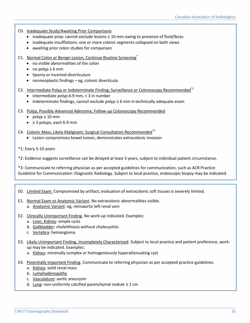
Canadian Association of Radiologists
CAR CT Colonography Standards 35
E0. Limited Exam. Compromised by artifact; evaluation of extracolonic soft tissues is severely limited.
E1. Normal Exam or Anatomic Variant. No extracolonic abnormalities visible. a. Anatomic Variant: eg, retroaortic left renal vein
E2. Clinically Unimportant Finding. No work-up indicated. Examples: a. Liver, Kidney: simple cysts b. Gallbladder: cholelithiasis without cholecystitis c. Vertebra: hemangioma
E3. Likely Unimportant Finding, Incompletely Characterized. Subject to local practice and patient preference, work-up may be indicated. Examples: a. Kidney: minimally complex or homogeneously hyperattenuating cyst
E4. Potentially Important Finding. Communicate to referring physician as per accepted practice guidelines. a. Kidney: solid renal mass b. Lymphadenopathy c. Vasculature: aortic aneurysm d. Lung: non-uniformly calcified parenchymal nodule ≥ 1 cm
C0. Inadequate Study/Awaiting Prior Comparisons • inadequate prep: cannot exclude lesions ≥ 10 mm owing to presence of fluid/feces • inadequate insufflations: one or more colonic segments collapsed on both views • awaiting prior colon studies for comparison
C1. Normal Colon or Benign Lesion; Continue Routine Screening* • no visible abnormalities of the colon • no polyp ≥ 6 mm • lipoma or inverted diverticulum • nonneoplastic findings – eg, colonic diverticula
C2. Intermediate Polyp or Indeterminate Finding; Surveillance or Colonoscopy Recommended*2 • intermediate polyp 6-9 mm, < 3 in number • indeterminate findings, cannot exclude polyp ≥ 6 mm in technically adequate exam
C3. Polyp, Possibly Advanced Adenoma; Follow-up Colonoscopy Recommended • polyp ≥ 10 mm • ≥ 3 polyps, each 6-9 mm
C4. Colonic Mass, Likely Malignant; Surgical Consultation Recommended*3 • Lesion compromises bowel lumen, demonstrates extracolonic invasion
*1: Every 5-10 years
*2: Evidence suggests surveillance can be delayed at least 3 years, subject to individual patient circumstance.
*3: Communicate to referring physician as per accepted guidelines for communication, such as ACR Practice Guideline for Communication: Diagnostic Radiology. Subject to local practice, endoscopic biopsy may be indicated.
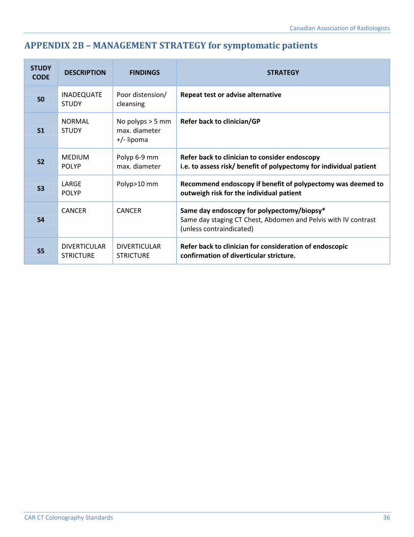
Canadian Association of Radiologists
CAR CT Colonography Standards 36
APPENDIX 2B – MANAGEMENT STRATEGY for symptomatic patients
STUDY CODE DESCRIPTION FINDINGS STRATEGY
S0 INADEQUATE STUDY
Poor distension/ cleansing
Repeat test or advise alternative
S1 NORMAL STUDY
No polyps > 5 mm max. diameter +/- lipoma
Refer back to clinician/GP
S2 MEDIUM POLYP
Polyp 6-9 mm max. diameter
Refer back to clinician to consider endoscopy i.e. to assess risk/ benefit of polypectomy for individual patient
S3 LARGE POLYP
Polyp>10 mm Recommend endoscopy if benefit of polypectomy was deemed to outweigh risk for the individual patient
S4 CANCER CANCER Same day endoscopy for polypectomy/biopsy*
Same day staging CT Chest, Abdomen and Pelvis with IV contrast (unless contraindicated)
S5 DIVERTICULAR STRICTURE
DIVERTICULAR STRICTURE
Refer back to clinician for consideration of endoscopic confirmation of diverticular stricture.
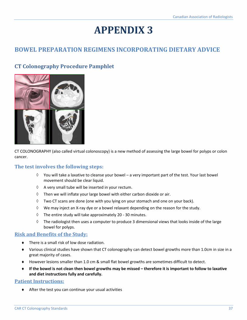
Canadian Association of Radiologists
CAR CT Colonography Standards 37
APPENDIX 3
BOWEL PREPARATION REGIMENS INCORPORATING DIETARY ADVICE
CT Colonography Procedure Pamphlet
CT COLONOGRAPHY (also called virtual colonoscopy) is a new method of assessing the large bowel for polyps or colon cancer.
The test involves the following steps: ◊ You will take a laxative to cleanse your bowel – a very important part of the test. Your last bowel
movement should be clear liquid. ◊ A very small tube will be inserted in your rectum. ◊ Then we will inflate your large bowel with either carbon dioxide or air. ◊ Two CT scans are done (one with you lying on your stomach and one on your back). ◊ We may inject an X-ray dye or a bowel relaxant depending on the reason for the study. ◊ The entire study will take approximately 20 - 30 minutes. ◊ The radiologist then uses a computer to produce 3 dimensional views that looks inside of the large
bowel for polyps.
Risk and Benefits of the Study: ♦ There is a small risk of low dose radiation. ♦ Various clinical studies have shown that CT colonography can detect bowel growths more than 1.0cm in size in a
great majority of cases. ♦ However lesions smaller than 1.0 cm & small flat bowel growths are sometimes difficult to detect. ♦ If the bowel is not clean then bowel growths may be missed – therefore it is important to follow to laxative
and diet instructions fully and carefully.
Patient Instructions: ♦ After the test you can continue your usual activities
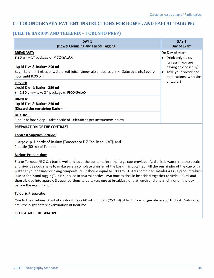
Canadian Association of Radiologists
CAR CT Colonography Standards 38
CT COLONOGRAPHY PATIENT INSTRUCTIONS FOR BOWEL AND FAECAL TAGGING
(DILUTE BARIUM AND TELEBRIX – TORONTO PREP)
DAY 1 (Bowel Cleansing and Faecal Tagging )
DAY 2 Day of Exam
BREAKFAST: 8:30 am – 1st package of PICO-SALAX Liquid Diet & Barium 250 ml Begin to drink 1 glass of water, fruit juice, ginger ale or sports drink (Gatorade, etc.) every hour until 8:00 pm
On Day of exam ♦ Drink only fluids
(unless if you are having colonoscopy)
♦ Take your prescribed medications (with sips of water)
LUNCH: Liquid Diet & Barium 250 ml ♦ 2:30 pm – take 2nd package of PICO-SALAX
DINNER: Liquid Diet & Barium 250 ml (Discard the remaining Barium)
BEDTIME: 1 hour before sleep – take bottle of Telebrix as per instructions below
PREPARATION OF THE CONTRAST
Contrast Supplies Include:
1 large cup, 1 bottle of Barium (Tomocat or E-Z Cat, Readi-CAT), and 1 bottle (60 ml) of Telebrix.
Barium Preparation:
Shake Tomocat/E-Z Cat bottle well and pour the contents into the large cup provided. Add a little water into the bottle and give it a good shake to make sure a complete transfer of the barium is obtained. Fill the remainder of the cup with water at your desired drinking temperature. It should equal to 1000 ml (1 litre) combined. Readi-CAT is a product which is used for "stool tagging". It is supplied in 450 ml bottles. Two bottles should be added together to yield 900 ml and then divided into approx. 3 equal portions to be taken, one at breakfast, one at lunch and one at dinner on the day before the examination.
Telebrix Preparation:
One bottle contains 60 ml of contrast. Take 60 ml with 8 oz (250 ml) of fruit juice, ginger ale or sports drink (Gatorade, etc.) the night before examination at bedtime.
PICO-SALAX IS THE LAXATIVE.
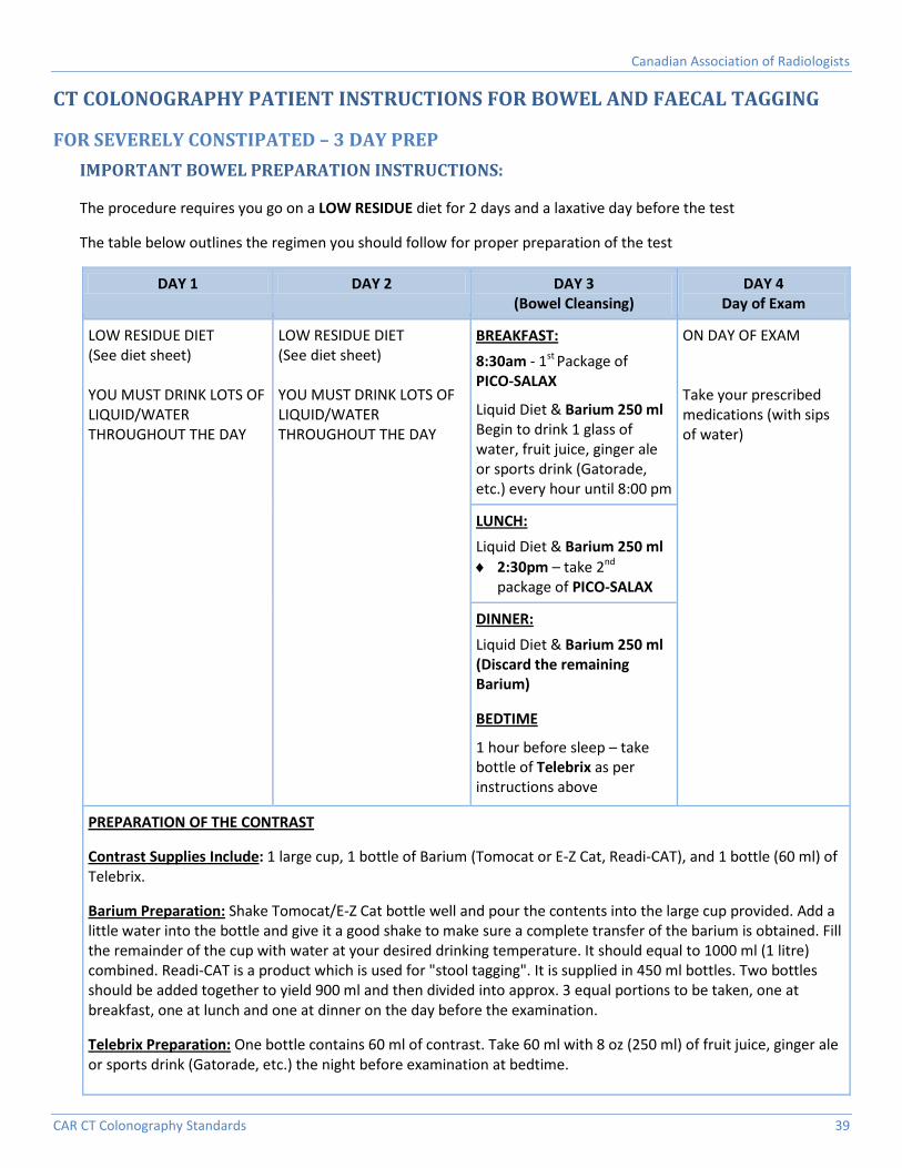
Canadian Association of Radiologists
CAR CT Colonography Standards 39
CT COLONOGRAPHY PATIENT INSTRUCTIONS FOR BOWEL AND FAECAL TAGGING
FOR SEVERELY CONSTIPATED – 3 DAY PREP IMPORTANT BOWEL PREPARATION INSTRUCTIONS:
The procedure requires you go on a LOW RESIDUE diet for 2 days and a laxative day before the test
The table below outlines the regimen you should follow for proper preparation of the test
DAY 1 DAY 2 DAY 3 (Bowel Cleansing)
DAY 4 Day of Exam
LOW RESIDUE DIET (See diet sheet) YOU MUST DRINK LOTS OF LIQUID/WATER THROUGHOUT THE DAY
LOW RESIDUE DIET (See diet sheet) YOU MUST DRINK LOTS OF LIQUID/WATER THROUGHOUT THE DAY
BREAKFAST: 8:30am - 1st Package of PICO-SALAX
Liquid Diet & Barium 250 ml Begin to drink 1 glass of water, fruit juice, ginger ale or sports drink (Gatorade, etc.) every hour until 8:00 pm
ON DAY OF EXAM Take your prescribed medications (with sips of water)
LUNCH: Liquid Diet & Barium 250 ml ♦ 2:30pm – take 2nd
package of PICO-SALAX
DINNER: Liquid Diet & Barium 250 ml (Discard the remaining Barium)
BEDTIME
1 hour before sleep – take bottle of Telebrix as per instructions above
PREPARATION OF THE CONTRAST
Contrast Supplies Include: 1 large cup, 1 bottle of Barium (Tomocat or E-Z Cat, Readi-CAT), and 1 bottle (60 ml) of Telebrix.
Barium Preparation: Shake Tomocat/E-Z Cat bottle well and pour the contents into the large cup provided. Add a little water into the bottle and give it a good shake to make sure a complete transfer of the barium is obtained. Fill the remainder of the cup with water at your desired drinking temperature. It should equal to 1000 ml (1 litre) combined. Readi-CAT is a product which is used for "stool tagging". It is supplied in 450 ml bottles. Two bottles should be added together to yield 900 ml and then divided into approx. 3 equal portions to be taken, one at breakfast, one at lunch and one at dinner on the day before the examination.
Telebrix Preparation: One bottle contains 60 ml of contrast. Take 60 ml with 8 oz (250 ml) of fruit juice, ginger ale or sports drink (Gatorade, etc.) the night before examination at bedtime.
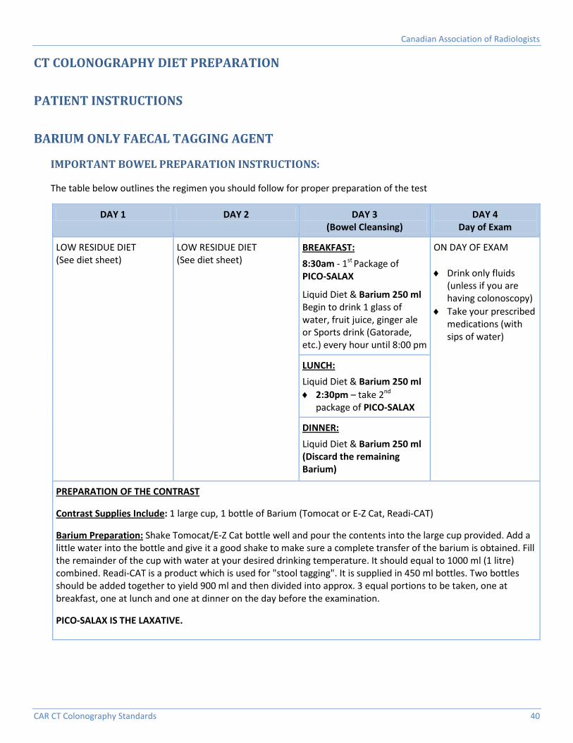
Canadian Association of Radiologists
CAR CT Colonography Standards 40
CT COLONOGRAPHY DIET PREPARATION
PATIENT INSTRUCTIONS
BARIUM ONLY FAECAL TAGGING AGENT
IMPORTANT BOWEL PREPARATION INSTRUCTIONS:
The table below outlines the regimen you should follow for proper preparation of the test
DAY 1 DAY 2 DAY 3 (Bowel Cleansing)
DAY 4 Day of Exam
LOW RESIDUE DIET (See diet sheet)
LOW RESIDUE DIET (See diet sheet)
BREAKFAST: 8:30am - 1st Package of PICO-SALAX
Liquid Diet & Barium 250 ml Begin to drink 1 glass of water, fruit juice, ginger ale or Sports drink (Gatorade, etc.) every hour until 8:00 pm
ON DAY OF EXAM ♦ Drink only fluids
(unless if you are having colonoscopy)
♦ Take your prescribed medications (with sips of water)
LUNCH: Liquid Diet & Barium 250 ml ♦ 2:30pm – take 2nd
package of PICO-SALAX
DINNER: Liquid Diet & Barium 250 ml (Discard the remaining Barium)
PREPARATION OF THE CONTRAST
Contrast Supplies Include: 1 large cup, 1 bottle of Barium (Tomocat or E-Z Cat, Readi-CAT)
Barium Preparation: Shake Tomocat/E-Z Cat bottle well and pour the contents into the large cup provided. Add a little water into the bottle and give it a good shake to make sure a complete transfer of the barium is obtained. Fill the remainder of the cup with water at your desired drinking temperature. It should equal to 1000 ml (1 litre) combined. Readi-CAT is a product which is used for "stool tagging". It is supplied in 450 ml bottles. Two bottles should be added together to yield 900 ml and then divided into approx. 3 equal portions to be taken, one at breakfast, one at lunch and one at dinner on the day before the examination.
PICO-SALAX IS THE LAXATIVE.
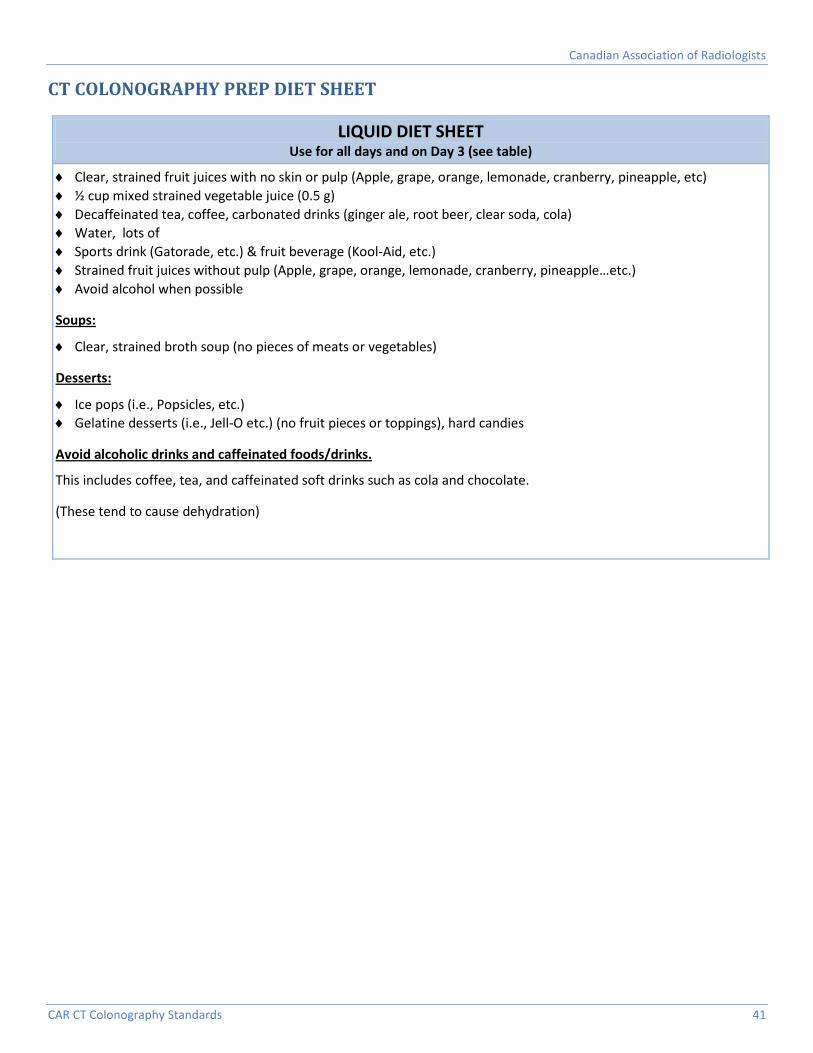
Canadian Association of Radiologists
CAR CT Colonography Standards 41
CT COLONOGRAPHY PREP DIET SHEET
LIQUID DIET SHEET Use for all days and on Day 3 (see table)
♦ Clear, strained fruit juices with no skin or pulp (Apple, grape, orange, lemonade, cranberry, pineapple, etc) ♦ ½ cup mixed strained vegetable juice (0.5 g) ♦ Decaffeinated tea, coffee, carbonated drinks (ginger ale, root beer, clear soda, cola) ♦ Water, lots of ♦ Sports drink (Gatorade, etc.) & fruit beverage (Kool-Aid, etc.) ♦ Strained fruit juices without pulp (Apple, grape, orange, lemonade, cranberry, pineapple…etc.) ♦ Avoid alcohol when possible
Soups:
♦ Clear, strained broth soup (no pieces of meats or vegetables)
Desserts:
♦ Ice pops (i.e., Popsicles, etc.) ♦ Gelatine desserts (i.e., Jell-O etc.) (no fruit pieces or toppings), hard candies
Avoid alcoholic drinks and caffeinated foods/drinks.
This includes coffee, tea, and caffeinated soft drinks such as cola and chocolate.
(These tend to cause dehydration)
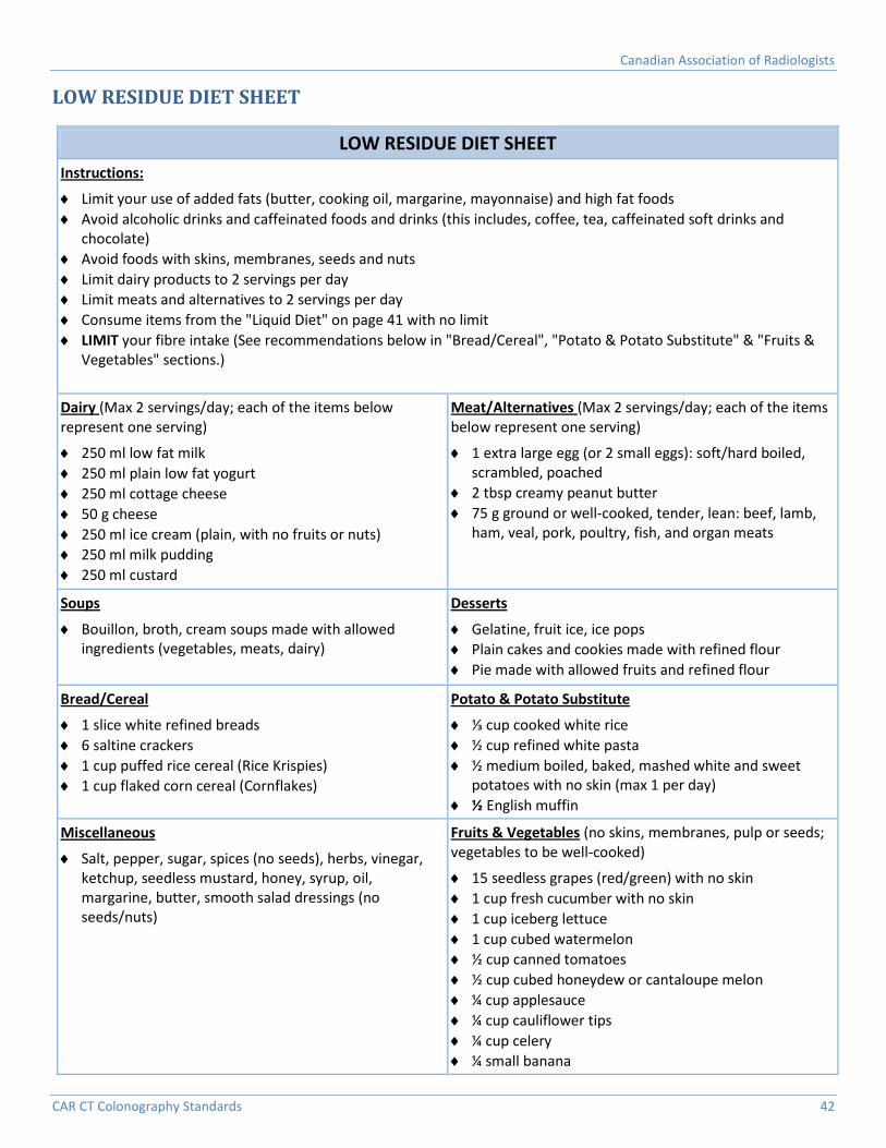
Canadian Association of Radiologists
CAR CT Colonography Standards 42
LOW RESIDUE DIET SHEET
LOW RESIDUE DIET SHEET Instructions:
♦ Limit your use of added fats (butter, cooking oil, margarine, mayonnaise) and high fat foods ♦ Avoid alcoholic drinks and caffeinated foods and drinks (this includes, coffee, tea, caffeinated soft drinks and
chocolate) ♦ Avoid foods with skins, membranes, seeds and nuts ♦ Limit dairy products to 2 servings per day ♦ Limit meats and alternatives to 2 servings per day ♦ Consume items from the "Liquid Diet" on page 41 with no limit ♦ LIMIT your fibre intake (See recommendations below in "Bread/Cereal", "Potato & Potato Substitute" & "Fruits &
Vegetables" sections.)
Dairy (Max 2 servings/day; each of the items below represent one serving)
♦ 250 ml low fat milk ♦ 250 ml plain low fat yogurt ♦ 250 ml cottage cheese ♦ 50 g cheese ♦ 250 ml ice cream (plain, with no fruits or nuts) ♦ 250 ml milk pudding ♦ 250 ml custard
Meat/Alternatives (Max 2 servings/day; each of the items below represent one serving)
♦ 1 extra large egg (or 2 small eggs): soft/hard boiled, scrambled, poached
♦ 2 tbsp creamy peanut butter ♦ 75 g ground or well-cooked, tender, lean: beef, lamb,
ham, veal, pork, poultry, fish, and organ meats
Soups
♦ Bouillon, broth, cream soups made with allowed ingredients (vegetables, meats, dairy)
Desserts
♦ Gelatine, fruit ice, ice pops ♦ Plain cakes and cookies made with refined flour ♦ Pie made with allowed fruits and refined flour
Bread/Cereal
♦ 1 slice white refined breads ♦ 6 saltine crackers ♦ 1 cup puffed rice cereal (Rice Krispies) ♦ 1 cup flaked corn cereal (Cornflakes)
Potato & Potato Substitute
♦ ⅓ cup cooked white rice ♦ ½ cup refined white pasta ♦ ½ medium boiled, baked, mashed white and sweet
potatoes with no skin (max 1 per day) ♦ ½ English muffin
Miscellaneous
♦ Salt, pepper, sugar, spices (no seeds), herbs, vinegar, ketchup, seedless mustard, honey, syrup, oil, margarine, butter, smooth salad dressings (no seeds/nuts)
Fruits & Vegetables (no skins, membranes, pulp or seeds; vegetables to be well-cooked)
♦ 15 seedless grapes (red/green) with no skin ♦ 1 cup fresh cucumber with no skin ♦ 1 cup iceberg lettuce ♦ 1 cup cubed watermelon ♦ ½ cup canned tomatoes ♦ ½ cup cubed honeydew or cantaloupe melon ♦ ¼ cup applesauce ♦ ¼ cup cauliflower tips ♦ ¼ cup celery ♦ ¼ small banana
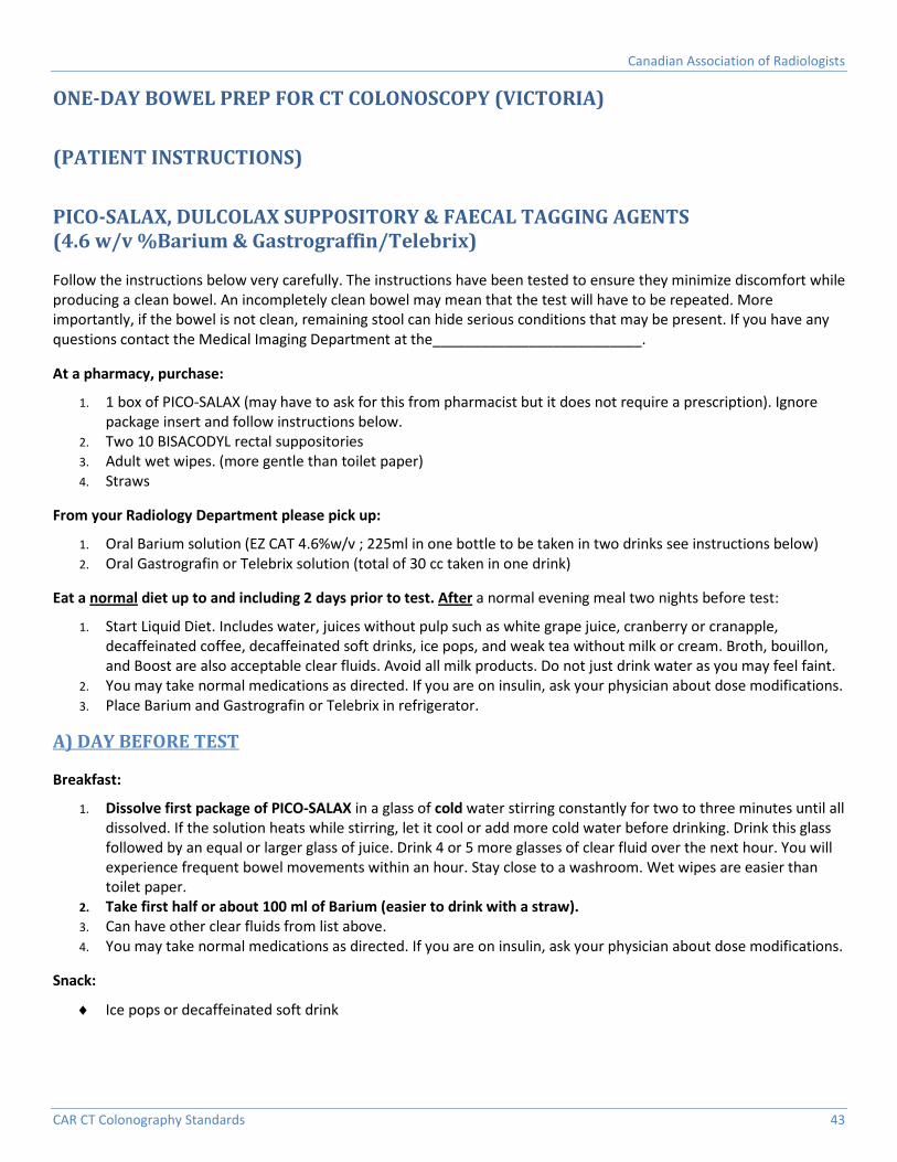
Canadian Association of Radiologists
CAR CT Colonography Standards 43
ONE-DAY BOWEL PREP FOR CT COLONOSCOPY (VICTORIA)
(PATIENT INSTRUCTIONS)
PICO-SALAX, DULCOLAX SUPPOSITORY & FAECAL TAGGING AGENTS (4.6 w/v %Barium & Gastrograffin/Telebrix)
Follow the instructions below very carefully. The instructions have been tested to ensure they minimize discomfort while producing a clean bowel. An incompletely clean bowel may mean that the test will have to be repeated. More importantly, if the bowel is not clean, remaining stool can hide serious conditions that may be present. If you have any questions contact the Medical Imaging Department at the__________________________.
At a pharmacy, purchase:
1. 1 box of PICO-SALAX (may have to ask for this from pharmacist but it does not require a prescription). Ignore package insert and follow instructions below.
2. Two 10 BISACODYL rectal suppositories 3. Adult wet wipes. (more gentle than toilet paper) 4. Straws
From your Radiology Department please pick up:
1. Oral Barium solution (EZ CAT 4.6%w/v ; 225ml in one bottle to be taken in two drinks see instructions below) 2. Oral Gastrografin or Telebrix solution (total of 30 cc taken in one drink)
Eat a normal diet up to and including 2 days prior to test. After a normal evening meal two nights before test:
1. Start Liquid Diet. Includes water, juices without pulp such as white grape juice, cranberry or cranapple, decaffeinated coffee, decaffeinated soft drinks, ice pops, and weak tea without milk or cream. Broth, bouillon, and Boost are also acceptable clear fluids. Avoid all milk products. Do not just drink water as you may feel faint.
2. You may take normal medications as directed. If you are on insulin, ask your physician about dose modifications. 3. Place Barium and Gastrografin or Telebrix in refrigerator.
A) DAY BEFORE TEST
Breakfast:
1. Dissolve first package of PICO-SALAX in a glass of cold water stirring constantly for two to three minutes until all dissolved. If the solution heats while stirring, let it cool or add more cold water before drinking. Drink this glass followed by an equal or larger glass of juice. Drink 4 or 5 more glasses of clear fluid over the next hour. You will experience frequent bowel movements within an hour. Stay close to a washroom. Wet wipes are easier than toilet paper.
2. Take first half or about 100 ml of Barium (easier to drink with a straw). 3. Can have other clear fluids from list above. 4. You may take normal medications as directed. If you are on insulin, ask your physician about dose modifications.
Snack:
♦ Ice pops or decaffeinated soft drink
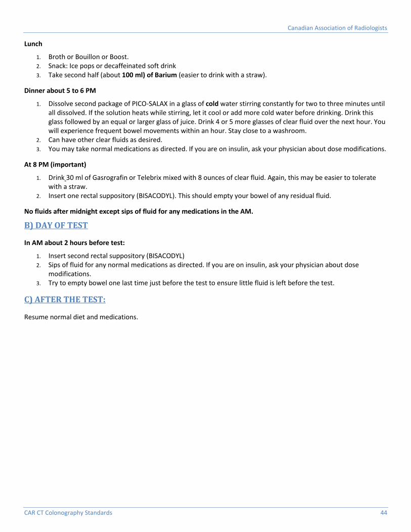
Canadian Association of Radiologists
CAR CT Colonography Standards 44
Lunch
1. Broth or Bouillon or Boost. 2. Snack: Ice pops or decaffeinated soft drink 3. Take second half (about 100 ml) of Barium (easier to drink with a straw).
Dinner about 5 to 6 PM
1. Dissolve second package of PICO-SALAX in a glass of cold water stirring constantly for two to three minutes until all dissolved. If the solution heats while stirring, let it cool or add more cold water before drinking. Drink this glass followed by an equal or larger glass of juice. Drink 4 or 5 more glasses of clear fluid over the next hour. You will experience frequent bowel movements within an hour. Stay close to a washroom.
2. Can have other clear fluids as desired. 3. You may take normal medications as directed. If you are on insulin, ask your physician about dose modifications.
At 8 PM (important)
1. Drink 30 ml of Gasrografin or Telebrix mixed with 8 ounces of clear fluid. Again, this may be easier to tolerate with a straw.
2. Insert one rectal suppository (BISACODYL). This should empty your bowel of any residual fluid.
No fluids after midnight except sips of fluid for any medications in the AM.
B) DAY OF TEST
In AM about 2 hours before test:
1. Insert second rectal suppository (BISACODYL) 2. Sips of fluid for any normal medications as directed. If you are on insulin, ask your physician about dose
modifications. 3. Try to empty bowel one last time just before the test to ensure little fluid is left before the test.
C) AFTER THE TEST:
Resume normal diet and medications.