Embed Size (px)
Citation preview
Carbohydrates
Today we will talk about carbohydrates ,metabolism
(digestion, absorption, and transport the carbohydrates)
*SLIDE (1):
This slides shows the*Dietary carbohydrates*that
need digestion
The carbohydrates are digestible
The carbohydrates almost constituent 50% of the
calories consume by human
Most of carbohydrates are complex carbohydrates in
the form of amylase and amylopectine which are
found in the grains ,potato ,rice ,and vegetables(are
the main source of carbohydrates)
Carbohydrates can be either polysaccharide(complex
molecule) or they can be disaccharide
Examples of polysaccharide:
Amylose :is polymer of glucose in alpha(1-4)glycosidic
linkage that is unbranched and non helical stright
chain (not stright)the importantce of it is to store
glucose for plant,and we use it as source of food
(calories)
Amylopectine: is polymer of alpha (1-4)glocose in each
(10-15)residue,there is abranch in this branching point
the
Amylose and Amylopectine are found in the starch
Examples of disaccharide:
Sucrose:
-that is found in vegetables ,fruits ,and
-All sugar is constituent a lot of calories for the body
-it is formed from: glucose and fructose, they are
connected by glycosidic bond of C1 of glucose with C2 of
fructose, alpha (1-2)
-the sugar food from animal origin is:
*LACTOSE
- Which found in milk.
-is formed from: glucose and galactose
-the glycosidic linkage is: beta (1-2) C1 of galactose with
C2 of glucose
***In addition to these, there are monosaccharides that
do not need digestion:
Glucose and fructose which found in some food like
honey and fruit
*SLIDE (2):
*The aim of digestion is to covert polysaccharide and
disaccharide to monosaccharide by breaking the
glycosidic linkage between sugar residues, this is
catalyzed by glycosidase which hydrolyze the bond by
addition of water
*The bond which connect the sugar residues together
called glycosidic bond which is between C1 (ANOMERIC
C) and any hydroxyl group (OH)
*In any sugar:
-C1 is linked to OXYGEN
-it has different ends (not symmetric)
1-REDUCING END
2-NON REDUCING END: it has (OH) on C4
-Sugar can be alpha or beta
1-ALPHA: if oxygen is below the ring
2-BETA: if oxygen is above the ring
***The glycosidases differ in their specificity with regard
to type of sugar and type of glycosidic bond , it is not one
type of glycosidases there are many type of glycosidases
that differ in their specificity (which sugar they can beta
glycosidic bond)
*SLIDE (3):
The enzymes that we are going to give it:

Alpha Amylase:
-it found in salivary, and disaccharidases (in the
intestine).
*NOTE:
-Not all carbohydrates are digested by human enzymes,
some of them like CELLULOSE cannot be digested by
human enzymes (because it is beta, and it is no specific
enzymes for cleaving beta bond).
-These dietary carbohydrates not necessary to
contribute to calories intake, they help in absorption of
water because they are hydrophilic so they facilitate the
transport of food in the small and large intestine and
some of them are digested by c-THE DIGESTION
PROCESS: colonic bacteria (bacteria which is found in
the colon)
***Human enzymes cannot digest the fiber
carbohydrates
*SLIDE (4):
1-During the MASTICATION the food that we eat (starch,
lactose, sucrose, and cellulose), the salivary gland
secrete an AMYLASE, which is the major enzyme in
saliva.
Note:
-the salivary gland secrete 1000 ml of saliva ,the major
enzymes of this saliva ,is ALPHA AMYLASE
2- Alpha Amylase begin acting in the mouth during
chewing of food but it rapidly will be inactivated when
the food reach the stomach because of low PH (1-1.5)
which stop the action of salivary amylase because of
denaturation so the importance of it :to get rid of any
food remain in the mouth.
3-Then the food enters the small intestine in the
Duodenum.
4-Then it enter the Pancreas that secrete its secretion
(Pancreatic juice which rich in bicarbohydrates that
neutralize the acid content of the stomach) so the
pancreatic amylase that is similar to salivary amylase
will continue the work by cutting the carbohydrates or
amino peptide into small pieces.
*Note:
-Alpha Amylase also works in alkaline media.
-Alpha Amylase cannot digest the isomaltose, maltose,
lactose,and sucrose .But they are digested by enzymes
that are found in small intestine.
5-The mucosal cell membrane-bound enzymes convert
the isomaltase, maltase ,lactase, and sucrose into
glucose , fructose , and galactose .
6- Then these products will be absorped through portal
circulation to reach the liver.
*SLIDE (5):
-This is a chain of starch connected by alpha (1-
4)glycosidic linkage but at the branching point the
glycosidic linkage is alpha (1-6).
-It has two kinds of different ends (reducing and non
reducing end)but in this case, it has one reducing end
and many non reducing end .
-The salivary amylase can cut this chain of starch
randomly so this process produce different product (it
may be produce maltose or isomaltose or dixtrine
depend on "how the salivary amylase cut the starch".
*SLIDE (6):
-This is a part of glycogen molecule
-glycogen has similar structure to the amylopectin ,but
glycogen is more branched .
-By cleavage of alpha (1-4), then the glycogen produce
the maltose and maltotriose.
-By cleavage of alpha (1-6) glycosidic linkage, then the
glycogen produce oligosaccharide .
*SLIDE (7):
-The disaccharidases are found in the intestinal brush
border which full of villi which have absorptive cells, the
aim of these cells is to increase the surface area and to
facilitate the digestion and absorption.
-The disaccharidases attach to the membrane of brush
border (they are produced by the cells but they are not
excretion, they do not leave the cell, they remain attach
to the membrane of brush border).
-We have 4 types of enzymes:
1-Glucoamylase.
2-Sucrase isomaltase.
3- Beta glycosidase .
4-Trehalase.
-These enzymes are found in Jejunum, and they have
different distribution but they mostly found in jejunum.
-The concentration of glucoamylase increase until it
reach to the ilium so the amount of enzymes in the
intestine increase as we go down ,but the maximum
concentration of glucomylase is found in jejunum in the
case of sucrose.
*SLIDE (8):
-GLUCOAMYLASE:
-is oligosaccharide and heavily glycosylated (full of sugar
residues which protect the enzymes, that are found on
the extracellular surface, from many proteases
,digestion enzymes , and trypsin (or from degradation ).
-It is similar structure to SUCRASE ISOMALTASE .
-It has 2 domain with similar activity .
-Glycoamylase is Exoglcosidase (opposite to
endoglucosidase ).
-This exoglcosidase act on the non reducing end .
*Note:
-If it acts only on "reducing end", there will be only one
end per molecule.
-But it acts on "non reducing end ",so there are large
number like in the case of dextrins.
-Exoglucosidase means it remove one glucose.
-The dextrins convert to isomaltose by this enzyme.
*SLIDE (9):
GLUCOAMYLASE ACTIVITY
-once the bond is cleaved by glucoamylase then the
bonds will be cleaved by maltase activity.
*SLIDE (10):
-SUCRASE ISOMALTASE COMPLEX STRUCTURE
-It is similar to glucoamylase
-It is attached to the membrane.
-It has 2 domain (sucrose domain and isomaltase
domain ).
-It has connecting segment (stalk).
-It has carbohydrates chain which protect from protease
action .
-It is hydrated so the enzymes protrude on the
membrane.
-It expose the protein and enzymes inside the lumen of
the small intestine.
(SUCRASE-ISOMALTASE COMPLEX) is 2 proteins because
it has C- terminal and N- terminal .
**Actually it synthesize as one protein but it cleaved .but
even it cleaved ,it remain attach to each other by "non
covalent interaction".
-Each subunit (sucrose-maltase domain and isomaltase-
maltase domain ) has maltase activity .
-Both of them will hydrolyze maltose as well.
Slide 11
Beta-glycosidase complex is a protein complex that
hydrolyze the carbon-carbon beta -glycosidic bond it's a
large glycolprotein attached to the brush border ,it
hydrolyzes lactose ,which is a galactose binding to
glucose via beta-glycosidic bond .this bond is hydrolyzed
by beta-glycosidase
So this enzyme catalyzes the hydrolysis of terminal non
reducing beta-galactose residues in lactose
Remember that glyco means ;sugar ,and this
enzyme recognize the beta-glycosidic bond
between galactose and glucose
This enzyme has two catalytic sites (two subunits),means
they are not similar.
The first subunit is called lactase; which has lactase
activity(hydrolysis of lactose as we mentioned before)
And the other subunit is called glucosylceramidase
A Brief explanation
Ceramide is a sphingolipid consist of a sphingosine
backbone attached to one fatty acid group ,while
cerebroside is a glycolipid contains ceramide and sugar
residues ,the sugar is either glucose or galactose .
So this enzyme (glucosylceramidase) ,hydrolyzes
ceramide attached to glucose or galactose ,(hydrolyzes
cerebrosides) ,breaking the bond between ceramide and
sugar.
Slide 12&13&14
Lactose intolerance;
Lactose is ingested from the food , and it founds
principally in milk and their products,lactase is an
enzyme that digest lactose ,which found in milk ,but
lactase level in the intestine start to decrease from the
first month of age ,so usually during the normal process
,level of lactase stay increasing during the late gestation
until the baby reaches the first month of his age, and
then start to decrease.
So the lactose intolerance due to lactase deficiency will
lead to pain ( abdominal pain ),nausea,flatulence.
Most of the population are non-persistent lactase ,the
adult level reaches 10% of the infant level at 5-7 age;
means that people who are between 5-7 years old will
have just 10% of the original level of lactase ,this case
roughly meets half of the world .
Asia world suffer from lactase deficiency ,while Europe
;west Europe ,like Holland ,the lactase level remains until
the adulthood in high levels .
The ingestion of lactose in milk for old people, will lead
to severe pain (abdominal pain),nausea and flatulence
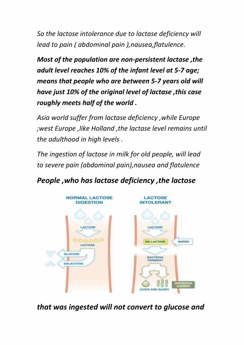
People ,who has lactase deficiency ,the lactose
that was ingested will not convert to glucose and
galactose ,so it will pass all the route to the large
intestine ,because the absorption of lactose in small
intestine doesn't occur ,now in the large intestine
,lactose will meet the bacteria of the large intestine ,this
bacteria utilize lactose and cause fermentation .
The fermentation will lead to the production of different
metabolites, which are:
2-carbon metabolites ( such as :acetic acid ), 3-
carbon metabolites ( such as :lactic acid ), CO2, H2,
in brief the products are a combination of gases.
Why diarrhea occurs in lactase deficiency people?
As we mentioned before lactose doesn't undergo
digestion and absorption, so it moves to the large
intestine and is subjected to fermentation by bacteria
,however all the products are small molecules (except
hydrogen),means they have a large osmotic pressure,
and water level inside intestine will be low in comparison
to metabolites ,consequentially ,water will move from
interstitial fluid to the intestine ,and this will increase the
level of water in the large intestine , the loss of that
water will be by diarrhea ,and this will lead to
dehydration.
10% of lactase level in adults is enough for drinking one
cup of milk ,but if you want to drink 10 cups ,you will
enjoy the pain (severe abdominal pain),and you will have
the chance for going to the hospital (if you are not from
European people)
One glass of milk is about 200 ml ,will resulted in
loss of one liter of extracellular fluid (water)
,dehydration occurs ,and a lot of fluid will lost by
lactase deficiency
Lactase deficiency might occur due diseases ; any
disease that affect or cause injury to small intestinal cells
, will lead to lactase ,sucrose and isomaltase deficiencies.
All these disaccharidases will be affected and will be lost
from the small intestine . the most enzyme by which
affected is lactase ,while the others(other
disaccharidases) can be affected in a transient form
,which means they recover rapidly.
Before discovering the proton pump inhibitor, which is a medicine
for peptic ulcer patients ,this patients were taking lactose in the
milk as a medicine ,but this way caused a terrible consequences ,as
diarrhea and flatulence so they prefer to tolerate the pain of peptic
ulcer ,instead of suffering from diarrhea and bloating
Slide 15,16,17
Absorption of sugars :
Sugars are highly polar molecules , so they can easily
bind to H2O by H bonds ,these hydrophilic molecules
(sugars which are hydrated by H2O ),will pass the lipid
by layer with the help of transporters; so there must be
transporters in the small intestine during the process of
absorption.
To carry this highly polar molecules across the
hydrophobic membrane into the cell , transport protein
are needed.
the transport of sugars across membrane is
achieved by two different mechanism ,which are:
** Active transport: there is a transporter called
NA+ dependent glucose transporter, this type of
transport requires energy (ATP).
** Facilitated transporter, which is facilitated
glucose transporters.
Two types of transporter are specific for
absorption of sugars across the intestinal cells
Study the picture that follows;
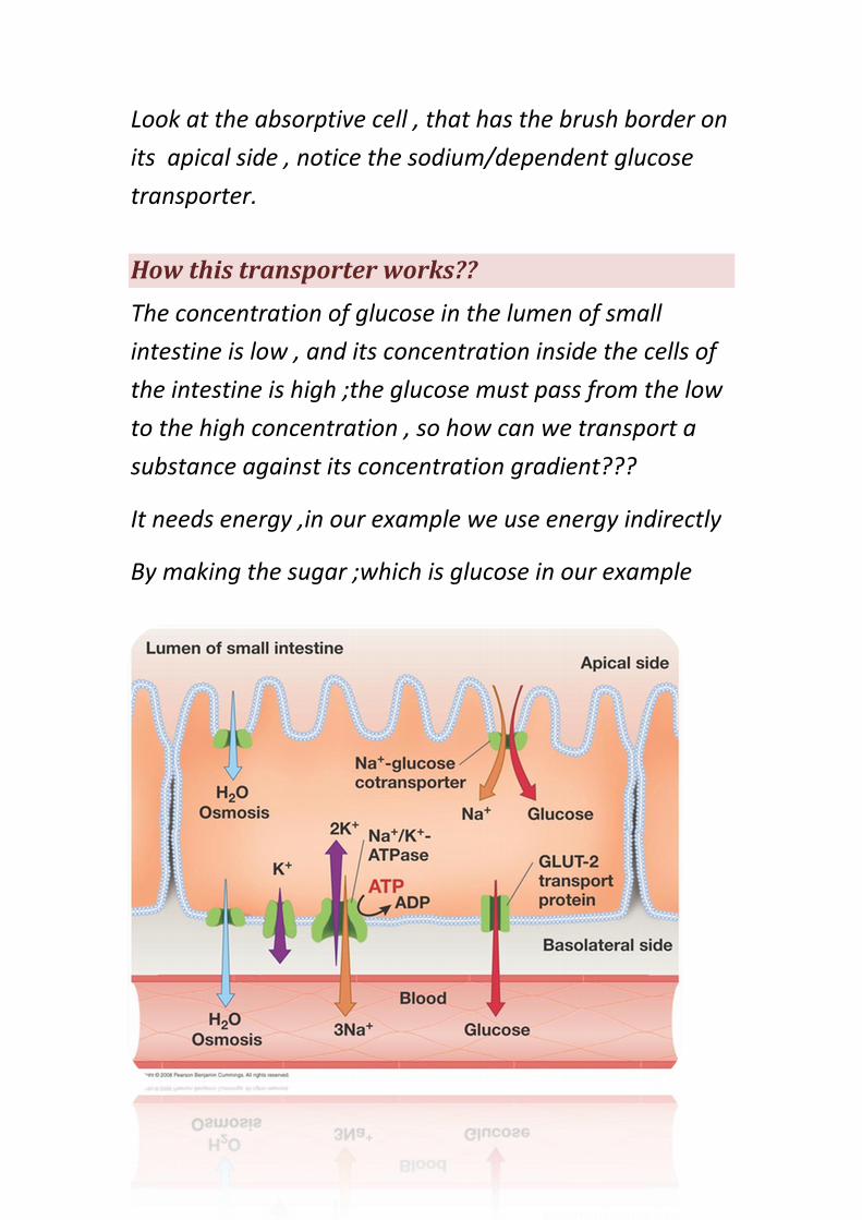
Look at the absorptive cell , that has the brush border on
its apical side , notice the sodium/dependent glucose
transporter.
How this transporter works??
The concentration of glucose in the lumen of small
intestine is low , and its concentration inside the cells of
the intestine is high ;the glucose must pass from the low
to the high concentration , so how can we transport a
substance against its concentration gradient???
It needs energy ,in our example we use energy indirectly
By making the sugar ;which is glucose in our example
;co-transported with a substance that is found in high
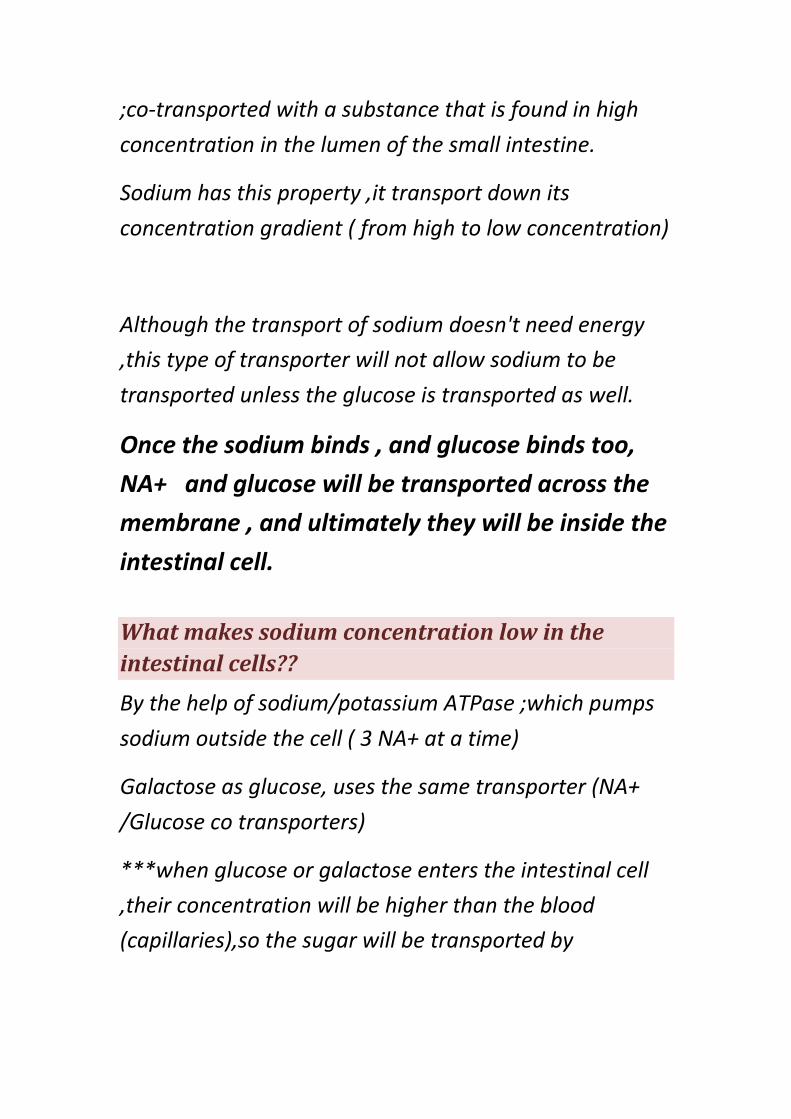
concentration in the lumen of the small intestine.
Sodium has this property ,it transport down its
concentration gradient ( from high to low concentration)
Although the transport of sodium doesn't need energy
,this type of transporter will not allow sodium to be
transported unless the glucose is transported as well.
Once the sodium binds , and glucose binds too,
NA+ and glucose will be transported across the
membrane , and ultimately they will be inside the
intestinal cell.
What makes sodium concentration low in the
intestinal cells??
By the help of sodium/potassium ATPase ;which pumps
sodium outside the cell ( 3 NA+ at a time)
Galactose as glucose, uses the same transporter (NA+
/Glucose co transporters)
***when glucose or galactose enters the intestinal cell
,their concentration will be higher than the blood
(capillaries),so the sugar will be transported by
facilitated diffusion across the serosal side from the cells
to capillaries (high to low concentration),
Glucose can be also transported to the intestine and out
of the intestine ( as we mentioned in the previous
paragraph) by facilitative diffusion .
When glucose is found in high concentration in the
lumen , it will use this property to transport across
membrane via facilitative transporters ,where we don't
need energy ,and as we mentioned before it can
transported across the serosal side going to the blood by
the same way.
Clinical Application
Before discovering the sodium / glucose dependent transporter,
when small babies are suffered from diarrhea and dehydration,
doctors were giving an intravenous rehydration solution,(in the
past they thought that sodium can't pass the intestinal cell ).
After discovering the transporter that pass the NA+ and glucose
together they found that if you give the baby a solution that
contains both sodium chloride and glucose ,the absorption of
sodium will occur (because of the transporter;NA+/Glucose
dependent transporter),and rehydration.
If you give to the patient just a solution of salt and water,
sodium will not be transported, why?????
*because sodium can't enter the cell without being co-
transported with glucose.
Doctors tend now to treat dehydration and diarrhea by giving
an oral rehydration that is available in pharmacies, by this way
patients can compensate for the body loss of fluids.
Facilitative transport of glucose :
The proteins that mediate the transport of glucose by
facilitated transport ,found in the membrane as integral
proteins, they possess outside and inside parts .
Mechanism of working:
glucose can bind to the transporter ,because it's opened
from its outside parts (the side toward the lumen ), once
glucose bind ,this will cause a conformational change in
the transporter in a way that it will close from outside
and open from the other side .
In the opposite side the glucose level is low , so it
dissociates (transported to capillaries ).
Glucose transported from the outside to inside , or from
the inside to the outside depending on the
concentration(from high to low).
*from outside to inside :from the lumen to cell
*from inside to outside : from cell to capillary
The facilitative transporters are a family of similar
proteins, have 12 membrane spanning domains, they
are not one protein, but number of proteins reach 14.
The most important are 5; they called GLUT (Glucose
transporter).
SLIDE (18):
*Please refer to the slide, the doctor read it all ,I want to
write the extra information related to it .
*We have numbers of protein which called (glucose
transporter) ,the most famous families are
GLUT(1,2,3,4,5).
*KM of enzyme: the concentration that is needed to
produce half of the maximum velocity.
-e.g.: the concentration of substrate (glucose) that cause
50% of the transporter to be occupied =1 ml .
-The normal concentration of glucose is (five times of
KM)=5 ml.
*The transporter work in full capacity.
*GLUT 1 is found in certain barrier function like in brain.
*As you notice, that in the liver there is GLUT 2 which
the KM=15 (high KM),why ?????...
-when the glucose in the blood is high after food intake ,
it is important to transfer glucose inside the liver so it is
active only when the concentration of glucose in the
blood is high .
*beta pancreatic (beta cells) is glucose sensor
-when glucose is enter to the beta cells of pancreatic
which is found in high concentration so the pancreatic
cell know that the blood glucose is high so it should
secrete the insulin.
*You notice that KM of GLUT 4 is 5 ml (low affinity)
why???...
-because the glucose transporters dependent on insulin
when insulin level increase, the amount of GLUT 4 is
increase .
-Glucose enter to adipose cells and muscles in only case
,when the glucose is found in high level.
*increase in glucose so the insulin is high (this is sensitive
to presence of insulin).
SLIDE (19)
STAIMULATION OF GLUCOSE TRANSPORT INTO MUSCLE
AND ADIPOSE CELLS BY Insulin.
**when insulin level is high so it is bind to its receptors
through number of events .
**GLUT are transported from vesicles (the initial ) ,then
it will be inserted to the membrane.
**when the insulin decrease, they will be recycled again
and they go to the GLUT vesicles and they aggregate in
the cytosol.
SLIDE (20)
*The doctor read exactly what we have in the slide .:
BEST WISHES……
DONE BY :Wafaa Shalan & Alaa Shqeirat