Embed Size (px)
Citation preview

217
Approximately half the dry mass of corn (maize) kernels is starch, a polysaccharide
that is broken down to glucose during digestion. Starch, along with the indigestible
polysaccharide cellulose, is also the starting material for production of the biofuel
ethanol. [© Sharon Dominick/iStockphoto]
C H A P T E R 8
CarbohydratesChapter Contents
1 MonosaccharidesA Monosaccharides Are Aldoses or Ketoses
B Monosaccharides Vary in Configuration and
Conformation
C Sugars Can Be Modified and Covalently Linked
2 PolysaccharidesA Lactose and Sucrose Are Disaccharides
B Cellulose and Chitin Are Structural
Polysaccharides
C Starch and Glycogen Are Storage
Polysaccharides
D Glycosaminoglycans Form Highly Hydrated Gels
3 GlycoproteinsA Proteoglycans Contain Glycosaminoglycans
B Bacterial Cell Walls Are Made of Peptidoglycan
C Many Eukaryotic Proteins Are Glycosylated
D Oligosaccharides May Determine Glycoprotein
Structure, Function, and Recognition
Carbohydrates or saccharides (Greek: sakcharon, sugar) are the most abun-dant biological molecules. They are chemically simpler than nucleotides oramino acids, containing just three elements—carbon, hydrogen, andoxygen—combined according to the formula (C � H2O)n, where n � 3. Thebasic carbohydrate units are called monosaccharides. There are numerous dif-ferent types of monosaccharides, which, as we discuss below, differ in theirnumber of carbon atoms and in the arrangement of the H and O atoms at-tached to the carbons. Furthermore, monosaccharides can be strung togetherin almost limitless ways to form polysaccharides.
Until the 1960s, carbohydrates were thought to have only passive roles asenergy sources (e.g., glucose and starch) and as structural materials (e.g., cellu-lose). Carbohydrates, as we will see, do not catalyze complex chemical reactionsas do proteins, nor do carbohydrates replicate themselves as do nucleic acids.And because polysaccharides are not built according to a genetic “blueprint,” asare nucleic acids and proteins, they tend to be much more heterogeneous—bothin size and in composition—than other biological molecules.
However, it has become clear that the innate structural variation in car-bohydrates is fundamental to their biological activity. The apparently haphaz-ard arrangements of carbohydrates on proteins and on the surfaces of cells arethe key to many recognition events between proteins and between cells. Anunderstanding of carbohydrate structure, from the simplest monosaccharidesto the most complex branched polysaccharides, is essential for appreciating thevaried functions of carbohydrates in biological systems.
JWCL460_c08_217-240.qxd 5/16/11 8:14 PM Page 217
CHO
HCOH
CH2OH
D-Glyceraldehyde
HCOH
CH2OH
CHO
HCOH
HCOH
CH2OH
CHO
HOCH
HCOH
CH2OH
HCOH
HCOH
CHO
HCOH
D-Allose
HCOH
CH2OH
HCOH
HCOH
CHO
HOCH
D-Altrose
HCOH
CH2OH
HCOH
HCOH
CHO
HOCH
D-Glucose(Glc)
HCOH
CH2OH
CHO
HOCH
D-Talose
HCOH
CH2OH
HCOH
CHO
HOCH
D-Mannose(Man)
HOCH
HCOH
CH2OH
HCOH
HCOH
CHO
HOCH
D-Gulose
HCOH
CH2OH
HCOH
CHO
HOCH
D-Idose
HOCH
HCOH
CH2OH
HCOH
CHO
HOCH
D-Galactose(Gal)
HOCH
HOCH
HOCH
D-Lyxose (Lyx)D-Ribose (Rib) D-Arabinose (Ara) D-Xylose (Xyl)
HCOH
CH2OH
HOCH
CHO
HOCH
HCOH
CH2OH
HOCH
CHO
HCOH
HCOH
CH2OH
HCOH
CHO
HOCH
HCOH
CH2OH
HCOH
CHO
HCOH
D-ThreoseD-Erythrose
Aldotriose
Aldotetroses
Aldopentoses
Aldohexoses
218
Chapter 8 Carbohydrates 1 MonosaccharidesK E Y C O N C E P T S
• The smallest sugars are aldoses or ketoses with the formula (C � H2O)n.• Monosaccharides cyclize to form � or � anomers.• The derivatives of monosaccharides include the aldonic acids, uronic acids,
alditols, deoxy sugars, and amino sugars.• Monosaccharides can be linked to each other or to other molecules by glycosidic
bonds.• Monosaccharides, or simple sugars, are synthesized from smaller precursors that
are ultimately derived from CO2 and H2O by photosynthesis.
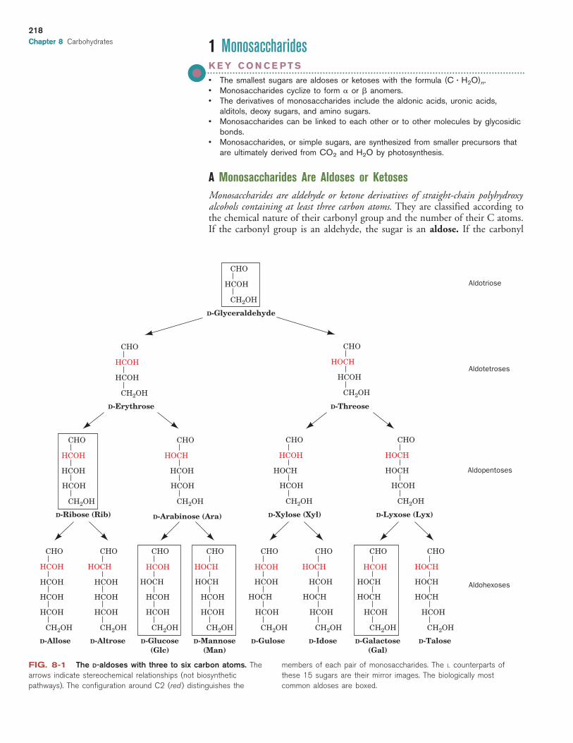
A Monosaccharides Are Aldoses or KetosesMonosaccharides are aldehyde or ketone derivatives of straight-chain polyhydroxyalcohols containing at least three carbon atoms. They are classified according tothe chemical nature of their carbonyl group and the number of their C atoms.If the carbonyl group is an aldehyde, the sugar is an aldose. If the carbonyl
Chapter 8
FIG. 8-1 The D-aldoses with three to six carbon atoms. The
arrows indicate stereochemical relationships (not biosynthetic
pathways). The configuration around C2 (red ) distinguishes the
members of each pair of monosaccharides. The L counterparts of
these 15 sugars are their mirror images. The biologically most
common aldoses are boxed.
JWCL460_c08_217-240.qxd 8/17/11 1:53 PM Page 218

group is a ketone, the sugar is a ketose. The smallest monosaccharides, thosewith three carbon atoms, are trioses. Those with four, five, six, seven, etc. C atoms are, respectively, tetroses, pentoses, hexoses, heptoses, etc.
The aldohexose D-glucose has the formula (C � H2O)6:
All but two of its six C atoms, C1 and C6, are chiral centers, so D-glu-cose is one of 24 � 16 possible stereoisomers. The stereochemistry andnomenclature of the D-aldoses are presented in Fig. 8-1. The assignmentof D or L is made according to the Fischer convention (Section 4-2): D
sugars have the same absolute configuration at the asymmetric center farthestfrom their carbonyl group as does D-glyceraldehyde (i.e., the ¬OH at C5 ofD-glucose is on the right in a Fischer projection). The L sugars are the mir-ror images of their D counterparts. Because L sugars are biologically muchless abundant than D sugars, the D prefix is often omitted.
Sugars that differ only by the configuration around one C atomare known as epimers of one another. Thus, D-glucose and D-man-nose are epimers with respect to C2. The most common aldoses in-clude the six-carbon sugars glucose, mannose, and galactose. Thepentose ribose is a component of the ribonucleotide residues ofRNA. The triose glyceraldehyde occurs in several metabolic path-ways.
The most common ketoses are those with their ketone functionat C2 (Fig. 8-2). The position of their carbonyl group gives ketoses oneless asymmetric center than their isomeric aldoses, so a ketohexose hasonly 23 � 8 possible stereoisomers (4 D sugars and 4 L sugars).The most common ketoses are dihydroxyacetone, ribulose, andfructose, which we will encounter in our studies of metabolism.
B Monosaccharides Vary in Configuration and ConformationAlcohols react with the carbonyl groups of aldehydes and ketones to formhemiacetals and hemiketals, respectively:
The hydroxyl and either the aldehyde or the ketone functions of monosaccha-rides can likewise react intramolecularly to form cyclic hemiacetals and
R� R�
R� C
Ketone
H
R�R�
C C
HOR
R OH +
+
Aldehyde HemiacetalAlcohol
R OH
OH
HemiketalAlcohol
O
R�
C
OR
OHO
OH
CH2OH
C H
C OH
OH
C
C
H
HO
H
H
2
1
3
4
5
6
D-Glucose
C
O H
219
Section 1 Monosaccharides
Dihydroxyacetone
D-TagatoseD-Psicose D-Fructose D-Sorbose
HCOH
CH2OH
HOCH
HOCH
HCOH
CH2OH
HOCH
HCOH
HCOH
CH2OH
HCOH
HOCH
HCOH
CH2OH
HCOH
C
HCOH
D-XyluloseD-Ribulose
CH2OH
O C
CH2OH
O C
CH2OH
O C
CH2OH
O
CH2OH
HCOH
HOCH
C
CH2OH
O
CH2OH
HCOH
C
HCOH
CH2OH
O
D-Erythrulose
CH2OH
HCOH
C
CH2OH
O
CH2OH
C
CH2OH
O
FIG. 8-2 The D-ketoses with three to six
carbon atoms. The configuration around C3
(red ) distinguishes the members of each pair.
The biologically most common ketoses are
boxed.
What is the number of L-ketoses??
JWCL460_c08_217-240.qxd 6/3/11 3:38 PM Page 219

hemiketals (Fig. 8-3). The configurations of the substituents of each carbonatom in these sugar rings are conveniently represented by their Haworth pro-jections, in which the heavier ring bonds project in front of the plane of thepaper and the lighter ring bonds project behind it.
A sugar with a six-membered ring is known as a pyranose in analogy withpyran (at left), the simplest compound containing such a ring. Similarly, sug-ars with five-membered rings are designated furanoses in analogy with furan(at left). The cyclic forms of glucose and fructose with six- and five-memberedrings are therefore known as glucopyranose and fructofuranose, respectively.
Cyclic Sugars Have Two Anomeric Forms. When a monosaccharide cy-clizes, the carbonyl carbon, called the anomeric carbon, becomes a chiralcenter with two possible configurations. The pair of stereoisomers that dif-fer in configuration at the anomeric carbon are called anomers. In the � anomer, the OH substituent of the anomeric carbon is on the oppositeside of the sugar ring from the CH2OH group at the chiral center that desig-nates the D or L configuration (C5 in hexoses). The other form is known asthe � anomer (Fig. 8-4).
The two anomers of D-glucose have slightly different physical and chem-ical properties, including different optical rotations (Section 4-2). The anomersfreely interconvert in aqueous solution, so at equilibrium, D-glucose is a mixtureof the � anomer (63.6%) and the � anomer (36.4%). The linear form is nor-mally present in only minute amounts.
Sugars Can Adopt Different Conformations. A given hexose or pentose canassume pyranose or furanose forms. In principle, hexoses and larger sugars can
220
O O
Pyran Furan
FIG. 8-3 Cyclization of glucose and fructose. (a) The linear form of D-glucose
yielding the cyclic hemiacetal �-D-glucopyranose. (b) The linear form of D-fructose yielding
the hemiketal �-D-fructofuranose. The cyclic sugars are shown as both Haworth projections
and in stick form embedded in their semitransparent space-filling models with C green, H
white, and O red.
O
OH
CH2OH
CH2OH
CH2OH
CH2OH
CH2OH
CH2OH
C H
C OH
OH
C
C
H
HO
H
H
1
2
3
4
5
6
OH H
H
H
H
HO
H OH
C
C
C C
HHOH
H
HO
OH
C
H
O
C
OHO
OHH
D-Glucose(linear form)
β-D-Glucopyranose(Haworth projection)
3
4
5
6
2
1
23
4
5
6
1
H(a)
O
C H
C OH
OH
C
C
HO
H
H
1
2
3
4
5
6
C
C C
H
HOH2C
HOH2C
OH
CO
OH H
D-Fructose(linear form)
β-D-Fructofuranose(Haworth projection)
4
5
6
2
(b)
HHO
3
1O
H OH
OH
H
OH H
6
2
34
5
CH2OH1
In a Haworth projection, which hydroxyl groups correspond to the hydroxyl
groups on the left in a Fischer projection?
?
JWCL460_c08_217-240.qxd 5/16/11 8:14 PM Page 220

6CH2OH
H
H O
H O
OHOH HH
HO
OHH
3
5
2
14
6CH2OH
OHH O
HOH HH
HO
OHH
3
5
2
14
1C
2CH
HO
OH
H3C
4CH OH
5CH OH
6CH2OH
α-D-Glucopyranose β-D-GlucopyranoseD-Glucose(linear form)
FIG. 8-4 � and � anomers. The monosaccharides �-D-glucopyranose and
�-D-glucopyranose, drawn as Haworth projections and ball-and-stick models, interconvert
through the linear form. They differ only by their configuration about the anomeric carbon, C1.
See Kinemage Exercise 7-1.
form rings of seven or more atoms, but such rings are rarely observed becauseof the greater stabilities of the five- and six-membered rings. The internal strainof three- and four-membered rings makes them less stable than the linear forms.
The use of Haworth formulas may lead to the erroneous impression thatfuranose and pyranose rings are planar. This cannot be the case, however, be-cause all the atomic orbitals in the ring atoms are tetrahedrally (sp3) hybridized.The pyranose ring, like the cyclohexane ring, can assume a chair conforma-tion, in which the substituents of each atom are arranged tetrahedrally. Of thetwo possible chair conformations, the one that predominates is the one inwhich the bulkiest ring substituents occupy equatorial positions rather thanthe more crowded axial positions (Fig. 8-5). Only �-D-glucose can simulta-neously have all five of its non-H substituents in equatorial positions. Perhapsthis is why glucose is the most abundant monosaccharide in nature.
Furanose rings can also adopt different conformations, whose stabilitiesdepend on the arrangements of bulky substituents. Note that a monosaccha-ride can readily shift its conformation, because no bonds are broken in theprocess. The shift in configuration between the � and � anomeric forms or be-tween the pyranose and furanose forms, which requires breaking and re-formingbonds, occurs slowly in aqueous solution. Other changes in configuration,such as epimerization, do not occur under physiological conditions withoutthe appropriate enzyme.
C Sugars Can Be Modified and Covalently LinkedBecause the cyclic and linear forms of aldoses and ketoses do interconvert,these sugars undergo reactions typical of aldehydes and ketones.
1. Oxidation of an aldose converts its aldehyde group to a carboxylic acidgroup, thereby yielding an aldonic acid such as gluconic acid (atright). Aldonic acids are named by appending the suffix -onic acid tothe root name of the parent aldose.
221
H
CH2OH
OH
OH
OH
OHH
HH
H
OO
H
H
H
OHHO
HO
CH2OH
H
HOH
FIG. 8-5 The two chair conformations of �-D-glucopyranose. In the conformation
on the left, which predominates, the relatively bulky OH and CH2OH substituents all occupy
equatorial positions, where they extend alternately above and below the ring. In the confor-
mation on the right (drawn in ball-and-stick form in Fig. 8-4, right), the bulky groups occupy
the more crowded axial (vertical) positions. See Kinemage Exercise 7-1.
D-Gluconic acid
1COOH2C OHH3C HHO4C OHH5C OHH6CH2OH
JWCL460_c08_217-240.qxd 5/16/11 8:14 PM Page 221
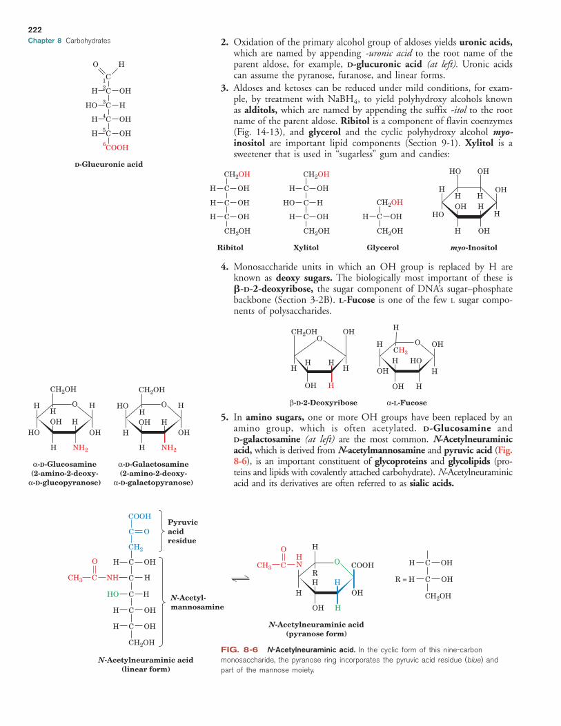
2. Oxidation of the primary alcohol group of aldoses yields uronic acids,which are named by appending -uronic acid to the root name of theparent aldose, for example, D-glucuronic acid (at left). Uronic acidscan assume the pyranose, furanose, and linear forms.
3. Aldoses and ketoses can be reduced under mild conditions, for exam-ple, by treatment with NaBH4, to yield polyhydroxy alcohols knownas alditols, which are named by appending the suffix -itol to the rootname of the parent aldose. Ribitol is a component of flavin coenzymes(Fig. 14-13), and glycerol and the cyclic polyhydroxy alcohol myo-inositol are important lipid components (Section 9-1). Xylitol is asweetener that is used in “sugarless” gum and candies:
4. Monosaccharide units in which an OH group is replaced by H areknown as deoxy sugars. The biologically most important of these is�-D-2-deoxyribose, the sugar component of DNA’s sugar–phosphatebackbone (Section 3-2B). L-Fucose is one of the few L sugar compo-nents of polysaccharides.
5. In amino sugars, one or more OH groups have been replaced by anamino group, which is often acetylated. D-Glucosamine and D-galactosamine (at left) are the most common. N-Acetylneuraminicacid, which is derived from N-acetylmannosamine and pyruvic acid (Fig.8-6), is an important constituent of glycoproteins and glycolipids (pro-teins and lipids with covalently attached carbohydrate). N-Acetylneuraminicacid and its derivatives are often referred to as sialic acids.
H
H
HOHO
HOOH
OH
H
OH H
O
H HHH
OH H
CH2OH
CH3
β-D-2-Deoxyribose α-L-Fucose
Ribitol
CH2OH
C OHH
C OHH
C OHH
CH2OH
Xylitol
CH2OH
C OHH
C HHO
C OHH
CH2OH
Glycerol myo-Inositol
CH2OH
C OHH
CH2OH
HO OH
OHH H
HOOH H
H
H OH
H
HO
OH
CH2OH
HH OH
H
H NH2
HH
OH
CH2OH
HHO
H
OH
H NH2
H
α-D-Glucosamine(2-amino-2-deoxy-
α-D-glucopyranose)
α-D-Galactosamine(2-amino-2-deoxy-
α-D-galactopyranose)
O O
222
Chapter 8 Carbohydrates
OH
COOH
N-Acetylneuraminic acid(pyranose form)
COOH
C O
CH2
H C OHO
CH3 C NH C H
HO C H
H C OH
H C OH
CH2OH
Pyruvicacidresidue
N-Acetyl-mannosamine
H
HNCCH3
O
N-Acetylneuraminic acid(linear form)
H C OH
OHCHR =
CH2OHHH
H
O
OH H
R
FIG. 8-6 N-Acetylneuraminic acid. In the cyclic form of this nine-carbon
monosaccharide, the pyranose ring incorporates the pyruvic acid residue (blue) and
part of the mannose moiety.
D-Glucuronic acid
1C2C OHH3C HHO4C OHH5C OHH6COOH
O H
JWCL460_c08_217-240.qxd 5/16/11 8:14 PM Page 222

223
Glycosidic Bonds Link the Anomeric Carbon to Other Compounds. Theanomeric group of a sugar can condense with an alcohol to form �- and�-glycosides (Greek: glykys, sweet; Fig. 8-7). The bond connecting the anomericcarbon to the alcohol oxygen is termed a glycosidic bond. N-Glycosidicbonds, which form between the anomeric carbon and an amine, are the bondsthat link D-ribose to purines and pyrimidines in nucleic acids:
Like peptide bonds, glycosidic bonds hydrolyze extremely slowly underphysiological conditions in the absence of appropriate hydrolytic enzymes.Consequently, an anomeric carbon that is involved in a glycosidic bond can-not freely convert between its � and � anomeric forms. Saccharides bearinganomeric carbons that have not formed glycosides are termed reducing sugars,because the free aldehyde group that is in equilibrium with the cyclic form ofthe sugar reduces mild oxidizing agents. Identification of a sugar as nonre-ducing is evidence that it is a glycoside.
–O
NH2
CH2 OP O
O
O
N
N
HH H
H
N-glycosidic bonds
O
OH
OH
O
HNN
N N
O
O
H H
...
...
H2N
H
–O CH2P O
O
O
H
OCH3
H
OH
CH2OH
HHO
H
OH
H OH
H
`-D-Glucose Methyl-`-D-glucoside
OCH3
Methyl-a-D-glucoside
+H
+
+ + H2OCH3OH
glycosidicbonds
OH
OH
CH2OH
HHO
H
H OH
H
OH
OH
CH2OH
HHO H
H OH
H
O
FIG. 8-7 Formation of glycosides. The acid-catalyzed condensation of �-D-glucose
with methanol yields an anomeric pair of methyl-D-glucosides.
C H E C K P O I N T
• How does an aldose differ from a ketose?• Draw a Fischer projection of D-glucose.
Draw two stereoisomers of this molecule,including one that is an epimer.
• Show how aldoses and ketoses can formfive- and six-membered rings.
• Draw a Haworth projection of D-glucoseand identify it as an � or � anomer.
• Explain why anomers of a monosaccharidecan readily interconvert whereas epimersdo not.
• Describe aldonic acids, uronic acids,alditols, deoxy sugars, and amino sugars.
• Explain why a sugar can form at least twodifferent glycosides.
Section 1 Monosaccharides
JWCL460_c08_217-240.qxd 8/17/11 1:53 PM Page 223

2 PolysaccharidesK E Y C O N C E P T S
• Disaccharides such as lactose and sucrose consist of two sugars linked byspecific glycosidic bonds.
• Cellulose and chitin are polymers of �(1S4)-linked glucose residues.• In starch and glycogen, glucose residues are linked mainly by �(1S4) bonds.• Glycosaminoglycans and other large heteropolysaccharides typically have a gel-
like structure.
Polysaccharides, which are also known as glycans, consist of monosaccharides linkedtogether by glycosidic bonds. They are classified as homopolysaccharides orheteropolysaccharides if they consist of one type or more than one type of mono-saccharide. Although the monosaccharide sequences of heteropolysaccharides can,in principle, be even more varied than those of proteins, many are composed ofonly a few types of monosaccharides that alternate in a repetitive sequence.
Polysaccharides, in contrast to proteins and nucleic acids, form branched aswell as linear polymers. This is because glycosidic linkages can be made to anyof the hydroxyl groups of a monosaccharide. Fortunately for structural bio-chemists, most polysaccharides are linear and those that branch do so in onlya few well-defined ways.
A complete description of an oligosaccharide or polysaccharide includesthe identities, anomeric forms, and linkages of all its component monosaccha-ride units. Some of this information can be gathered through the use of spe-cific exoglycosidases and endoglycosidases, enzymes that hydrolyzemonosaccharide units in much the same way that exopeptidases and endopep-tidases cleave amino acid residues from polypeptides (Section 5-3B). NMRmeasurements are also invaluable in determining both sequences and confor-mations of polysaccharides.
A Lactose and Sucrose Are DisaccharidesOligosaccharides containing three or more residues are relatively rare, occur-ring almost entirely in plants. Disaccharides, the simplest polysaccharides, aremore common. Many occur as the hydrolysis products of larger molecules.However, two disaccharides are notable in their own right. Lactose (at left),for example, occurs naturally only in milk, where its concentration ranges from0 to 7% depending on the species (Box 8-1). The systematic name for lac-tose, O -�-D-galactopyranosyl-(1S4)-D-glucopyranose, specifies its monosac-charides, their ring types, and how they are linked together. The symbol(1S4) combined with the � in the prefix indicates that the glycosidic bondlinks C1 of the � anomer of galactose to O4 of glucose. Note that lactose hasa free anomeric carbon on its glucose residue and is therefore a reducing sugar.
224
Chapter 8 Carbohydrates
Glucose
HOH
HO
H OH
H
O
3 2 3 2
HH
H
H
O
O
OH
Galactose
Lactose
44H
CH2OH CH2OH
OHH
H
6
5
6
1(β) 1(β)
OH5
Box 8-1 Biochemistryin Health and Disease
Lactose Intolerance
In infants, lactose (also known as milk sugar) is hydrolyzed by the
intestinal enzyme �-D-galactosidase (or lactase) to its compo-
nent monosaccharides for absorption into the bloodstream. The
galactose is enzymatically converted (epimerized) to glucose,
which is the primary metabolic fuel of many tissues.
Since mammals are unlikely to encounter lactose after they
have been weaned, most adult mammals have low levels of
�-galactosidase. Consequently, much of the lactose they might
ingest moves through their digestive tract to the colon, where
bacterial fermentation generates large quantities of CO2, H2, and
irritating organic acids. These products cause the embarrassing
and often painful digestive upset known as lactose intolerance.
Lactose intolerance, which was once considered a metabolic
disturbance, is actually the norm in adult humans, particularly
those of African and Asian descent. Interestingly, however,
�-galactosidase levels decrease only mildly with age in descendants
of populations that have historically relied on dairy products for
nutrition throughout life. Modern food technology has come to the
aid of milk lovers who develop lactose intolerance: Milk in which the
lactose has been hydrolyzed enzymatically is widely available.
JWCL460_c08_217-240.qxd 8/17/11 1:53 PM Page 224

The most abundant disaccharide is sucrose (at right), the major form inwhich carbohydrates are transported in plants. Sucrose is familiar to us as com-mon table sugar (see Kinemage Exercise 7-2). The systematic name for su-crose, O -�-D-glucopyranosyl-(1S2)-�-D-fructofuranoside, indicates that theanomeric carbon of each sugar (C1 in glucose and C2 in fructose) participatesin the glycosidic bond, and hence sucrose is not a reducing sugar.Noncarbohydrate molecules that mimic the taste of sucrose are used as sweet-ening agents in foods and beverages (Box 8-2). FructoseGlucose
H
H
OH H
H
H
HO
O
4 HO
OH
HO
O
Sucrose
2
1
5
4 63
5
6
OH
CH2OH
H HOCH2
H
23
1(α) (β)
CH2OH
225
Box 8-2 Perspectivesin Biochemistry
Artificial Sweeteners
Artificial sweeteners are added to processed foods and beverages
to impart a sweet taste without adding calories. This is possible
because the compounds mimic sucrose in its interactions with
taste receptors but either are not metabolized or contribute very
little to energy metabolism because they are used at such low
concentrations.
Naturally occurring saccharides, such as fructose, are slightly
sweeter than sucrose. Honey, which contains primarily fructose,
glucose, and maltose (a glucose disaccharide), is about 1.5 times as
sweet as sucrose. How is sweetness measured? There is no substi-
tute for the human sense of taste, so a panel of individuals sample
solutions of a compound and compare them to a reference solution
containing sucrose. The very sweet compounds listed below must
be diluted significantly before testing in this manner.
Compound Sweetness Relative to Sucrose
Acesulfame 200
Alitame 2000
Aspartame 180
Saccharin 350
Sucralose 600
One of the oldest artificial sweeteners
is saccharin, discovered in 1879 and
commonly consumed as Sweet’N Low®. In
the 1970s, extremely high doses of sac-
charin were found to cause cancer in
laboratory rats. Such doses are now con-
sidered to be so far outside of the range
used for sweetening as to be of insignificant concern to users.
Aspartame, the active ingredient in NutraSweet® and Equal®, was
approved for human use in 1981 and is currently the market leader:
Aspartylphenylalanine methyl ester (aspartame)
O
C+H3N CHCH
CH2
CH3
COO–
NH
CH2
O
OC
Unlike saccharin, which is not metabolized by the human body,
aspartame is broken down into its components: aspartate
(green), phenylalanine (red ), and methanol (blue). The Asp and
Phe, like all amino acids, can be metabolized, so aspartame is
not calorie-free. Methanol in large amounts is toxic; however, the
amount derived from an aspartame-sweetened drink is compara-
ble to the amount naturally present in the same volume of fruit
juice. Individuals with the genetic disease phenylketonuria, who
are unable to metabolize phenylalanine, are advised to avoid
ingesting excess Phe in the form of aspartame (or any other
polypeptide). The greatest drawback of aspartame may be its
instability to heat, which makes it unsuitable for baking. In
addition, aspartame in soft drinks hydrolyzes over a period of
months and hence loses its flavor.
Acesulfame is sometimes used in combination with aspar-
tame, since the two compounds act synergistically (i.e., their
sweetness when combined is greater than the sum of their
individual sweetnesses).
Other artificial sweeteners are derivatives of sugars, such as su-
cralose (Splenda®; see Problem 8-12), or of aspartame (e.g.,
alitame). Some plant extracts (e.g., Stevia) are also used as artifi-
cial sweeteners.
The market for artificial sweeteners is worth several billion
dollars annually. But surprisingly, the most successful sweetening
agents have not been the result of dedicated research efforts.
Instead, they were discovered by chance or mishap. For example,
aspartame was discovered in 1965 by a synthetic chemist who
unknowingly got a small amount of the compound on his fingers
and happened to lick them. Sucralose came to light in 1975 when
a student was asked to “test” a compound and misunderstood the
directions as “taste” the compound.
O
Saccharin
N
SO22�
O
OAcesulfame
N
SO2H3C 2�
JWCL460_c08_217-240.qxd 5/16/11 8:14 PM Page 225

B Cellulose and Chitin Are Structural PolysaccharidesPlants have rigid cell walls that can withstand osmotic pressure differences be-tween the extracellular and intracellular spaces of up to 20 atm. In large plants,such as trees, the cell walls also have a load-bearing function. Cellulose, theprimary structural component of plant cell walls (Fig. 8-8), accounts for overhalf of the carbon in the biosphere: Approximately 1015 kg of cellulose is es-timated to be synthesized and degraded annually.
Cellulose is a linear polymer of up to 15,000 D-glucose residues linked by�(1S4) glycosidic bonds:
X-Ray and other studies of cellulose fibers reveal that cellulose chains are flatribbons in which successive glucose rings are turned over 180° with respect toeach other. This permits the C3¬OH group of each glucose residue to form ahydrogen bond with the ring oxygen (O5) of the next residue. Parallel cellulosechains form sheets with interchain hydrogen bonds, including O2¬H���O6 andO6¬H���O3 bonds (Fig. 8-9). Stacks of these sheets are held together by hy-drogen bonds and van der Waals interactions. This highly cohesive structuregives cellulose fibers exceptional strength and makes them water insoluble de-spite their hydrophilicity. In plant cell walls, the cellulose fibers are embeddedin and cross-linked by a matrix containing other polysaccharides and lignin, aplasticlike phenolic polymer. The resulting composite material can withstandlarge stresses because the matrix evenly distributes the stresses among the cellu-lose reinforcing elements. The difficulty of removing these other substances,
Glucose
HOH
H OH
H
O
HH
H
H
O
O
OH
O
Glucose
Cellulose
HH
CH2OH CH2OH
OH
H
n
226
Chapter 8 Carbohydrates
FIG. 8-8 Electron micrograph of cellulose
fibers. The cellulose fibers in this sample of cell
wall from the alga Chaetomorpha are arranged
in layers. [Biophoto Associates/ Photo
Researchers.]
FIG. 8-9 Model of cellulose. Cellulose
fibers consist of �40 parallel, extended glycan
chains. Each of the �(1S4)-linked glucose units
in a chain is rotated 180� with respect to its
neighboring residues and is held in this position
by intrachain hydrogen bonds (dashed lines). The
glycan chains line up laterally to form sheets,
and these sheets stack vertically so that they are
staggered by half the length of a glucose unit.
The entire assembly is stabilized by
intermolecular hydrogen bonds. Hydrogen atoms
not participating in hydrogen bonds have been
omitted for clarity. [Illustration, Irving Geis. Image
from the Irving Geis Collection, Howard Hughes
Medical Institute. Reprinted with permission.]
JWCL460_c08_217-240.qxd 7/19/11 9:57 AM Page 226

however, is one of the main reasons why the cellulose in wood and agriculturalwaste, despite its abundance, cannot be easily converted to biofuels.
Although vertebrates themselves do not possess an enzyme capable of hy-drolyzing the �(1S4) linkages of cellulose, the digestive tracts of herbivorescontain symbiotic microorganisms that secrete a series of enzymes, collectivelyknown as cellulases, that do so. The same is true of termites. Nevertheless,the degradation of cellulose is a slow process because its tightly packed andhydrogen-bonded glycan chains are not easily accessible to cellulase and donot separate readily even after many of their glycosidic bonds have beenhydrolyzed. Thus, cows and other ruminants must chew their cud.
Chitin is the principal structural component of the exoskeletons of inverte-brates such as crustaceans, insects, and spiders and is also present in the cell wallsof most fungi and many algae. It is therefore almost as abundant as cellulose.Chitin is a homopolymer of �(1S4)-linked N-acetyl-D-glucosamine residues:
It differs chemically from cellulose only in that each C2¬OH group is re-placed by an acetamido function. X-Ray analysis indicates that chitin and cel-lulose have similar structures.
C Starch and Glycogen Are Storage PolysaccharidesStarch is a mixture of glycans that plants synthesize as their principal energyreserve. It is deposited in the chloroplasts of plant cells as insoluble granulescomposed of �-amylose and amylopectin. �-Amylose is a linear polymer ofseveral thousand glucose residues linked by �(1S4) bonds.
Note that although �-amylose is an isomer of cellulose, it has verydifferent structural properties. While cellulose’s �-glycosidic link-ages cause it to assume a tightly packed, fully extended conforma-tion (Fig. 8-9), �-amylose’s �-glycosidic bonds cause it to adopt anirregularly aggregating helically coiled conformation (Fig. 8-10).
nGlucoseGlucose
H
H
OH H
H
H
O
H
OH
HO
O O
�-Amylose
OH
CH2OH
H H
OHH
CH2OH
N-Acetylglucosamine
HOH
H NHCCH3
H
O
HH
H
H
O
O O
N-Acetylglucosamine
Chitin
HH
CH2OH CH2OH
OH
H
n
O
NHCCH3
O
O
227
Section 2 Polysaccharides
FIG. 8-10 �-Amylose. This regularly repeating polymer forms a left-handed helix.
Note the great differences in structure and properties that result from changing �-amylose’s
�(1S4) linkages to the �(1S4) linkages of cellulose (Fig. 8-9). [Illustration, Irving
Geis/Geis Archives Trust. Copyright Howard Hughes Medical Institute. Reproduced with
permission.]
JWCL460_c08_217-240.qxd 5/16/11 8:15 PM Page 227

Amylopectin (at left) consists mainly of �(1S4)-linked glucose residuesbut is a branched molecule with �(1S6) branch points every 24 to 30 glu-cose residues on average. Amylopectin molecules contain up to 106 glucoseresidues, making them some of the largest molecules in nature. The storage ofglucose as starch greatly reduces the large intracellular osmotic pressure thatwould result from its storage in monomeric form, because osmotic pressure isproportional to the number of solute molecules in a given volume (Section 2-1D). Starch is a reducing sugar, although it has only one residue, called thereducing end, that lacks a glycosidic bond.
The digestion of starch, the main carbohydrate source in the human diet,begins in the mouth. Saliva contains an amylase, which randomly hydrolyzesthe �(1S4) glycosidic bonds of starch. Starch digestion continues in the smallintestine under the influence of pancreatic amylase, which degrades starch toa mixture of small oligosaccharides. Further hydrolysis by an �-glucosidase,which removes one glucose residue at a time, and by a debranching enzyme,which hydrolyzes �(1S6) as well as �(1S4) bonds, produces monosaccha-rides that are absorbed by the intestine and transported to the bloodstream.
Glycogen, the storage polysaccharide of animals, is present in all cells butis most prevalent in skeletal muscle and in liver, where it occurs as cytoplas-mic granules (Fig. 8-11). The primary structure of glycogen resembles that ofamylopectin, but glycogen is more highly branched, with branch points oc-curring every 8 to 14 glucose residues. In the cell, glycogen is degraded formetabolic use by glycogen phosphorylase, which phosphorolytically cleavesglycogen’s �(1S4) bonds sequentially inward from its nonreducing ends.Glycogen’s highly branched structure, which has many nonreducing ends, permitsthe rapid mobilization of glucose in times of metabolic need. The �(1S6)branches of glycogen are cleaved by glycogen debranching enzyme (glycogenbreakdown is discussed further in Section 16-1).
D Glycosaminoglycans Form Highly Hydrated GelsThe extracellular spaces, particularly those of connective tissues such as cartilage,tendon, skin, and blood vessel walls, contain collagen (Section 6-1C) and otherproteins embedded in a gel-like matrix that is composed largely ofglycosaminoglycans. These unbranched polysaccharides consist of alternatinguronic acid and hexosamine residues. Solutions of glycosaminoglycans have aslimy, mucuslike consistency that results from their high viscosity and elasticity.
Hyaluronate Acts as a Shock Absorber and Lubricant. Hyaluronic acid(hyaluronate) is an important glycosaminoglycan component of connectivetissue, synovial fluid (the fluid that lubricates joints), and the vitreous humorof the eye. Hyaluronate molecules are composed of 250 to 25,000 �(1S4)-linked disaccharide units that consist of D-glucuronic acid and N-acetyl-D-glucosamine (GlcNAc) linked by a �(1S3) bond (Fig. 8-12). Hyaluronateis an extended, rigid molecule whose numerous repelling anionic groups bindcations and water molecules. In solution, hyaluronate occupies a volume�1000 times that in its dry state.
Hyaluronate solutions have a viscosity that is shear dependent (an objectunder shear stress has equal and opposite forces applied across its oppositefaces). At low shear rates, hyaluronate molecules form tangled masses thatgreatly impede flow; that is, the solution is quite viscous. As the shear stressincreases, the stiff hyaluronate molecules tend to line up with the flow andthus offer less resistance to it. This viscoelastic behavior makes hyaluronate so-lutions excellent biological shock absorbers and lubricants.
Some Glycosaminoglycans Are Sulfated. The other common glycosamino-glycans shown in Fig. 8-12 consist of 50 to 1000 sulfated disaccharide units.Chondroitin-4-sulfate and chondroitin-6-sulfate differ only in the sulfation
228
Chapter 8 Carbohydrates
FIG. 8-11 Glycogen granules in a liver
cell. In this photomicrograph, glycogen granules
are pink, the greenish objects are mitochondria,
and the yellow object is a fat globule. The
glycogen content of liver may reach 10% of its
net weight. [CNRI/Science Photo Library/Photo
Researchers.]
H
H
OH H
H
H
O
H
OH
HO
O
OH
CH2OH
H
H
OHH
O
CH2
O
O...
...
...
α(1n6) branch point
Amylopectin
JWCL460_c08_217-240.qxd 5/16/11 8:15 PM Page 228
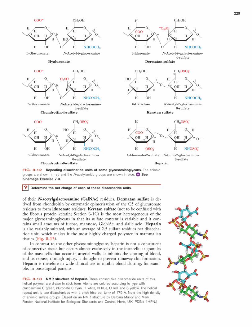
of their N-acetylgalactosamine (GalNAc) residues. Dermatan sulfate is de-rived from chondroitin by enzymatic epimerization of the C5 of glucuronateresidues to form iduronate residues. Keratan sulfate (not to be confused withthe fibrous protein keratin; Section 6-1C) is the most heterogeneous of themajor glycosaminoglycans in that its sulfate content is variable and it con-tains small amounts of fucose, mannose, GlcNAc, and sialic acid. Heparinis also variably sulfated, with an average of 2.5 sulfate residues per disaccha-ride unit, which makes it the most highly charged polymer in mammaliantissues (Fig. 8-13).
In contrast to the other glycosaminoglycans, heparin is not a constituentof connective tissue but occurs almost exclusively in the intracellular granulesof the mast cells that occur in arterial walls. It inhibits the clotting of blood,and its release, through injury, is thought to prevent runaway clot formation.Heparin is therefore in wide clinical use to inhibit blood clotting, for exam-ple, in postsurgical patients.
229
FIG. 8-12 Repeating disaccharide units of some glycosaminoglycans. The anionic
groups are shown in red and the N-acetylamido groups are shown in blue. See
Kinemage Exercise 7-3.
OH H
HH
H OH
H
O
4 1
COO–
H
HH
HO
H
H
O
3
CH2OH CH2OH
1
O
β
NHCOCH3 NHCOCH3
Oβ
D-Glucuronate N-Acetyl-D-glucosamine N-Acetyl-D-galactosamine-4-sulfate
N-Acetyl-D-galactosamine- 4-sulfate
N-Acetyl-D-glucosamine-6-sulfate
N-Acetyl-D-galactosamine-6-sulfate
N-Sulfo-D-glucosamine-6-sulfate
L-Iduronate-2-sulfate
Hyaluronate
OH H
H
H
H OH
H
O
4 1COO–
HH
H
H
O
3
1
O
α Oβ
L-Iduronate
Dermatan sulfate
–O3SO
–O3SO
H
OH H
HH
H OH
H
O
4 1
COO–
COO–
COO–
HH
H
H
O
3
1
O
β Oβ
D-Glucuronate
D-Glucuronate
Chondroitin-4-sulfate
H
HO
H OH
H
O
3
3
HH
H
H
O
1Oβ
NHCOCH3NHCOCH3
NHCOCH3 NHOSO3–
Oβ
D-Galactose
Keratan sulfate
OH H
HH
H OH
H
O
4 1 H
HOH
H
H
O
1
O
β Oβ
Chondroitin-6-sulfate
OH H
H
H
H
O
4 1 HH
H
HO
2
1Oα Oα
Heparin
H
H
4
4
4
4
4
H
CH2OHCH2OH CH2OSO3–
CH2OSO3– CH2OSO3
–
OSO3–
OHH
H
6
H
H
OH2
FIG. 8-13 NMR structure of heparin. Three consecutive disaccharide units of this
helical polymer are drawn in stick form. Atoms are colored according to type with
glucosamine C green, iduronate C cyan, H white, N blue, O red, and S yellow. The helical
repeat unit is two disaccharides with a pitch (rise per turn) of 17.5 Å. Note the high density
of anionic sulfate groups. [Based on an NMR structure by Barbara Mulloy and Mark
Forster, National Institute for Biological Standards and Control, Herts, U.K. PDBid 1HPN.]
Determine the net charge of each of these disaccharide units.?
JWCL460_c08_217-240.qxd 6/3/11 3:39 PM Page 229

Heparan sulfate, a ubiquitous cell-surface component as well as an extra-cellular substance in blood vessel walls and brain, resembles heparin but hasa far more variable composition with fewer N- and O -sulfate groups and moreN-acetyl groups. Heparan sulfate plays a critical role in development and inwound healing. Various growth factors bind to heparan sulfate, and the for-mation of complexes of the glycosaminoglycan, the growth factor, and thegrowth factor receptor is required to initiate cell differentiation and prolifera-tion. Specific sulfation patterns on heparan sulfate are required for the forma-tion of these ternary complexes.
Plants Produce Pectin. Plants do not synthesize glycosaminoglycans, but thepectins, which are major components of cell walls, may function similarly asshock absorbers. Pectins are heterogeneous polysaccharides with a core of�(1S4)-linked galacturonate residues interspersed with the hexose rhamnose.
The galacturonate residues may be modified by the addition of methyl andacetyl groups. Other polysaccharide chains, some containing the pentoses ara-binose and xylose and other sugars, are attached to the galacturonate. Theaggregation of pectin molecules to form bundles requires divalent cations(usually Ca2�), which form cross-links between the anionic carboxylate groupsof neighboring galacturonate residues. The tendency for pectin to form highlyhydrated gels is exploited in the manufacture of jams and jellies, to whichpectin is often added to augment the endogenous pectin content of the fruit.
Bacterial Biofilms Are a Type of Extracellular Matrix. Outside the labora-tory, bacteria are most often found growing on surfaces as a biofilm, an as-sociation of cells in a semisolid matrix (Fig. 8-14). The extracellular materialof the biofilm consists mostly of highly hydrated polysaccharides such as an-ionic poly-D-glucuronate, poly-N-acetylglucosamine, cellulose-like molecules,and acetylated glycans. A biofilm is difficult to characterize, as it typicallyhouses a mixture of species, and the proportions of its component polysaccha-rides can vary over time and space.
The gel-like consistency of a biofilm, for example, the plaque that formson teeth, prevents bacterial cells from being washed away and protects themfrom desiccation. Biofilms that develop on medical apparatus, such ascatheters, are problematic because they offer a foothold for pathogenic organ-isms and create a barrier to soluble antimicrobial agents.
3 GlycoproteinsK E Y C O N C E P T S
• Proteoglycans are large, glycosaminoglycan-containing proteins.• Bacterial cell walls consist of glycan chains cross-linked by peptides.• The oligosaccharide chains covalently attached to eukaryotic glycoproteins may
play a role in protein structure and recognition.
Many proteins are actually glycoproteins, with carbohydrate contents varyingfrom �1% to �90% by weight. Glycoproteins occur in all forms of life andhave functions that span the entire spectrum of protein activities, including
HO
HO
CH3
H
HH H
HO
RhamnoseOH
OH
230
Chapter 8 Carbohydrates
C H E C K P O I N T
• Describe the monosaccharide units andtheir linkages in the common disaccha-rides and polysaccharides.
• Explain why the systematic name of anoligosaccharide must include more thanjust the names of the componentmonosaccharides.
• Compare and contrast the structures andfunctions of cellulose, chitin, starch, andglycogen.
• How do the physical properties ofglycosaminoglycans and similar moleculesrelate to their biological roles?
FIG. 8-14 A Pseudomonas aeruginosa
biofilm. Bacterial colonies growing on the
surface of an agar plate form a biofilm with
complex architecture. [Courtesy of Roberto
Kolter, Harvard Medical School.]
JWCL460_c08_217-240.qxd 8/17/11 1:54 PM Page 230

those of enzymes, transport proteins, receptors, hormones, and structural pro-teins. The polypeptide chains of glycoproteins, like those of all proteins, aresynthesized under genetic control. Their carbohydrate chains, in contrast, areenzymatically generated and covalently linked to the polypeptide without therigid guidance of nucleic acid templates. For this reason, glycoproteins tendto have variable carbohydrate composition, a phenomenon known as micro-heterogeneity. Characterizing the structures of carbohydrates—and theirvariations—is one goal of the field of glycomics, which complements the studiesof genomics (for DNA) and proteomics (for proteins).
A Proteoglycans Contain GlycosaminoglycansProteins and glycosaminoglycans in the extracellular matrix aggregate cova-lently and noncovalently to form a diverse group of macromolecules knownas proteoglycans. Electron micrographs (Fig. 8-15a) and other evidence indi-cate that proteoglycans have a bottlebrush-like molecular architecture, with“bristles” noncovalently attached to a filamentous hyaluronate “backbone.”The bristles consist of a core protein to which glycosaminoglycans, mostoften keratan sulfate and chondroitin sulfate, are covalently linked (Fig. 8-15b).The interaction between the core protein and the hyaluronate is stabilized bya link protein. Smaller oligosaccharides are usually attached to the core pro-tein near its site of attachment to hyaluronate. These oligosaccharides are gly-cosidically linked to the protein via the amide N of specific Asn residues (andare therefore known as N-linked oligosaccharides; Section 8-3C). The ker-atan sulfate and chondroitin sulfate chains are glycosidically linked to the coreprotein via oligosaccharides that are covalently bonded to side chain O atomsof specific Ser or Thr residues (i.e., O -linked oligosaccharides).
Altogether, a central strand of hyaluronate, which varies in length from4000 to 40,000 Å, can have up to 100 associated core proteins, each of whichbinds �50 keratan sulfate chains of up to 250 disaccharide units and �100chondroitin sulfate chains of up to 1000 disaccharide units each. This accountsfor the enormous molecular masses of many proteoglycans, which range up totens of millions of daltons.
The extended brushlike structure of proteoglycans, together with the polyan-ionic character of their keratan sulfate and chondroitin sulfate components, causethese complexes to be highly hydrated. Cartilage, which consists of a meshworkof collagen fibrils that is filled in by proteoglycans, is characterized by its highresilience: The application of pressure on cartilage squeezes water away fromthe charged regions of its proteoglycans until charge–charge repulsions preventfurther compression. When the pressure is released, the water returns. Indeed,the cartilage in the joints, which lacks blood vessels, is nourished by this flowof liquid brought about by body movements. This explains why long periodsof inactivity cause cartilage to become thin and fragile.
B Bacterial Cell Walls Are Made of PeptidoglycanBacteria are surrounded by rigid cell walls that give them theircharacteristic shapes (Fig. 1-7) and permit them to live in hypo-tonic (less than intracellular salt concentration) environmentsthat would otherwise cause them to swell osmotically until their
231
Section 3 Glycoproteins
Core protein
Hyaluronate
Chondroitin sulfate
Keratan sulfate
N-Linked oligosaccharides
(b)
(a)
FIG. 8-15 A proteoglycan. (a) Electron micrograph showing a central
strand of hyaluronate, which supports numerous projections. [From Caplan, A.I., Sci.
Am. 251(4), 87 (1984). Copyright © Scientific American, Inc. Used by permission.]
(b) Bottlebrush model of the proteoglycan shown in Part a. Numerous core proteins are
noncovalently linked to the central hyaluronate strand. Each core protein has three
saccharide-binding regions.
JWCL460_c08_217-240.qxd 5/16/11 8:15 PM Page 231

plasma (cell) membranes lysed (burst). Bacterial cell walls are of consider-able medical significance because they are, in part, responsible for bacterialvirulence (disease-evoking power). In fact, the symptoms of many bacterialdiseases can be elicited in animals merely by injecting bacterial cell walls.Furthermore, bacterial cell wall components are antigenic (Section 7-3B), sosuch injections often invoke immunity against these bacteria.
Bacteria are classified as gram-positive or gram-negative according towhether or not they take up Gram stain (a procedure developed in 1884 byChristian Gram in which heat-fixed cells are successively treated with the dyecrystal violet and iodine and then destained by ethanol or acetone). Gram-positive bacteria (Fig. 8-16a) have a thick cell wall (�250 Å) surroundingtheir plasma membrane, whereas gram-negative bacteria (Fig. 8-16b) have athin cell wall (�30 Å) covered by a complex outer membrane. This outermembrane functions, in part, to exclude substances toxic to the bacterium, in-cluding Gram stain. This accounts for the observation that gram-negative bac-teria are more resistant to antibiotics than are gram-positive bacteria.
The cell walls of bacteria consist of covalently linked polysaccharide andpolypeptide chains, which form a baglike macromolecule that completely en-cases the cell. This framework, whose structure was elucidated in large part byJack Strominger, is known as a peptidoglycan. Its polysaccharide componentconsists of linear chains of alternating �(1S4)-linked GlcNAc and N-acetylmuramic acid (Latin: murus, wall). The lactic acid group of N-acetylmuramic acid forms an amide bond with a D-amino acid–containingtetrapeptide to form the peptidoglycan repeating unit (Fig. 8-17). Neighboringparallel peptidoglycan chains are covalently cross-linked through their tetrapep-tide side chains, although only �40% of possible cross-links are made.
In the bacterium Staphylococcus aureus, whose tetrapeptide has thesequence L-Ala-D-isoglutamyl-L-Lys-D-Ala, the cross-link consists of a pen-taglycine chain that extends from the terminal carboxyl group of onetetrapeptide to the e-amino group of the Lys in a neighboring tetrapeptide.To explore this structure, Simon Foster has used atomic force microscopy(AFM), an imaging technique that reports the variation in the force betweena probe that is several nanometers in diameter and a surface of interest as theprobe is scanned over the surface; its resolution is as little as several ångstroms.Foster’s model of the cell wall of the gram-negative bacterium Bacillus subtilisis shown in Figure 8-18. Several glycan chains are cross-linked much as de-scribed above to form a peptidoglycan “rope,” which due to its natural twist,forms an �50-nm-diameter helical cable up to 50 �m in length that coilsaround the long axis of the bacterium to form its cell wall. This structure ispresumably stabilized by the formation of covalent cross-links between neigh-boring segments of the coil. The cell walls of gram-negative bacteria appear to
232
Chapter 8 Carbohydrates
FIG. 8-16 Bacterial cell walls. This diagram compares the cell envelopes of
(a) gram-positive bacteria and (b) gram-negative bacteria.
Peptidoglycan(cell wall)
Plasmamembrane
Cytoplasm
Peptidoglycan(cell wall)
Plasmamembrane
Cytoplasm
Periplasmicspace
Outer membrane
(a) Gram-positive bacteria (b) Gram-negative bacteria
JWCL460_c08_217-240.qxd 5/16/11 8:15 PM Page 232

be only one layer thick, whereas as those of gram-positive bacteria are postu-lated to consist of several such layers. How the peptidoglycan imposes cellshape is unknown.
The D-amino acids of peptidoglycans render them resistant to proteases,which are mostly specific for L-amino acids. However, lysozyme, an enzymethat is present in tears, mucus, and other vertebrate body secretions, as wellas in egg whites, catalyzes the hydrolysis of the �(1S4) glycosidic linkage be-tween N-acetylmuramic acid and N-acetylglucosamine (the structure andmechanism of lysozyme are examined in detail in Section 11-4). The cell wallis also compromised by antibiotics that inhibit its biosynthesis (Box 8-3).
233
FIG. 8-17 Peptidoglycan. (a) The repeating unit of peptidoglycan is an
N-acetylglucosamine–N-acetylmuramic acid disaccharide whose lactyl side chain forms an
amide bond with a tetrapeptide. The tetrapeptide of S. aureus is shown. The isoglutamyl
residue is so designated because it forms a peptide link via its �-carboxyl group. (b) The
S. aureus bacterial cell wall peptidoglycan, showing its pentaglycine connecting bridges
(purple).
FIG. 8-18 Model of the B. subtilis cell wall. The cell wall consists of a right-handed
helical cable composed of several peptidoglycan strands that wraps about the bacterium’s
plasma membrane. The cell is �3 �m long. [Courtesy of Simon Foster, University of
Sheffield, U.K.]
OH H
H
H
H
H
O
CH2OH
CH
C O
H
H
H
H
H
O
CH2OH
O
NHCOCH3
COO–
NHCOCH3
O
H3C CH C O
O
OC
NH
CH3
NH
CH
CH3
CH2
CH2
NH
CH (CH2)4 NH3+
OC
NH
CH
COO–
L-Ala
Isoglutamyl
L-Lys
N-Acetylglucosamine N-Acetylmuramic acid
D-Ala
Peptidechain
Pentaglycinebridge
N -Acetylmuramic acid
N -Acetylglucosamine
(a) (b)
JWCL460_c08_217-240.qxd 5/16/11 8:15 PM Page 233

C Many Eukaryotic Proteins Are GlycosylatedAlmost all the secreted and membrane-associated proteins of eukaryotic cellsare glycosylated. Oligosaccharides are covalently attached to proteins byeither N-glycosidic or O -glycosidic bonds.
N-Linked Oligosaccharides Are Attached to Asparagine. In N-linkedoligosaccharides, GlcNAc is invariably �-linked to the amide nitrogen of an Asnresidue in the sequence Asn-X-Ser or Asn-X-Thr, where X is any amino acid ex-cept Pro and only rarely Asp, Glu, Leu, or Trp.
N-Glycosylation occurs cotranslationally, that is, while the polypeptide is be-ing synthesized. Proteins containing N-linked oligosaccharides typically areglycosylated and then processed as elucidated, in large part, by Stuart Kornfeld(Fig. 8-19):
1. An oligosaccharide containing 9 mannose residues, 3 glucose residues,and 2 GlcNAc residues is attached to the Asn of a growing polypep-tide chain that is being synthesized by a ribosome associated with theendoplasmic reticulum (Section 9-4D).
2. Some of the sugars are removed during processing, which begins in thelumen (internal space) of the endoplasmic reticulum and continues inthe Golgi apparatus (Fig. 1-8). Enzymatic trimming is accomplishedby glucosidases and mannosidases.
HOH H
H
H
O
NHCOCH3
CH2OH
NH
H
C
C O
O
CH2 CH
NH
HO
Ser or Thr
X
GlcNAc
Asn
234
Box 8-3 Biochemistryin Health and Disease
Peptidoglycan-Specific Antibiotics
In 1928, Alexander Fleming noticed
that the chance contamination of a
bacterial culture plate with the mold
Penicillium notatum resulted in the
lysis of the bacteria in the vicinity of
the mold. This was caused by the
presence of penicillin, an antibiotic
secreted by the mold. Penicillin con-
tains a thiazolidine ring (red ) fused to
a �-lactam ring (blue). A variable
R group is bonded to the �-lactam
ring via a peptide link.
Penicillin specifically binds to and inactivates enzymes that
cross-link the peptidoglycan strands of bacterial cell walls.
Since cell wall expansion in growing cells requires that their
rigid cell walls be opened up for the insertion of new cell wall
material, exposure of growing bacteria to penicillin results in
cell lysis. However, since no human enzyme binds penicillin
specifically, it is not toxic to humans and is therefore therapeu-
tically useful.
Most bacteria that are resistant to penicillin secrete the enzyme
penicillinase (also called �-lactamase), which inactivates peni-
cillin by cleaving the amide bond of its �-lactam ring. Attempts to
overcome this resistance have led to the development of �-lacta-
mase inhibitors such as sulbactam that are often administered in
combination with penicillin derivatives.
Multiple drug resistant bacteria are a growing problem. For
many years, vancomycin, the so-called antibiotic of last resort,
has been used to treat bacterial infections that do not succumb
to other antibiotics. Vancomycin inhibits the transpeptidation
(cross-linking) reaction of bacterial cell wall synthesis by binding
to the peptidoglycan precursor. However, bacteria can become
resistant to vancomycin by acquiring a gene that allows cell wall
synthesis from a slightly different precursor sequence, to which
vancomycin binds much less effectively.
One limitation of drugs such as vancomycin and penicillin,
particularly for slow-growing bacteria, is that the drug may halt
bacterial growth without actually killing the cells. For this reason,
effective antibacterial treatments may require combinations of
antibiotics over a course of several weeks.
C
HN
HC C CH S
N CHC
O
O
R
Penicillin
CH3
CH3
COO–
JWCL460_c08_217-240.qxd 5/16/11 8:15 PM Page 234

3. Additional monosaccharide residues, including GlcNAc, galactose, fucose, and sialic acid, are added by the action of specific glycosyl-transferases in the Golgi apparatus.
The exact steps of N-linked oligosaccharide processing vary with the iden-tity of the glycoprotein and the battery of endoglycosidases in the cell, but allN-linked oligosaccharides have a common core pentasaccharide with the fol-lowing structure:
In some glycoproteins, processing is limited, leaving “high-mannose”oligosaccharides; in other glycoproteins, extensive processing generates largeoligosaccharides containing several kinds of sugar residues. There is enormous di-versity among the oligosaccharides of N-linked glycoproteins. Indeed, even glycopro-teins with a given polypeptide chain exhibit considerable microheterogeneity,
Man �(1n6)
Man �(1n3)Man �(1n4) GlcNAc �(1n4) GlcNAc
235
14-residue oligosaccharide is attached to Asn of a polypeptide.
Removal of monosaccharide units produces a (mannose)3(GlcNAc)2 oligosaccharide. This (mannose)3(GlcNAc)2 coreis found in all N-linked oligosaccharides.
Further trimming and addition of other sugars yields a variety of N-linked oligosaccharides.
Polypeptide Asn
Corepentasaccharide
N-Acetylglucosamine
Mannose
Galactose
Glucose
Sialic acid
L-Fucose
FIG. 8-19 Synthesis of N-linked oligosaccharides. The
addition of a (mannose)9(glucose)3(GlcNAc)2 oligosaccharide is
followed by removal of monosaccharides as catalyzed by glycosidases,
and the addition of other monosaccharides as catalyzed by
glycosyltransferases. The core pentasaccharide occurs in all N-linked
oligosaccharides. [Adapted from Kornfeld, R. and Kornfeld, S., Annu.
Rev. Biochem. 54, 640 (1985).] See Kinemage Exercise 7-4.
JWCL460_c08_217-240.qxd 5/16/11 8:15 PM Page 235
presumably as a consequence of incomplete glycosylation and lack of absolutespecificity on the part of glycosidases and glycosyltransferases.
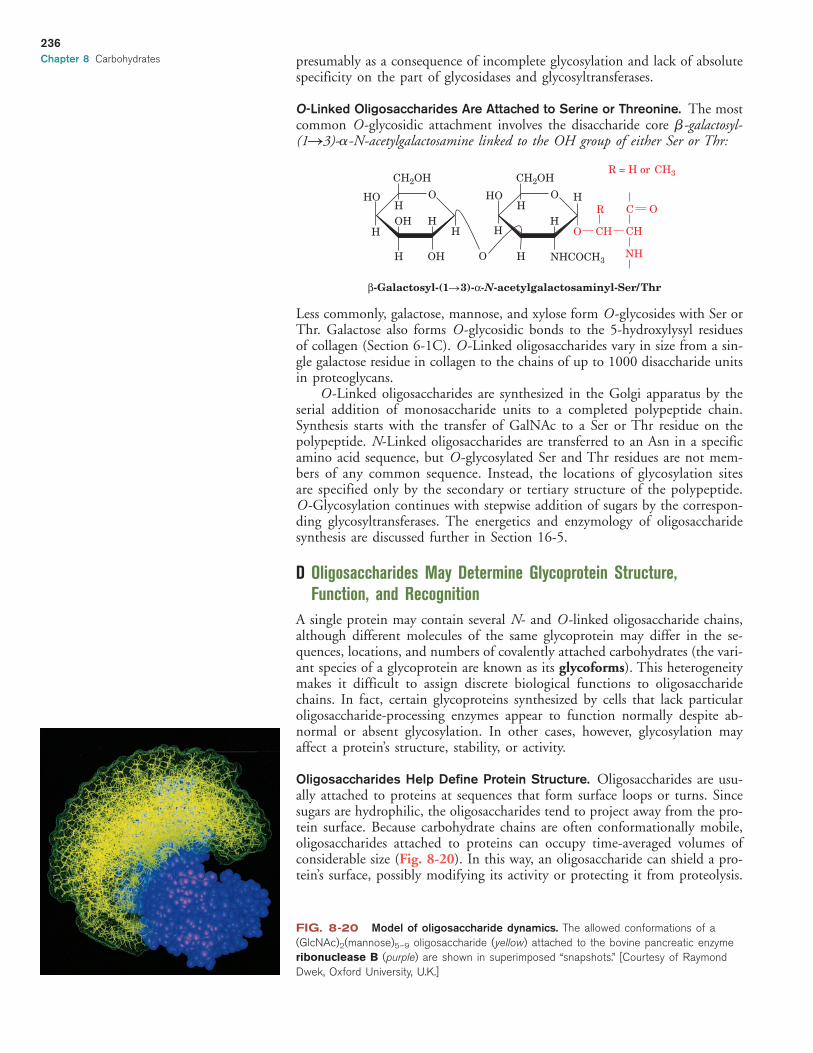
O-Linked Oligosaccharides Are Attached to Serine or Threonine. The mostcommon O-glycosidic attachment involves the disaccharide core �-galactosyl-(1S3)-�-N-acetylgalactosamine linked to the OH group of either Ser or Thr:
Less commonly, galactose, mannose, and xylose form O -glycosides with Ser orThr. Galactose also forms O -glycosidic bonds to the 5-hydroxylysyl residuesof collagen (Section 6-1C). O -Linked oligosaccharides vary in size from a sin-gle galactose residue in collagen to the chains of up to 1000 disaccharide unitsin proteoglycans.
O -Linked oligosaccharides are synthesized in the Golgi apparatus by theserial addition of monosaccharide units to a completed polypeptide chain.Synthesis starts with the transfer of GalNAc to a Ser or Thr residue on thepolypeptide. N-Linked oligosaccharides are transferred to an Asn in a specificamino acid sequence, but O -glycosylated Ser and Thr residues are not mem-bers of any common sequence. Instead, the locations of glycosylation sitesare specified only by the secondary or tertiary structure of the polypeptide.O -Glycosylation continues with stepwise addition of sugars by the correspon-ding glycosyltransferases. The energetics and enzymology of oligosaccharidesynthesis are discussed further in Section 16-5.
D Oligosaccharides May Determine Glycoprotein Structure, Function, and Recognition
A single protein may contain several N- and O -linked oligosaccharide chains,although different molecules of the same glycoprotein may differ in the se-quences, locations, and numbers of covalently attached carbohydrates (the vari-ant species of a glycoprotein are known as its glycoforms). This heterogeneitymakes it difficult to assign discrete biological functions to oligosaccharidechains. In fact, certain glycoproteins synthesized by cells that lack particularoligosaccharide-processing enzymes appear to function normally despite ab-normal or absent glycosylation. In other cases, however, glycosylation mayaffect a protein’s structure, stability, or activity.
Oligosaccharides Help Define Protein Structure. Oligosaccharides are usu-ally attached to proteins at sequences that form surface loops or turns. Sincesugars are hydrophilic, the oligosaccharides tend to project away from the pro-tein surface. Because carbohydrate chains are often conformationally mobile,oligosaccharides attached to proteins can occupy time-averaged volumes ofconsiderable size (Fig. 8-20). In this way, an oligosaccharide can shield a pro-tein’s surface, possibly modifying its activity or protecting it from proteolysis.
OH HH
H
H
O
CH2OH CH2OH
HO
CH
NH
CH
CH
H O
HO
NHCOCH3
β-Galactosyl-(1n3)-α-N-acetylgalactosaminyl-Ser/ Thr
HO
OH
HR O
R = H or CH3
H
HO
236
Chapter 8 Carbohydrates
FIG. 8-20 Model of oligosaccharide dynamics. The allowed conformations of a
(GlcNAc)2(mannose)5–9 oligosaccharide (yellow) attached to the bovine pancreatic enzyme
ribonuclease B (purple) are shown in superimposed “snapshots.” [Courtesy of Raymond
Dwek, Oxford University, U.K.]
JWCL460_c08_217-240.qxd 5/16/11 8:15 PM Page 236

In addition, some oligosaccharides may play structural roles by limitingthe conformational freedom of their attached polypeptide chains. SinceN-linked oligosaccharides are added as the protein is being synthesized, theattachment of an oligosaccharide may help determine how the protein folds.In addition, the oligosaccharide may help stabilize the folded conformation ofa polypeptide by reducing backbone flexibility. In particular, O -linkedoligosaccharides, which are usually clustered in heavily glycosylated segmentsof a protein, may help stiffen and extend the polypeptide chain.
Oligosaccharides Mediate Recognition Events. The many possible waysthat carbohydrates can be linked together to form branched structures givesthem the potential to carry more biological information than either nucleicacids or proteins of similar size. For example, two different nucleotides canmake only two distinct dinucleotides, but two different hexoses can combine in36 different ways (although not all possibilities are necessarily realized in nature).
The first evidence that unique combinations of carbohydrates might beinvolved in intercellular communication came with the discovery that all cellsare coated with sugars in the form of glycoconjugates such as glycoproteinsand glycolipids. The oligosaccharides of glycoconjugates form a fuzzy layer upto 1400 Å thick in some cells (Fig. 8-21).
Additional evidence that cell-surface carbohydrates have recognition func-tions comes from lectins (proteins that bind carbohydrates), which are ubiq-uitous in nature and frequently appear on the surfaces of cells. Lectins areexquisitely specific: They can recognize individual monosaccharides in partic-ular linkages to other sugars in an oligosaccharide (this property also makeslectins useful laboratory tools for isolating glycoproteins and oligosaccharides).Protein–carbohydrate interactions are typically characterized by extensive hydro-gen bonding (often including bridging water molecules) and the van der Waalspacking of hydrophobic sugar faces against aromatic side chains (Fig. 8-22).
Proteins known as selectins mediate the attachment between leukocytes(circulating white blood cells) and the surfaces of endothelial cells (the cellsthat line cavities, in this case blood vessels). Leukocytes constitutively (contin-ually) express selectins on their surface; endothelial cells transiently displaytheir own selectins in response to tissue damage from infection or mechanicalinjury. The selectins recognize and bind specific oligosaccharides on cell-surface glycoproteins. Reciprocal selectin–oligosaccharide interactions betweenthe two cell types allow the endothelial cells to “capture” circulating leukocytes,which then crawl past the endothelial cells on their way to eliminate theinfection or help repair damaged tissues.
Other cell–cell recognition phenomena depend on oligosaccharides. Forexample, proteins on the surface of mammalian spermatozoa recognizeGlcNAc or galactose residues on the glycoproteins of the ovum as part of thebinding and activation events during fertilization. Many viruses, bacteria, andeukaryotic parasites invade their target tissues by first binding to cell-surfacecarbohydrates.
Oligosaccharides Are Antigenic Determinants. The carbohydrates oncell surfaces are some of the best known immunochemical markers. For ex-
ample, the ABO blood group antigens are oligosaccharide components ofglycoproteins and glycolipids on the surfaces of an individual’s cells (not just
237
FIG. 8-21 Electron micrograph of the
erythrocyte surface. Its thick (up to 1400 Å)
carbohydrate coat, which is called the
glycocalyx, consists of closely packed
oligosaccharides attached to cell-surface proteins
and lipids. [Courtesy of Harrison Latta, UCLA.]
FIG. 8-22 Carbohydrate binding by a lectin. Human galectin-2 binds �-galactosides,
such as lactose, primarily through their galactose residue. The galactose and glucose
residues are shown with C green and O red, and the lectin amino acid side chains
are shown in violet. Hydrogen bonds between the side chains and the sugar residues are
shown as dashed yellow lines. [Courtesy of Hakon Leffler, University of California at
San Francisco. PDBid 1HLC.]
Section 3 Glycoproteins
JWCL460_c08_217-240.qxd 5/16/11 8:15 PM Page 237
red blood cells). Individuals with type A cells have A antigens on their cellsurfaces and carry anti-B antibodies in their blood; those with type B cells,which bear B antigens, carry anti-A antibodies; those with type AB cells, whichhave both A and B antigens, carry neither anti-A nor anti-B antibodies; andtype O individuals, whose cells bear neither antigen, carry both anti-A andanti-B antibodies. Consequently, the transfusion of type A blood into a typeB individual, for example, results in an anti-A antibody–A antigen reaction,which agglutinates (clumps together) the transfused erythrocytes, resulting inan often fatal blockage of blood vessels.
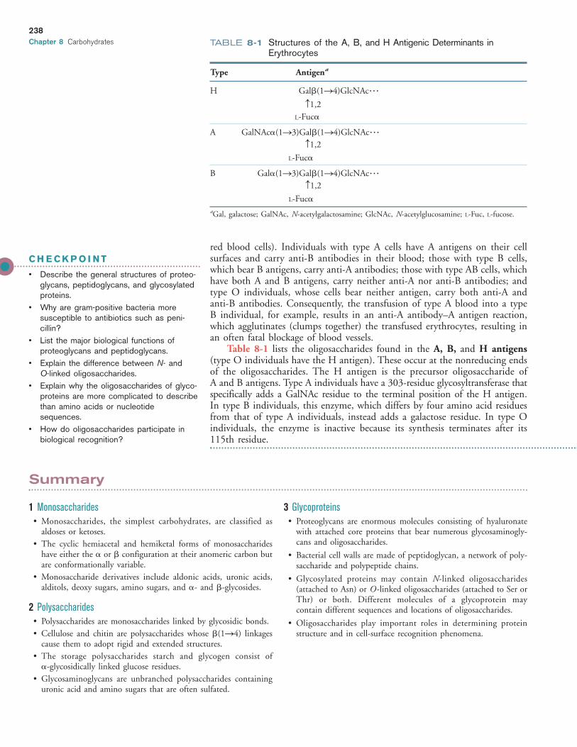
Table 8-1 lists the oligosaccharides found in the A, B, and H antigens(type O individuals have the H antigen). These occur at the nonreducing endsof the oligosaccharides. The H antigen is the precursor oligosaccharide of A and B antigens. Type A individuals have a 303-residue glycosyltransferase thatspecifically adds a GalNAc residue to the terminal position of the H antigen.In type B individuals, this enzyme, which differs by four amino acid residuesfrom that of type A individuals, instead adds a galactose residue. In type Oindividuals, the enzyme is inactive because its synthesis terminates after its115th residue.
238
Chapter 8 Carbohydrates
C H E C K P O I N T
• Describe the general structures of proteo-glycans, peptidoglycans, and glycosylatedproteins.
• Why are gram-positive bacteria moresusceptible to antibiotics such as peni-cillin?
• List the major biological functions of proteoglycans and peptidoglycans.
• Explain the difference between N- and O-linked oligosaccharides.
• Explain why the oligosaccharides of glyco-proteins are more complicated to describethan amino acids or nucleotidesequences.
• How do oligosaccharides participate inbiological recognition?
Summary
1 Monosaccharides• Monosaccharides, the simplest carbohydrates, are classified as
aldoses or ketoses.• The cyclic hemiacetal and hemiketal forms of monosaccharides
have either the � or � configuration at their anomeric carbon butare conformationally variable.
• Monosaccharide derivatives include aldonic acids, uronic acids,alditols, deoxy sugars, amino sugars, and �- and �-glycosides.
2 Polysaccharides• Polysaccharides are monosaccharides linked by glycosidic bonds.• Cellulose and chitin are polysaccharides whose �(1S4) linkages
cause them to adopt rigid and extended structures.• The storage polysaccharides starch and glycogen consist of
�-glycosidically linked glucose residues.• Glycosaminoglycans are unbranched polysaccharides containing
uronic acid and amino sugars that are often sulfated.
3 Glycoproteins• Proteoglycans are enormous molecules consisting of hyaluronate
with attached core proteins that bear numerous glycosaminogly-cans and oligosaccharides.
• Bacterial cell walls are made of peptidoglycan, a network of poly-saccharide and polypeptide chains.
• Glycosylated proteins may contain N-linked oligosaccharides (attached to Asn) or O -linked oligosaccharides (attached to Ser orThr) or both. Different molecules of a glycoprotein may contain different sequences and locations of oligosaccharides.
• Oligosaccharides play important roles in determining proteinstructure and in cell-surface recognition phenomena.
TABLE 8-1 Structures of the A, B, and H Antigenic Determinants inErythrocytes
Type Antigena
H Gal�(1S4)GlcNAc���
c1,2L-Fuc�
A GalNAc�(1S3)Gal�(1S4)GlcNAc���c1,2
L-Fuc�
B Gal�(1S3)Gal�(1S4)GlcNAc���c1,2
L-Fuc�
aGal, galactose; GalNAc, N-acetylgalactosamine; GlcNAc, N-acetylglucosamine; L-Fuc, L-fucose.
JWCL460_c08_217-240.qxd 8/17/11 1:54 PM Page 238

239
Key Terms
carbohydrate 217monosaccharide 217polysaccharide 217aldose 218ketose 219epimer 219hemiacetal 219hemiketal 219Haworth projection 220pyranose 220furanose 220anomeric carbon 220� anomer 220� anomer 220
oligosaccharide 224exoglycosidase 224endoglycosidase 224disaccharide 224starch 227glycogen 228glycosaminoglycan 228growth factor 230biofilm 230microheterogeneity 231glycomics 231proteoglycan 231N-linked oligosaccharide 231O -linked oligosaccharide 231
aldonic acid 221uronic acid 222alditol 222deoxy sugar 222amino sugar 222glycoprotein 222glycolipid 222�-glycoside 223�-glycoside 223glycosidic bond 223reducing sugar 223glycan 224homopolysaccharide 224heteropolysaccharide 224
gram-positive 232gram-negative 232peptidoglycan 232atomic force microscopy 232glycosylation 234oligosaccharide processing 234glycoforms 236glycoconjugate 237lectin 237leukocyte 237ABO blood group antigens 237
Problems
1. How many stereoisomers are possible for (a) a ketopentose, (b) a ketohexose, and (c) a ketoheptose?
2. Are (a) D-glucitol, (b) D-galactitol, and (c) D-glycerol opticallyactive?
3. Which of the following pairs of sugars are epimers of each other?
(a) D-sorbose and D-psicose
(b) D-sorbose and D-fructose
(c) D-erythrose and D-arabinose
4. Which of the following pairs of sugars are epimers of each other?
(a) D-fructose and L-fructose
(b) D-arabinose and D-ribose
(c) D-ribose and D-ribulose
5. Draw the furanose and pyranose forms of D-ribose.
6. The sucrose substitute tagatose (Fig. 8-2) is produced by hy-drolyzing lactose and then chemically converting one of the tworesulting aldoses to a ketose. Which residue of lactose gives riseto tagatose?
7. Draw a Fischer projection of L-fucose. L-Fucose is the 6-deoxyform of which L-hexose?
8. What type of sugar derivative is rhamnose?
9. How many different disaccharides of D-glucopyranose are possible?
10. In addition to lactose, how many other heterodisaccharides canD-galactose and D-glucose form?
11. (a) Deduce the structure of the disaccharide trehalose from thefollowing information: Complete hydrolysis yields only D-glucose; it is hydrolyzed by �-glucosidase but not �-glucosidase;and it does not reduce Cu2� to Cu�. (b) When exposed todehydrating conditions, many plants and invertebrates synthesizelarge amounts of trehalose, which enables them to survive pro-longed desiccation. What properties of the trehalose moleculemight allow it to act as a water substitute?
12. The artificial sweetener sucralose is a derivative of sucrose withthe formal name 1,6-dichloro-1,6-dideoxy-�-D-fructofuranosyl-4-chloro-4-deoxy-�-D-galactopyranoside. Draw its structure.
13. Draw the structure of the mannose trisaccharide that is part ofthe core N-linked oligosaccharide.
14. Identify the monosaccharides and their linkages in the followingdisaccharide.
15. Cellulose is treated with methanol, which methylates freeanomeric carbons. (a) How many methyl groups would be incor-porated per cellulose chain? (b) Cellulose is treated with dimethylsulfate, which adds a methyl group to all free hydroxyl groups.The cellulose is then hydrolyzed to release all its monosaccha-rides. Draw their structure.
16. Glycogen is treated with dimethyl sulfate, which adds a methylgroup to every free OH group. Next, the molecule is hydrolyzedto break all the glycosidic bonds between glucose residues. Thereaction products are then chemically analyzed.
(a) How many different types of methylated glucose moleculesare obtained?
(b) Draw the structure of the one that is most abundant.
17. How many reducing ends are in a molecule of glycogen that con-tains 10,000 residues with a branch every 10 residues?
18. Is amylose or amylopectin more likely to be a long-term storagepolysaccharide in plants?
19. “Nutraceuticals” are products that are believed to have some ben-eficial effect but are not strictly defined as either food or drug.Why might an individual suffering from osteoarthritis betempted to consume the nutraceutical glucosamine?
20. The core of pectin molecules is a polymer of �(1S4)-linkedD-galacturonate. Draw one of its residues.
21. Calculate the net charge of a chondroitin-4-sulfate molecule con-taining 100 disaccharide units.
22. Basic fibroblast growth factor binds to glycosaminoglycans in theextracellular matrix. Which amino acids would you expect to beabundant in this protein?
H
H
OH HH
H
HO O
HO
HO
OHOO
OH
HOCH2
H H
CH2
HCH2OH
JWCL460_c08_217-240.qxd 5/16/11 8:15 PM Page 239

240
23. Following their synthesis, many proteoglycans are stored in ahighly condensed state in intracellular compartments. Thesecompartments also contain many Ca2� ions, which are rapidlypumped out as the proteoglycans are released extracellularly.Explain the function of the Ca2� ions.
24. The peptidoglycan cell walls of bacteria contain pores that allowthe passage of substances with diameters less than 200 Å. Whyis it important that the cell wall be porous rather than solid?
25. Draw the structure of the O-type oligosaccharide (the H antigen,described in Table 8-1).
26. During gel electrophoresis, glycoproteins migrate as relatively dif-fuse bands, whereas nonglycosylated proteins typically migrate asnarrow, well-defined bands. Explain the difference in elec-trophoretic behavior.
BIOINFORMATICS www.wiley.com/college/voet
Project 5 Glycomics and the H1N1 Flu1. Looking at branched carbohydrates. Explore some well-known
glycans using MarvinSketch, PubChem, and the KEGG N-glycansynthesis reference pathway.
2. The role of glycans in swine flu. Review the structures of twoproteins (the H and N in H1N1) that are critical to swine flu infections and vaccine development, including the glycans thatare attached to these proteins. Then explore the glycan targetsfor swine flu.
Chitosan is a substance pro-duced from the chitin of crustacean shells (mainly from shrimp andcrab). How is the chitin chemically modified, and how does thistreatment affect its physical and chemical properties? What aresome industrial and medical uses for chitosan? Are all the adver-tised uses for chitosan supported by scientific data?
MORE TO EXPLORE
References
Aoki-Kinoshita, K.F., An introduction to bioinformatics for glycomicsresearch, PLoS Comput. Biol. 4, e1000075 (2008). [Describes someof the approaches used to characterize carbohydrate structures.]
Branda, S.S., Vik, A., Friedman, L., and Kolter, R., Biofilms: the matrixrevisited, Trends Microbiol. 13, 20–26 (2005). [Summarizes the gen-eral features of biofilms and ways of studying them.]
Esko, J.D. and Lindahl, U., Molecular diversity of heparan sulfate, J. Clin. Invest. 108, 169–173 (2001). [Reviews the structure, function,and biosynthesis of heparan sulfate–containing proteoglycans.]
Meroueh, S.O., Bencze, K.Z., Hesek, D., Lee, M., Fisher, J.F., Stemmler,T.L., and Mobashery, S., Three-dimensional structure of the bac-terial cell wall peptidoglycan, Proc. Natl. Acad. Sci. 103, 4404–4409(2006).
Mitra, N., Sinha, S., Ramya, T.N.C., and Surolia, A., N-Linked oligosac-charides as outfitters for glycoprotein folding, form and function,
Trends Biochem. Sci. 31, 156–163 and 251 (2006). [Summarizes theways in which oligosaccharides can influence glycoprotein structure.]
Sharon, N. and Lis, H., History of lectins: from hemagglutinins to bio-logical recognition molecules, Glycobiology 14, 53R–62R (2004). [A historical account of lectin research and applications.]
Spiro, R.G., Protein glycosylation: nature, distribution, enzymatic forma-tion, and disease implications of glycopeptide bonds, Glycobiology12, 43R–56R (2002). [Catalogs the various ways in which saccha-rides are linked to proteins, and describes the enzymes involved inglycoprotein synthesis.]
Taylor, M.E. and Drickamer, K., Introduction to Glycobiology, OxfordUniversity Press (2011).
Varki, A., Cummings, R.D., Esko, J.D., Freeze, H.H., Stanley, S., Bertozzi, C.R., Hart, G.W., and Etzler, M.E. (Eds.), Essentials ofGlycobiology (2nd ed.), Cold Spring Harbor Laboratory Press (2009).
JWCL460_c08_217-240.qxd 9/7/11 11:24 AM Page 240


![Nov 23 2018.ppt [Kompatibilitätsmodus] - Limes-Institut-Bonn · Figure 11-5 The anomeric monosaccharides -D-glucopyranose and -D-glucopyranose, drawn as both Haworth projections](https://img.pdfslide.net/doc/110x75/5f7f813523e6cd4b030ea89a/nov-23-2018ppt-kompatibilittsmodus-limes-institut-bonn-figure-11-5-the-anomeric.jpg)