Embed Size (px)
Citation preview
Carboxyamidotriazole-Orotate Inhibits the Growth ofImatinib-Resistant Chronic Myeloid Leukaemia Cells andModulates Exosomes-Stimulated AngiogenesisChiara Corrado1., Anna Maria Flugy1., Simona Taverna1., Stefania Raimondo1, Giuliana Guggino1,
Rashida Karmali2, Giacomo De Leo1", Riccardo Alessandro1*"
1 Dipartimento di Biopatologia e Biotecnologie Mediche e Forensi, Sezione di Biologia e Genetica, Universita di Palermo, Italy, 2 Tactical Therapeutics Inc., New York, New
York, United States of America
Abstract
The Bcr/Abl kinase has been targeted for the treatment of chronic myelogenous leukaemia (CML) by imatinib mesylate.While imatinib has been extremely effective for chronic phase CML, blast crisis CML are often resistant. New therapeuticoptions are therefore needed for this fatal disease. Although more common in solid tumors, increased microvessel densitywas also reported in chronic myelogenous leukaemia and was associated with a significant increase of angiogenic factors,suggesting that vascularity in hematologic malignancies is a controlled process and may play a role in the leukaemogenicprocess thus representing an alternative therapeutic target. Carboxyamidotriazole-orotate (CTO) is the orotate salt form ofcarboxyamidotriazole (CAI), an orally bioavailable signal transduction inhibitor that in vitro has been shown to possessantileukaemic activities. CTO, which has a reduced toxicity, increased oral bioavailability and stronger efficacy whencompared to the parental compound, was tested in this study for its ability to affect imatinib-resistant CML tumor growth ina xenograft model. The active cross talk between endothelial cells and leukemic cells in the bone marrow involvingexosomes plays an important role in modulating the process of neovascularization in CML. We have thus investigated theeffects of CTO on exosome-stimulated angiogenesis. Our results indicate that CTO may be effective in targeting both cancercell growth and the tumor microenvironment, thus suggesting a potential therapeutic utility for CTO in leukaemia patients.
Citation: Corrado C, Flugy AM, Taverna S, Raimondo S, Guggino G, et al. (2012) Carboxyamidotriazole-Orotate Inhibits the Growth of Imatinib-Resistant ChronicMyeloid Leukaemia Cells and Modulates Exosomes-Stimulated Angiogenesis. PLoS ONE 7(8): e42310. doi:10.1371/journal.pone.0042310
Editor: Sonja Loges, University Hospital Hamburg-Eppendorf, Germany
Received January 20, 2012; Accepted July 5, 2012; Published August 3, 2012
Copyright: � 2012 Corrado et al. This is an open-access article distributed under the terms of the Creative Commons Attribution License, which permitsunrestricted use, distribution, and reproduction in any medium, provided the original author and source are credited.
Funding: This work was supported by Tactical Therapeutics Inc, New York, United States of America; University of Palermo (International Cooperation) to RA; ex60% Ministero per l’Universita e per la Ricerca Scientifica e Tecnologica to RA, AF and to GDL. No additional external funding received for this study. The fundershad no role in study design, data collection and analysis, decision to publish, or preparation of the manuscript.
Competing Interests: RA received a grant research from Tactical Therapeutics that owns the anticancer drug (CTO) studied in the research. However, this doesnot alter the authors’ adherence to all the PLoS ONE policies on sharing data and materials.
* E-mail: [email protected]
. These authors contributed equally to this work.
" These authors also contributed equally to this work.
Introduction
Chronic myeloid leukaemia is characterized by the Philadelphia
(Ph) chromosome encoding the chimeric Bcr–Abl oncoprotein
with a constitutive tyrosine kinase activity that drives disease
pathogenesis by stimulating a number of downstream signalling
cascades [1,2]. Whereas CML can be effectively treated, during
chronic phase (CP), with tyrosine kinase inhibitors (TKIs) such as
imatinib [3], the acquisition of imatinib resistance, mainly due to
point mutations, causes disease progression (blast crisis) that can be
fatal within months. To circumvent resistance, more potent TKIs,
such as nilotinib and dasatinib, have been recently approved [4].
However, these compounds do not have therapeutic activity
against all imatinib-resistant mutants of Bcr-Abl and therefore, a
long-term tolerability problem has emerged [5]. Combination
strategies of imatinib with drugs that target downstream signalling
molecules have shown some success in the treatment of the
imatinib-resistant cells in in vitro settings and in mouse models but
have not been studied in clinical trials yet [6,7]. Therefore, there is
an urgent need for new anticancer agents and combinations that
can improve responses and survival rates for CML.
Carboxyamidotriazole-orotate (CTO) is the orotate salt form of
carboxyamidotriazole (CAI), an orally bioavailable small molecule
that was recently shown to decrease in vitro cell viability and to
augment apoptosis in three different imatinib-resistant CML cell
lines through the down-regulation of Bcr-Abl protein, inhibition of
tyrosine phosphorylation of Bcr-Abl, STAT5, CrkL, as well as
inhibition of ERK1/2 phosphorylation [8,9]. CTO shows a
reduced toxicity, increased oral bioavailability and achieves higher
plasma concentrations and stronger efficacy when compared to
the parental compound [10].
We have recently demonstrated that LAMA84 CML cells
release exosomes and that the addition of those microvesicles to
HUVEC affects several steps of in vitro angiogenesis including
motility, cytokine production, cell adhesion, and cell signalling as
well as in vivo angiogenesis in nude mice [11]. A number of studies
have recently described exosomes as new players in modulating
the tumor microenvironment, promoting angiogenesis and tumor
PLoS ONE | www.plosone.org 1 August 2012 | Volume 7 | Issue 8 | e42310
development [12]; furthermore, neovascularization is known to
exert an important role in the progression of chronic myeloid
leukaemia and may represent a valid alternative target for therapy.
Taking these data into account, the aims of our study were (i) to
test if CTO is able to inhibit in vivo the growth of imatinib-resistant
CML cells and (ii) to investigate the ability of CTO to affect tumor
microenvironment by modulating exosome-stimulated angiogen-
esis in vitro and in vivo. Our results indicate that administration of
CTO to a CML xenograft model in NOD/SCID mice may
increase survival and that CTO reduces in a dose- and time-
dependent fashion exosomes-stimulated angiogenic process. Fur-
ther work is necessary to demonstrate in a clinical setting, the
possible use of CTO as an alternative therapeutical option in the
treatment of imatinib-resistant forms of chronic myelogenous
leukaemia.
Materials and Methods
Ethic StatementAll animal experiments were conducted in full compliance with
Universita’ di Palermo and Italian Legislation for Animal Care
and Dipartimento di Biopatologia e Biotecnologie Mediche e
Forensi (DiBiMef) review board has approved this study.
Cell Culture and ReagentsImatinib resistant LAMA84 and K562 cells (LAMA84R and
K562R) were kindly provided by Dr. P. Vigneri, Universita di
Catania [13]. Cells were cultured in RPMI 1640 medium
(Euroclone, UK) supplemented with 10% fetal bovine serum
(Euroclone, UK), 2 mM L-glutamine, 100 U/ml penicillin and
100 mg/ml streptomycin (Euroclone, UK) and 1 mM of imatinib to
maintain the resistance. HUVEC were obtained from Lonza and
grown in Endothelial Growth Medium (EGM) according to
supplier’s information (Clonetics, Verviers, Belgium). Imatinib
mesylate (Selleck chemicals, Houston, TX, USA) was prepared as
a 1 mM stock solution in sterile phosphate-buffered saline (PBS);
CAI orotate (CTO) from Tactical Therapeutics Inc, New York,
USA was solubilized in DMSO at 0.1 M for in vitro assay. For
in vivo assay CTO was prepared at 50 mg/ml in 80% PEG-100,
sonicated, aliquoted and kept at 220uC; imatinib was dissolved in
PBS at a concentration of 2 mg/ml. All other reagents were
purchased from Sigma (St. Louis, MO), if not cited otherwise.
Proliferation Assay (MTT Assay)Methyl-thiazol-tetrazolium (MTT) assay was done as previously
described [8], cells were plated in triplicate or quadruplicate at
1.56104 per well and exposed to escalating doses of CTO for up to
4 days. Means and standard deviations generated from 3 to 4
independent experiments are reported as the percentage of
growth. Cell proliferation curves were derived from these data
by using Microsoft Excel software.
Western BlotTotal protein cell lysates or exosome lysates were obtained and
analyzed by SDS-PAGE followed by Western blotting as
previously described [8]. Antibodies used in the experiments
were: c-Abl, phospho-Abl, phospho-CrkL, Erk 1/2, phospho Erk
1/2, Hsc70, b-actin (all from Cell Signalling Technology, MA,
USA); anti-CrkL, VCAM1-FITC, ICAM1-FITC and anti-CD63
(all from Santa Cruz Biotechnology, CA, USA).
CML Mouse XenograftMale NOD/SCID mice four-to-five week old were purchased
from Charles River (Charles River Laboratories International, Inc,
MA, USA) and acclimated for a week prior to experimentation.
Mice received filtered water and sterilized diet ad libitum. Animals
were observed daily and clinical signs were noted.
Each mouse was inoculated subcutaneously (sc) in the right
flank with viable single cells (16107) suspended in 0.2 ml of PBS.
The day of injection was considered as Day 0. On Day 7, when
tumors were palpable, mice were randomly assigned to groups of
ten and were treated with imatinib administered intraperitoneally
(i.p) (50 mg/Kg, three days a week for two rounds) or with its
vehicle (PBS) in combination either with CTO 342 mg/kg
(Q1D65 for two rounds) or with CTO 513 mg/kg (Q1D65 for
two rounds) or with their vehicle (80% PEG-100). All mice
received both p.o. and i.p. doses of the vehicle to control for
morbidity associated with the treatment. Tumor xenografts were
measured and the mice were weighed three times a week starting
on Day 7. Tumor volume was determined by calliper by using the
following formula: L6W2/2 = mm3 where L and W are the
longest and shortest perpendicular measurements in millimeters,
respectively. The same formula was used to calculate tumor
weights assuming that 1 mm3 = 1 mg. Due to Universita di
Palermo rules and Italian legislation, animals were euthanized
when sc tumor xenografts reached 4000 mg in weight.
Exosome Isolation and CharacterizationExosomes produced by LAMA84R cells during a 24 h culture
period, were isolated from conditioned culture medium by
different centrifugations as described previously [11]. Exosome
protein content was determined by the Bradford assay (Pierce,
Rockford, IL, USA). The activity of acetylcholinesterase, an
exosome marker protein, was determined as described by Savina
et al [14]. Briefly a total of 10 mg of exosomes or 10 mg of total cell
lysate in 100 ml of PBS were resuspended in a solution of 1.25 mM
acetylthiocoline and 0.1 mM 5,59-dithiobis (2-nitrobenzoic acid) in
a final volume of 1 ml. The incubation was carried out in cuvettes
at 37uC and the change in absorbance at 412 nm was followed at
different time points (from 0 to 180 min).
Isolated exosomes were observed with a scanning electron
microscope. They were fixed with 2% glutaraldehyde in PBS for
10 min, attached onto stubs, coated with gold in a sputterer
(Sputter Coater 150A, Edwards, UK) and observed using a field
emission scanning electron microscope (FEGESEM QUANTA
200 FEI) at a working voltage of 30 kV.
RNA InterferenceSmall interfering RNAs (siRNA) targeting IL8 or scramble
siRNA were purchased from Dharmacon (ON-TARGET plus
SMART pool, Human, Dharmacon inc. CO, USA) and used to
transfect HUVEC by employing oligofectamine (Invitrogen, UK),
according to the suggested protocol. Briefly, 85% confluent cells
were incubated with a mix of oligofectamine- Opti-MEM
containing 75 pmol of siRNA or equal volume of PBS (CN).
After 24 h, the conditioned medium was collected for ELISA assay
and cells were lysed for RNA extraction. Knockdown efficiency
was determined by Real-time PCR and ELISA quantitation of IL8
release.
RNA Extraction and Real-time PCRHUVEC were grown to confluence in 12-well plates, pre-
treated or not with CTO (5 and 10 mM) for 24 h or 48 h and
incubated for 6 h with exosomes (20 and 50 mg/ml). To
investigate efficacy of IL8 silencing, HUVEC were grown to
confluence in 6-well plates, transfected with siRNAs, incubated for
6 h with 50 mg/ml of exosomes and then lysed to extract RNA.
For all experiments, IL8, VCAM1, ICAM1 transcript levels were
CTO Modulates Exosome-Stimulated Angiogenesis
PLoS ONE | www.plosone.org 2 August 2012 | Volume 7 | Issue 8 | e42310
measured by reverse transcription (RT) and TaqMan real-time
quantitative polymerase chain reaction (RQ-PCR) and were
analyzed as previously described [11]. The following primers
were used: IL8 Hs00174103 m1, VCAM1 Hs00174239 m1,
ICAM1 Hs00277001 m1 and GAPDH Hs99999905 m1 (Applied
Biosystems, Foster City, CA, USA) used as internal controls.
Flow CytometryExpression of HUVEC cell surface VCAM1 and ICAM1 was
determined by flow cytometry analysis. HUVEC were pre-treated
or not with 10 mM CTO for 24 h and incubated over night with
50 mg/ml of LAMA84R-exosomes in a low serum medium
(EGM:RPMI, 1:9). 56105 cells were washed in PBS and
incubated with 0.5 mg anti VCAM1-FITC or ICAM1-FITC
(Santa Cruz Biotechnology, CA, USA) for 15 min at 4uCaccording to manufacturer’s recommendations. Isotype-matched
irrelevant antibodies were used as a negative control. Viable cells
were gated by forward and side scatter and analysis was performed
on 100,000 acquired events for each sample. Samples were
analyzed on a FACS Calibur with the use of the CellQuest
software (BD Biosciences, NJ, USA).
ElisaHUVEC conditioned medium (CM) was collected from cells
pre-treated or not for 24 h with 10 mM CTO and then stimulated
for 6 h with indicated treatments; HUVEC CM was also collected
from cells after 24 h of transfection with siRNA (scramble or IL8)
and 6 h of incubation with 50 mg/ml of exosomes; CM aliquots
were centrifuged to remove cellular debris and afterwards IL8
protein concentrations were quantified using an ELISA kit (R&D
Systems, MN, USA), according to manufacturer’s protocol. IL8
was also measured directly in LAMA84R exosomes.
Migration AssayMigration assays were performed in Transwell chemotaxis
chambers assay (NeuroProbe, Cabin John, MD, USA) [8]. Briefly,
HUVEC (26106/ ml) were suspended in serum-free RPMI 1640
medium supplemented with 0.1% BSA with or without CTO (1, 5,
10 mM), in transwell chemotaxis chamber equipped with 8 mm
pore filters and exposed to chemoattractants with exosomes (20–
50 mg/ml), 10 ng/ml of recombinant IL8 or neutralizing antibod-
ies anti IL8 (5 mg/ml) (R&D system, MN, USA) as indicated. To
evaluate the migration ability of HUVEC transfected with siRNA,
endothelial cells were suspended in RPMI 1640 medium
supplemented with 0.1% BSA with or without 10 mM CTO,
were exposed to chemoattractants with 50 mg/ml of exosomes and
were processed as above. Filters were removed after 6 h, were
fixed in methanol and were stained with Diff-Quick (Medion
Diagnostics GmbH, Dudingen, Switzerland). Each test group was
tested in three independent experiments; the number of migrating
cells in five high-power fields per well were counted at 400X
magnification.
Adhesion AssayAdhesion assays were performed as previously described [11].
Briefly, HUVEC were pre-treated or not with 5–10 mM of CTO
for 24 h and HUVEC monolayer was incubated for 6 h with
indicated conditions, as described in the results. After treatment,
cells were washed with PBS and CML cells were added for 2 h at
37uC. Adherent cells were stained with hematoxylin/eosin, each
test group was assayed in triplicate; five high power (400X) fields
were counted for each condition.
Tube Formation of HUVEC on MatrigelMatrigel was used to test the effects of exosomes on in vitro
vascular tube formation as described [11,15]. 50 mg/ml of
exosomes were added to HUVEC plated on Matrigel in low
serum medium and 10 mM CTO. For HUVEC transfected with
siRNA the same experiment were performed after 24 h of
transfection. Cells were incubated for 6 h and then evaluated by
phase-contrast microscopy and photographed. The length of the
cables was measured manually with the IMAGE-J software
(http://rsbweb.nih.gov/ij/) [16].
Matrigel Plug AssayGroups of six NOD/SCID mice (4 weeks) (Charles River) were
injected subcutaneously with 400 mL of Matrigel (BD Biosciences
Pharmingen, San Diego, CA, USA) as follows: respectively,
animals in group 1 and 2 were injected with matrigel plus PBS
(PBS) or plus 100 mg LAMA84R-derived exosomes (Exo), and
were treated with 80% PEG-100 and PBS. Animals in group 3-4-5
were injected with matrigel plus 100 mg LAMA84R-derived
exosomes and were treated either with CTO 513 mg/Kg/inj
and imatinib vehicle (group 3, Exo + CTO) or with CTO
513 mg/Kg/inj and imatinib 50 mg/kg (group 4, Exo + IM +CTO) or with CTO vehicle and imatinib 50 mg/kg (group 5, Exo
+ IM). Animals in group 6 and 7 were injected with matrigel plus
50 ng of recombinant IL8 and were respectively treated either
with vehicles of CTO and imatinib (Rec IL8) or with CTO
513 mg/Kg/inj and imatinib vehicle (Rec IL8+ CTO), The
degree of vascularization was evaluated by determination of
hemoglobin content using the Drabkin method (Drabkin’s reagent
kit) [17].
StatisticsData were expressed as means 6 SEMs of the indicated number
of experiments. Statistical analysis was performed by using a
paired samples t test. Differences were considered to be significant
when P values were smaller than 0.05.
Results
Effects of CTO on Growth of Human CML Cells and Bcr-Abl Mediated Tyrosine Phosphorylation
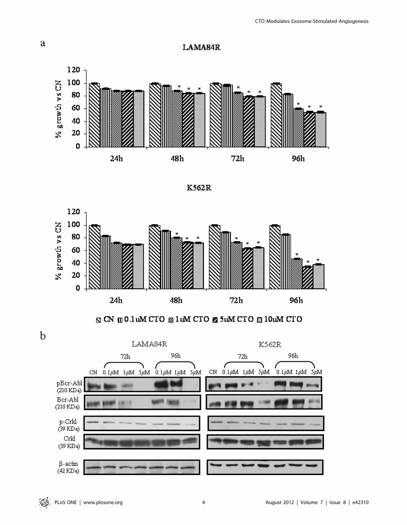
MTT assays were performed to determine the antiproliferative
effects of CTO on LAMA84R and on K562R cells. Data
presented in figure 1 (panel a) show results of 4 days treatment.
CTO inhibits cell growth of LAMA84R and K562R in the low
micromolar range in a dose dependent fashion (p,0.001). The
results herein show a 50% growth reduction of the CML lines with
5 mM CTO at 96 h time point. In order to correlate the
antiproliferative effects of CTO on CML cells with the Bcr-Abl
activity, cells were incubated with increasing concentrations of
CTO, were harvested and were subjected to immunoblotting with
antibodies against phosphorylated Bcr-Abl and CrkL. As shown in
figure 1 (panel b), a dose-dependent inhibition of both total and
phosphorylated Bcr-Abl levels was observed after 72 and 96 h of
drug exposure. Consistent with this conclusion, CTO inhibits the
phosphorylation of a selected target of Bcr-Abl kinase; tyrosine
phosphorylation of CrkL was reduced by 5 mM CTO treatment.
Effects of CTO on Tumor Xenograft GrowthOn the basis of the in vitro growth and Bcr-Abl signalling
inhibitory effects of CTO, we further examined the antineoplastic
effect of CTO on LAMA84R using a xenograft CML tumor
model. Otherwise CTO–treated mice seemed healthy and did not
CTO Modulates Exosome-Stimulated Angiogenesis
PLoS ONE | www.plosone.org 3 August 2012 | Volume 7 | Issue 8 | e42310
CTO Modulates Exosome-Stimulated Angiogenesis
PLoS ONE | www.plosone.org 4 August 2012 | Volume 7 | Issue 8 | e42310
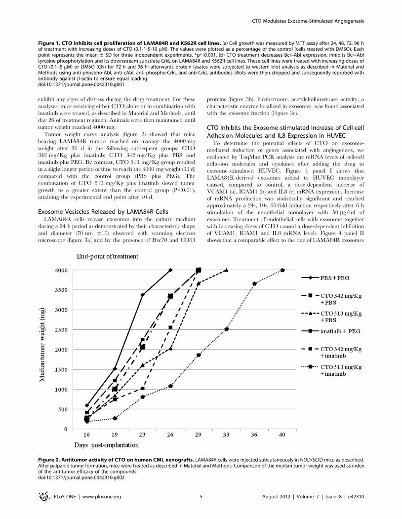
exhibit any signs of distress during the drug treatment. For these
analyses, mice receiving either CTO alone or in combination with
imatinib were treated, as described in Material and Methods, until
day 26 of treatment regimen. Animals were then maintained until
tumor weight reached 4000 mg.
Tumor weight curve analysis (figure 2) showed that mice
bearing LAMA84R tumor- reached on average the 4000 mg
weight after 26 d in the following subsequent groups: CTO
342 mg/Kg plus imatinib; CTO 342 mg/Kg plus PBS and
imatinib plus PEG. By contrast, CTO 513 mg/Kg group resulted
in a slight longer period of time to reach the 4000 mg weight (33 d)
compared with the control group (PBS plus PEG). The
combination of CTO 513 mg/Kg plus imatinib slowed tumor
growth to a greater extent than the control group (P,0.01),
attaining the experimental end point after 40 d.
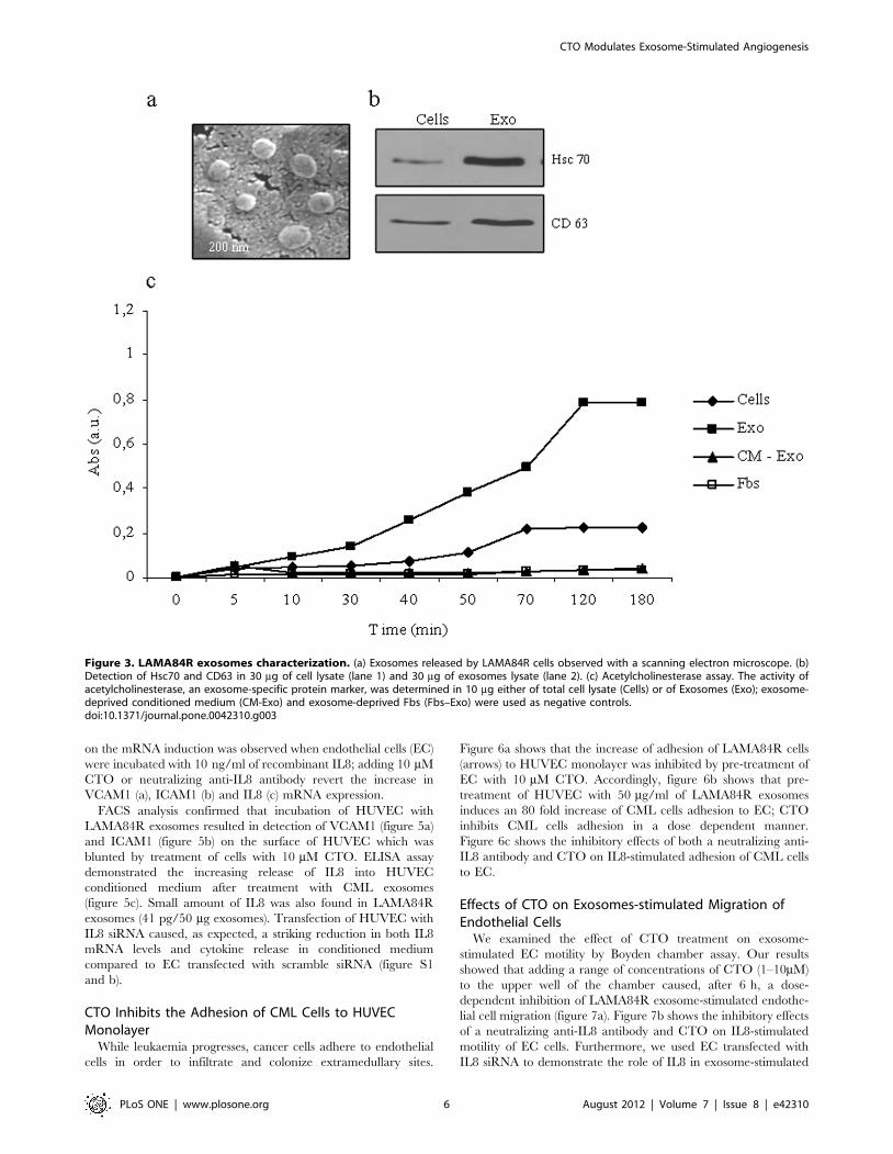
Exosome Vesiscles Released by LAMA84R CellsLAMA84R cells release exosomes into the culture medium
during a 24 h period as demonstrated by their characteristic shape
and diameter (70 nm 610) observed with scanning electron
microscope (figure 3a) and by the presence of Hsc70 and CD63
proteins (figure 3b). Furthermore, acetylcholinesterase activity, a
characteristic enzyme localized in exosomes, was found associated
with the exosome fraction (Figure 3c).
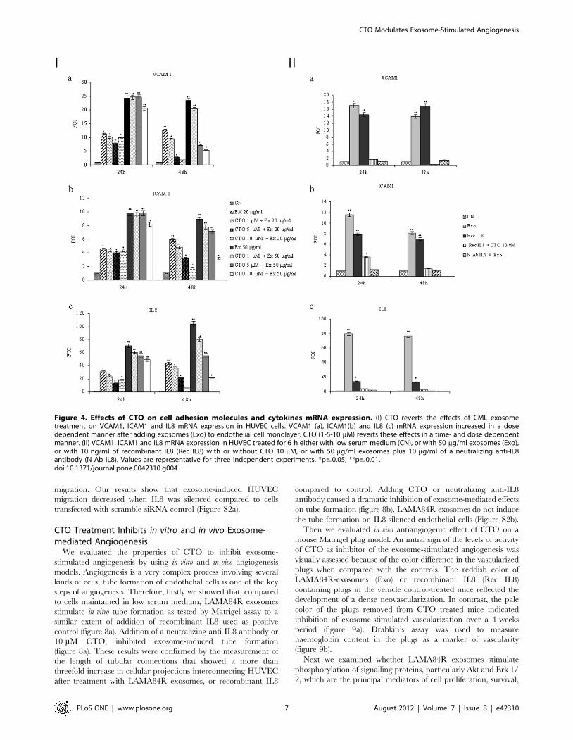
CTO Inhibits the Exosome-stimulated Increase of Cell-cellAdhesion Molecules and IL8 Expression in HUVEC
To determine the potential effects of CTO on exosome-
mediated induction of genes associated with angiogenesis, we
evaluated by TaqMan PCR analysis the mRNA levels of cell-cell
adhesion molecules and cytokines after adding the drug to
exosome-stimulated HUVEC. Figure 4 panel I shows that
LAMA84R-derived exosomes added to HUVEC monolayer
caused, compared to control, a dose-dependent increase of
VCAM1 (a), ICAM1 (b) and IL8 (c) mRNA expression. Increase
of mRNA production was statistically significant and reached
approximately a 24-, 10-, 60-fold induction respectively after 6 h
stimulation of the endothelial monolayer with 50 mg/ml of
exosomes. Treatment of endothelial cells with exosomes together
with increasing doses of CTO caused a dose-dependent inhibition
of VCAM1, ICAM1 and IL8 mRNA levels. Figure 4 panel II
shows that a comparable effect to the one of LAMA84R exosomes
Figure 1. CTO inhibits cell proliferation of LAMA84R and K562R cell lines. (a) Cell growth was measured by MTT assay after 24, 48, 72, 96 hof treatment with increasing doses of CTO (0.1-1-5-10 mM). The values were plotted as a percentage of the control (cells treated with DMSO). Eachpoint represents the mean 6 SD for three independent experiments. *p#0.001. (b) CTO treatment decreases Bcr–Abl expression, inhibits Bcr–Abltyrosine phosphorylation and its downstream substrate CrkL on LAMA84R and K562R cell lines. These cell lines were treated with increasing doses ofCTO (0.1–5 mM) or DMSO (CN) for 72 h and 96 h; afterwards protein lysates were subjected to western blot analysis as described in Material andMethods using anti-phospho-Abl, anti-cAbl, anti-phospho-CrkL and anti-CrkL antibodies. Blots were then stripped and subsequently reprobed withantibody against b-actin to ensure equal loading.doi:10.1371/journal.pone.0042310.g001
Figure 2. Antitumor activity of CTO on human CML xenografts. LAMA84R cells were injected subcutaneously in NOD/SCID mice as described.After palpable tumor formation, mice were treated as described in Material and Methods. Comparison of the median tumor weight was used as indexof the antitumor efficacy of the compounds.doi:10.1371/journal.pone.0042310.g002
CTO Modulates Exosome-Stimulated Angiogenesis
PLoS ONE | www.plosone.org 5 August 2012 | Volume 7 | Issue 8 | e42310
on the mRNA induction was observed when endothelial cells (EC)
were incubated with 10 ng/ml of recombinant IL8; adding 10 mM
CTO or neutralizing anti-IL8 antibody revert the increase in
VCAM1 (a), ICAM1 (b) and IL8 (c) mRNA expression.
FACS analysis confirmed that incubation of HUVEC with
LAMA84R exosomes resulted in detection of VCAM1 (figure 5a)
and ICAM1 (figure 5b) on the surface of HUVEC which was
blunted by treatment of cells with 10 mM CTO. ELISA assay
demonstrated the increasing release of IL8 into HUVEC
conditioned medium after treatment with CML exosomes
(figure 5c). Small amount of IL8 was also found in LAMA84R
exosomes (41 pg/50 mg exosomes). Transfection of HUVEC with
IL8 siRNA caused, as expected, a striking reduction in both IL8
mRNA levels and cytokine release in conditioned medium
compared to EC transfected with scramble siRNA (figure S1
and b).
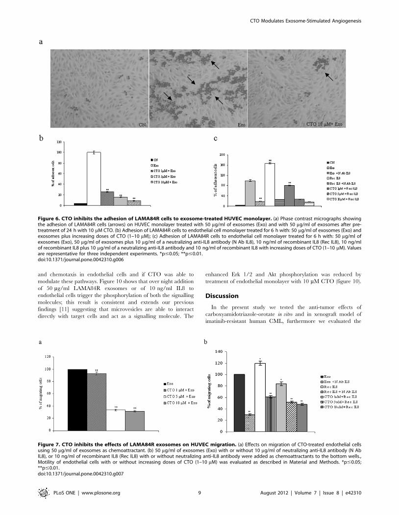
CTO Inhibits the Adhesion of CML Cells to HUVECMonolayer
While leukaemia progresses, cancer cells adhere to endothelial
cells in order to infiltrate and colonize extramedullary sites.
Figure 6a shows that the increase of adhesion of LAMA84R cells
(arrows) to HUVEC monolayer was inhibited by pre-treatment of
EC with 10 mM CTO. Accordingly, figure 6b shows that pre-
treatment of HUVEC with 50 mg/ml of LAMA84R exosomes
induces an 80 fold increase of CML cells adhesion to EC; CTO
inhibits CML cells adhesion in a dose dependent manner.
Figure 6c shows the inhibitory effects of both a neutralizing anti-
IL8 antibody and CTO on IL8-stimulated adhesion of CML cells
to EC.
Effects of CTO on Exosomes-stimulated Migration ofEndothelial Cells
We examined the effect of CTO treatment on exosome-
stimulated EC motility by Boyden chamber assay. Our results
showed that adding a range of concentrations of CTO (1–10mM)
to the upper well of the chamber caused, after 6 h, a dose-
dependent inhibition of LAMA84R exosome-stimulated endothe-
lial cell migration (figure 7a). Figure 7b shows the inhibitory effects
of a neutralizing anti-IL8 antibody and CTO on IL8-stimulated
motility of EC cells. Furthermore, we used EC transfected with
IL8 siRNA to demonstrate the role of IL8 in exosome-stimulated
Figure 3. LAMA84R exosomes characterization. (a) Exosomes released by LAMA84R cells observed with a scanning electron microscope. (b)Detection of Hsc70 and CD63 in 30 mg of cell lysate (lane 1) and 30 mg of exosomes lysate (lane 2). (c) Acetylcholinesterase assay. The activity ofacetylcholinesterase, an exosome-specific protein marker, was determined in 10 mg either of total cell lysate (Cells) or of Exosomes (Exo); exosome-deprived conditioned medium (CM-Exo) and exosome-deprived Fbs (Fbs–Exo) were used as negative controls.doi:10.1371/journal.pone.0042310.g003
CTO Modulates Exosome-Stimulated Angiogenesis
PLoS ONE | www.plosone.org 6 August 2012 | Volume 7 | Issue 8 | e42310
migration. Our results show that exosome-induced HUVEC
migration decreased when IL8 was silenced compared to cells
transfected with scramble siRNA control (Figure S2a).
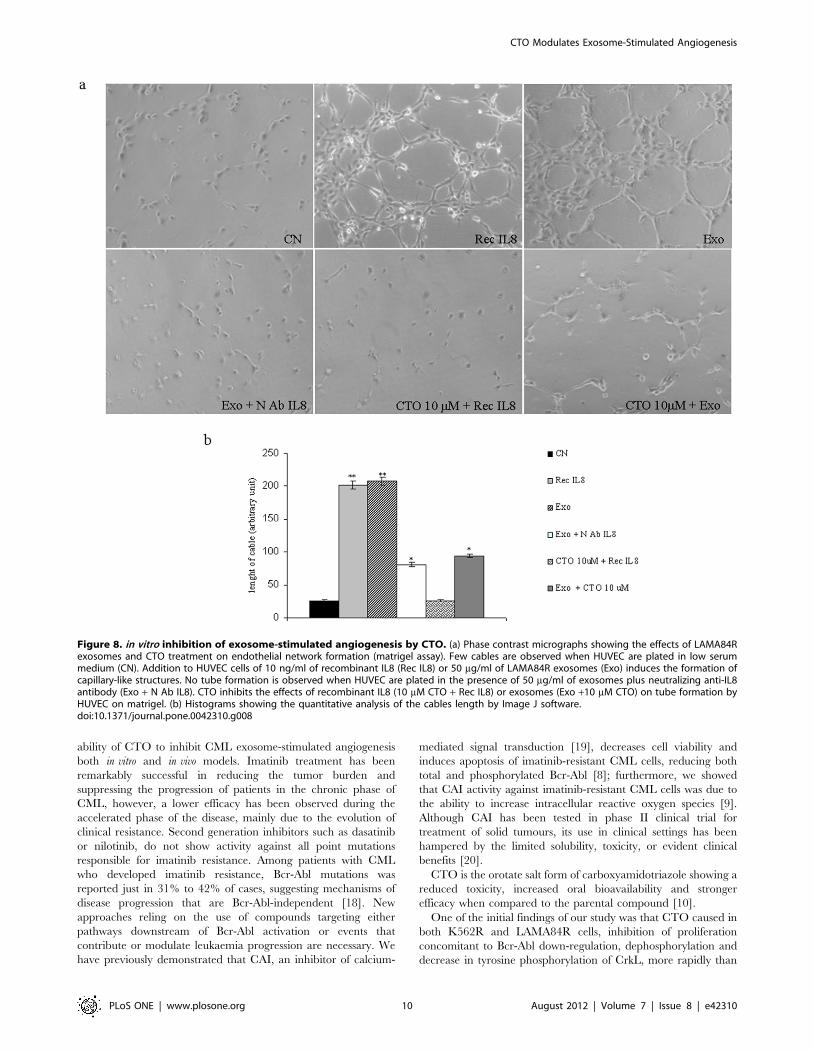
CTO Treatment Inhibits in vitro and in vivo Exosome-mediated Angiogenesis
We evaluated the properties of CTO to inhibit exosome-
stimulated angiogenesis by using in vitro and in vivo angiogenesis
models. Angiogenesis is a very complex process involving several
kinds of cells; tube formation of endothelial cells is one of the key
steps of angiogenesis. Therefore, firstly we showed that, compared
to cells maintained in low serum medium, LAMA84R exosomes
stimulate in vitro tube formation as tested by Matrigel assay to a
similar extent of addition of recombinant IL8 used as positive
control (figure 8a). Addition of a neutralizing anti-IL8 antibody or
10 mM CTO, inhibited exosome-induced tube formation
(figure 8a). These results were confirmed by the measurement of
the length of tubular connections that showed a more than
threefold increase in cellular projections interconnecting HUVEC
after treatment with LAMA84R exosomes, or recombinant IL8
compared to control. Adding CTO or neutralizing anti-IL8
antibody caused a dramatic inhibition of exosome-mediated effects
on tube formation (figure 8b). LAMA84R exosomes do not induce
the tube formation on IL8-silenced endothelial cells (Figure S2b).
Then we evaluated in vivo antiangiogenic effect of CTO on a
mouse Matrigel plug model. An initial sign of the levels of activity
of CTO as inhibitor of the exosome-stimulated angiogenesis was
visually assessed because of the color difference in the vascularized
plugs when compared with the controls. The reddish color of
LAMA84R-exosomes (Exo) or recombinant IL8 (Rec IL8)
containing plugs in the vehicle control-treated mice reflected the
development of a dense neovascularization. In contrast, the pale
color of the plugs removed from CTO–treated mice indicated
inhibition of exosome-stimulated vascularization over a 4 weeks
period (figure 9a). Drabkin’s assay was used to measure
haemoglobin content in the plugs as a marker of vascularity
(figure 9b).
Next we examined whether LAMA84R exosomes stimulate
phosphorylation of signalling proteins, particularly Akt and Erk 1/
2, which are the principal mediators of cell proliferation, survival,
Figure 4. Effects of CTO on cell adhesion molecules and cytokines mRNA expression. (I) CTO reverts the effects of CML exosometreatment on VCAM1, ICAM1 and IL8 mRNA expression in HUVEC cells. VCAM1 (a), ICAM1(b) and IL8 (c) mRNA expression increased in a dosedependent manner after adding exosomes (Exo) to endothelial cell monolayer. CTO (1-5-10 mM) reverts these effects in a time- and dose dependentmanner. (II) VCAM1, ICAM1 and IL8 mRNA expression in HUVEC treated for 6 h either with low serum medium (CN), or with 50 mg/ml exosomes (Exo),or with 10 ng/ml of recombinant IL8 (Rec IL8) with or without CTO 10 mM, or with 50 mg/ml exosomes plus 10 mg/ml of a neutralizing anti-IL8antibody (N Ab IL8). Values are representative for three independent experiments. *p#0.05; **p#0.01.doi:10.1371/journal.pone.0042310.g004
CTO Modulates Exosome-Stimulated Angiogenesis
PLoS ONE | www.plosone.org 7 August 2012 | Volume 7 | Issue 8 | e42310
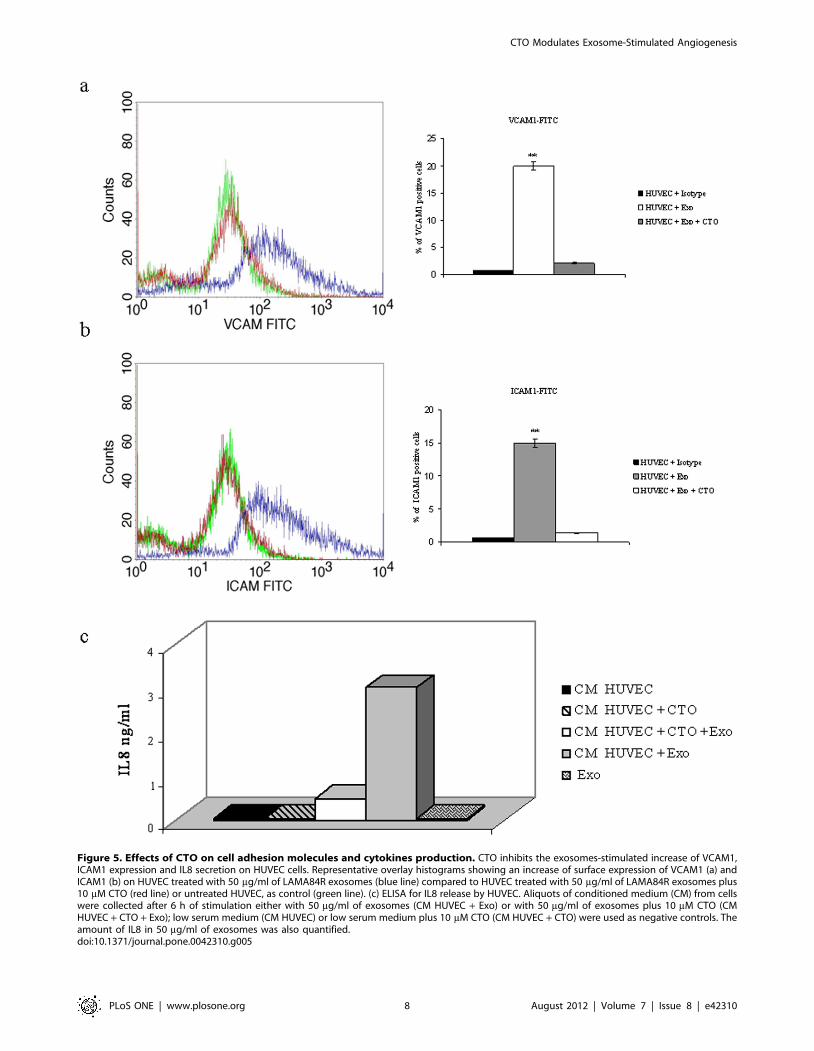
Figure 5. Effects of CTO on cell adhesion molecules and cytokines production. CTO inhibits the exosomes-stimulated increase of VCAM1,ICAM1 expression and IL8 secretion on HUVEC cells. Representative overlay histograms showing an increase of surface expression of VCAM1 (a) andICAM1 (b) on HUVEC treated with 50 mg/ml of LAMA84R exosomes (blue line) compared to HUVEC treated with 50 mg/ml of LAMA84R exosomes plus10 mM CTO (red line) or untreated HUVEC, as control (green line). (c) ELISA for IL8 release by HUVEC. Aliquots of conditioned medium (CM) from cellswere collected after 6 h of stimulation either with 50 mg/ml of exosomes (CM HUVEC + Exo) or with 50 mg/ml of exosomes plus 10 mM CTO (CMHUVEC + CTO + Exo); low serum medium (CM HUVEC) or low serum medium plus 10 mM CTO (CM HUVEC + CTO) were used as negative controls. Theamount of IL8 in 50 mg/ml of exosomes was also quantified.doi:10.1371/journal.pone.0042310.g005
CTO Modulates Exosome-Stimulated Angiogenesis
PLoS ONE | www.plosone.org 8 August 2012 | Volume 7 | Issue 8 | e42310
and chemotaxis in endothelial cells and if CTO was able to
modulate these pathways. Figure 10 shows that over night addition
of 50 mg/ml LAMA84R exosomes or of 10 ng/ml IL8 to
endothelial cells trigger the phosphorylation of both the signalling
molecules; this result is consistent and extends our previous
findings [11] suggesting that microvesicles are able to interact
directly with target cells and act as a signalling molecule. The
enhanced Erk 1/2 and Akt phosphorylation was reduced by
treatment of endothelial monolayer with 10 mM CTO (figure 10).
Discussion
In the present study we tested the anti-tumor effects of
carboxyamidotriazole-orotate in vitro and in xenograft model of
imatinib-resistant human CML, furthermore we evaluated the
Figure 6. CTO inhibits the adhesion of LAMA84R cells to exosome-treated HUVEC monolayer. (a) Phase contrast micrographs showingthe adhesion of LAMA84R cells (arrows) on HUVEC monolayer treated with 50 mg/ml of exosomes (Exo) and with 50 mg/ml of exosomes after pre-treatment of 24 h with 10 mM CTO. (b) Adhesion of LAMA84R cells to endothelial cell monolayer treated for 6 h with: 50 mg/ml of exosomes (Exo) andexosomes plus increasing doses of CTO (1–10 mM); (c) Adhesion of LAMA84R cells to endothelial cell monolayer treated for 6 h with: 50 mg/ml ofexosomes (Exo), 50 mg/ml of exosomes plus 10 mg/ml of a neutralizing anti-IL8 antibody (N Ab IL8), 10 ng/ml of recombinant IL8 (Rec IL8), 10 ng/mlof recombinant IL8 plus 10 mg/ml of a neutralizing anti-IL8 antibody and 10 ng/ml of recombinant IL8 with increasing doses of CTO (1–10 mM). Valuesare representative for three independent experiments. *p#0.05; **p#0.01.doi:10.1371/journal.pone.0042310.g006
Figure 7. CTO inhibits the effects of LAMA84R exosomes on HUVEC migration. (a) Effects on migration of CTO-treated endothelial cellsusing 50 mg/ml of exosomes as chemoattractant. (b) 50 mg/ml of exosomes (Exo) with or without 10 mg/ml of neutralizing anti-IL8 antibody (N AbIL8), or 10 ng/ml of recombinant IL8 (Rec IL8) with or without neutralizing anti-IL8 antibody were added as chemoattractants to the bottom wells.,Motility of endothelial cells with or without increasing doses of CTO (1–10 mM) was evaluated as described in Material and Methods. *p#0.05;**p#0.01.doi:10.1371/journal.pone.0042310.g007
CTO Modulates Exosome-Stimulated Angiogenesis
PLoS ONE | www.plosone.org 9 August 2012 | Volume 7 | Issue 8 | e42310
ability of CTO to inhibit CML exosome-stimulated angiogenesis
both in vitro and in vivo models. Imatinib treatment has been
remarkably successful in reducing the tumor burden and
suppressing the progression of patients in the chronic phase of
CML, however, a lower efficacy has been observed during the
accelerated phase of the disease, mainly due to the evolution of
clinical resistance. Second generation inhibitors such as dasatinib
or nilotinib, do not show activity against all point mutations
responsible for imatinib resistance. Among patients with CML
who developed imatinib resistance, Bcr-Abl mutations was
reported just in 31% to 42% of cases, suggesting mechanisms of
disease progression that are Bcr-Abl-independent [18]. New
approaches reling on the use of compounds targeting either
pathways downstream of Bcr-Abl activation or events that
contribute or modulate leukaemia progression are necessary. We
have previously demonstrated that CAI, an inhibitor of calcium-
mediated signal transduction [19], decreases cell viability and
induces apoptosis of imatinib-resistant CML cells, reducing both
total and phosphorylated Bcr-Abl [8]; furthermore, we showed
that CAI activity against imatinib-resistant CML cells was due to
the ability to increase intracellular reactive oxygen species [9].
Although CAI has been tested in phase II clinical trial for
treatment of solid tumours, its use in clinical settings has been
hampered by the limited solubility, toxicity, or evident clinical
benefits [20].
CTO is the orotate salt form of carboxyamidotriazole showing a
reduced toxicity, increased oral bioavailability and stronger
efficacy when compared to the parental compound [10].
One of the initial findings of our study was that CTO caused in
both K562R and LAMA84R cells, inhibition of proliferation
concomitant to Bcr-Abl down-regulation, dephosphorylation and
decrease in tyrosine phosphorylation of CrkL, more rapidly than
Figure 8. in vitro inhibition of exosome-stimulated angiogenesis by CTO. (a) Phase contrast micrographs showing the effects of LAMA84Rexosomes and CTO treatment on endothelial network formation (matrigel assay). Few cables are observed when HUVEC are plated in low serummedium (CN). Addition to HUVEC cells of 10 ng/ml of recombinant IL8 (Rec IL8) or 50 mg/ml of LAMA84R exosomes (Exo) induces the formation ofcapillary-like structures. No tube formation is observed when HUVEC are plated in the presence of 50 mg/ml of exosomes plus neutralizing anti-IL8antibody (Exo + N Ab IL8). CTO inhibits the effects of recombinant IL8 (10 mM CTO + Rec IL8) or exosomes (Exo +10 mM CTO) on tube formation byHUVEC on matrigel. (b) Histograms showing the quantitative analysis of the cables length by Image J software.doi:10.1371/journal.pone.0042310.g008
CTO Modulates Exosome-Stimulated Angiogenesis
PLoS ONE | www.plosone.org 10 August 2012 | Volume 7 | Issue 8 | e42310
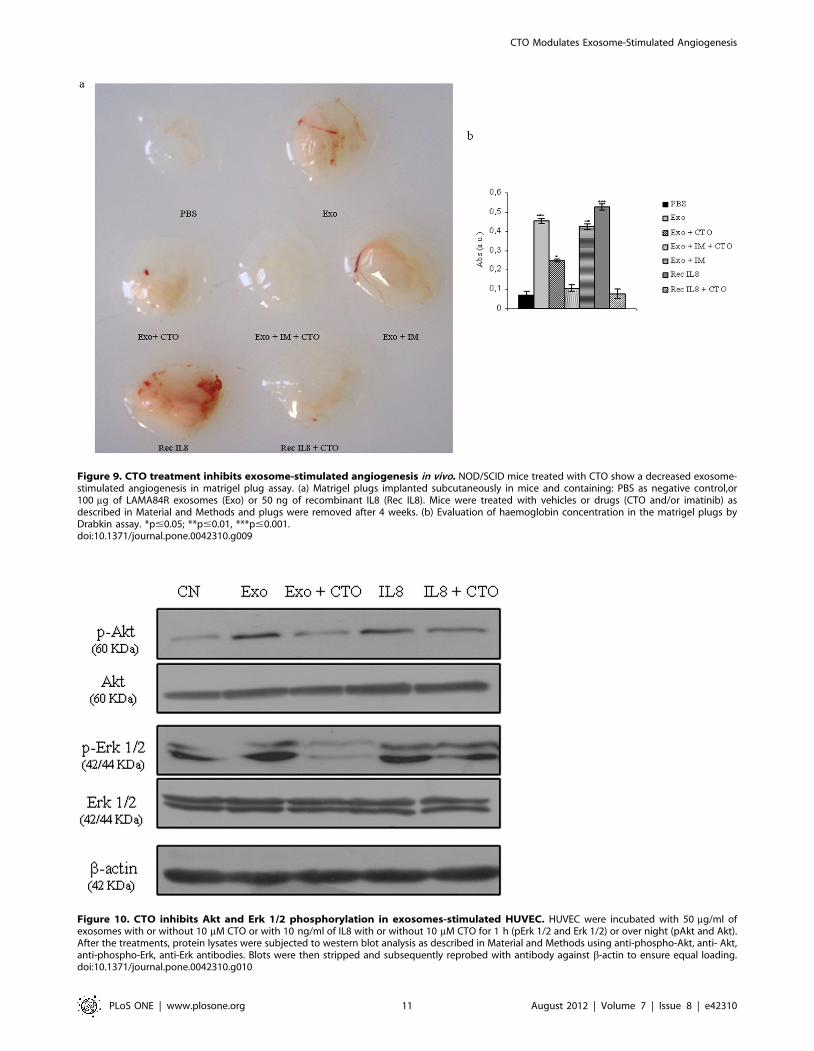
Figure 9. CTO treatment inhibits exosome-stimulated angiogenesis in vivo. NOD/SCID mice treated with CTO show a decreased exosome-stimulated angiogenesis in matrigel plug assay. (a) Matrigel plugs implanted subcutaneously in mice and containing: PBS as negative control,or100 mg of LAMA84R exosomes (Exo) or 50 ng of recombinant IL8 (Rec IL8). Mice were treated with vehicles or drugs (CTO and/or imatinib) asdescribed in Material and Methods and plugs were removed after 4 weeks. (b) Evaluation of haemoglobin concentration in the matrigel plugs byDrabkin assay. *p#0.05; **p#0.01, ***p#0.001.doi:10.1371/journal.pone.0042310.g009
Figure 10. CTO inhibits Akt and Erk 1/2 phosphorylation in exosomes-stimulated HUVEC. HUVEC were incubated with 50 mg/ml ofexosomes with or without 10 mM CTO or with 10 ng/ml of IL8 with or without 10 mM CTO for 1 h (pErk 1/2 and Erk 1/2) or over night (pAkt and Akt).After the treatments, protein lysates were subjected to western blot analysis as described in Material and Methods using anti-phospho-Akt, anti- Akt,anti-phospho-Erk, anti-Erk antibodies. Blots were then stripped and subsequently reprobed with antibody against b-actin to ensure equal loading.doi:10.1371/journal.pone.0042310.g010
CTO Modulates Exosome-Stimulated Angiogenesis
PLoS ONE | www.plosone.org 11 August 2012 | Volume 7 | Issue 8 | e42310
the parental compound. Furthermore, it showed to be more active
than CAI on a molar basis [8]. Rapid reduction of Bcr-Abl protein
coupled with kinase inactivation, as seen with CTO, can be
particularly advantageous because of the multiple Bcr-Abl
domains that mediate protein interactions triggering different
signalling pathways responsible for cell proliferation, adhesion,
and inhibition of apoptosis [21]. The inhibitory effects of CTO
against CML cells in culture, is mirrored by its activity against
CML xenografts in NOD/SCID model. Tumor growth retarda-
tion in mice treated with CTO 513 mg/kg plus imatinib was
evident suggesting that CTO was acting in this model as an
antileukaemic agent. Recently, there are increasing data showing
that angiogenesis plays an important role in the development and
progression of chronic myeloid leukaemia [22,23]. The bone
marrow of patients with CML exhibit marked neovascularization
and increased number of endothelial cells [24]; the cross-talk
between tumor cells and endothelial cells leads to enhanced tumor
growth, metastasis and altered response to anti-cancer therapy
[25]. Recently, a number of studies have recently described
exosomes as new players in modulating tumor microenvironment,
promoting angiogenesis and tumor progression [12]. Our group
and other collaborators have shown that exosomes released by
imatinib-sensitive LAMA84 [11] and K562 CML cells [26] have a
potential to influence in vitro and in vivo angiogenesis by affecting
directly endothelial cells properties. One of the findings of the
present study was the confirmation, by morphological and
biochemical analysis, that LAMA84R CML cells secrete exosomes
and that these vesiscles are able to modulate angiogenesis in vitro
and in vivo.
These findings drove us to investigate if CTO could target both
tumor cells and the tumor microenvironment. Therefore, we
focused on the inhibitory effects of CTO on in vitro selected
functional steps of angiogenesis as well as on in vivo angiogenesis in
NOD/SCID mice. Our in vitro studies with HUVEC demonstrat-
ed that CTO inhibits exosome stimulated motility, cytokines and
cell-adhesion molecules (ICAM1 and VCAM1) expression of
endothelial cells; moreover CTO inhibits exosomes activated
signalling pathways and capillary-like structure formation. The
matrigel plug assay that mimics physiological neo-angiogenesis,
was used as in vivo model; our study showed that CTO drastically
decreased exosome-stimulated angiogenesis. To investigate on the
possible molecular mechanisms of the CTO-mediated antiangio-
genic effect, we examined whether CTO inhibited the activation
of intracellular signalling pathways involved in endothelial cell
activation. Treatment of the EC with CTO blocked significantly
the exosome-induced phosphorylation of signalling proteins,
particularly Akt and Erk 1/2, which are the principal mediators
of cell proliferation, survival, and chemotaxis in endothelial cells
[27]. Kinase-dependent and kinase-independent mechanisms are
known to contribute to the abnormal adhesion and migration of
CML progenitors, thus the effect of CTO on both endothelial cells
and leukemic cells may concomitantly inhibit adhesion of
leukaemia cells to vascular endothelium and conditions that
favour leukostasis and tissue infiltration. IL8 is a member of the
CXC family of chemokines, a potent proangiogenic factor [28],
and its plasma levels are found significantly higher in patients
affected by chronic myelogenous leukaemia [29]. Interestingly we
showed, through the use of IL8 neutralizing antibodies and short
interfering RNAs, that IL8 was in part responsible for the effects of
LAMA84R exosomes on EC activation; furthermore, treatment of
EC with CTO inhibited the IL8-stimulated angiogenic phenotype.
It is conceivable to hypothesize that IL8 secreted by EC stimulated
with CML exosomes, may modulate both myeloid malignant cells
and endothelial cells, thus generating a paracrine machinery
between hematopoietic malignant cells and newly generated
endothelium. In this tumor microenvironment, CTO could inhibit
the angiogenic process through blocking the exosome-mediated
crosstalk, thus causing the interruption of a reciprocal stimulatory
loop between leukemic and endothelial cells.
Other groups have pointed their attention on the close
relationship between exosome production and tumor microenvi-
ronment modulation; Hood and collaborators demonstrated that
exosomes released by melanoma cells modulate both angiogenic
and immunological cytokine signalling, thus serving as paracrine
nanocarriers that might prepare distal sites for the arrest of
metastatic cells [30]. In this context, the inhibition of either
exosomes shedding or modulation of their function has been
proposed as worthwhile approach to cancer therapy. Al-Nedawi
et al. showed that the treatment of A431 tumor xenografts with
Diannexin, which inhibits the uptake of the A431 (human
squamous cell carcinoma cell line)-derived microvesicles into
endothelial cells, led to a reduction of tumor growth rate and
microvascular density [31].
As far as we are aware, this is the first study that demonstrates
the inhibitory effect of an anticancer drug on angiogenesis
stimulated by exosomes released from drug-resistant cancer cells;
collectively, our findings generate a rationale for investigating
clinical efficacy of molecules such as CTO that are endowed with
antitumor and antiangiogenic properties.
Supporting Information
Figure S1 IL8 siRNA inhibits IL8 mRNA expression andcytokine release from HUVEC. IL8 mRNA expression levels
(a) or IL8 protein release in conditioned medium (b) were
evaluated in HUVEC transfected either with oligofectamine (CN),
or with scramble siRNA or with IL8 siRNA. HUVEC transfected
were treated or not for 6 h with 50 mg of LAMA84R exosomes
(Exo).
(TIF)
Figure S2 IL8 siRNA inhibits the effects of LAMA84Rexosomes on migration and tube formation capabilitiesof HUVEC. (a) Addition of exosomes to the bottom wells of
Boyden chamber increases the migration of either HUVEC and
HUVEC transfected with scramble siRNA while concomitant
CTO treatment reverts this effect. On the contrary exosomes have
not significative effects on migration of IL8-silenced HUVEC.(b)
Phase contrast micrographs showing the effects of LAMA84R
exosomes and CTO treatment on endothelial network formation
after silencing of HUVEC with IL8 siRNA (matrigel assay).
Exosomes (Exo) induce formation of capillary-like structures on
HUVEC transfected with scramble siRNA compared to control
cells (siRNA scramble). No tube formation is observed when
exosomes stimulated EC were silenced for IL 8 mRNA expression
with short interfering RNAs.
(TIF)
Author Contributions
Conceived and designed the experiments: CC ST AF GDL RA. Performed
the experiments: CC AF ST SR GG. Analyzed the data: CC AF ST SR
GG RA. Contributed reagents/materials/analysis tools: RK. Wrote the
paper: CC AF ST RA.
CTO Modulates Exosome-Stimulated Angiogenesis
PLoS ONE | www.plosone.org 12 August 2012 | Volume 7 | Issue 8 | e42310
References
1. Rowley J (1973) A new consistent chromosomal abnormality in chronic
myelogenous leukaemia identified by quinacrine fluorescence and Giemsa
staining. Nature 243: 290–293.
2. Sonoyama J, Matsumura I, Ezoe S, Satoh Y, Zhang X, et al. (2002) Functional
cooperation among Ras, STAT5, and phosphatidylinositol 3-kinase is required
for full oncogenic activities of BCR/ABL in K562 cells. J Biol Chem 277: 8076–
8082.
3. Druker BJ, Guilhot F, O’Brien SG, Gathmann I, Kantarjian H, et al. (2006)
Five-year follow-up of patients receiving imatinib for chronic myeloid leukemia.
N Engl J Med 355: 2408–2417.
4. Quintas-Cardama A, Kantarjian H, Cortes J (2007) Flying under the radar: the
new wave of BCR-ABL inhibitors. Nat Rev Drug Discov 6: 834–848.
5. Jabbour E, Branford S, Saglio G, Jones D, Cortes J, et al. (2011) Practical advice
for determining the role of BCR-ABL mutations in guiding tyrosine kinase
inhibitor therapy in patients with chronic myeloid leukemia. Cancer 117: 1800–
1811.
6. Zhang J, Adrian FJ, Jahnke W, Cowan-Jacob SW, Li AG, et al. (2010) Targeting
Bcr-Abl by combining allosteric with ATP-binding-site inhibitors. Nature 463:
501–506.
7. Zhang H, Trachootham D, Lu W, Carew J, Giles FJ, et al. (2008) Effective
killing of Gleevec-resistant CML cells with T315I mutation by a natural
compound PEITC through redox-mediated mechanism. Leukemia 22: 1191–
1199.
8. Alessandro R, Fontana S, Giordano M, Corrado C, Colomba P, et al. (2008)
Effects of carboxyamidotriazole on in vitro models of imatinib-resistant chronic
myeloid leukemia. J Cell Physiol 215: 111–121.
9. Corrado C, Raimondo S, Flugy AM, Fontana S, Santoro A, et al. (2011)
Carboxyamidotriazole inhibits cell growth of imatinib-resistant chronic myeloid
leukaemia cells including T315I Bcr-Abl mutant by a redox-mediated
mechanism. Cancer Lett 300: 205–214.
10. Grover G, Kelly J, Moore G, Jacoby H, Karmali R, et al. (2007) Comparative
pharmacokinetic profile of carboxyamidotriazole and carboxyamidotriazole-
orotate. Cancer Therapy 5: 437–442.
11. Taverna S, Flugy A, Saieva L, Kohn EC, Santoro A, et al. (2011) Role of
exosomes released by chronic myelogenous leukemia cells in angiogenesis.
Int J Cancer.
12. Anderson HC, Mulhall D, Garimella R (2010) Role of extracellular membrane
vesicles in the pathogenesis of various diseases, including cancer, renal diseases,
atherosclerosis, and arthritis. Lab Invest.
13. D’Asaro M, La Mendola C, Di Liberto D, Orlando V, Todaro M, et al. (2010) V
gamma 9V delta 2 T lymphocytes efficiently recognize and kill zoledronate-
sensitized, imatinib-sensitive, and imatinib-resistant chronic myelogenous
leukemia cells. J Immunol 184: 3260–3268.
14. Savina A, Furlan M, Vidal M, Colombo MI (2003) Exosome release is regulated
by a calcium-dependent mechanism in K562 cells. J Biol Chem 278: 20083–
20090.
15. Kohn EC, Alessandro R, Spoonster J, Wersto RP, Liotta LA (1995)
Angiogenesis: role of calcium-mediated signal transduction. Proc Natl AcadSci U S A 92: 1307–1311.
16. Rodriguez LG, Wu X, Guan JL (2005) Wound-healing assay. Methods Mol Biol294: 23–29.
17. Wysoczynski M, Ratajczak M (2009) Lung cancer secreted microvesicles:
Underappreciated modulators of microenvironment in expanding tumors.International Jounal of Cancer 125: 1595–1603.
18. Muller M, Cortes J, Kim D, Druker B, Erben P, et al. (2009) Dasatinibtreatment of chronic-phase chronic myeloid leukemia: analysis of responses
according to preexisting BCR-ABL mutations. Blood 114: 4944–4953.
19. Hussain M, Kotz H, Minasian L, Premkumar A, Sarosy G, et al. (2003) Phase IItrial of carboxyamidotriazole in patients with relapsed epithelial ovarian cancer.
J Clin Oncol 21: 4356–4363.20. Johnson E, Marks R, Mandrekar S, Hillman S, Hauge M, et al. (2008) Phase III
randomized, double-blind study of maintenance CAI or placebo in patients withadvanced non-small cell lung cancer (NSCLC) after completion of initial therapy
(NCCTG 97–24–51). Lung Cancer 60: 200–207.
21. Naka K, Hoshii T, Hirao A (2010) Novel therapeutic approach to eradicatetyrosine kinase inhibitor resistant chronic myeloid leukemia stem cells. Cancer
Sci 101: 1577–1581.22. Schmidt T, Carmeliet P (2011) Angiogenesis: a target in solid tumors, also in
leukemia? Hematology Am Soc Hematol Educ Program 2011: 1–8.
23. Aguayo A, Kantarjian H, Manshouri T, Gidel C, Estey E, et al. (2000)Angiogenesis in acute and chronic leukemias and myelodysplastic syndromes.
Blood 96: 2240–2245.24. Kvasnicka HM, Thiele J, Staib P, Schmitt-Graeff A, Griesshammer M, et al.
(2004) Reversal of bone marrow angiogenesis in chronic myeloid leukemiafollowing imatinib mesylate (STI571) therapy. Blood 103: 3549–3551.
25. Hu M, Polyak K (2008) Microenvironmental regulation of cancer development.
Curr Opin Genet Dev 18: 27–34.26. Mineo M, Garfield SH, Taverna S, Flugy A, De Leo G, et al. (2011) Exosomes
released by K562 chronic myeloid leukemia cells promote angiogenesis in a src-dependent fashion. Angiogenesis.
27. Patel-Hett S, D’Amore P (2011) Signal transduction in vasculogenesis and
developmental angiogenesis. Int J Dev Biol;(): 55: 353–363.28. Waugh DJ, Wilson C (2008) The interleukin-8 pathway in cancer. Clin Cancer
Res 14: 6735–6741.29. Negaard HF, Iversen N, Bowitz-Lothe IM, Sandset PM, Steinsvik B, et al. (2009)
Increased bone marrow microvascular density in haematological malignancies isassociated with differential regulation of angiogenic factors. Leukemia 23: 162–
169.
30. Hood JL, Pan H, Lanza GM, Wickline SA (2009) Paracrine induction ofendothelium by tumor exosomes. Lab Invest 89: 1317–1328.
31. Al-Nedawi K, Meehan B, Kerbel RS, Allison AC, Rak J (2009) Endothelialexpression of autocrine VEGF upon the uptake of tumor-derived microvesicles
containing oncogenic EGFR. Proc Natl Acad Sci U S A 106: 3794–3799.
CTO Modulates Exosome-Stimulated Angiogenesis
PLoS ONE | www.plosone.org 13 August 2012 | Volume 7 | Issue 8 | e42310