Embed Size (px)
Citation preview
Published 04/26/2012
© Copyright 2012Wolf et al. This is an open accessarticle distributed under the terms ofthe Creative Commons AttributionLicense CC-BY 3.0., which permitsunrestricted use, distribution, andreproduction in any medium,provided the original author andsource are credited.
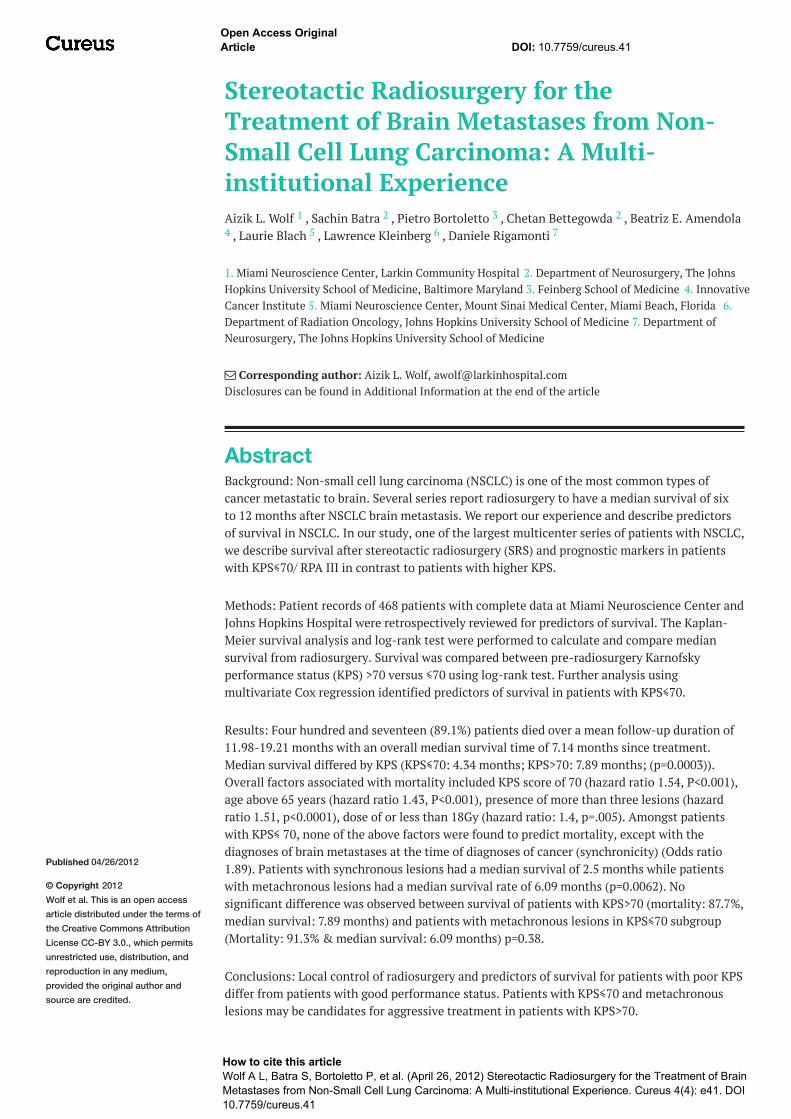
Stereotactic Radiosurgery for theTreatment of Brain Metastases from Non-Small Cell Lung Carcinoma: A Multi-institutional ExperienceAizik L. Wolf , Sachin Batra , Pietro Bortoletto , Chetan Bettegowda , Beatriz E. Amendola , Laurie Blach , Lawrence Kleinberg , Daniele Rigamonti
1. Miami Neuroscience Center, Larkin Community Hospital 2. Department of Neurosurgery, The JohnsHopkins University School of Medicine, Baltimore Maryland 3. Feinberg School of Medicine 4. InnovativeCancer Institute 5. Miami Neuroscience Center, Mount Sinai Medical Center, Miami Beach, Florida 6.Department of Radiation Oncology, Johns Hopkins University School of Medicine 7. Department ofNeurosurgery, The Johns Hopkins University School of Medicine
Corresponding author: Aizik L. Wolf, [email protected] Disclosures can be found in Additional Information at the end of the article
AbstractBackground: Non-small cell lung carcinoma (NSCLC) is one of the most common types ofcancer metastatic to brain. Several series report radiosurgery to have a median survival of sixto 12 months after NSCLC brain metastasis. We report our experience and describe predictorsof survival in NSCLC. In our study, one of the largest multicenter series of patients with NSCLC,we describe survival after stereotactic radiosurgery (SRS) and prognostic markers in patientswith KPS≤70/ RPA III in contrast to patients with higher KPS.
Methods: Patient records of 468 patients with complete data at Miami Neuroscience Center andJohns Hopkins Hospital were retrospectively reviewed for predictors of survival. The Kaplan-Meier survival analysis and log-rank test were performed to calculate and compare mediansurvival from radiosurgery. Survival was compared between pre-radiosurgery Karnofskyperformance status (KPS) >70 versus ≤70 using log-rank test. Further analysis usingmultivariate Cox regression identified predictors of survival in patients with KPS≤70.
Results: Four hundred and seventeen (89.1%) patients died over a mean follow-up duration of11.98-19.21 months with an overall median survival time of 7.14 months since treatment.Median survival differed by KPS (KPS≤70: 4.34 months; KPS>70: 7.89 months; (p=0.0003)).Overall factors associated with mortality included KPS score of 70 (hazard ratio 1.54, P<0.001),age above 65 years (hazard ratio 1.43, P<0.001), presence of more than three lesions (hazardratio 1.51, p<0.0001), dose of or less than 18Gy (hazard ratio: 1.4, p=.005). Amongst patientswith KPS≤ 70, none of the above factors were found to predict mortality, except with thediagnoses of brain metastases at the time of diagnoses of cancer (synchronicity) (Odds ratio1.89). Patients with synchronous lesions had a median survival of 2.5 months while patientswith metachronous lesions had a median survival rate of 6.09 months (p=0.0062). Nosignificant difference was observed between survival of patients with KPS>70 (mortality: 87.7%,median survival: 7.89 months) and patients with metachronous lesions in KPS≤70 subgroup(Mortality: 91.3% & median survival: 6.09 months) p=0.38.
Conclusions: Local control of radiosurgery and predictors of survival for patients with poor KPSdiffer from patients with good performance status. Patients with KPS≤70 and metachronouslesions may be candidates for aggressive treatment in patients with KPS>70.
1 2 3 2
4 5 6 7
Open Access OriginalArticle DOI: 10.7759/cureus.41
How to cite this articleWolf A L, Batra S, Bortoletto P, et al. (April 26, 2012) Stereotactic Radiosurgery for the Treatment of BrainMetastases from Non-Small Cell Lung Carcinoma: A Multi-institutional Experience. Cureus 4(4): e41. DOI10.7759/cureus.41
Categories: Radiation Oncology, NeurosurgeryKeywords: Stereotactic Radiosurgery, Outcomes, progression, mortality, non-small cell lung carcinoma
IntroductionNon-small cell lung carcinoma (NSCLC) is one of the most common types of cancer thatmetastasize to the brain. Autopsy studies have revealed 8-41% prevalence of brain metastasisin patients with bronchogenic carcinoma [1]. When treated conservatively with steroids andmedical management, the survival for brain metastasis is one to two months, independent ofhistology [2]. Several studies have demonstrated that radiosurgery enhances the survival to sixto 12 months in patients with NSCLC [3-5]. This efficacy, however, may not be observed in asubclass of patients with poor functional/performance status, often described as Karnofskyperformance scores (KPS) ≤70. KPS is one of the most consistently observed predictors of poorsurvival and is a component of recursive partition analysis (RPA) classification, a most widelyused parameter to select patients for aggressive treatment. The role of other factors, such asnumber of lesions, location of lesions, volume, and gender as prognostic factors, is not clear [2,6-8]. Other factors like delay in onset of metastatic disease are not considered in RPAclassification during the selection of patients for radiosurgical or surgical treatment [2, 4, 6-9].Furthermore, selection bias towards the aggressive management of patients with acceptedfavorable prognostic factors prevents evaluation of subgroups with poor prognosis [10]. So far,only a few series have analyzed predictors of survival in patients with poor KPS [9, 11-12].
Materials And MethodsPatients and methodsPrior to initiation, approval was gained from the Johns Hopkins Institutional Review Board toperform this study. We retrospectively reviewed data collected from chart review of 482 patientswho received stereotactic radiosurgery to treat metastatic brain lesions from NSCLC. Fourteenpatients did not return for follow-up after SRS and were excluded, yielding 468 patients thatwere eligible for analysis. Amongst these patients, 383 patients were treated atMiami Neuroscience Center, Miami, FL (MNC) from 1993 to 2009, while 85 patients weretreated at Johns Hopkins Hospital, Baltimore, MD (JHH) between 2003 and 2007. Thehistopathological diagnosis was confirmed by a biopsy of the primary tumor or of a metastaticlesion at an extracranial site or from tissue obtained during craniotomy. Patients wereevaluated for neurologic and radiographic progression initially at four weeks post-radiosurgeryand subsequently at three month intervals. Any local recurrence or new metastatic disease wastreated with subsequent sessions of radiosurgery or craniotomy.
Gamma KnifeAll lesions were treated using Leksel I Gamma Knife model U, model B or model C (ElektaInstruments, Stockholm, Sweden). The decision to treat with SRS was determined afterconsultation with a neurosurgeon and radiation oncologist at the respective centers. Patientsharboring multiple lesions were sometimes treated in multiple sessions. The prescription dosewas based on volume, number, and location of lesions, with deep lesions and those located inproximity to critical neurovascular lesions receiving a lower dose. The median prescription doseat MNC was 15 Gy (I QR: 15-18 Gy), whereas patients at JHH received a significantly higher dosewith a median of 18 Gy (IQR: 16 Gy-20 Gy) (p<0.001).
Statistical analysisThe demographic and clinical features of patients included in this study are summarized inTable 1.
2012 Wolf et al. Cureus 4(4): e41. DOI 10.7759/cureus.41 2 of 16
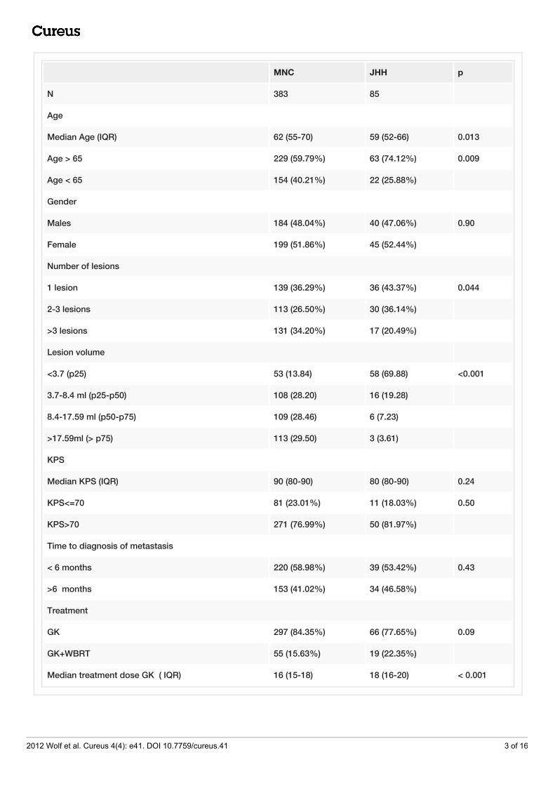
MNC JHH p
N 383 85
Age
Median Age (IQR) 62 (55-70) 59 (52-66) 0.013
Age > 65 229 (59.79%) 63 (74.12%) 0.009
Age < 65 154 (40.21%) 22 (25.88%)
Gender
Males 184 (48.04%) 40 (47.06%) 0.90
Female 199 (51.86%) 45 (52.44%)
Number of lesions
1 lesion 139 (36.29%) 36 (43.37%) 0.044
2-3 lesions 113 (26.50%) 30 (36.14%)
>3 lesions 131 (34.20%) 17 (20.49%)
Lesion volume
<3.7 (p25) 53 (13.84) 58 (69.88) <0.001
3.7-8.4 ml (p25-p50) 108 (28.20) 16 (19.28)
8.4-17.59 ml (p50-p75) 109 (28.46) 6 (7.23)
>17.59ml (> p75) 113 (29.50) 3 (3.61)
KPS
Median KPS (IQR) 90 (80-90) 80 (80-90) 0.24
KPS<=70 81 (23.01%) 11 (18.03%) 0.50
KPS>70 271 (76.99%) 50 (81.97%)
Time to diagnosis of metastasis
< 6 months 220 (58.98%) 39 (53.42%) 0.43
>6 months 153 (41.02%) 34 (46.58%)
Treatment
GK 297 (84.35%) 66 (77.65%) 0.09
GK+WBRT 55 (15.63%) 19 (22.35%)
Median treatment dose GK ( IQR) 16 (15-18) 18 (16-20) < 0.001
2012 Wolf et al. Cureus 4(4): e41. DOI 10.7759/cureus.41 3 of 16
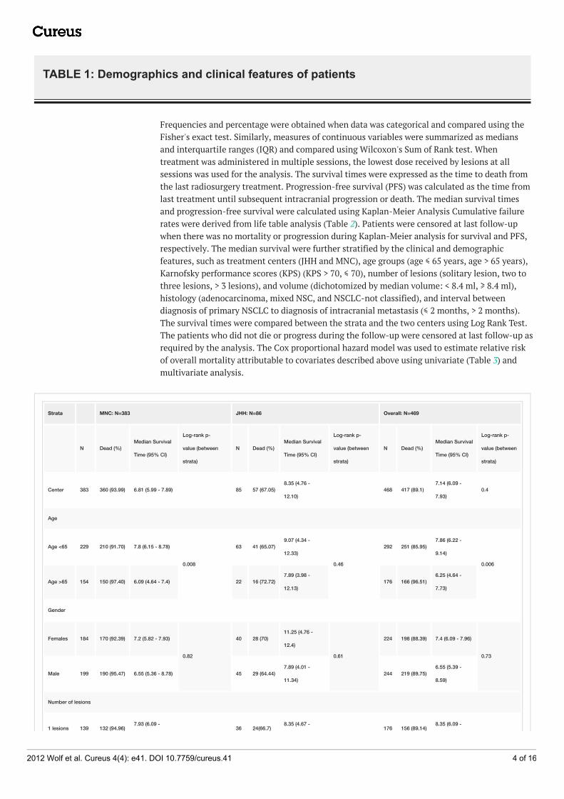
TABLE 1: Demographics and clinical features of patients
Frequencies and percentage were obtained when data was categorical and compared using theFisher's exact test. Similarly, measures of continuous variables were summarized as mediansand interquartiIe ranges (IQR) and compared using Wilcoxon's Sum of Rank test. Whentreatment was administered in multiple sessions, the lowest dose received by lesions at allsessions was used for the analysis. The survival times were expressed as the time to death fromthe last radiosurgery treatment. Progression-free survival (PFS) was calculated as the time fromlast treatment until subsequent intracranial progression or death. The median survival timesand progression-free survival were calculated using Kaplan-Meier Analysis Cumulative failurerates were derived from life table analysis (Table 2). Patients were censored at last follow-upwhen there was no mortality or progression during Kaplan-Meier analysis for survival and PFS,respectively. The median survival were further stratified by the clinical and demographicfeatures, such as treatment centers (JHH and MNC), age groups (age ≤ 65 years, age > 65 years),Karnofsky performance scores (KPS) (KPS > 70, ≤ 70), number of lesions (solitary lesion, two tothree lesions, > 3 lesions), and volume (dichotomized by median volume: < 8.4 ml, ≥ 8.4 ml),histology (adenocarcinoma, mixed NSC, and NSCLC-not classified), and interval betweendiagnosis of primary NSCLC to diagnosis of intracranial metastasis (≤ 2 months, > 2 months).The survival times were compared between the strata and the two centers using Log Rank Test.The patients who did not die or progress during the follow-up were censored at last follow-up asrequired by the analysis. The Cox proportional hazard model was used to estimate relative riskof overall mortality attributable to covariates described above using univariate (Table 3) andmultivariate analysis.
Strata MNC: N=383 JHH: N=86 Overall: N=469
N Dead (%)Median Survival
Time (95% CI)
Log-rank p-
value (between
strata)
N Dead (%)Median Survival
Time (95% CI)
Log-rank p-
value (between
strata)
N Dead (%)Median Survival
Time (95% CI)
Log-rank p-
value (between
strata)
Center 383 360 (93.99) 6.81 (5.99 - 7.89) 85 57 (67.05)8.35 (4.76 -
12.10) 468 417 (89.1)
7.14 (6.09 -
7.93)0.4
Age
Age <65 229 210 (91.70) 7.8 (6.15 - 8.78)
0.008
63 41 (65.07)9.07 (4.34 -
12.33)
0.46
292 251 (85.95)7.86 (6.22 -
9.14)
0.006
Age >65 154 150 (97.40) 6.09 (4.64 - 7.4) 22 16 (72.72)7.89 (3.98 -
12.13)176 166 (96.51)
6.25 (4.64 -
7.73)
Gender
Females 184 170 (92.39) 7.2 (5.82 - 7.93)
0.82
40 28 (70)11.25 (4.76 -
12.4)
0.61
224 198 (88.39) 7.4 (6.09 - 7.96)
0.73
Male 199 190 (95.47) 6.55 (5.36 - 8.78) 45 29 (64.44)7.89 (4.01 -
11.34)244 219 (89.75)
6.55 (5.39 -
8.59)
Number of lesions
1 lesions 139 132 (94.96)7.93 (6.09 -
36 24(66.7)8.35 (4.67 -
176 156 (89.14)8.35 (6.09 -
2012 Wolf et al. Cureus 4(4): e41. DOI 10.7759/cureus.41 4 of 16
10.82)
0.001
12.43)
0.09
10.82)
0.00052-3
lesions113 105 (92.92)
7.14 (4.97 -
10.03)30 19 (63.3) 10.82 (3.9) 143 124 (86.71) 7.76 (5.3 - 10.2)
>3
lesions131 123 (93.89) 6.25 (4.41 - 7.73) 17 12 (70.58)
5.82 (1.89 -
11.25)148 135 (91.21)
5.99 (4.41 -
7.66)
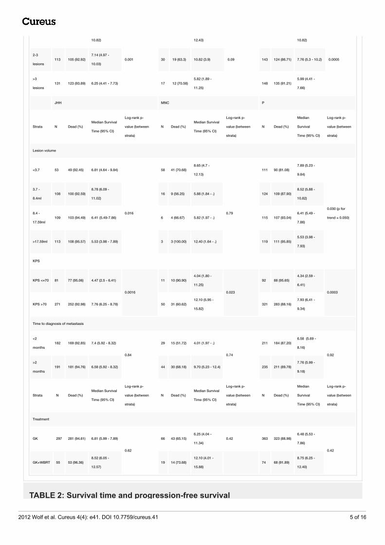
JHH MNC P
Strata N Dead (%)Median Survival
Time (95% CI)
Log-rank p-
value (between
strata)
N Dead (%)Median Survival
Time (95% CI)
Log-rank p-
value (between
strata)
N Dead (%)
Median
Survival
Time (95% CI)
Log-rank p-
value (between
strata)
Lesion volume
<3.7 53 49 (92.45) 6.81 (4.64 - 9.84)
0.016
58 41 (70.68)8.65 (4.7 -
12.13)
0.79
111 90 (81.08)7.89 (5.23 -
9.84)
0.030 (p for
trend = 0.050)
3.7 -
8.4ml108 100 (92.59)
8.78 (6.09 -
11.02)16 9 (56.25) 5.88 (1.84 - .) 124 109 (87.90)
8.52 (5.88 -
10.82)
8.4 -
17.59ml109 103 (94.49) 6.41 (5.49-7.86) 6 4 (66.67) 5.82 (1.97 - .) 115 107 (93.04)
6.41 (5.49 -
7.86)
>17.59ml 113 108 (95.57) 5.53 (3.98 - 7.89) 3 3 (100.00) 12.40 (1.64 - .) 119 111 (95.85)5.53 (3.98 -
7.93)
KPS
KPS <=70 81 77 (95.06) 4.47 (2.5 - 6.41)
0.0016
11 10 (90.90)4.04 (1.80 -
11.25)
0.023
92 88 (95.65)4.34 (2.59 -
6.41)
0.0003
KPS >70 271 252 (92.98) 7.76 (6.25 - 8.78) 50 31 (60.62)12.10 (5.95 -
15.82)321 283 (88.16)
7.93 (6.41 -
9.34)
Time to diagnosis of metastasis
<2
months182 169 (92.85) 7.4 (5.92 - 8.32)
0.84
29 15 (51.72) 4.01 (1.97 - .)
0.74
211 184 (87.20)6.58 (5.69 -
8.16)
0.92
>2
months191 181 (94.76) 6.58 (5.92 - 8.32) 44 30 (68.18) 9.70 (5.23 - 12.4) 235 211 (89.78)
7.76 (5.99 -
9.18)
Strata N Dead (%)Median Survival
Time (95% CI)
Log-rank p-
value (between
strata)
N Dead (%)Median Survival
Time (95% CI)
Log-rank p-
value (between
strata)
N Dead (%)
Median
Survival
Time (95% CI)
Log-rank p-
value (between
strata)
Treatment
GK 297 281 (94.61) 6.81 (5.99 - 7.89)
0.62
66 43 (65.15)6.25 (4.04 -
11.34)0.42 363 323 (88.98)
6.48 (5.53 -
7.86)
0.42
GK+WBRT 55 53 (96.36)8.52 (6.05 -
12.57)19 14 (73.68)
12.10 (4.01 -
15.88) 74 68 (91.89)
8.75 (6.25 -
12.40)
TABLE 2: Survival time and progression-free survival
2012 Wolf et al. Cureus 4(4): e41. DOI 10.7759/cureus.41 5 of 16
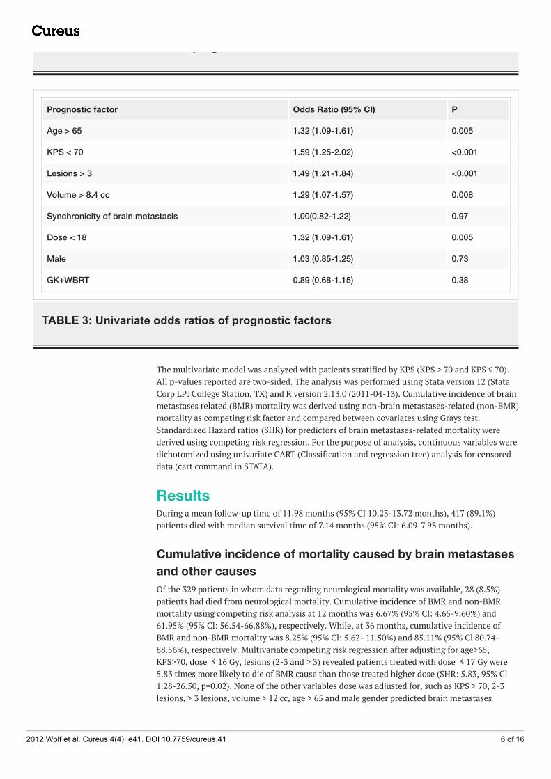
TABLE 2: Survival time and progression-free survival
Prognostic factor Odds Ratio (95% CI) P
Age > 65 1.32 (1.09-1.61) 0.005
KPS < 70 1.59 (1.25-2.02) <0.001
Lesions > 3 1.49 (1.21-1.84) <0.001
Volume > 8.4 cc 1.29 (1.07-1.57) 0.008
Synchronicity of brain metastasis 1.00(0.82-1.22) 0.97
Dose < 18 1.32 (1.09-1.61) 0.005
Male 1.03 (0.85-1.25) 0.73
GK+WBRT 0.89 (0.68-1.15) 0.38
TABLE 3: Univariate odds ratios of prognostic factors
The multivariate model was analyzed with patients stratified by KPS (KPS > 70 and KPS ≤ 70).All p‑values reported are two-sided. The analysis was performed using Stata version 12 (StataCorp LP: College Station, TX) and R version 2.13.0 (2011-04-13). Cumulative incidence of brainmetastases related (BMR) mortality was derived using non-brain metastases-related (non-BMR)mortality as competing risk factor and compared between covariates using Grays test.Standardized Hazard ratios (SHR) for predictors of brain metastases-related mortality werederived using competing risk regression. For the purpose of analysis, continuous variables weredichotomized using univariate CART (Classification and regression tree) analysis for censoreddata (cart command in STATA).
ResultsDuring a mean follow-up time of 11.98 months (95% CI 10.23-13.72 months), 417 (89.1%)patients died with median survival time of 7.14 months (95% CI: 6.09-7.93 months).
Cumulative incidence of mortality caused by brain metastasesand other causes Of the 329 patients in whom data regarding neurological mortality was available, 28 (8.5%)patients had died from neurological mortality. Cumulative incidence of BMR and non-BMRmortality using competing risk analysis at 12 months was 6.67% (95% Cl: 4.65-9.60%) and61.95% (95% Cl: 56.54-66.88%), respectively. While, at 36 months, cumulative incidence ofBMR and non-BMR mortality was 8.25% (95% Cl: 5.62- 11.50%) and 85.11% (95% Cl 80.74-88.56%), respectively. Multivariate competing risk regression after adjusting for age>65,KPS>70, dose ≤ 16 Gy, lesions (2-3 and > 3) revealed patients treated with dose ≤ 17 Gy were5.83 times more likely to die of BMR cause than those treated higher dose (SHR: 5.83, 95% Cl1.28-26.50, p=0.02). None of the other variables dose was adjusted for, such as KPS > 70, 2-3lesions, > 3 lesions, volume > 12 cc, age > 65 and male gender predicted brain metastases
2012 Wolf et al. Cureus 4(4): e41. DOI 10.7759/cureus.41 6 of 16
related mortality with standardized hazard ratios (SHR) of 0.67 (95% Cl: 0.28 - 1.58, p=0.36),1.58 (95% Cl: 0.53 - 4.69, p=0.40 ), 1.63 (95% Cl: 0.60 - 4.42, p=0.33), 1.97 (0.93 - 4.20, p=0.07),1.04 (95% Cl: 0.42- 2.56, p=0.92) and 1.12 (0.50 - 2.47, p=0.77), respectively. The two centershad comparable survival times with patients at JHH and MNC surviving for a median of 8.35months (95% CI: 4.76-12.10 months, 67.05% mortality) and 6.81 (5.99-7.89 months, 93.99%mortality), respectively (p=0.34). At one year, cumulative mortality was 70.6% (95% CI: 66.41%-74.64%) while at 18 months 80.75% (95% CI: 76.84%-84.37%) patients were not alive.
Survival was subsequently compared by various predictors of mortality. Patients who were olderthan 65 years had a median survival of 6.25 months (95% CI: 4.64-7.73 months, 96.51%mortality) as compared to 7.83 months (95% CI: 6.22-9.14 months, 85.95% mortality) inpatients who were younger (p=0.004) (Figure 1).
FIGURE 1: Survival by age
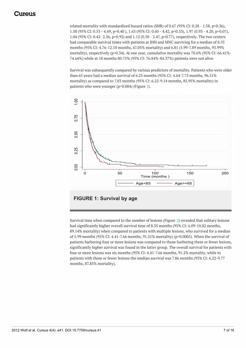
Survival time when compared to the number of lesions (Figure 2) revealed that solitary lesionshad significantly higher overall survival time of 8.35 months (95% CI: 6.09-10.82 months,89.14% mortality) when compared to patients with multiple lesions, who survived for a medianof 5.99 months (95% Cl: 4.41-7.66 months, 91.21% mortality) (p=0.0005). When the survival ofpatients harboring four or more lesions was compared to those harboring three or fewer lesions,significantly higher survival was found in the latter group. The overall survival for patients withfour or more lesions was six months (95% Cl: 4.41-7.66 months, 91.2% mortality, while inpatients with three or fewer lesions the median survival was 7.86 months (95% Cl: 6.22-9.77months, 87.85% mortality).
2012 Wolf et al. Cureus 4(4): e41. DOI 10.7759/cureus.41 7 of 16
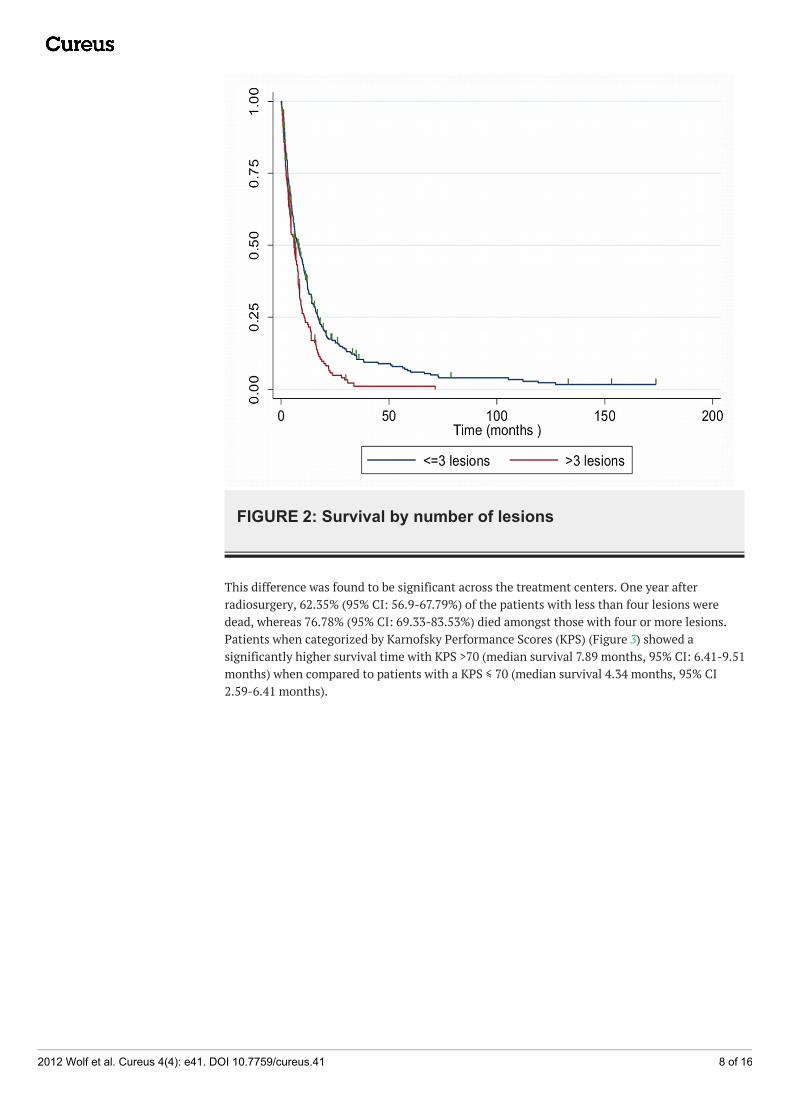
FIGURE 2: Survival by number of lesions
This difference was found to be significant across the treatment centers. One year afterradiosurgery, 62.35% (95% CI: 56.9-67.79%) of the patients with less than four lesions weredead, whereas 76.78% (95% CI: 69.33-83.53%) died amongst those with four or more lesions.Patients when categorized by Karnofsky Performance Scores (KPS) (Figure 3) showed asignificantly higher survival time with KPS >70 (median survival 7.89 months, 95% CI: 6.41-9.51months) when compared to patients with a KPS ≤ 70 (median survival 4.34 months, 95% CI2.59-6.41 months).
2012 Wolf et al. Cureus 4(4): e41. DOI 10.7759/cureus.41 8 of 16
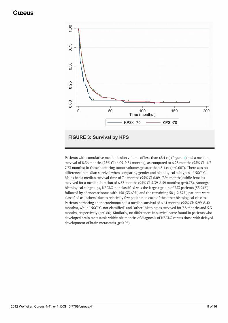
FIGURE 3: Survival by KPS
Patients with cumulative median lesion volume of less than (8.4 cc) (Figure 4) had a mediansurvival of 8.36 months (95% CI: 6.09-9.84 months), as compared to 6.28 months (95% CI: 4.7-7.73 months) in those harboring tumor volumes greater than 8.4 cc (p=0.007). There was nodifference in median survival when comparing gender and histological subtypes of NSCLC.Males had a median survival time of 7.4 months (95% CI 6.09- 7.96 months) while femalessurvived for a median duration of 6.55 months (95% CI 5.39-8.59 months) (p=0.73). Amongsthistological subgroups, NSCLC-not classified was the largest group of 253 patients (53.94%)followed by adenocarcinoma with 158 (33.69%) and the remaining 58 (12.37%) patients wereclassified as "others" due to relatively few patients in each of the other histological classes.Patients harboring adenocarcinoma had a median survival of 6.61 months (95% Cl: 5.99-8.42months), while "NSCLC-not classified" and "other" histologies survived for 7.8 months and 5.3months, respectively (p=0.66). Similarly, no differences in survival were found in patients whodeveloped brain metastasis within six months of diagnosis of NSCLC versus those with delayeddevelopment of brain metastasis (p=0.95).
2012 Wolf et al. Cureus 4(4): e41. DOI 10.7759/cureus.41 9 of 16
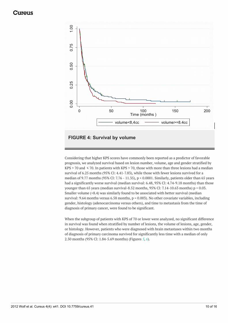
FIGURE 4: Survival by volume
Considering that higher KPS scores have commonly been reported as a predictor of favorableprognosis, we analyzed survival based on lesion number, volume, age and gender stratified byKPS > 70 and ≤ 70. In patients with KPS > 70, those with more than three lesions had a mediansurvival of 6.25 months (95% Cl: 4.41-7.83), while those with fewer lesions survived for amedian of 9.77 months (95% Cl: 7.76 - 11.35), p < 0.0001. Similarly, patients older than 65 yearshad a significantly worse survival (median survival: 6.48, 95% Cl: 4.74-9.18 months) than thoseyounger than 65 years (median survival-8.52 months, 95% Cl: 7.14-10.63 months) p = 0.03.Smaller volume (<8.4) was similarly found to be associated with better survival (mediansurvival: 9.64 months versus 6.58 months, p = 0.003). No other covariate variables, includinggender, histology (adenocarcinoma versus others), and time to metastasis from the time ofdiagnosis of primary cancer, were found to be significant.
When the subgroup of patients with KPS of 70 or lower were analyzed, no significant differencein survival was found when stratified by number of lesions, the volume of lesions, age, gender,or histology. However, patients who were diagnosed with brain metastases within two monthsof diagnosis of primary carcinoma survived for significantly less time with a median of only2.50 months (95% Cl: 1.84-5.69 months) (Figures 5, 6).
2012 Wolf et al. Cureus 4(4): e41. DOI 10.7759/cureus.41 10 of 16
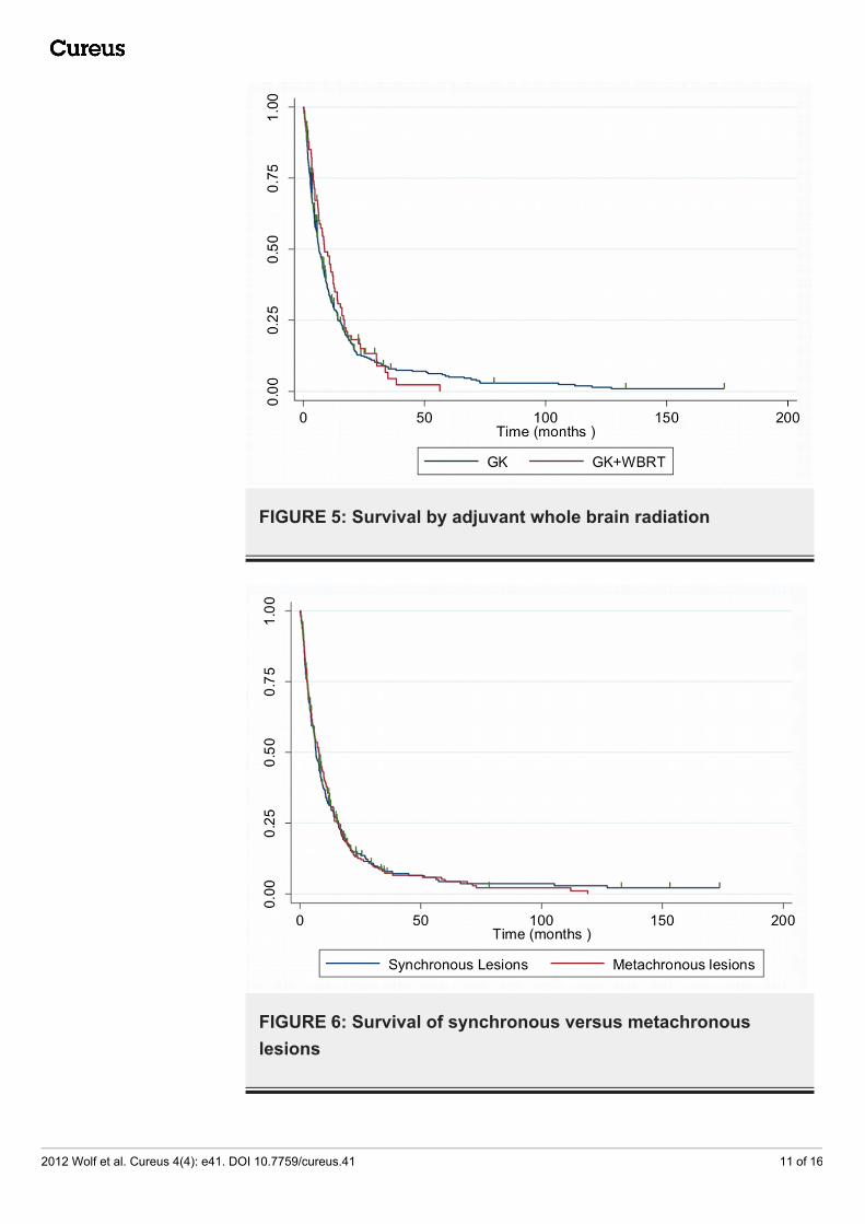
FIGURE 5: Survival by adjuvant whole brain radiation
FIGURE 6: Survival of synchronous versus metachronouslesions
2012 Wolf et al. Cureus 4(4): e41. DOI 10.7759/cureus.41 11 of 16
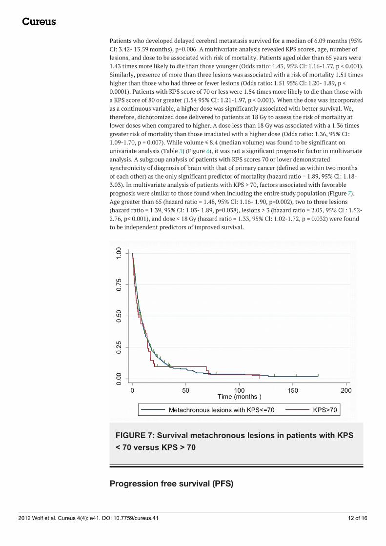
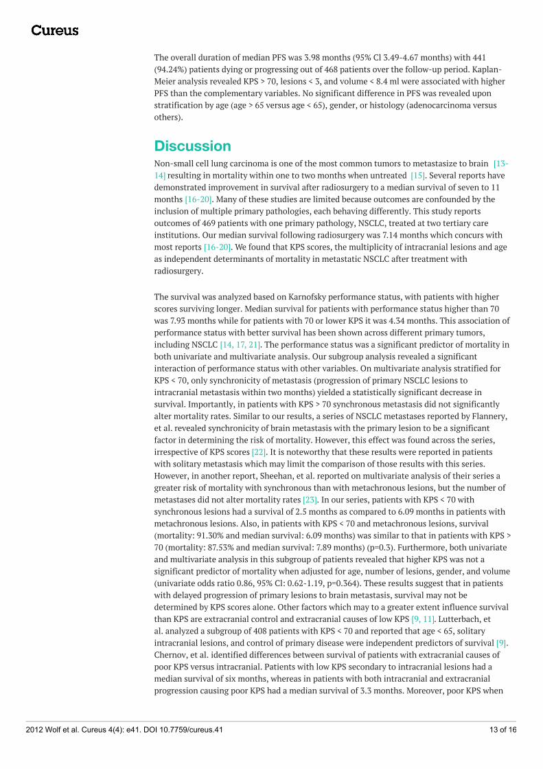
Patients who developed delayed cerebral metastasis survived for a median of 6.09 months (95%Cl: 3.42- 13.59 months), p=0.006. A multivariate analysis revealed KPS scores, age, number oflesions, and dose to be associated with risk of mortality. Patients aged older than 65 years were1.43 times more likely to die than those younger (Odds ratio: 1.43, 95% Cl: 1.16-1.77, p < 0.001).Similarly, presence of more than three lesions was associated with a risk of mortality 1.51 timeshigher than those who had three or fewer lesions (Odds ratio: 1.51 95% Cl: 1.20- 1.89, p <0.0001). Patients with KPS score of 70 or less were 1.54 times more likely to die than those witha KPS score of 80 or greater (1.54 95% CI: 1.21-1.97, p < 0.001). When the dose was incorporatedas a continuous variable, a higher dose was significantly associated with better survival. We,therefore, dichotomized dose delivered to patients at 18 Gy to assess the risk of mortality atlower doses when compared to higher. A dose less than 18 Gy was associated with a 1.36 timesgreater risk of mortality than those irradiated with a higher dose (Odds ratio: 1.36, 95% CI:1.09-1.70, p = 0.007). While volume ≤ 8.4 (median volume) was found to be significant onunivariate analysis (Table 3) (Figure 6), it was not a significant prognostic factor in multivariateanalysis. A subgroup analysis of patients with KPS scores 70 or lower demonstratedsynchronicity of diagnosis of brain with that of primary cancer (defined as within two monthsof each other) as the only significant predictor of mortality (hazard ratio = 1.89, 95% Cl: 1.18-3.03). In multivariate analysis of patients with KPS > 70, factors associated with favorableprognosis were similar to those found when including the entire study population (Figure 7).Age greater than 65 (hazard ratio = 1.48, 95% Cl: 1.16- 1.90, p=0.002), two to three lesions(hazard ratio = 1.39, 95% Cl: 1.03- 1.89, p=0.038), lesions > 3 (hazard ratio = 2.05, 95% Cl : 1.52-2.76, p< 0.001), and dose < 18 Gy (hazard ratio = 1.33, 95% Cl: 1.02-1.72, p = 0.032) were foundto be independent predictors of improved survival.
FIGURE 7: Survival metachronous lesions in patients with KPS< 70 versus KPS > 70
Progression free survival (PFS)
2012 Wolf et al. Cureus 4(4): e41. DOI 10.7759/cureus.41 12 of 16
The overall duration of median PFS was 3.98 months (95% Cl 3.49-4.67 months) with 441(94.24%) patients dying or progressing out of 468 patients over the follow-up period. Kaplan-Meier analysis revealed KPS > 70, lesions < 3, and volume < 8.4 ml were associated with higherPFS than the complementary variables. No significant difference in PFS was revealed uponstratification by age (age > 65 versus age < 65), gender, or histology (adenocarcinoma versusothers).
DiscussionNon-small cell lung carcinoma is one of the most common tumors to metastasize to brain [13-14] resulting in mortality within one to two months when untreated [15]. Several reports havedemonstrated improvement in survival after radiosurgery to a median survival of seven to 11months [16-20]. Many of these studies are limited because outcomes are confounded by theinclusion of multiple primary pathologies, each behaving differently. This study reportsoutcomes of 469 patients with one primary pathology, NSCLC, treated at two tertiary careinstitutions. Our median survival following radiosurgery was 7.14 months which concurs withmost reports [16-20]. We found that KPS scores, the multiplicity of intracranial lesions and ageas independent determinants of mortality in metastatic NSCLC after treatment withradiosurgery.
The survival was analyzed based on Karnofsky performance status, with patients with higherscores surviving longer. Median survival for patients with performance status higher than 70was 7.93 months while for patients with 70 or lower KPS it was 4.34 months. This association ofperformance status with better survival has been shown across different primary tumors,including NSCLC [14, 17, 21]. The performance status was a significant predictor of mortality inboth univariate and multivariate analysis. Our subgroup analysis revealed a significantinteraction of performance status with other variables. On multivariate analysis stratified forKPS < 70, only synchronicity of metastasis (progression of primary NSCLC lesions tointracranial metastasis within two months) yielded a statistically significant decrease insurvival. Importantly, in patients with KPS > 70 synchronous metastasis did not significantlyalter mortality rates. Similar to our results, a series of NSCLC metastases reported by Flannery,et al. revealed synchronicity of brain metastasis with the primary lesion to be a significantfactor in determining the risk of mortality. However, this effect was found across the series,irrespective of KPS scores [22]. It is noteworthy that these results were reported in patientswith solitary metastasis which may limit the comparison of those results with this series.However, in another report, Sheehan, et al. reported on multivariate analysis of their series agreater risk of mortality with synchronous than with metachronous lesions, but the number ofmetastases did not alter mortality rates [23]. In our series, patients with KPS < 70 withsynchronous lesions had a survival of 2.5 months as compared to 6.09 months in patients withmetachronous lesions. Also, in patients with KPS < 70 and metachronous lesions, survival(mortality: 91.30% and median survival: 6.09 months) was similar to that in patients with KPS >70 (mortality: 87.53% and median survival: 7.89 months) (p=0.3). Furthermore, both univariateand multivariate analysis in this subgroup of patients revealed that higher KPS was not asignificant predictor of mortality when adjusted for age, number of lesions, gender, and volume(univariate odds ratio 0.86, 95% Cl: 0.62-1.19, p=0.364). These results suggest that in patientswith delayed progression of primary lesions to brain metastasis, survival may not bedetermined by KPS scores alone. Other factors which may to a greater extent influence survivalthan KPS are extracranial control and extracranial causes of low KPS [9, 11]. Lutterbach, etal. analyzed a subgroup of 408 patients with KPS < 70 and reported that age < 65, solitaryintracranial lesions, and control of primary disease were independent predictors of survival [9].Chernov, et al. identified differences between survival of patients with extracranial causes ofpoor KPS versus intracranial. Patients with low KPS secondary to intracranial lesions had amedian survival of six months, whereas in patients with both intracranial and extracranialprogression causing poor KPS had a median survival of 3.3 months. Moreover, poor KPS when
2012 Wolf et al. Cureus 4(4): e41. DOI 10.7759/cureus.41 13 of 16
caused by only extracranial disease, had a median survival of one month. Although our resultswere not adjusted for these possible confounders, this study, along with other series describedabove, suggest that a subclass of patients with low KPS/RPA III should be treated aggressivelywith a goal to control both extracranial and intracranial disease instead of common practice ofpalliation of patients in RPA III. Furthermore, in patients with synchronous disease wheresurvival is associated with control of both intracranial and extracranial disease [24], thedecision to treat brain metastasis may be guided by the ability to control the primary tumor.
The predictors of survival in patients with KPS scores higher that 70 were different frompatients with KPS < 70. Multiple metastasis (> 3) and age greater than 65 were the clinicalfeatures found to be significantly associated with poor outcomes for univariate and multivariateanalysis in patients with KPS > 70. Unlike patients with poorer KPS, the synchronicity of brainmetastasis was not associated with worse outcomes in patients with KPS > 70.
Median survival was 7.86 months (95% CI: 6.22-9.77 months) in patients with three or fewerlesions which was significantly higher than the patients with greater than three lesions (5.99months 95% CI: 4.41-7.66 months), with a log-rank value of p=0.0001. In agreement with ourresults, other series have also reported higher survival with solitary metastasis and pooroutcomes with multiple lesions [4, 9, 16]. Di Luna, et al. reported that in 336 patients withmetastatic tumors (36% NSCLC), patients with greater than four lesions had a median survivalof 6.1 months, while patients with patients with fewer lesions survived for a median of ninemonths. In that report, 113 patients harboring metastatic NSCLC had a median survival of 12.9months, 12.0 months, 13.1 months and eight months (since radiosurgery) when there weresolitary, two lesions, three lesions and greater than three lesions, respectively. Though thetrend is consistent with our results, their study shows overall higher survival rates that may beattributed to 93% of their patients having KPS of 70 or higher and 68% being younger than 65years. Flannery, et al., in their series, reported a median survival of 33 months versus 8.6months in metachronous versus synchronous metastasis [22]. These results are in agreementwith our results of higher survival for patients with solitary metachronous lesions.
A major limitation of the study is lack of detailed data regarding extracranial disease. We were,however, able to observe a lower neurological mortality rate in metastases of NSCLC (8.5%)than observed in our entire brain metastases experience consisting of all histologies(12.30%).This suggests that brain metastases from NSCLC may be more sensitive to SRS thanother histologies.
ConclusionsStereotactic radiosurgery provides an efficacious treatment option for patients with NSCLCmetastatic to the brain in patients with fewer lesions (< 3 lesions), younger age (< 65 years) andhigher KPS scores (> 70). SRS may also be effective in prolonging survival in patients withmetachronous lesions despite low KPS scores.
Additional InformationDisclosuresHuman subjects: Consent was obtained by all participants in this study. The Johns HopkinsInstitutional Review Board issued approval N/A. Animal subjects: All authors have confirmedthat this study did not involve animal subjects or tissue. Conflicts of interest: In compliancewith the ICMJE uniform disclosure form, all authors declare the following: Payment/servicesinfo: All authors have declared that no financial support was received from any organizationfor the submitted work. Financial relationships: We declare(s) a grant from Elekta AB.Education grant. Other relationships: All authors have declared that there are no other
2012 Wolf et al. Cureus 4(4): e41. DOI 10.7759/cureus.41 14 of 16
relationships or activities that could appear to have influenced the submitted work.
References1. Hazra T, Mullins GM, Lott S: Management of cerebral metastasis from bronchogenic
carcinoma. Johns Hopkins Med J. 1972, 130:377-383.2. Gaspar L, Scott C, Rotman M, et al: Recursive partitioning analysis (RPA) of prognostic factors
in three Radiation Therapy Oncology Group (RTOG) brain metastases trials. Int J Radiat OncolBiol Phys. 1997, 37:745-751. 10.1016/S0360-3016(96)00619-0
3. Abrahams JM, Torchia M, Putt M, et al: Risk factors affecting survival after brain metastasesfrom non-small cell lung carcinoma: a follow-up study of 70 patients. J Neurosurg. 2001,95:595-600. 10.3171/jns.2001.95.4.0595
4. Zabel A, Milker-Zabel S, Thilmann C, et al: Treatment of brain metastases in patients withnon- small cell lung cancer (NSCLC) by stereotactic linac-based radiosurgery: prognosticfactors. Lung Cancer. 2002, 37:87-94. 10.1016/S0169-5002(02)00030-2
5. Flannery TW, Suntharalingam M, Kwok Y, et al: Gamma Knife stereotactic radiosurgery forsynchronous versus metachronous solitary brain metastases from non-small cell lung cancer.Lung Cancer. 2003, 42:327-333. 10.1016/S0169-5002(03)00357-X
6. Frazier JL, Batra S, Kapor S, et al: Stereotactic radiosurgery in the management of brainmetastases: an institutional retrospective analysis of survival. Int J Radiat Oncol Biol Phys.2010, 76:1486-1492. 10.1016/j.ijrobp.2009.03.028
7. Lorenzoni J, Devriendt D, Massager N, et al: Radiosurgery for treatment of brain metastases:estimation of patient eligibility using three stratification systems. Int J Radiat Oncol Biol Phys.2004, 60:218-224. 10.1016/j.ijrobp.2004.02.017
8. Gaspar LE, Scott C, Murray K, et al: Validation of the RTOG recursive partitioning analysis(RPA) classification for brain metastases. Int J Radiat Oncol Biol Phys. 2000, 47:1001-1006.10.1016/S0360-3016(00)00547-2
9. Lutterbach J, Bartelt S, Stancu E, et al: Patients with brain metastases: hope for recursivepartitioning analysis (RPA) class 3. Radiother Oncol. 2002, 63:339-345. 10.1016/S0167-8140(02)00119-6
10. Buchsbaum J, Suh J, Lee S, et al: Survival by radiation therapy oncology group recursivepartitioning analysis class and treatment modality in patients with brain metastases frommalignant melanoma: a retrospective study. Cancer. 2002, 94:2265-2272. 10.1002/cncr.10426
11. Chernov M, Nakaya K, Izawa M, et al: Outcome after radiosurgery for brain metastases inpatients with low Karnofsky performance scale (KPS) scores. Int J Radiat Oncol Biol Phys.2007, 67:1492-1498. 10.1016/j.ijrobp.2006.11.023
12. Weltman E, Salvajoli J, Brandt R, et al: Radiosurgery for brain metastases: who may notbenefit?. Int J Radiat Oncol Biol Phys. 2001, 51:1320-1327. 10.1016/S0360-3016(01)01696-0
13. Nussbaum E, Djalilian H, Cho K, et al: Brain metastases. Histology, multiplicity, surgery, andsurvival. Cancer. 1996, 78:1781-1788. 10.1002/(SICI)1097-0142(19961015)78:8<1781::AID-CNCR19>3.0.CO;2-U
14. Karlsson B, Hanssens P, Wolff R, et al: Thirty years' experience with Gamma Knife surgery formetastases to the brain. J Neurosurg. 2009, 111:449-457. 10.3171/2008.10.JNS08214
15. Hazra T, Mullins G, Lott S: Management of cerebral metastasis from bronchogenic carcinoma .Johns Hopkins Med J. 1972, 130:377-383.
16. Hoffman R, Sneed P. McDermott M, et al: Radiosurgery for brain metastases from primarylung carcinoma. Cancer J. 2001, 7:121-131.
17. Sperduto P. Chao S, Sneed P. et al: Diagnosis-Specific Prognostic Factors, Indexes, andTreatment Outcomes for Patients with Newly Diagnosed Brain Metastases: A Multi-Institutional Analysis of 4,259 Patients. Int J Radiat Oncol Biol Phys. 2009, 77:655-61.10.1016/j.ijrobp.2009.08.025
18. Flickinger J, Kondziolka D, Lunsford L, et al: A multi-institutional experience withstereotactic radiosurgery for solitary brain metastasis. Int J Radiat Oncol Biol Phys. 1994,28:797-802. 10.1016/0360-3016(94)90098-1
19. Kano H, Kondziolka D, Zorro 0, et al: The results of resection after stereotactic radiosurgeryfor brain metastases. J Neurosurg. 2009, 111:825-831. 10.3171/2009.4.JNS09246
20. Chen J, Petrovich Z, O'Day S, et al: Stereotactic radiosurgery in the treatment of metastaticdisease to the brain. Neurosurg. 2000, 47:268-279. 10.1097/00006123-200008000-00003
2012 Wolf et al. Cureus 4(4): e41. DOI 10.7759/cureus.41 15 of 16
21. Lagerwaard F, Levendag P, Nowak P, et al: Identification of prognostic factors in patients withbrain metastases: a review of 1292 patients. Int J Radiat Oncol Biol Phys. 1999, 43:795-803.10.1016/S0360-3016(98)00442-8
22. Flannery T, Suntharalingam M, Kwok Y, et al: Gamma knife stereotactic radiosurgery forsynchronous versus metachronous solitary brain metastases from non-small cell lung cancer.Lung Cancer. 2003, 42:327-333. 10.1016/S0169-5002(03)00357-X
23. Sheehan J, Sun M, Kondziolka D, et al: Radiosurgery for non-small cell lung carcinomametastatic to the brain: long-term outcomes and prognostic factors influencing patientsurvival time and local tumor control. J Neurosurg. 2002, 97:1276-1281.10.3171/jns.2002.97.6.1276
24. Kong D, Lee J, Nam D, et al: Prognosis of non-small cell lung cancer with synchronous brainmetastases treated with Gamma Knife radiosurgery. J Korean Med Sci. 2006, 21:527-532.10.3346/jkms.2006.21.3.527
2012 Wolf et al. Cureus 4(4): e41. DOI 10.7759/cureus.41 16 of 16


![Skin Metastases from Ovarian Carcinoma · small and isolated radiotherapy has been described as an option [7]. Our patient has developed . Abstract. Skin metastases from malignant](https://img.pdfslide.net/doc/110x75/5f477e02ffcea70dc574f589/skin-metastases-from-ovarian-small-and-isolated-radiotherapy-has-been-described.jpg)


![CASE REPORT Open Access Metastatic colorectal carcinoma ...the female reproductive system most commonly affected by metastases [3]. Ovarian metastases occur in 3 to 8% of women with](https://img.pdfslide.net/doc/110x75/60df4bb305bcd923ec2815ba/case-report-open-access-metastatic-colorectal-carcinoma-the-female-reproductive.jpg)