Embed Size (px)
Citation preview

CARCINOMA CERVIX

CARCINOMA CERVIX
• 1,25,000 new patients in India every year• Incidence varies from 15 – 48 / 100,000 women• Carcinoma cervix is preventable
– Health education– Screening programmes
• Risk factors for Carcinoma cervix– Early age at intercourse– Repeated / Frequent births– Multiple sexual partners– HPV infections (Type 16 & 18 highly oncogenic)– Low socio-economic status– Smoking

CARCINOMA CERVIX• Site
– Ectocervix 80%– Endocervix 20%
• Gross lesion– Occult– Proliferative : Friable growth– Ulcerative : Erodes the cervix to form an irregular crater– Infiltrative : Expands the cervix
• Histopathology– Squamous cell carcinoma (80-90%)
• Large cell keratinizing• Large cell non-keratinizing• Small cell
– Adenocarcinoma (10-20%)• Endocervical• Clear cell• Adeno-squamous• Adeno-acanthoma

CARCINOMA CERVIX : SPREAD
• Direct– Vagina– Uterus– Parametrium
• Lymphatic– Primary nodes
• Obturator• Internal & External Iliac• Sacral
– Secondary nodes• Common Iliac• Para-aortic• Inguinal
• Haematogenous– Lungs– Liver– Bone

CARCINOMA CERVIX : STAGING
• Staging is clinical
• Investigations permitted– Cystoscopy / Proctoscopy– Intra-venous Urography– X-ray chest – Imaging studies (USG / CT / MRI)
• Preinvasive carcinoma– Stage 0 : Carcinoma in situ – Not to be included in therapeutic statistics

CARCINOMA CERVIX : STAGING OF INVASIVE DISEASE
• Stage I : Carcinoma confined to the cervix (Extension to the corpus should be disregarded)
• Stage Ia : Invasive carcinoma diagnosed only by microscopy
– Stage Ia1: Minimal microscopic stromal invasion, maximum depth 3mm from basement membrane
– Stage Ia2: Microscopic stromal invasion > 3mm from basement membrane, but less than 5mm. Maximum horizontal
spread < 7mm. Larger lesions should be staged Ib
• Stage Ib : Invasive carcinoma confined to the cervix, greater than Ia2 whether seen clinically or
not– Stage Ib1 : Preclinical lesions greater than Ia2 or clinical lesions not
exceeding 4cm in size– Stage Ib2 : Clinical lesions > 4 cm in size
CARCINOMA CERVIX : STAGING OF INVASIVE DISEASE
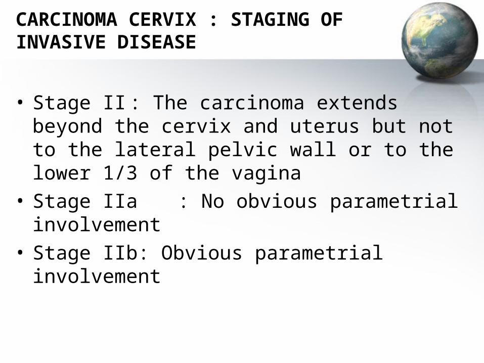
• Stage II : The carcinoma extends beyond the cervix and uterus but not to the lateral pelvic wall or to the lower 1/3 of the vagina
• Stage IIa : No obvious parametrial involvement
• Stage IIb: Obvious parametrial involvement

CARCINOMA CERVIX : STAGING OF INVASIVE DISEASE
• Stage III : The carcinoma extends to the lateral pelvic wall, or to the lower 1/3 of the vagina, or causes Hydronephrosis or non-functioning Kidney
• Stage IIIa: The carcinoma involves the lower 1/3 of the vagina. No extention to the lateral pelvic wall
• Stage IIIb: The carcinoma extends to the lateral pelvic wall, or causes Hydronephrosis or non functioning kidney

CARCINOMA CERVIX : STAGING OF INVASIVE DISEASE
• Stage IV : The carcinoma extends beyond the true pelvis or has clinically involved the mucosa of the bladder or rectum (biopsy proven)
A bullous edema of the bladder / rectal mucosa as such, does not permit a case to be allotted to Stage IV
• Stage IVa: Spread of carcinoma to adjacent organs
• Stage IVb: Spread of carcinoma to distant organs

Diagnosis of carcinoma cervix
• Preclinical (Stage Ia & some patients of Stage Ib1 with absence of obvious growth)– Asymptomatic– Detected on screening
• If microinvasion is detected on targeted biopsy or endocervical curettage, a conization of the cervix is mandatory to exclude the presence of invasive carcinoma

Diagnosis of carcinoma cervix
• Clinical (Stage Ib1 onwards)– Early symptoms
• Abnormal bleeding– Post coital– Inter-menstrual– Post-menopausal
• Abnormal discharge– Blood stained– Dirty– Foul smelling
– Late symptoms• Pelvic pain• Urinary symptoms• Rectal symptoms
Diagnosis of carcinoma cervix
• Signs– Abnormal area / growth on cervix– Induration– Friability– Bleeding on touch– Fixity
• Confirmation of diagnosis– Diagnosis is confirmed by Histopathological
examination of the biopsy sample

Differential diagnosis
• Fibroid polyp
• Chronic inversion of the uterus
• Cervical Tuberculosis
• Cervical ectopic pregnancy

Complications
• Pyometra
• Haemorrhage
• Pyelonephritis
• Vesico-vaginal fistula
• Uraemia
• Recto-vaginal fistula
Prognosis
• Staging
• Histologic type
• Differentiation
• Tumor volume
• Lymph node involvement

Cause of death
• Uraemia
• Haemorrhage
• Sepsis

Prevention of Carcinoma cervix
• Health education– Avoid early marriage– Avoid early intercourse– Avoid promiscuity– Proper hygiene– Use of barrier contraception
• Screening programs– Screening for pre-malignant lesions– Screening for early diagnosis

Investigations
• For confirmation of diagnosis– Biopsy
• From obvious growth or abnormal area• Directed biopsy in very early lesions• Cone biopsy
• For staging of disease• Intravenous Urography• Abdominal Ultrasonography• Cystoscopy• Proctosigmoidoscopy• Examination under anaesthesia (EUA)• CT / MRI
• Base line investigations of general condition

Treatment
• Factors– Stage of disease– Age of patient– General condition / Associated problems– Tumor configuration
• Modalities– Surgery– Radiotherapy– Combined– Chemo-radiation
Surgery for Carcinoma cervix
• Curative surgery can be performed in Ca Cx upto Stage IIa
• Surgery is preferred in– Young patients– Patients with prolapse– Patients with uteri distorted by fibroids– Co-existing pelvic pathology
• Stage Ia1 disease– Conization may be both diagnostic and therapeutic– Simple extra fascial hysterectomy

Surgery for Carcinoma cervix
• Stage Ia2 IIa disease– Wertheim’s / Meig’s hysterectomy
(Extended hysterectomy with pelvic lymphadenectomy)• Uterus including cervix
• Adnexae (Ovaries spared in the young)
• Wide resection of the parametrium
• Removal of vaginal cuff
• Dissection of peri-ureteral tissues
• Pelvic lymphadenectomy
• Stage IV a disease– Exenteration

Surgery for Carcinoma cervix
• Advantages– Preservation of ovarian function– Preservation of vaginal function– Lesser long term morbidity– Complications correctable
• Complications– Haemorrhage – Infection– Lymphocyst formation– Ureteric injury / fistula
• Traumatic
• Ischaemic– Bladder injury– Neurogenic bladder dysfunction
Radiotherapy for Carcinoma cervix
• Advantages– Applicable for all stages of disease– As effective as surgery in early stages– Lesser primary mortality and immediate morbidity as
compared to surgery– Preferred in patients unfit for surgery because of
medical conditions or extreme obesity
• Techniques– Brachytherapy– Teletherapy

Brachytherapy
• Radiation sources placed adjacent to the tumor by means of intra-uterine tandems and vaginal colpostats
• Inverse square law : The dose of radiation at any given point is inversely proportional to the square of the distance from the source of the radiationThe dose decreases rapidly as the distance from the applicator increases
• Personnel protected by afterloading techniques• Computerized dosimetry plots isodose curves by taking
into account tumor geometry and placement of radiation sources
• Brachytherapy helps in achieving central control of the tumor


Brachytherapy
• Point A– It is a paracervical area located 2 cm lateral to the cervical canal and
2 cm above the external os– It corresponds to the crossing of the ureters under the uterine artery– Adequate summated dose to point A to achieve central control of the
tumor is ~ 7500 – 8000 cGy
• Point B– It is located 3 cm lateral to point A on the same horizontal plane– It corresponds to the site of the Obturator lymph nodes on the lateral
pelvic wall– The prescribed dose to point B is 4500 – 6000 cGy depending upon
the bulk of parametrial and side wall disease

Techniques of Brachytherapy
• Low dose radiation (LDR)– Paris technique
• One application : 120 hrs
– Manchester technique• Two applications : 72 hrs each repeated after 7
days
– Stockholm technique• Three applications : 24 hrs each at weekly intervals
• High dose radiation (HDR)– Five fractions of 700 cGy each to Point A daily

Teletherapy
• Radiation is directed towards tumor tissue from external sources like Cobalt 60, Caesium 137 or Linear accelerators
• Usual dosage is 900 cGy / week in 5 fractions of 180 cGy each, given with or without central shielding
• Teletherapy is usually given by parallel opposing fields or multiple external fields to decrease damage to normal tissues

Complications of radiotherapy
• Radiation damages adjacent normal pelvic tissues in addition to malignant cells
• Ideal radiation treatments aims to achieve a delicate balance between complete tumor kill without exceeding the tolerance dosage for normal tissues
• The dose limiting tissues within the pelvis are the rectum, bladder and any loops of the small intestine within the radiation field
• The radiation dosage to the bladder and rectum should be kept less than 6000 cGy

Complications of radiotherapy
• Radiation effects may be immediate or delayed
• Immediate effects are inflammation and ulceration
• Delayed effects may appear after months or years
• Delayed effects are due to ischaemic endarteritis. These effects are progressive, irreversible and dose dependant
• Vagina, Bladder and Rectum are effected with fibrosis, stricture, vasculitis and fistula formation

Combined surgery and radiotherapy
• Minimal role, except in bulky endophytic lesions (Stage Ib2 and IIa)
• Long term survival is not improved using combined radiotherapy and surgery
• Complications of combined radiotherapy and surgery are higher
Chemo-radiation
• Adjuvant chemotherapy– Cisplatin initially used as an adjuvant to
improve results with radiotherapy or shrink tumor size before surgery
– Radiotherapy is now combined with adjuvant Cisplatin chemotherapy in a chemo-radiation protocol

Results of therapy
Stage 5 year survival rate
I 85%
II 55%
III 38%
IV 15%


![Unusual Metastasis from Carcinoma Cervix · Carcinoma cervix is the most prevalent malignancy in Indian women with incidence of about 19–44 per 100,000 women [3]. The standard treatment](https://img.pdfslide.net/doc/110x75/5f704683f3e5dc1d486aa6c3/unusual-metastasis-from-carcinoma-cervix-carcinoma-cervix-is-the-most-prevalent.jpg)