Embed Size (px)
Citation preview
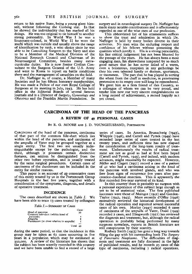
562 T H E B R I T I S H J O U R N A L O F S U R G E R Y
return to his native State, being a young giant him- self, without following the Cushing star. In that he showed the individuality that has marked all his doings. He was too original to tie himself to another strong personality. In World War I he was a lieutenant-colonel, in World War I1 he wore the mufti which was outside and usually above the range of identification by rank, a wise choice since he was able to be Consulting Surgeon to the Navy and also to be a Member of the Medical Division of the National Research Council and Chairman of its Neurosurgical Committee, besides many extra- curricular duties. He is now Senior Civilian Con- sultant to the Surgeon General of the U.S. Army and went to Korea to report on battle conditions there and the management of casualties on the field.
Dr. Naffziger is, of course, a Member of many Societies and he has fifteen honorary memberships. He was made a Fellow of our own Royal College of Surgeons at its meeting in July, 1943. He has held office in the Editorial Boards of several famous journals and is a Director of Surgery, Gynecology and Obstetrics and the Franklin Martin Foundation. In
surgery and in neurological surgery Dr. Naffziger has made many notable contributions and is affectionately regarded as one of the wise men of our profession.
This abbreviated list of his attainments suffices to show the trust and admiration in which Dr. Naffziger is held by many bodies diverse in their activities. No man wins the enduring respect and confidence of his fellows without possessing the qualities which justify it. His is a strong personality, his fine critical judgement has not been blunted by the passage of the years. He has always been a most engaging man, his shrewdness tempered by so much good nature that he has never failed of a warm, even a boisterous, reception in any gatherings be they his own talented family or doctors, sportsmen, or statesmen. The part that he has played in sorting the wheat from the chaff in medicine, in penetrating pretension to its empty core will long be remembered. We greet him as a firm friend of this Country, as a colleague of whom we can be very proud, and tender him now our very sincere congratulations on a long record of achievement, a record happily nct yet closed.
CARCINOMA OF THE HEAD OF THE PANCREAS A REVIEW OF 49 PERSONAL CASES
BY R. G. MOORE AND J. D. YOUNGHUSBAND, PORTSMOUTH
CARCINOMA of the head of the pancreas, carcinoma of that part of the common bile-duct which lies within the head of the pancreas, and carcinoma of the ampulla of Vater may be grouped together as a single entity. The first two are usually indis- tinguishable except by the pathologist, and the third, although it possesses certain distinctive features, can seldom be distinguished from the other two before operation, and is usually treated by the same surgical procedures. Certain cases of carcinoma of the duodenum can be included in the entity for similar reasons.
This paper is an account of 49 consecutive cases of this entity treated by us in the Portsmouth Group Hospitals in the last five years, together with a consideration of the symptoms, diagnosis, and details of operative treatment.
INCIDENCE The cases described are shown in Table I. We
have been able to trace 13 cases treated by colleagues Table Z.-SUMMARY OF CASES
Cases Head of pancreas 33 Common bile-duct (within head of pancreas) 7
Ampulla 8 Duodenum (in close relation to ampulla) I -
Total 49
during the same period, so that the incidence in this group may be taken as 62 cases occurring in five years in a population which may be estimated at 350,000~ A review of the literature has shown that the subject has been scantily recorded in this country and we have been unable to find a record of a large
series of cases. In America, Brunschwig (1943), Whipple (1946)~ and Cattell and Pyrtek (1949) have recorded large series of cases treated in the last twenty years, and sufficient time has now elapsed for consideration of the long-term results of treat- ment. Unfortunately the results of radical treat- ment are not as good as had at first been hoped (Cattell and Pyrtek, 1949), and indeed, with modem advances, might reasonably be expected. However, Miller and Clagett (1951) record a case of a patient of 41 who had a carcinoma arising in the head of the pancreas with involved glands, and who was free from signs of recurrence five years after pan- creatico-duodenal resection. This is apparently the first recorded five-year survival of its kind.
In this country there is probably no surgeon with a personal experience of this subject large enough as yet to be of statistical value. The first published successes were those of Maingot (1941) and Gordon- Taylor (1942, 1943). More recently d’Offay (1946) extensively reviewed the historical development of the radical operation and reported several successful cases of his own. Morley (1947) recorded 6 cases of carcinoma of the ampulla of Vater, Dodd (1949) recorded 2 cases, and Illingworth (195 I) has reviewed the diagnosis and treatment, but, although the radical operation is probably being performed by many surgeons nowadays, reports in British literature are still conspicuous by their scarcity.
Rodney Smith (1953) has gone a long way towards filling the gap with his outstanding book The Surgery of Pancreatic Neoplasms. The difficulties of diag- nosis and treatment are fully discussed in the light of published results, and he records 25 cases of this type of carcinoma, including 7 radical operations.
C A R C I N O M A O F H E A D O F P A N C R E A S 563
Another valuable contribution to the subject is the work of Falconer and Griffiths (I950), who have made a detailed study of the vascular supply of the pancreas and its frequent variations.
Our purpose in writing this paper is to record our experiences and difficulties, and the results of treatment, rather than to describe all aspects of the disease, and for this reason we have not thought it necessary to repeat much of the ground so thoroughly covered by these authors.
CLINICAL ASPECTS Sex Incidence.-In this series there were 21
males and 28 females. This contrasts with the generally accepted ratio of males/females of 2-4 to I (Brunschwig, 1942) or 3-4 to I (Rodney Smith,
Age Incidence.-This is given in Table II. It will be seen that four-fifths of the cases were over 60 years of age, and one-third were over 70.
Table II.-AGE INCIDENCE
1953).
Carer 30140 2 40150 2 sol60 6 60170 22 70180 I4 80i90 3
Symptoms.-Jaundice is the outstanding symp- tom of this disease. It was present in every case in this series. Only two cases were referred for symp- toms other than jaundice, one of whom complained of right-sided abdominal pain for five months, the other of backache for three months. Both these patients developed jaundice just after they were first seen by one of us. Fourteen patients had pre- icteric symptoms of three months' duration or more, consisting of anorexia, abdominal pain, backache, loss of weight, constipation, or diarrhoea, in some cases due to or coincident with intercurrent disease such as chronic cholecystitis. In the remainder, jaundice appeared within a week or two of the onset of illness, which often took the form of a mild digestive disturbance, or a sudden chill. Several patients noticed dark-coloured urine and pale stools as their first symptom, followed in a few days by clinical jaundice.
None of these pre-icteric symptoms can be said to be specific to carcinoma of the pancreas, but when a patient gives a short history of one or more of them this diagnosis must be considered. Cattell (1945) has stated that loss of weight, asthenia, and anorexia are early features in carcinoma of the head of the pancreas and usually precede the jaundice, but are less marked and usually follow the jaundice in ampullary carcinoma. We can confirm Cattell's findings in this series.
Jaundice.-It is fortunate, therefore, that the vast majority of these tumours obstruct the bile-duct at a relatively early stage. The jaundice which results has several important features. Commonly it is steadily progressive and may increase in depth slowly or surprisingly rapidly. In some cases, however, it is intermittent, and the colour of the stools and urine may vary from time to time. In a few cases, as Illingworth (1951) describes, the jaundice may disappear completely after a week or two, and this is a dangerous feature if it is allowed to deceive the
medical attendant into concluding that a carcinoma cannot be present.
Pain.-Older text-books state that pain is absent, but modern writers agree that it is a prominent symptom. Brunschwig (1942) states that pain is a major complaint and may be the initial symptom in carcinoma of the head of the pancreas, whereas with ampullary growths mild epigastric discomfort may herald the onset of jaundice but does not persist. Most authors agree with this view. It is described as variable in position, type, and degree. In this series 22 patients had pain in the form of vague epigastric discomfort, severe right-sided or central epigastric pain, biliary colic, or backache. Three of these cases had chronic cholecystitis and gall-stones. We were not able to find any distinctive feature in the type of pain described by our patients.
Itch.-This symptom has received scant atten- tion. It is usually described as a sequel of obstruc- tive jaundice. In this series the incidence of itch was very variable. In most cases it coincided with the onset of jaundice and was severe and persistent as the jaundice deepened. In some cases, however, it disappeared after a few days and did not return even though the jaundice progressed. In each of the 8 cases of ampullary tumour generalized itching preceded the appearance of jaundice by an interval of from two to six weeks, and was the first symptom noted by the patient. Illingworth (1951) describes a case in which itching occurred for three months before jaundice appeared, the patient having a small malignant ulcer on the duodenal aspect of the ampulla. He suggests that the obstruction of the bile-duct was sufficient to raise the bile-salt in the blood to the ' itching level ' without raising the bile- pigments enough to produce clinical jaundice. The assumption that excess of bile-salts causes itching is not generally accepted. Itching also occurs in jaundice due to other causes, so that it does not appear to be of much diagnostic value except that, when it precedes the onset of obstructive jaundice by several weeks, it suggests that the site of obstruc- tion is the ampulla.
However desirable it may be to make the diag- nosis before jaundice has occurred, this aim is seldom achieved. Symptoms arising before icterus will depend largely on the site of origin of the tumour. For example, if the pancreatic duct is obstructed early the only symptoms may be vague dyspepsia, alteration in bowel habit and character of the stools, and backache. These symptoms are vague and non- specific, and are similar to those due to carcinoma arising in the body or tail of the pancreas, and it is significant that the diagnosis of growths in those situations is seldom made at an early or operable stage.
Examination.-Probably the most important finding in the clinical examination is a palpable gall- bladder. In this series it was palpable in 29 cases, or 63 per cent (Table ZII). I t is generally accepted that it can be palpated in 6-70 per cent of cases, When clearly palpable in a jaundiced patient it is almost diagnostic of neoplastic obstruction of the common duct, since in cholelithiasis the gall-bladder is nearly always too thick walled to be distended by back-pressure. Possible exceptions to this state- ment are cystic duct obstruction (mucocele of the gall-bladder) by a stone which also compresses the
564 T H E B R I T I S H J O U R N A L O F S U R G E R Y
common duct sufficiently to cause jaundice, or mucocele associated with jaundice of some inde- pendent cause.
The liver is almost invariably enlarged and some- times tender. We were able to feel a mass, separable
Table III.-STATE OF THE GALL-BLADDER
Impalpable 17 (37 per cent)
Previously removed 3
Palpable zg (63 per cent)
Found distended at operation 15 Small and full of gall-stones z
from the liver and gall-bladder, in 5 of our cases and, as might be expected, each proved to have a large inoperable growth in the head of the pancreas. When the pancreatic duct is obstructed, the pancreas is found at operation to be enlarged and firm, but we have not been able to palpate it clinically.
The stools are usually putty coloured, although pigment may appear in them at times if local condi- tions in the tumour allow bile to leak temporarily past the obstruction in the bile-duct. The urine contains bile-pigment once jaundice has developed, and in 3 of our cases contained urobilinogen in normal amounts, but never in excess. In the remainder urobilinogen was absent.
INVESTIGATIONS AND DIAGNOSIS When a patient presents symptoms such as those
described as pre-icteric in this series, the diagnosis of carcinoma arising in the pancreas should be borne in mind. The examination and investigation of such a case will naturally be along the usual lines, but tests which may assist in locating the tumour in the pancreas are occult blood examinations, duodenal intubation, the secretin test, and radiology. Occult blood may be present in the stools in cases of ampul- lary carcinoma or carcinoma of the head of the pancreas which is invading the duodenum. I t is less likely to be detected when the growth is confined to the pancreas. In this series frank melaena occurred in 3 cases, in each of which the duodenum was involved. Duodenal intubation aims at the demon- stration of blood, mucus, and malignant cells in the fluid removed. When combined with the injection of secretin, the composition of pancreatic secretions may be studied. Parsons (1951) con- cludes that the demonstration of cancer cells is likely to be successful if ulceration is present, but that the study of pancreatic ferments is not helpful. Rodney Smith (1953) shows that skilled radiology is capable of demonstrating tumours of the ampulla and tumours in the head of the pancreas provided that some invasion or distortion of the duodenum has occurred.
When jaundice is present the problem becomes one of distinguishing between pure obstruction and prolonged hepatitis. Either may be ushered in by a gastro-intestinal disturbance, and either may be painless. An enlarged tender liver may be found in both, together with dark urine and pale stools. Unless the case is seen early the phase of excessive excretion of urobilinogen in the urine in hepatitis may have passed, but if it is still present it excludes pure obstruction.
We have found tests of liver function of great value, and out of the large number available have
used only a few as routine procedures, namely, the total and differential plasma-proteins, the thymol turbidity test, the gold and thymol flocculations, and the alkaline phosphatase. In our cases of pure obstruction without secondary liver damage the flocculations have been invariably negative and the thymol turbidity (normal 0-4 units) never above 4 units.
The alkaline phosphatase is raised early in obstructive jaundice and late in severe hepatitis. In early cases, therefore, a high figure indicates obstruction and a dividing line of 30 units (normal 4-13 units) has been suggested (Sherlock, 1946)~ but this is not clear cut. In 30 of our cases the alkaline phosphatase was over 30 (commonly 50, 60, or 70, and in 2 cases over IOO), and in these, with negative flocculations, the diagnosis was re- garded as virtually certain. In 17 cases, however, it was below 30, and had not risen when repeated after a short interval, but in these cases the floccula- tions remained negative and the serum-bilirubin increased, and this has not been observed to occur in persistent hepatitis. In a few cases the alkaline phosphatase level was found to decrease slowly while the serum bilirubin was rising.
Ascending cholangitis may give rise to difficulty. It presents the picture of obstructive jaundice, but is usually associated with rigors, or at least with fever higher than the small rise in temperature found in some cases of carcinoma of the pancreas.
We do not consider that the differential diagnosis is difficult if the patient is seen soon after the onset of jaundice. During the period in which this series of cases was seen, one case of hepatitis was incorrectly diagnosed as obstructive jaundice and laparotomy performed. At the time of operation the liver function tests were doubtful, and in the light of our later experience we would delay operation in such a case. When the tests were repeated ten days later they showed clear evidence of hepatitis.
Every case in this series, except two, was jaun- diced when first seen by one of us, and the jaundice could be labelled ‘ obstructive ’ at the first examina- tion with the aid of liver function tests in the majority, and in the remainder after a short interval. Once the diagnosis of obstructive jaundice has been made we believe that operation should not be delayed in order that further investigations may be carried out in an attempt to reach a more exact diagnosis.
PREPARATION FOR OPERATION It is very important to give the patient vitamin
K for two or three days before operation in order to restore the plasma-prothrombin level. It may be given by mouth combined with bile-salts, or by injection. The electrolyte balance must be restored by suitable means if it is disordered, and anaemia corrected by transfusion unless it is only slight. A high protein intake should be given if the plasma- proteins are low. Breathing exercidbs and postural drainage of the bronchial tree are very important, especially as many of these patients are elderly.
CHOICE OF OPERATION D’Offay (1946) has given an excellent review of
the history of the development of the operation of
C A R C I N O M A O F H E A D O F P A N C R E A S 565
pancreatico-duodenectomy. An even fuller review is given by Child (1949).
In the gradual evolution of the present-day operation a great variety of methods was tried. This was largely owing to the need to find a final arrangement of the parts concerned which would function, was free from complications such as entry of stomach contents into the gall-bladder, and was so planned that part could be done as a first stage when the patient was too ill to have it all done at once, and the rest when his condition had improved
The advantages of a single operation are of course obvious; the avoidance of delay, a clean field, a single anaesthetic, and the psychological effect on the patient. These advantages are reversed in a two-stage plan, although the general condition at the time of the second stage should on average be better than in those considered fit for a single opera- tion, since the jaundice will have been relieved.
A theoretical disadvantage of external chole- cystostomy is that a disaster, such as coronary throm- bosis, might occur to prevent the second stage and
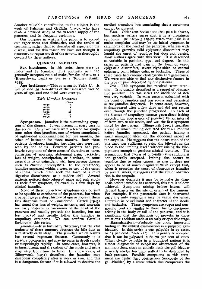
Table ZV.-TWO-STAGE PANCREATECTOMY. SERUM-BILIRUBIN READINGS IN RELATION TO INTERVAL BETWEEN STAGES
=I 2 20’0 10’1
14 I 4.2 . . . 16.0 . . . I S 1 7 ’ 3 . . . 1 0 8 . . . 14’7.
3’0 12.3
I 6 13.i. . . 9.0 30 1 15.8. . . 3.0 33 8.3 . . . 15.3 . . . 4’5
INTERVAL BETWEEN STAGES
days 22 22 2 1 16 37 28 30 31 13 28 14 25 41
CLINICAL JAUNDICE
Clear Clear Clear Trace Clear Clear Clear + + Clear Trace Clear Clear
RESULT
Recovered Recovered Recovered Died Recovered Recovered Died Died Died Recovered Died Recovered Recovered
sufficiently as a result of what had been done in the first operation. The first stage had to consist of anastomosis of the gall-bladder to the stomach or jejunum. This was apt to leave the surgeon with some awkward ‘ends’, such as the common bile-duct, ‘ left over’ at the end of the second stage, and sometimes to result in troublesome adhesions.
Opinion as to whether a one-stage or a two-stage operation should be performed is gradually reaching uniformity and is perhaps best summarized by Cattell (1945) when he says :-
a. In most instances a one-stage procedure is preferable.
b. The criteria used as indications for a two-stage operation are : severe debility, a serum-bilirubin of 5 mg. or more, and severe impairment of liver function.
We have taken the view that if the patient’s condition is not good enough to stand a single-stage operation, then the simpler the procedure at the f is t stage the better. We have, therefore, performed simple external cholecystostomy as the first-stage plan, and believe that its advantages over internal bile drainage are considerable. I t is simple, it leaves the field of subsequent resection undisturbed (the tube is placed as far out in the flank as possible), it leaves the surgeon free to employ any form of reconstruction of the alimentary tract that he desires at the second stage, it can be easily converted to cholecyst-duodenostomy or cholecyst-jejunostomy if for any reason it is found at the second stage that resection cannot be completed, and it is free from the disadvantage of internal cholecystostomy men- tioned by d’Offay in which the patient feels so much better as a result of the relief of jaundice that he refuses to undergo the second stage.
the patient would thus be condemned to a permanent ‘tube and bottle ’. We do not think that this risk should weigh very heavily against an operation which has so many advantages.
In z of our cases choledochostomy was performed because in one the gall-bladder had been previously removed, and in the other it was so grossly diseased that it could not be used. In several cases the gall- bladder contained multiple calculi but was only moderately thickened, and in these the calculi were removed and the cholecystostomy performed in the usual way. We believe that it is very important to preserve the gall-bladder in this disease whenever possible. Two patients were gravely ill with chol- zmia when first seen. Both had large palpable gall- bladders and simple cholecystostomy was performed through a small incision without any attempt being made to explore the abdomen or establish an exact diagnosis. Both recovered and underwent successful resection later.
The loss of bile from the alimentary tract is compensated to some extent by giving bile-salts by mouth, and it is of the greatest importance that the electrolyte balance should be maintained. The progress of patients treated in this way has been satisfactory, though we have found that the serum- bilirubin may rise for a time after cholecystostomy, and no appreciable redu.ction in clinical jaundice is usually seen for two weeks. Table I V shows the behaviour of the serum-bilirubin in our two-stage cases. The table suggests that the deaths (with the exception of Case 12, in whom the cause of death was a pancreatic fistula) were related to the presence of jaundice and a short interval between the stages, and it seems reasonable to draw the conclusion that the interval between stages should be not less than three weeks.
566 T H E B R I T I S H J O U R N A L O F S U R G E R Y
DIAGNOSIS AT OPERATION The first problem confronting the surgeon on
opening the abdomen is that of making a firm diag- nosis which will justify the major steps he is about to take. All writers stress this difficulty.
Tumours of the ampulla can be directly inspected by opening the duodenum, and immediate frozen- section biopsy may be done if doubt remains. Benign tumours of the ampulla have been recorded, but are extremely rare (Child, 1949).
Tumours of the lower end of the bile-duct may closely resemble a stone when palpated-through the surrounding tissues. When the common duct is opened and a probe is passed down to the obstruction and pressed against it, biliary mud and portions of a stone may escape and proclaim the diagnosis, but this procedure may detach malignant cells from a carcinoma of the duct and there is at least a theoretical danger of their implantation in the operation field. If it is impossible to make a decision by palpation alone, and the surgeon feels that he must open the bile-duct, the opening should be sited carefully so as not to interfere with the use of the duct for anastomosis later, and on no account should the fluid in the duct be allowed to escape on to the surrounding tissues.
When the biliary tract and the remainder of the pancreas are healthy, it is unlikely that a small area of pancreatitis will be found capable of completely obstructing the bile-duct, and in such a case a small constricting neoplasm is almost the only possible diagnosis. When cholelithiasis is present, or the history includes previous attacks of jaundice, the possibilities are wider, as the following two cases illustrate.
Female, aged 62, seen in attack of typical cholecystitis. No jaundice, but jaundice had been observed in previous attacks. X rays showed multiple gall-stones. At opera- tion, gall-stones in contracted gall-bladder, common duct not dilated, small hard lump felt just deep to ampulla. Duodenum opened, ampulla normal, lump just deep to it. Gall-bladder opened, stones removed, probe passed through cystic duct down common duct, but would not pass lump to enter duodenum. External chole- cystostomy, enlarged lymph-gland removed for biopsy. Biopsy showed reactive hyperplasia only. Second opera- tion two weeks later with full preparation for pancreat- ectomy. Lump as before. Considered insufficient evidence to justify radical operation. Cholecyst-duoden- ostomy. This patient has remained fit and well for three years (except for developing a primary carcinoma of the breast treated by radical mastectomy).
Female, aged 63, flatulent dyspepsia, attacks of epigastric pain and vomiting, pains R. shoulder, all for 6-9 months. X rays ehowed multiple gall-stones. Jaundice appeared two days before operation. At opera- tion, gall-stones in contracted gall-bladder, common duct dilated, small area of vague thickening in head of pancreas. Gall-bladder opened, stones removed, probe passed through cystic duct down common duct but would not enter duodenum. Duodenum opened, ampulla normal. Cholecyst-duodenostomy. Patient remained well for three months, then developed abdominal pains, constipation, anorexia, and loss of weight. Operation, hard lump in head of pancreas obviously malignant, no secondaries, but radical operation abandoned because mesenteric vessels frozen. Death two months later. Biopsy of local lymph-gland rem:ved at second operation -' replaced by adenocarcinoma .
The essential difference between these two cases is that in the second case the common bile-duct was dilated. Cattell stresses this point and states that in his cases of cancer of the head of the pancreas the common duct was always dilated, and he concludes that the radical operation (when malignancy cannot be proved at the operating table) should not be done unless it is.
When larger tumours are found in the head of the pancreas there may be great difficulty in differ- entiating neoplasm from chronic pancreatitis. Either may be localized or diffise, and both may be very hard. A scirrhous type of growth may show charac- teristic retraction in one or other aspect or may be commencing to invade the duodenum. When the pancreatic duct is obstructed the body and tail of the pancreas are much firmer than normal and may be swollen, but retain their natural shape and have not been in our experience as hard or as irregular as the region of the growth. However, chronic pancreatitis of any degree from mild thickening to cavitation and frank abscess formation, or even calcification, may be found, and a small carcinoma in such a gland may well be impossible to detect.
Direct biopsy of the suspected part may fail to obtain a portion of the growth and there is at least a theoretical objection to incising directly into the growth even if the surgeon is prepared to proceed to pancreatectomy there and then. Biopsy through the duodenum, as described by Rodney Smith (1953), certainly seems safer than biopsy through the anterior surface of the gland.
Carlson (1950) discusses the difficulty of estab- lishing the diagnosis of tumours in the head of the pancreas at operation and describes 3 cases in which radical operation for non-malignant conditions might well have been carried out on clinical and operation findings only. He concludes that radical operation should not be undertaken unless histological proof of carcinoma is obtained, and stresses the operative mortality of the radical procedure (20-60 per cent) compared to a palliative operation (Cattell, o out of 22 recent cases) and the small extra length of average sur- vival of radical operations compared to palliative ones (quoting Cattell, I I months as against 9.2 months).
We consider that this attitude is unnecessarily gloomy. In many cases a confident diagnosis can be made by palpation alone. We have not used frozen-section biopsy in this series, yet every case submitted to radical operation has been proved malignant, and no case has been diagnosed malignant at operation incorrectly. Biopsy should have been performed in one patient in whom an early carcinoma was missed. We firmly believe in the principle of obtaining histological proof of malignancy before performing any form of radical operation whenever possible, but in this instance the obtaining of positive biopsy material may not always be successful, and furthermore may introduce dangers of its own. Biopsy should only be done in doubtful cases, and if it fails to show carcinoma the right course is undoubtedly a palliative operation.
OPERABILITY The next difficulty facing the surgeon is the
operability of the growth. The interesting anatomy of the relationship of the pancreas to the portal vein
C A R C I N O M A O F H E A D O F P A N C R E A S 567
provides perhaps the most anxious moment in the radical operation. Tumours which arise near the neck of the pancreas, or in the uncinate process, may involve the portal vein or superior mesenteric vessels before they involve the bile-duct. For this reason a case with. a short history and relatively small tumour may be inoperable solely because the superior mesenteric vessels are ‘ frozen ’.
When the tumour lies in this region a careful investigation of the superior mesenteric vein should be done before any major step is taken. A constant large vein which collects tributaries from the anterior aspect of the head of the pancreas joins the superior mesenteric vein from the right side just at the lower border of the neck of the pancreas. Dissection here is very delicate and it is necessary to be able to ligate this vein flush with the superior mesenteric if resection is to be possible.
Attempts to separate the gland from the main vein are usually made first and sooner or later a tear may occur with severe bleeding, the controlling of which results in harm to the patient’s condition and to a surgeon who has lost much of his freshness and still has much to do (d’Offay, 1946). (Cases 7 and 31, Table VZ.)
Any surgeon undertaking this operation must decide whether he is prepared to resect part of the mesenteric (or portal) vein when its involvement is the only ‘inoperable’ feature. If so, an early decision to do so will save time and perhaps the results of a brisk hzemorrhage. When a short seg- ment of vein is removed repair by end-to-end anasto- mosis is possible (Martin, 1951)~ but portocaval anastomosis may be the only way of dealing with a large gap.
Skirting past the portal vein, as Child (1949) points out, is one of the weaknesses of the radical bperation, since the vein may lie within a millimetre or two of growth. Moreover, handling of the growth during resection may liberate malignant emboli into the rich venous plexus which drains the pancreas.
Another weakness of the radical operation is the failure to remove the whole pancreas. However small and well defined the growth in the head may appear to be, it is impossible to determine how much of the gland to remove and how much may safely be left. Sections through the divided edge of the pancreas were unfortunately not made in all the specimens in this series, but out of 14 so examined growth was demonstrated in 4, and in 3 of these it was thought that a sufficient margin of healthy pancreas was being removed. In the fourth it was suspected that the resection might be inadequate, but in this case, for other reasons, radical resection had already been abandoned. Thus, in 3 cases out of 14, so-called radical operation was doomed to failure from the very beginning.
Parsons (1951) describes the finding of cancer cells in the fluid from an obstructed pancreatic duct, and also satellite foci of cancer in the distal part of the pancreatic duct. The prognosis of patients who survive the radical operation is discussed later, but most authors with extensive experience are dis- appointed at the high incidence of early recurrence, and Cattell and Pyrtek (1g4g), after reviewing 30 cases which survived the operation for carcinoma of the head of the pancreas, conclude : “ . . . in the
light of present experience we must modify the operative procedure or abandon radical surgery . . . we wish to suggest total pancreatectomy. . . .”
The metabolic effects of total pancreatectomy are considerable, and discussion of them is beyond the scope of this paper, but reports are not entirely unfavourable (Whitfield, Gourevitch, and Garfield Thomas, 1952). A more extensive removal of the pancreas, with the more thorough removal of lymph- glands which this entails, certainly seems to demand
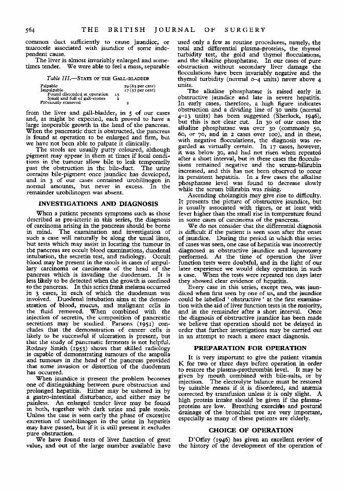
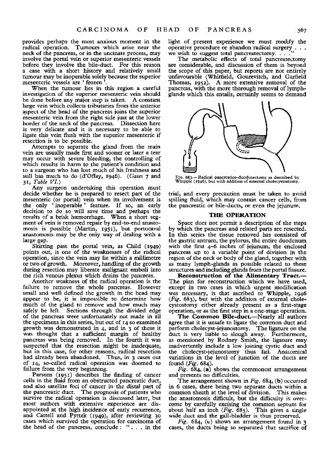
FIG. 683.- Radical pancreatico-duodenectomy as described by Whipple (1946), but with addition of external cholecystostomy.
trial, and every precaution must be taken to avoid spilling fluid, which may contain cancer cells, from the pancreatic or bile-ducts, or even the jejunum.
THE OPERATION Space does not permit a description of the steps
by which the pancreas and related parts are resected. In this series the tissue removed has consisted of the gastric antrum, the pylorus, the entire duodenum with the first 4-6 inches of jejunum, the enclosed pancreas up to a variable point of division in the region of the neck or body of the gland, together with as many lymph-glands as possible related to these structures and including glands from the portal fissure.
Reconstruction of the Alimentary Tract.- The plan for reconstruction which we have used, except in two cases in which urgent modification was necessary, is that ascribed to Whipple, 1946 (Fig. 683), but with the addition of external chole- cystostomy either already present as a first-stage operation, or as the first step in a one-stage operation.
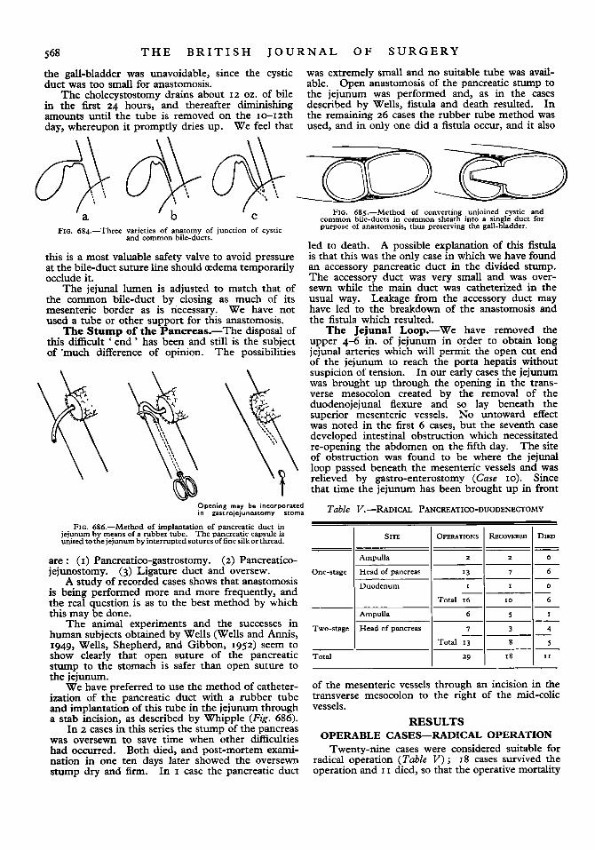
The Common Bile-duct.-Nearly all authors agree that it is unsafe to ligate the common duct and perform cholecyst-jejunostomy. The ligature on the duct is very liable to slough away. Furthermore, as mentioned by Rodney Smith, the ligature may inadvertently include a low joining cystic duct and the cholecyst-jejunostomy thus fail. Anatomical variations in the level of junction of the ducts are found (Fig. 684).
Fig. 684, (a) shows the commonest arrangement and presents no difficulties.
The arrangement shown in Fig. 684, (b) occurred in 6 cases, there being two separate ducts within a common sheath at the level of division. This makes the anastomosis difficult, but the difficulty is over- come by carefully excising the common septum for about half an inch (Fig. 685). This gives a single wide duct and the gall-bladder is thus preserved.
Fig. 684, (c ) shows an arrangement found in 3 cases, the ducts being so separated that sacrifice of
568 T H E B R I T I S H J O U R N A L O F S U R G E R Y
the gall-bladder was unavoidable, since the cystic duct was too small for anastomosis.
The cholecystostomy drains about 12 oz. of bile in the first 24 hours, and thereafter diminishing amounts until the tube is removed on the 10-12th day, whereupon it promptly dries up. We feel that
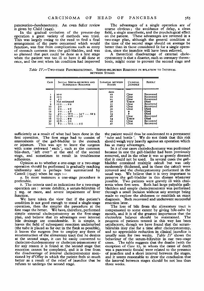
was extremely small and no suitable tube was avail- able. Open anastomosis of the pancreatic stump to the jejunum was performed and, as in the cases described by Wells, fistula and death resulted. In the remaining 26 cases the rubber tube method was used, and in only one did a fistula occur, and it also
FIG. 686.-Method of implantation of pancreatic duct in jejunum b means of a rubber tube. The pancreatic capsule is SITE united to& jejunum by interrupted sutures of fine silk or thread.
are : (I) Pancreatico-gastrostomy. (2) Pancreatico- Ampulla - jejunostomy. (3) Ligature duct and oversew. One-stage Head of pancreas
Duodenum A study of recorded cases shows that anastomosis is being performed more and more frequently, and the real question is as to the best method by which this may be done. Ampulla
human subjects obtained by Wells (Wells and Annis, 1949, Wells, Shepherd, and Gibbon, 1952) seem to show clearly that open suture of the pancreatic Total
The animal experiments and the successes in Two-stage Head of
stump to the stomach is safer than open suture to
OPERATIONS RECOVERED DIED
2 2 0
I3 7 6
I I 0
Total 16 I 0 6
-- -
I -- 6 S
7 3 4 - __ Total 13 8 5
29 I 8 I 1
Tabl
e VZ
.-SuM
MAR
y OF 2
9 RA
DICA
L PANCREATICO-DUODENECTOMIES
M. 3
8
F. 6
6
F. 66
F. 7
3
--
--
j
CA
SE
No.
-
--
I 2 _
_
3
-
2
Hea
d -
-
56 -
Clin
ical
seco
ndar
ies
-
-
z H
ead
Ade
noca
rcin
oma
Low
N
otex
amin
ed
Non
e -
IS
-
Jeju
nal f
istu
la
-
-
I C
.B.D
. A
deno
carc
inom
a A
vera
ge
1-4
2Ist
day
-
-
Panc
reat
ic fi
stul
a -
Gal
l-st
ones
I
Hea
d A
deno
carc
inom
a A
vera
ge
Not
exa
min
ed
0-1
14
th d
ay
-
Mon
ths
Mon
ths
-
-
Stag
e A
deno
carc
inom
a A
vera
ge
Not
exa
min
ed
Non
e -
--
-~
~
--_
__
__
_--
0
--
~_
__
~
---
RE
WK
S
Ope
RA
- D
EA
TH
D
EG
RE
E
GR
OW
TH
IN
1 SIT
E 1 T
YP
E
I OF
1 CUT EN
D OF
1 GLAN
DS I
1 1 SURVIV
AL 1 CAU
SE O
F D
EA
TH
D
ISE
ASE
M
AL
IGN
AN
CY
PA
NC
REA
S
M. 6
9
F. 7
3
M. 76
--
--
- -~
--
-
-
2s
-
I C
.B.D
. A
deno
carc
inom
a A
vera
ge
Not
exa
min
ed
2-5
-
-
-
I C
.B.D
. A
deno
carc
inom
a A
vera
ge
-
-
Bro
nchi
tis,
cho-
I
Hea
d A
deno
carc
inom
a A
vera
ge
+ 0
-1
13th
day
-
--
~-
-
0
-
-
20
2-4 -
~~
-
~-
--
-
Cho
lzm
ia
-
-~
~-
la
ngiti
s - ---
M. 6
0
F. 67
0
-
-
1s
0-3
Ioth
day
-
-
Liv
er n
ecro
sis
Res
ecti
on p
ort
al
+ ve
in ; p
offo
-cav
al
-
-
-
2
Am
pulla
Pa
pil!a
ry
aden
o-
Low
0-
6
-
I H
ead
Ade
noca
rcin
oma
Ave
rage
-~
~~
ca
rcm
oma
-
anas
tom
osis
5 6 7 -
Not
exam
ined
1-1
-
6 -
Clin
ical
sec
onda
ries
-
2
Am
pulla
A
napl
astic
H
igh
-~
--
Cir
cula
tory
failu
re
Res
ecti
on p
ort
al
1 Adenoca
rcin
oma
1 Average
I Not e
xam
ined
I 0-5
I 48 hr.
1 -
I -
1 F.
58
I 2 1 Head
I vem 9
__
I0
-
F. 8
4 I G
all-
ston
es
I 2
I Hea
d I A
deno
carc
inom
a I A
\.era
ge
1 +
I 0-2
I -
I 12
1
-
I Clin
ical
sec
onda
ries
I Pa
pilla
ry a
deno
- ca
rcin
oma
Ave
rage
N
ot e
xam
ined
Hig
h N
ot ex
amin
ed
Non
e
Non
e
-
-
33
-
-
---
-
-
Cir
cula
tory
fai
lure
-
zoth
day
-
-
Panc
reat
ic fi
stul
a -
48 h
r. ---
I1
Ade
noca
rcin
oma
I2
Hig
h I N
otex
amin
ed
2-6
Ade
noca
rcin
oma
Ade
noca
rcin
oma
Papi
llary
ade
no-
-
8thd
av I
-
I -
I Anu
ria
I A
vera
ge
Not
exa
min
ed
Ave
rage
I N
ot e
xam
ined
Non
e
1-3
-
M. 7
4 I D
iabe
tes
I 2
I Hea
d I A
deno
carc
inom
a I L
ow
I o
I N
one
I26t
hday
I -
I -
I Liv
er f
ailu
re
I
22
26
27
28
-
30
I-
I-
F. 60
I -
/ I
I Ampu
lla ~
~~
Nor
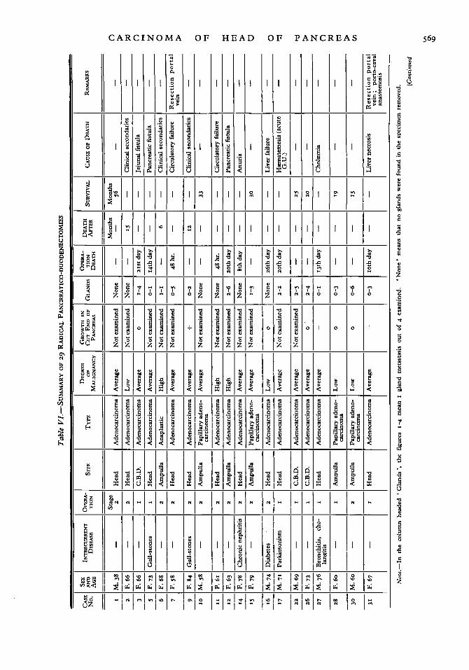
e.-In
th
e co
lum
n he
aded
' G
land
s ',
the
figur
es I
-4 m
ean
I gl
and
met
asta
sis
out
of 4
exa
min
ed.
'Non
e '
mea
ns t
hat
no g
land
s w
ere
foun
d in
the
spe
cim
en re
mov
ed. [C
ontin
ued
570
W P
c
la
v) c
.- a
.sj 8 a
P e fi
a 9 2 '
u M
e P <
T H E
I
VI
I
- W rD
u - m m
T I S H J O U R N A L O F S U R G E R Y
was 38 per cent. More detailed results are given in Table VI.
Operation Deaths.-These number I I, 8 occurring in the first 14 operations, only 3 in the next 15. Because this figure is high, a detailed analysis of the cause of death is given in Table VZZ.
The mortality-rate for any major operation is closely related to the standards of operability of the individual surgeon as well as to his technical skill, particulary so in this type of case. If the radical operation is reserved for the younger patient, fit in other respects, not deeply jaundiced and with a small tumour, a high recovery rate is to be expected, but the operability rate will be low. The alternative to radical operation is a palliative operation designed merely to relieve jaundice, leaving the carcinoma to advance unchecked into the surrounding tissues, and it is doubtful whether the term ' palliative ' should be used for such operations at all. In this series the standards of operability have been both optimistic and enthusiastic, as consideration of Table VIZ will no doubt show, and our reasons for attempting radical operation whenever possible were firstly, the miserably short benefit which palliative procedures give (Table VZIZ), which is discussed later, and secondly the fact that the radical operation, compared to other major abdominal operations, is as yet rela- tively untried. I t is interesting to note that, in the matter of age limit, the three oldest patients who underwent radical operation, aged 79, 80, and 84 respectively, all made uneventful recoveries.
Recovery from Operation, since died.-Five cases which survived the operation died 5, 6, 8, 12, and 15 months later, respectively, and in each death was due to clinical carcinomatosis. In two of these the outcome was certain, since in one histological examination of the specimen removed showed growth present in the cut edge of the pancreas, and in the other a single tiny nodule removed from the liver for biopsy showed carcinoma. One case (Case 32, Table VZ) is of especial interest. The primary in this case was a large ulcer of the medial wall of the second part of the duodenum.
Case p.-Female, aged 47, one-stage radical operation with uneventful recovery. Keport on specimen: " A shallow. ulcer in the second part of the duodenum measuring 3 x 5 cm. ; its base is formed by the pancreas, and the ampulla has been completely destroyed. Sections from the edge of the ulcer show infiltration by a poorly differentiated adenocarcinoma ; the pancreas forming its base is free from growth, as are the termination of the bile and pancreatic ducts. No invaded glands can be found. I do not think it is possible to be certain whether the growth arose from the ampulla or from the duodenal mucosa ; on the whole the appearances and the histology favour the latter."
After good progress for three months, recurrence of jaundice and spontaneous re-opening of cholecystostomy. Operation : carcinomatous involvement of jejunal loop used in the reconstruction, especially in relation to the sites of the three anastomoses. No evidence of carcinoma found elsewhere. Cholecyst-jejunostomy the only possible palliation. Temporary relief of jaundice. Death 8 months after original operation.
The early and unexpected recurrence of growth in this case led us to suspect, in view of the sites in which it was found, that it might have been the result of direct implantation of malignant cells from
(Dr. A. Clay.)
C A R C I N O M A O F H E A D O F P A N C R E A S 57 1
the lumen of the jejunum on to the raw suture lines of the anastomoses, such as has been shown to occur by Goligher, Dukes, and Bussey (1951) in cases of conservative resection of the rectum.
Recovery from Operation, alive.-Thirteen cases survived the operation and all were well and free from signs of recurrence when last seen. One of these, who had a carcinoma of the head of the pancreas, has survived 56 months.
INOPERABLE CASES-PALLIATIVE OPERATION
Eighteen cases out of 49 were considered to be inoperable, and in a fimher 2 cases cholecystostomy with a view to possible radical operation was followed
35 in which, owing to misdiagnosis, cholecyst- duodenostomy was performed when the tumour was at an essentially operable stage. Yet death occurred after 11 months. With this case excepted, the longest survival after palliative operation has been a mere 25 weeks (Table VZIZ).
It is of course clear that if borderline cases are submitted to radical operation, those deemed in- operable will be advanced cases in whom long survival after palliative operations can hardly be expected. Yet if the mortality-rate for radical operation appears formidable and unjustifiable, and if the successful radical operation gives an average survival of 11 months (Cattell and Pyrtek, 1949), which is not significantly longer than that of simple
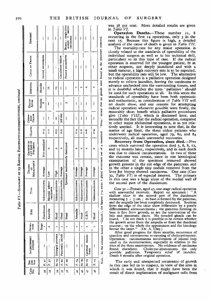
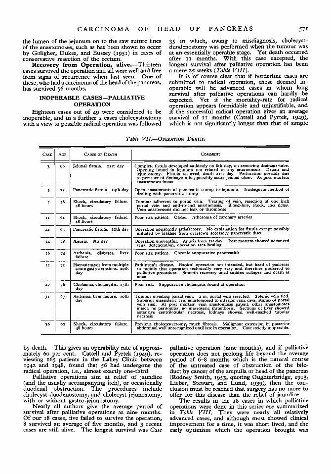
Table VZZ.-OPERATION DEATHS =
CASE - -
3
AGE
66 - -
COMMENT
Complete fistula developed suddenly on 8th day, on removing drainage-tube. Opening found in jejunum not related to any anastomosis. Repair and jejunostomy. Fistula recurred, death z1st day. Perforation possibly due to pressure of drainage-tube, possibly acute jejunal ulcer. At post mortem anastomoses intact
Inadequate method of dealing with pancreatic stump
- __ Open anastomosis of pancreatic stump to jejunum.
CAUSE OF DEATH --
Jejunal fistvla. 2 1 s day
5 73 Pancreatic fistula. 14th day
7 58 Shock, circulatory failure. 48 hours
Tumour adherent to portal vein. Tearing of vein resection of one inch Blood~losr, shock, and delay. portal vein and end-to-end anastomosis.
Vein anastomosis did not leak or thrombose ___- Poor risk patient. Obese. Atheroma of coronary arteries I1
__ I 2
__ 14
61 Shock, circulatory failure. 48 hours
63
78 -
Operation apparently satisfactory. No explanation for fistula except possibly
Operation uneventful. Anuria from 1st day. Post mortem showed advanced
Poor risk patient.
initiated by leakage from oversewn accessory pancreatic duct
renal degeneration, operation area healing
Chronic suppurative pancreatitis
Pancreatic fistula. 20th day
Anuria. 8th day --
16 74 Asthenia, diabetes, liver failure
17 71 Hematemesis from multiple acute gastric erosions. 20th day
Parkinson’s disease. Radical operation not intended, but head of pancreas so mobile that operation technically very easy and therefore preferred to palliative procedure. Smooth recovery until sudden collapse and death at once
Poor risk. Suppurative cholangitis found at operation 27 76 Cholzmia, cholangitis. 13th day
67 Asthenia, liver failure. 10th day
Tumour invading portal vein. 2 in. portal vein resected. Splenic vein tied. Superior mesenteric vein anastomosed to inferior vena cava, stump of ponal vein tied. At post mortem vein anastomosis patent, other anastomoses intact, no peritonitis, no mesenteric thrombosis. Sections of liver showed extensive centrilobular necrosis, kidneys showed well-marked tubular necrosis
36 60 Shock, circulatory failure. 48 hours
Previous cbolecystectomy much fibrosis. Malignant extension in posterior Case strictly inoperable. abdominal wall unrecognhed until late in operation.
by death. This gives an operability rate of approxi- mately 60 per cent. Cattell and Pyrtek (1949), re- viewing 165 patients in the Lahey Clinic between 1942 and 1948, found that 56 had undergone the radical operation, i.e., almost exactly one-third.
Palliative operations aim at relief of jaundice (and the usually accompanying itch), or occasionally duodenal obstruction. The procedures include cholecyst-duodenostomy, and cholecyst-jejunostomy, with or without gastro-jejunostomy.
Nearly all authors give the average period of survival after palliative operations as nine months. Of our 18 cases, five failed to survive the operation, 8 survived an average of five months, and 3 recent cases are still alive. The longest survival was Case
palliative operation (nine months), and if palliative operation does not prolong life beyond the average period of 6-8 months which is the natural course of the untreated case of obstruction of the bile- duct by cancer of the ampulla or head of the pancreas (Rodney Smith, 1953, quoting Oughterbridge, 1913, Lieber, Stewart, and Lund, 1939), then the con- clusion must be reached that surgery has no more to offer for this disease than the relief of jaundice.
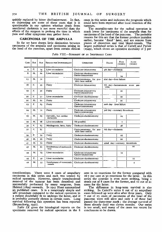
The results in the 18 cases in which palliative operations were done in this series are summarized in Table VZZZ. They were nearly all relatively advanced cases, and although most showed clinical improvement for a time, it was short lived, and the early optimism which the operation brought was
572 T H E B R I T I S H J O U R N A L O F S U R G E R Y
Cholecyst-duodenostomy
Cholecyst-jejunostomy
Cholecyst-jejunostomy Gastro-jejunostomy
quickly replaced by bitter disillusionment. In fact, so depressing are some of these cases that it is questionable in our opinion whether death from unrelieved cholasmia is not more merciful than the efforts of the surgeon to prolong the time in which pain and other symptoms may gather force.
CARCINOMA OF THE AMPULLA So far we have drawn little distinction between
carcinoma of the ampulla and carcinoma arising in the head of the pancreas, apart from certain clinical
20
171 20th day-liver failure
cases in this series and indicates the prognosis which could have been expected after local resection of the tumours.
The mortality-rate for the radical operation is much lower for carcinoma of the ampulla than for carcinoma of the head of the pancreas. The probable reasons for this are that the former produce jaundice earlier, become ‘ fixed ’ later, and are remote from planes of dissection and vital blood-vessels. The largest published series is that of Cattell and Pyrtek (I949), which shows an operative mortality of 5 per
Table VIII.--SUMMARY OF 20 INOPERABLE CASES
AGE REASON FOR INOPERABILITY
74 Liver secondaries Cholecyst-jejunostomy 4th day-cholomia
Cholecyst-duodenostomy Gastro-jejunostomy
“01 72
Liver secondaries
Cholecystostomy, for pos- 2 1 s day-liver failure sible later radical
:I 52 Fixity
Fixity Cholecyst-jejunostomy -1 7th tritis day-hynat;:; acute gas-
Cholecyst-jejunostomy Gastro-jejunostomy
37 I Liver secondaries 62 I Fixity
23 -
24
63 I Fixity
72 I Fixity Cholecyst-jejunostomy I 3rd day-coronary thrombosis Gastro-ieiunostomy
25 Cholecyst-duodenostomy
Nil possible I IKI 65 Operable, but cardiac aneurysm
68 Liver secondaries
63 Misdiagnosis, later, fixity Cholecyst-duodenostomy I I 48 1 Cholecystostomy, for pos- 7th day-choliemia sible later radical
Fixity Cholecyst-duodenostomy
Cholecyst-duodenostomy
Cholecyst-duodenostomy I 4znd day-coronary thrombosis 70 Fixity
61 Involvement of mesenteric vein
69 Liver secondaries
42 Liver secondaries
-- -
-~ 43 Cholecyst-duodenostomy 22
Cholecyst-j ejunostomy Gastro-jejunostomy
44 -_
45 Cholecyst-duodenostomy I I I 15
49
- nvolvement of mesenteric
53 I ‘vessels I l 4 Cholecyst-duodenostomy I I
considerations. There were 8 cases of ampullary carcinoma in this series and each was treated by radical operation. However, simple transduodenal removal of the tumour has been described and successfully performed by many surgeons, from Halsted (1899) onwards. In I941 Hunt summarized 93 published cases. It is a temptingly simple and safe procedure compared to the radical operation in a patient doubtfully fit to undergo the latter, and it is probably correctly chosen in certain cases. Long survival following this operation has been reported (Brunschwig, 1942).
Table ZX gives the histological reports on the specimens removed by radical operation in the 8
cent in 20 resections for the former compared with 16.5 per cent in 30 resections for the latter. In this series the contrast is even more striking, being I death out of 8 cases for the former, and 10 out of 21 cases for the latter.
The difference in long-term survival is also striking. In Cattell’s series 6 out of 12 ampullary cases followed up were alive after three years. Only 7 out of 25 cases of carcinoma of the head of the pancreas were still alive and only I of these had passed the three-year mark ; the average survival of those already dead was eleven months. Our series is too small, and many of the cases too recent for conclusions to be drawn.
C A R C I N O M A O F H E A D O F P A N C R E A S 573
CONCLUSIONS In the operation of pancreatico-duodenectomy a
very considerable alteration of physiology and function is produced. The pylorus, the sphincter of Oddi, the duodenum, and about half the pancreas
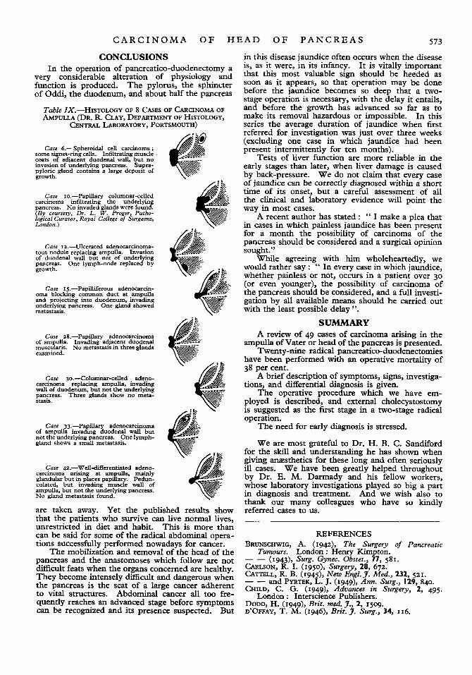
Table ZX.-HISTOLOGY OF 8 CASES OF CARCINOMA OF AMPULLA (DR. R. CLAY, DEPARTMENT OF HISTOLOGY,
CENTRAL LABORATORY, PORTSMOUTH)
Case 6.--Spheroidal cell carcinoma ; some signet-ring cells. Infiltrating muscle coats of adjacent duodenal wall, but no invasion of underlying pancreas. Supra- pyloric gland contains a large deposit of growth.
Case 10.-Papillary columnar-celled carcinoma infiltrating the underlying pancreas. No invaded glands were found. (By courtesy, Dr. L. W . Proger, Patho- logical Curator, Royal College of Surgeons, London.)
Case 12.-Ulcerated adenocarcinoma- tous nodule replacing ampulla. Invasion of duodenal wall but not of underlying pancreas. One lymph-node replaced by growth.
Case 15.-Papilliferous adenocarcin- oma blocking common duct at ampulla and projecting into duodenum, invading underlying pancreas. One gland showed metastasis.
Case zS.-Papillary adenocarcinoma of ampulla. Invading adjacent duodenal muscularis. No metastasis in three glands examined.
Case go.-Columnar-celled adeno- carcmoma replacing ampulla, invading wall of duodenum, but not the underlying pancreas. Three glands show no meta- stam.
Case 33.,-Papillary adenocarcinoma of ampulla invadmg duodenal wall but not the underlying pancreas. One lymph- gland shows a small metastasis.
Case 4z.-Well-dSerentiated adeno- carcinoma arising at ampulla, mainly glandular but in places papillary. Pedun- culated, but invading muscle wall of ampulla, but not the underlying pancreas. No gland metastasis found.
are taken away. Yet the published results show that the patients who survive can live normal lives, unrestricted in diet and habit. This is more than can be said for some of the radical abdominal opera- tions successfully performed nowadays for cancer.
The mobilization and removal of the head of the pancreas and the anastomoses which follow are not difficult feats when the organs concerned are healthy. They become intensely difficult and dangerous when the pancreas is the seat of a large cancer adherent to vital structures. Abdominal cancer all too fre- quently reaches an advanced stage before symptoms can be recognized and its presence suspected. But
in this disease jaundice often occurs when the disease is, as it were, in its infancy. It is vitally important that this most valuable sign should be heeded as soon as it appears, so that operation may be done before the jaundice becomes so deep that a two- stage operation is necessary, with the delay it entails, and before the growth has advanced so far as to make its removal hazardous or impossible. In this series the average duration of jaundice when f is t referred for investigation was just over three weeks (excluding one case in which jaundice had been present intermittently for ten months).
Tests of liver function are more reliable in the early stages than later, when liver damage is caused by back-pressure. We do not claim that every case of jaundice can be correctly diagnosed within a short time of its onset, but a careful assessment of all the clinical and laboratory evidence will point the way in most cases.
A recent author has stated : " I make a plea that in cases in which painless jaundice has been present for a month the possibility of carcinoma of the pancreas should be considered and a surgical opinion sought."
While agreeing with him wholeheartedly, we would rather say : " In every case in which jaundice, whether painless or not, occurs in a patient over 30 (or even younger), the possibility of carcinoma of the pancreas should be considered, and a full investi- gation by all available means should be carried out with the least possible delay ".
SUMMARY A review of 49 cases of carcinoma arising in the
ampulla of Vater or head of the pancreas is presented. Twenty-nine radical pancreatico-duodenectomies
have been performed with an operative mortality of 38 per cent.
A brief description of symptoms, signs, investiga- tions, and differential diagnosis is given.
The operative procedure which we have em- ployed is described, and external cholecystostomy is suggested as the first stage in a two-stage radical operation.
The need for early diagnosis is stressed.
We are most grateful to Dr. H. B. C. Sandiford for the skill and understanding he has shown when giving anaesthetics for these long and often seriously ill cases. We have been greatly helped throughout by Dr. E. M. Darmady and his fellow workers, whose laboratory investigations played so big a part in diagnosis and treatment. And we wish also to thank our many colleagues who have so kindly referred cases to us.
REFERENCES BRUNSCHWIG, A. (I942), The Surgery of Pancreatic
Turnours. London : Henry Kimpton. _ _ (1943), Surg. Gynec. Obstet., 77, 581. CARLSON, R. I. (1950)~ Surgery, 28, 672. CATTELL, R. B. (1945)~ New Eng1.J. Med., 232, 521. -- and PYRTEK, L. J. (1949), Ann. Surg., 129, 840. CHILD, C. G. (1949), Advances in Surgery, 2, 495.
London : Interscience Publishers. DODD, H. (1949), Brir. med. J., 2, 1509. D'OFFAY, T. M. (1946)~ Brit. J. Surg., 34, 116.
574 T H E B R I T I S H J O U R N A L O F S U R G E R Y
FALCONER, C. W. A., and GRIFFITHS, E. (1950), Ibid., 37,
GOLIGHER, J. C., DUKES, C. E., and BUSSEY, H. J., Ibid.,
GORDON-TAYLOR, G. (I942), Brit. med. J., 2, 119. _ _ (1943), Practitioner, 40, 257. HALSTEAD, W. S. (1899), Boston rned. surg. J., 141, 645. HUNT, V. (1941), Ann. Surg., I 14, 570. ILLINGWORTH, C. F. W. (1951), Ann. R. Coll. Surg., 8,
LIEBER, M., STEWART, H., and LUND, H. (1939)~ Ann.
MAINGOT, R. (1941), Lancet, 2, 798. MARTIN, P. (1951), Brit. med.J., 2, 586.
334-
39, 199.
53.
Surg., 109, 219.
MILLER, E. M., and CLAGETT, 0. T. (1951), Ann. Surg.,
MORLEY, J. (1947), Brit. J . Surg., 35, 146. OUGHTERBRIDGE, G. ( I S I ~ ) , Ann. Surg., 57, 402. PARSONS, W. B. ( I ~ S I ) , Rev. Gastroent., 18, 715. SHERLOCK, S. (1946),J. Path. Bacr., 53, 523. SMITH, RODNEY (1953), The Surgery of Pancreatic Neo-
WELLS, C. A., and ANNIS, D. (1949), Lancet, I, 97. _ _ SHEPHERD, J. A., and GIBBON, N. (1g52), Ibid.,
WHIPPLE, A. 0. (1946), Surg. Gynec. Obstet., 82, 623. WHITFIELD, A. G. W., GOUREVITCH, A., and THOMAS, G.
134, 1013.
plasms.
I, 588.
(195z), Lancet, 2, 180.
Edinburgh: E. & S. Livingstone.
CYSTADENOMA OF THE PANCREAS DESCRIPTION OF A CASE AND REVIEW OF SYMPTOMATOLOGY
BY DAVID H. TRAPNELL, READING
CYSTADENOMA of the pancreas is a very uncommon breathless on exertion and slight swelling of both ankles condition. As far as I can discover only 2 cases was present at the end of the day, but disappeared at have been reported in this c o w t r y (Carling and fight. The appetite was bad, particularly for meat, Hicks, 1925) and I in ( p h g l e , 1925), and she had lost some weight recently. She had no
indigestion. There was no relevant family or past while a further 2 cases described in the BRITISH JOURNAL OF SURGERY (Janes, 1936) were, in fact, ON EXAMINATION.-she was pale with a slightly both American cases. In 1926, Miller (quoted by sallow complexion. A hard mass could be felt in the Janes) could find only 24 cases in medical literature. epigastrium which was just mobile and tender to pressure.
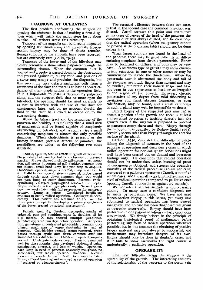
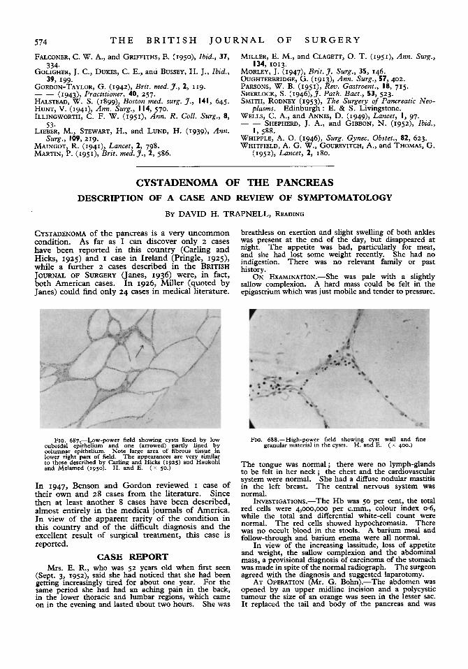
FIG. 687.-Low-power field showing cysts lined. by low cuboidal epithelium and one (arrowed) partly lined by columnar epithelium. Note large area of fibrous tissue in lower right part of field. The appearances are very similar to those described by Carling and Hicks (1925) and Haukohl and Melamed (1950). H. and E. ( x so.)
In 1947, Benson and Gordon reviewed I case of their own and 28 cases from the literature. Since then at least another 8 cases have been described, almost entirely in the medical journals of America. In view of the apparent rarity of the condition in this country and of the difficult diagnosis and the excellent result of surgical treatment, this case is reported.
CASE REPORT Mrs. E. R., who was 52 years old when first seen
(Sept. 3, 1952), said she had noticed that she had been getting increasingly tired for about one year. For the same period she had had an aching pain in the back, in the lower thoracic and lumbar regions, which came on in the evening and lasted about two hours. She was
FIG. 688.-High-power field showing cyst wall and fine granular material in the cysts. H. and E. ( K 400.)
The tongue was normal; there were no lymph-glands to be felt in her neck; the chest and the cardiovascular system were normal. She had a diffuse nodular mastitis in the left breast. The central nervous system was normal.
INVESTIGATIONS.-The H b was 50 per cent, the total red cells were 4,000,000 per c.mm., colour index 0.6, while the total and differential white-cell count were normal. The red cells showed hypochromasia. There was no occult blood in the stools. A barium meal and follow-through and barium enema were all normal.
In view of the increasing lassitude, loss of appetite and weight, the sallow complexion and the abdominal mass, a provisional diagnosis of carcinoma of the stomach was made in spite of the normal radiograph. The surgeon agreed with the diagnosis and suggested laparotomy.
AT OPERATION (Mr. G. Bohn).-The abdomen was opened by an upper midline incision and a polycystic tumour the size of an orange was seen in the lesser sac. It replaced the tail and body of the pancreas and was