Embed Size (px)
Citation preview

Cardiac comorbidity is an independent risk factor for radiation-induced lung
toxicity in lung cancer patients
Running head: Cardiac Comorbidity predicts RILT
Georgi Nalbantov1*, Bas Kietselaer2, Katrien Vandecasteele3, Cary Oberije1, Maaike
Berbee1, Esther Troost1, Anne-Marie Dingemans4, Angela van Baardwijk1, Kim Smits1,
André Dekker1, Johan Bussink5, Dirk De Ruysscher1,6, Yolande Lievens3, Philippe
Lambin1
1 Department of Radiation Oncology (Maastro Clinic), GROW - School for Oncology and
Developmental Biology, Maastricht University Medical Centre, Maastricht, the
Netherlands
2 Departments of Cardiology and Radiology, Cardiovascular Research Institute
Maastricht (CARIM), Maastricht, The Netherlands
3 Department of Radiation Oncology, Ghent University Hospital, Ghent, Belgium
4 Department of Pulmonology, GROW - School for Oncology and Developmental
Biology, Maastricht University Medical Center, Maastricht, The Netherlands
5 Department of Radiation Oncology, Radboud University Nijmegen Medical Center,
Nijmegen, The Netherlands
6 Radiation Oncology, University Hospitals Leuven/ KU Leuven, Leuven, Belgium

* Corresponding author. Address: Maastricht Radiation Oncology (MAASTRO),
Maastricht University, P.O. Box 616, 6200 MD Maastricht, The Netherlands.
Tel: +31 (0)88 445 5666; fax: +31 (0)88 4455667; email: [email protected]
Total number of pages: 19
Number of tables: 3
Number of figures: 2
Keywords: Lung cancer; Cardiac comorbidity; Radiotherapy; Dyspnea; Radiation-
Induced Lung Toxicity
Abstract
Purpose: To test the hypothesis that cardiac comorbidity before the start of
radiotherapy (RT) is associated with an increased risk of radiation-induced lung toxicity
(RILT) in lung cancer patients.
Material and methods: A retrospective analysis was performed of a prospective cohort
of 259 patients with locoregionally lung cancer treated with definitive
radio(chemo)therapy between 2007 and 2011 (ClinicalTrials.gov Identifiers:
NCT00572325 and NCT00573040). We defined RILT as dyspnea CTCv.3.0 grade ≥ 2
within 6 months after RT, and cardiac comorbidity as a recorded treatment of a cardiac
pathology at a cardiology department. Univariate and multivariate analyses, as well as
external validation, were performed. The model-performance measure was the area
under the receiver operating characteristic curve (AUC).
Results: Prior to RT, 75/259 (28.9%) patients had cardiac comorbidity, 44% of whom
(33/75) developed RILT. The odds ratio of developing RILT for patients with cardiac
comorbidity was 2.58 (p<0.01). The cross-validated AUC of a model with cardiac
comorbidity, tumor location, forced expiratory volume in 1 second, sequential
chemotherapy and pretreatment dyspnea score was 0.72 (p<0.001) on the training set,
and 0.67 (p<0.001) on the validation set.

Conclusion: Cardiac comorbidity is an important risk factor for developing RILT after
definite radio(chemo)therapy of lung cancer patients.

Introduction
Radiation-induced lung toxicity (RILT) is an important dose-limiting complication of
radical thoracic radiotherapy (RT). While high radiation doses are expected to provide
better locoregional control, associated toxicity, such as RILT, may have a major impact
on the quality of life and can even be lethal. About 10%-20% of all lung cancer patients
treated with radio(chemo)therapy, R(CH)T, develop RILT within 6 months after start of
treatment, with clinical symptoms of dyspnea, cough, and sometimes fever [1]. Notably,
the degree of RILT varies greatly among patients treated with similar dose levels to the
healthy lung. Identification of patients’ susceptibility to RILT prior to RT based on
baseline characteristics may permit (1) dose escalation for low-risk patients, potentially
leading to better survival rates at reduced/similar levels of treatment-related side effects
[2] and (2) dose reduction/redistribution for high-risk patients to avoid side effects.
Traditional risk factors for RILT include mean lung dose (MLD), V20 Gy (volume
of lung receiving at least 20 Gy), age, smoking status, gender, World Health
Organization (WHO) performance status, chemotherapy, and the location of the primary
lung tumor ([3-14], among others). Unfortunately, prognostic models based on these
factors have not provided consistent performance across different studies. Blood
biomarkers have likewise shown controversial results, [15-17]. Recently, preclinical [18]
and clinical [19] studies have demonstrated a short-term effect of irradiation of a healthy
heart on pulmonary dysfunction. Left-sided heart failure is known to lead to dyspnea
due to an elevated end-diastolic pressure of the left ventricle, which perpetrates to the
pulmonary capillaries and leads to pulmonary edema, [20,21]. Moreover, cardiac
comorbidity at the start of treatment among 3864 lung cancer patients with a mean age

of 67 years has been found to be the most frequent concomitant disease, with incidence
twice as high (23%) as in the general population, [22]. We therefore hypothesized that
patients with recorded historical treatment of cardiac pathologies are at greater risk of
developing RILT after R(CH)T.
Material and Methods
Patient population and inclusion criteria
Between 2008 and 2011 a total of 399 lung cancer patients, all treated in two hospitals
with cardiology departments, were referred to our institute for radiation treatment and
used in our retrospective study. Of these, 259 patients retrospectively met the inclusion
criteria of the study, namely: stage I-IIIB, (chemo)radiotherapy with curative intent,
radiation fraction dose ≤ 3 Gy. Stereotactic body irradiation treatments were excluded.
All patients underwent a FDG-PET/CT scan for treatment planning purposes, on which
the heart and lungs (manual contouring in either mediastinal or lung WW/WL-setting as
appropriate) were delineated. The treatment planning system used was XiO (4.3.4,
CMS, St. Louis, USA) using the superposition dose calculation algorithm. Patients were
treated with concurrent or sequential chemoradiotherapy, (postoperative) radiotherapy
with subsequent adjuvant chemotherapy, or with radiation alone. Sequential
chemotherapy consisted of carboplatin on day 1 and gemcitabine on day 1 and 8. The
majority of the patients received 3 cycles (range 1-6). Concurrent chemo radiation
consisted of cisplatin on day 1 and 8 and etoposide on day 1-3 of a three-weekly cycle.
In total three cycles were given. The patients were examined weekly during RT and

every three months thereafter by either the radiation oncologist or the chest physician.
Patient characteristics for the training (n=259) as well as the validation dataset (n=107
from Ghent University Hospital and n=44 from Radboud University Nijmegen Medical
Centre) are given in Table 1.
Toxicity Scoring
RILT was scored using the Common Terminology Criteria for Adverse Events version
3.0 (CTCAEv3.0) before, weekly during and every 3 months after RT by either a chest
physician or a radiation oncologist. A value of dyspnea ≥ 2 within 6 months after RT was
considered as acute manifestation of RILT and used as primary endpoint in the
analysis. In the CTCAE3.0 system, grade 0 is no dyspnea; grade 1 is dyspnea on
exertion, but can walk 1 flight of stairs without stopping; grade 2 is dyspnea on exertion
but unable to walk 1 flight of stairs or 100 meters without stopping; grade 3 is dyspnea
with ADLs (Activities of Daily Living. Basic ADLs include eating, dressing, getting into or
out of a bed or chair, taking a bath or shower, and using the toilet.); grade 4 is dyspnea
at rest, intubation/ventilator indicated; and grade 5 is death.
Cardiac comorbidity scoring
Cardiac comorbidity was defined as a recorded historical treatment of any cardiac
disorder at a cardiology department before the start of RT, irrespective of its severity.
Cardiac comorbidity for all patients was scored by a cardiologist from the academic
hospital azM Maastricht using the cardiac specific anamnesis from the cardiology
departments. Patient dyspnea scores were not provided to the cardiologist.

Statistical Analysis
We tested four statistical hypotheses:
1) the independence of cardiac comorbidity and post-treatment dyspnea ≥ 2, our
main clinical hypothesis being that we reject such independence;
2) the independence of cardiac comorbidity and post-treatment dyspnea ≥ 2,
given pretreatment dyspnea < 2, to determine whether cardiac comorbidity
might be a risk factor only for patients who already have high dyspnea levels
at the start of RT;
3) the independence of cardiac comorbidity and post-treatment dyspnea ≥ 3, to
determine the robustness of the first hypothesis, in case it is not rejected;
4) the independence of cardiac comorbidity and pretreatment dyspnea, In case
cardiac comorbidity is a risk factor for post-RT dyspnea, it may be also be
more likely that presence of cardiac comorbidity is associated with higher
levels of pretreatment dyspnea.
The univariate and multivariate logistic regression analyses were performed in SPSS
version 19 (IBM Corp., Armonk, NY) and MATLAB (MathWorks Inc., Natick, MA). The
following variables were considered as inputs for the prediction models: MLD, existing
cardiac comorbidity at the start of radiotherapy, smoking status, type of chemotherapy,
age, gender, forced expiratory volume in 1 second adjusted for gender and age (FEV1,
in %), lung surgery performed in the past before RT, WHO performance status
(WHOps), tumor location, lung volume, prescribed tumor dose expressed as equivalent
radiation dose in 2 Gy fractions corrected for overall treatment time (EQD2,t) [23] using

α/β=10 Gy and accelerated repopulation kick-off time of 28 days, overall treatment time,
and the level of dyspnea at the start of RT. In addition, mean heart dose (MHD) was
available for the patients in the training set. The variables for the multivariate model
were selected via a wrapper feature selection procedure, [24], on the training set using
a 10-fold cross validation with AUC set as a performance criterion. This feature
selection method was performed in WEKA (Waikato Environment for Knowledge
Analysis, [25]). An alpha value of 0.05 was used as a threshold for statistical
significance. P-values for nominal variables were computed using a chi-square test.
Model performance was evaluated using the area under the receiver operator
characteristic curve (AUC) estimated from a 10-fold cross-validation procedure to avoid
the problem of overfitting; p-values for the difference in AUC vis-à-vis AUC=0.5 (random
model) were calculated using 1000 bootstrap samples. Univariate and multivariate
analyses were performed on the training set and validated on the validation set.
Results
Among the 259 patients from the training dataset, 76 (29.3%) had a maximum dyspnea
score ≥ 2 after RT. Out of them, 33 (43.4%) had a cardiac comorbidity at the start of RT.
As the total number of patients with cardiac comorbidity was 75, this means that 44%
(33/75) of the cardiac-comorbidity patients developed RILT. Conversely, 23.4% (43/184)
of the patients without cardiac comorbidity experienced RILT.

Cardiac comorbidity and dyspnea ≥ 2. The odds ratio of post-RT dyspnea ≥ 2 for
patients with versus without cardiac comorbidity was 2.6 (p = 0.0009, 95% confidence
interval (CI): 1.5 – 4.5; Table 3 and Supplement Figure 1). These findings were
confirmed on the combined validation set from two university hospitals (n=151), with
corresponding odds ratio of 2.3 (p = 0.039, 95% CI: 1.03 – 4.9).The relative risk of RILT
in the validation dataset was 50% for the patients with cardiac comorbidity (17/34).
Cardiac comorbidity and maximal dyspnea ≥ 2, given baseline dyspnea <2. The odds
ratio of post-RT dyspnea in this case turned out to be similar to the one found without
the restriction on baseline dyspnea grade: 2.6 (p = 0.005, 95% CI: 1.3 –5.1; Table 3).
Cardiac comorbidity and maximal dyspnea ≥ 3. With this test we checked the
robustness of the main finding, which is confirmed: the odds ratio is 2.5 (p=0.009, 95%
CI: 1.2 – 5.2, see Table 3). Even though the corresponding odds ratio on the validation
set was 2.1, the chi-square test was not performed due to the insufficient (less than 5)
number of patients with both cardiac comorbidity and maximal dyspnea ≥ 3.
Cardiac comorbidity and baseline dyspnea. Cardiac comorbidity is an independent
cause of dyspnea, so therefore it is likely that presence of cardiac comorbidity is
associated with higher levels of baseline dyspnea. In this study, we rejected the
independence of these two factors on the training set: the odds ratio of observing
baseline dyspnea ≥ 2 for patients with cardiac comorbidity was 2.2 (p=0.02, 95% CI: 1.1
– 4.5; Supplement Table 1).

MHD, cardiac comorbidity and dyspnea >=2. We performed a Kruskal-Wallis test for
significant difference between median MHDs across the following four groups in the
training set: patients with/without cardiac comorbidity and with/without maximal dyspnea
≥ 2. The p-value of the test was 0.71, suggestive of no significant difference across the
four groups (Supplement Figure 2).
Cardiac comorbidity and maximal dyspnea ≥ 2, given that the patient received
chemotherapy. Considering the subgroup of patients who received chemotherapy: the
odds ratio of post-RT dyspnea ≥ 2 for patients with versus without cardiac comorbidity
was 2.6 (p = 0.005, 95% CI: 1.3 – 5.1; Supplement Table 2). These findings were
confirmed on the combined validation set (n=139), with corresponding odds ratio of 2.3
(p = 0.05, 95% CI: 0.97 – 5.3).
Cardiac comorbidity and smoking status. We tested for an association between cardiac
comorbidity and smoking status, which we refuted on both the training and validation
sets (Supplement Table 1).
Univariate and multivariate analyses: cardiac comorbidity, baseline dyspnea score, and
FEV1 were found to be statistically significant factors in univariate logistic regression
analysis (Table 2). Regarding chemotherapy, sequential chemotherapy was marginally
significant and concurrent was significant (having opposite sign of the sequential). The
wrapper-based feature selection on the training set resulted in the following subset of

variables, which we used in our multivariate model: cardiac comorbidity, tumor location,
FEV1, lung volume, and value of pretreatment dyspnea. The AUC’s for the multivariate
model were 0.72 (p<0.001) on the training set, and 0.67 (p<0.001) on the validation set.
Cardiac comorbidity, lung volume and baseline dyspnea score came out as having the
lowest p-values in the model. The AUCs of the “golden standard” model having only
MLD as input were 0.49 and 0.37 on the training and the validation set, respectively. A
nomogram and the ROC curves for the multivariate model on the training set (computed
using 10-fold cross-validation) and test sets are shown in Figure 1 and Figure 2,
respectively. Figure 2 depicts also the ROC curve for the MLD-only model. The
multivariate model will be available on www.predictcancer.org. Supplementary Figure 3
further demonstrates the added value of the model at two specific points on the ROC
curve of the training-set model.
Discussion
In this study we tested the hypothesis that pretreatment cardiac comorbidity is
associated with the development of RILT after high-dose R(CH)T for lung cancer.
Traditionally, the effects of radiation on both organs have not been considered in
tandem, most probably due to the short-term (up to 6-9 months) pulmonary toxicity
effect and the longer term (5-15 years) cardiac toxicity effect. Short term cardiac toxicity
has been considered in the context of high-dose chemotherapy, [26-29], mainly for
breast cancer patients. Recently, preclinical [18] and clinical [19] studies discussed the
short-term effect of irradiation of a healthy heart on pulmonary dysfunction. They

demonstrated that the heart and lungs interact in their reaction to radiation in that
excessive heart irradiation leads to pulmonary disorders. This is somewhat surprising,
given that the heart has long been considered as the prototype (radiation) dose-
resistant organ, based on the long (more than 5 years) rather than short-term increased
risk of cardiac events, [30,31]. The short-term pulmonary toxicity from radiation may,
however, may heavily depend on both the prior condition of the lungs and the heart.
Regarding the dose delivered to the heart, we tested whether MHD might be a
confounding factor and therefore patients with relatively higher MHD were actually those
who developed dyspnea ≥ 2. The respective Kruskal-Wallis test for independence
between four groups patients - with/without cardiac comorbidity and with/without
maximal dyspnea ≥ 2 - had a rather high p-value, suggestive of no significant difference
between median MHDs across the four groups.
The model describing the association of cardiac comorbidity and RILT was built on
one dataset and externally validated on 151 patients. A major finding of our study is that
cardiac comorbidity accounts for approximately 45% of all cases with dyspnea ≥ 2 after
RT, even when adjusting for patients with dyspnea grade 0 and 1 at the start of
treatment (Table 3). We confirmed the robustness of the results by considering the
endpoint of maximal dyspnea ≥ 3 and also by imposing the condition that only patients
with pretreatment dyspnea <2 are included. The biological mechanism underpinning the
short-term interplay between existing cardiac disorders and the effect of radiation on
lung toxicity is not clear cut and should be elucidated in the future. Furthermore, more
detailed subgroup analyses involving different types of cardiac comorbidity,
standardized across multiple institutions, have to be performed in a bigger future study,

taking into account also the severity of the cardiac disorder type. Such analyses should
be cast against the analyses of cardiac functions and parameters of lung cancer
patients who have never been treated for cardiac pathologies in a cardiology
department.
Choosing a suitable measure of RILT is likewise challenging, [6,32-35]. Dyspnea is a
clinical outcome measured qualitatively, and can be brought about by non-radiotherapy
related causes. A chest X-ray or a CT scan can be used as well, whereby radiation
pneumonitis (1-6 months) and/or fibrosis (1-2 years) could be detected after lung
irradiation, both of which cause dyspnea, [36]. However, in many cases radiation
pneumonitis is clinically asymptomatic [6]. We therefore chose for dyspnea as a
measure for RILT, since it is clinically the most relevant factor.
One study, [37], evaluated treatment-related cardiac toxicity in 64 breast cancer
patients treated with sequential RCHT. None of the patients had cardiovascular history
or echocardiographic abnormalities at the start of RT. Left ventricular ejection fraction
(LVEF) was measured at baseline as well as during follow-up. Twenty one patients
(32.8%) had a short-term decrease in LVEF, with a median decrease of 10%. These
and our findings suggest that future research could concentrate on performing
echocardiography before the start of radiotherapy to detect and/or quantify cardiac
pathologies and to test whether asymptomatic heart problems are likewise associated
with RILT in the same fashion as the symptomatic ones we examined. Another study,
[38], did not find a statistically significant association between LVEF ≤ 50% and
radiation pneumonitis grade III/IV according to the Common Toxicity Criteria, although
the reported odds ratio was 2.15; the overall incidence of radiation pneumonitis was

however low: only 3 out of 130 patients (2.3%). Cardiac toxicity blood biomarkers for
patients undergoing R(CH)T could also be considered, such as increases in troponin
and brain natriuretic peptide, [39,40], but such studies have shown conflicting results,
[31].
Patient’s smoking status is a potential confounding factor, which could arguably
provide an alternative explanation of our main findings. However, current smoking
status was not found to be significant in univariate analysis. On the other hand, smoking
status is strongly associated with the presence of cardiac disorders, but has also been
shown to be protective of RILT, [41]. In general it is the cause of about 15% of all cases
of heart failure, [42]. In our (training) dataset, approximately 22% of the patients with
cardiac comorbidity were current smokers at the start of RT. We tested therefore for an
association between cardiac comorbidity and smoking status, which we refuted.
Similarly, we tested for the chemotherapy being a confounding factor and found that for
the subgroup of patients who received chemotherapy the odds ratio for developing RILT
was virtually the same as for the patients in the original datasets.
The most commonly used predictive dosimetric parameter for RILT, the mean lung
dose, was not found to be significant in our cohort. This is to be expected, as prescribed
MLD levels had a relatively low mean with a small standard deviation (15.65 Gy ± 4.4)
due to the fact that MLD was used as a dose-limiting factor during radiotherapy
planning. That is why there is arguably insufficient data to detect a stable upward
sloping dose-toxicity response relationship, such as reported in the QUANTEC study,
[3]. This relative stability of the MLD is actually quite advantageous for the clinical
quality of our prediction models, as it gives the opportunity to find important factors for
RILT that are not confounded by largely-varying levels of MLD.
The role of ACE (angiotensin-converting-enzyme) inhibitors could also be further
investigated. They have been demonstrated to be both vascular protective and
preventive of new acute cardiovascular events, and have been proven to play central
role in afterload reduction in patients with congestive heart failure and improvement of
cardiac-driven dyspnea complaints, [43,44]. The role of ACE inhibitors for RILT is not
clear cut, however. While a decrease in RILT is reported in preclinical [45] and clinical
[46] studies with ACE inhibitors, other investigations did not reveal a protective effect at
the dose used for the treatment of hypertension [47].
The role of potential interdependency of cardiac comorbidity, pulmonary toxicity and
overall survival should be further investigated, as higher incidence of comorbidities in
general could influence both endpoints. In their study, [48], Firat et al. found that
comorbidity influenced overall survival when the Cumulative Illness Rating Scale for
Geriatrics (CIRS-G) was used to rate the comorbidity. However, this finding was not
observed with the Charlson scale.
The existence of cardiac comorbidity at the start of high-dose RT of lung cancer
patients is a major factor for development of RILT (defined as maximal dyspnea ≥ 2
after radio(chemo)therapy. Moreover, cardiac comorbidity accounted for 43.4% of the
incidence of RILT. These results suggest that (1) individualized treatment should be
considered for patients with cardiac comorbidity and (2) excluding these patients from
dose escalation studies will potentially allow for better targeting/escalation of the rest of

the patients. It should further be investigated whether asymptomatic patients with
cardiac comorbidity develop RILT to the same extent as the symptomatic ones.
Conflict of interest
None.
Acknowledgements
We acknowledge financial support from the CTMM framework (AIRFORCE project), EU
6th and 7th framework program (ARTFORCE and METOXIA program), Interreg
(www.eurocat.info), STW (DuCAT), the Kankeronderzoekfonds Limburg from the Health
Foundation Limburg and the Dutch Cancer Society (KWF UM 2011-5020, KWF UM
2009-4454).

References
[1] Rodrigues G, Lock M, D'Souza D, Yu E Van Dyk J. Prediction of radiation
pneumonitis by dose - volume histogram parameters in lung cancer--a
systematic review. Radiother Oncol 2004;71:127-138.
[2] van Baardwijk A, Wanders S, Boersma L, et al. Mature results of an
individualized radiation dose prescription study based on normal tissue
constraints in stages I to III non-small-cell lung cancer. J Clin Oncol
2010;28:1380-1386.
[3] Marks LB, Bentzen SM, Deasy JO, et al. Radiation dose-volume effects in
the lung. Int J Radiat Oncol Biol Phys 2010;76:S70-76.
[4] Dehing-Oberije C, De Ruysscher D, van Baardwijk A, Yu S, Rao B Lambin
P. The importance of patient characteristics for the prediction of
radiation-induced lung toxicity. Radiother Oncol 2009;91:421-426.
[5] Kim M, Lee J, Ha B, Lee R, Lee KJ Suh HS. Factors predicting radiation
pneumonitis in locally advanced non-small cell lung cancer. Radiat
Oncol J 2011;29:181-190.
[6] Palma DA, Senan S, Tsujino K, et al. Predicting radiation pneumonitis
after chemoradiation therapy for lung cancer: an international
individual patient data meta-analysis. Int J Radiat Oncol Biol Phys
2013;85:444-450.
[7] Kwa SL, Theuws JC, Wagenaar A, et al. Evaluation of two dose-volume
histogram reduction models for the prediction of radiation pneumonitis.
Radiother Oncol 1998;48:61-69.
[8] Vinogradskiy Y, Tucker SL, Liao Z Martel MK. A novel method to
incorporate the spatial location of the lung dose distribution into
predictive radiation pneumonitis modeling. Int J Radiat Oncol Biol Phys
2012;82:1549-1555.
[9] Hope AJ, Lindsay PE, El Naqa I, et al. Modeling radiation pneumonitis
risk with clinical, dosimetric, and spatial parameters. Int J Radiat
Oncol Biol Phys 2006;65:112-124.
[10] Jenkins P Watts J. An improved model for predicting radiation
pneumonitis incorporating clinical and dosimetric variables. Int J
Radiat Oncol Biol Phys 2011;80:1023-1029.
[11] Stenmark MH, Cai XW, Shedden K, et al. Combining physical and biologic
parameters to predict radiation-induced lung toxicity in patients with
non-small-cell lung cancer treated with definitive radiation therapy.
Int J Radiat Oncol Biol Phys 2012;84:e217-222.
[12] Madani I, De Ruyck K, Goeminne H, De Neve W, Thierens H Van Meerbeeck
J. Predicting risk of radiation-induced lung injury. J Thorac Oncol
2007;2:864-874.
[13] Rancati T, Ceresoli GL, Gagliardi G, Schipani S Cattaneo GM. Factors
predicting radiation pneumonitis in lung cancer patients: a
retrospective study. Radiother Oncol 2003;67:275-283.
[14] Jin H, Tucker SL, Liu HH, et al. Dose-volume thresholds and smoking
status for the risk of treatment-related pneumonitis in inoperable non-
small cell lung cancer treated with definitive radiotherapy. Radiother
Oncol 2009;91:427-432.
[15] Voets AM, Oberije C, Struijk RB, et al. No association between TGF-
beta1 polymorphisms and radiation-induced lung toxicity in a European
cohort of lung cancer patients. Radiother Oncol 2012;105:296-298.
[16] Novakova-Jiresova A, Van Gameren MM, Coppes RP, Kampinga HH Groen HJ.
Transforming growth factor-beta plasma dynamics and post-irradiation
lung injury in lung cancer patients. Radiother Oncol 2004;71:183-189.
[17] Iwata H, Shibamoto Y, Baba F, et al. Correlation between the serum KL-6
level and the grade of radiation pneumonitis after stereotactic body
radiotherapy for stage I lung cancer or small lung metastasis.
Radiother Oncol 2011;101:267-270.
[18] Ghobadi G, van der Veen S, Bartelds B, et al. Physiological interaction
of heart and lung in thoracic irradiation. Int J Radiat Oncol Biol Phys
2012;84:e639-646.
[19] Huang EX, Hope AJ, Lindsay PE, et al. Heart irradiation as a risk
factor for radiation pneumonitis. Acta Oncol 2011;50:51-60.
[20] Theresa A.McDonaghConsultant Cardiologist KsCH, London, UK Roy
S.GardnerConsultant Cardiologist, Scottish National Advanced Heart
Failure Service, Golden Jubilee National Hospital, UK Andrew
L.ClarkConsultant Cardiologist, Castle Hill Hospital, Cottingham, UK
HenryDargieConsultant Cardiologist, Golden Jubilee National Hospital,
Glasgow, UK. Oxford Textbook of Heart Failure. New York, NY: 'Oxford
University Press'.
[21] Guyton AC Lindsey AW. Effect of elevated left atrial pressure and
decreased plasma protein concentration on the development of pulmonary
edema. Circ Res 1959;7:649-657.
[22] Janssen-Heijnen ML, Schipper RM, Razenberg PP, Crommelin MA Coebergh
JW. Prevalence of co-morbidity in lung cancer patients and its
relationship with treatment: a population-based study. Lung Cancer
1998;21:105-113.
[23] Fowler JF, Tome WA, Fenwick JD Mehta MP. A challenge to traditional
radiation oncology. Int J Radiat Oncol Biol Phys 2004;60:1241-1256.
[24] Kohavi R John GH. Wrappers for feature subset selection. Artif. Intell.
1997;97:273-324.
[25] Witten IH Frank E. Data mining : practical machine learning tools and
techniques. Amsterdam ; Boston, MA: Morgan Kaufman, 2005.
[26] Cardinale D, Colombo A, Sandri MT, et al. Prevention of high-dose
chemotherapy-induced cardiotoxicity in high-risk patients by
angiotensin-converting enzyme inhibition. Circulation 2006;114:2474-
2481.
[27] Tallaj JA, Franco V, Rayburn BK, et al. Response of doxorubicin-induced
cardiomyopathy to the current management strategy of heart failure. J
Heart Lung Transplant 2005;24:2196-2201.
[28] Bird BR Swain SM. Cardiac toxicity in breast cancer survivors: review
of potential cardiac problems. Clin Cancer Res 2008;14:14-24.
[29] Shaffer R, Tyldesley S, Rolles M, Chia S Mohamed I. Acute
cardiotoxicity with concurrent trastuzumab and radiotherapy including
internal mammary chain nodes: a retrospective single-institution study.
Radiother Oncol 2009;90:122-126.
[30] Aleman BM, van den Belt-Dusebout AW, De Bruin ML, et al. Late
cardiotoxicity after treatment for Hodgkin lymphoma. Blood
2007;109:1878-1886.
[31] Yusuf SW, Sami S Daher IN. Radiation-induced heart disease: a clinical
update. Cardiol Res Pract 2011;2011:317659.
[32] Faria SL, Aslani M, Tafazoli FS, Souhami L Freeman CR. The challenge of
scoring radiation-induced lung toxicity. Clin Oncol (R Coll Radiol)
2009;21:371-375.
[33] Petit SF, van Elmpt WJ, Oberije CJ, et al. [(1)(8)F]fluorodeoxyglucose
uptake patterns in lung before radiotherapy identify areas more
susceptible to radiation-induced lung toxicity in non-small-cell lung
cancer patients. Int J Radiat Oncol Biol Phys 2011;81:698-705.
[34] Lambin P, van Stiphout RG, Starmans MH, et al. Predicting outcomes in
radiation oncology--multifactorial decision support systems. Nat Rev
Clin Oncol 2013;10:27-40.

[35] De Ruysscher D, Dehing C, Yu S, et al. Dyspnea evolution after high-
dose radiotherapy in patients with non-small cell lung cancer.
Radiother Oncol 2009;91:353-359.
[36] De Ruysscher D, Houben A, Aerts HJ, et al. Increased (18)F-deoxyglucose
uptake in the lung during the first weeks of radiotherapy is correlated
with subsequent Radiation-Induced Lung Toxicity (RILT): a prospective
pilot study. Radiother Oncol 2009;91:415-420.
[37] Magne N, Castadot P, Chargari C, Di Leo A, Philippson C Van Houtte P.
Special focus on cardiac toxicity of different sequences of adjuvant
doxorubicin/docetaxel/CMF regimens combined with radiotherapy in breast
cancer patients. Radiother Oncol 2009;90:116-121.
[38] Semrau S, Klautke G Fietkau R. Baseline cardiopulmonary function as an
independent prognostic factor for survival of inoperable non-small-cell
lung cancer after concurrent chemoradiotherapy: a single-center
analysis of 161 cases. Int J Radiat Oncol Biol Phys 2011;79:96-104.
[39] Kozak KR, Hong TS, Sluss PM, et al. Cardiac blood biomarkers in
patients receiving thoracic (chemo)radiation. Lung Cancer 2008;62:351-
355.
[40] Nellessen U, Zingel M, Hecker H, Bahnsen J Borschke D. Effects of
radiation therapy on myocardial cell integrity and pump function: which
role for cardiac biomarkers? Chemotherapy 2010;56:147-152.
[41] Vogelius IR Bentzen SM. A literature-based meta-analysis of clinical
risk factors for development of radiation induced pneumonitis. Acta
Oncol 2012;51:975-983.
[42] He J, Ogden LG, Bazzano LA, Vupputuri S, Loria C Whelton PK. Risk
factors for congestive heart failure in US men and women: NHANES I
epidemiologic follow-up study. Arch Intern Med 2001;161:996-1002.
[43] Michel JB. Relationship between decrease in afterload and beneficial
effects of ACE inhibitors in experimental cardiac hypertrophy and
congestive heart failure. Eur Heart J 1990;11 Suppl D:17-26.
[44] Neal B, MacMahon S Chapman N. Effects of ACE inhibitors, calcium
antagonists, and other blood-pressure-lowering drugs: results of
prospectively designed overviews of randomised trials. Blood Pressure
Lowering Treatment Trialists' Collaboration. Lancet 2000;356:1955-1964.
[45] Molteni A, Moulder JE, Cohen EF, et al. Control of radiation-induced
pneumopathy and lung fibrosis by angiotensin-converting enzyme
inhibitors and an angiotensin II type 1 receptor blocker. Int J Radiat
Biol 2000;76:523-532.
[46] Kharofa J, Cohen EP, Tomic R, Xiang Q Gore E. Decreased risk of
radiation pneumonitis with incidental concurrent use of angiotensin-
converting enzyme inhibitors and thoracic radiation therapy. Int J
Radiat Oncol Biol Phys 2012;84:238-243.
[47] Wang LW, Fu XL, Clough R, et al. Can angiotensin-converting enzyme
inhibitors protect against symptomatic radiation pneumonitis? Radiat
Res 2000;153:405-410.
[48] Firat S, Byhardt RW Gore E. Comorbidity and Karnofksy performance score
are independent prognostic factors in stage III non-small-cell lung
cancer: an institutional analysis of patients treated on four RTOG
studies. Radiation Therapy Oncology Group. Int J Radiat Oncol Biol Phys
2002;54:357-364.

Tables
Table 1. Patient Characteristics.
No. of patients (%)
Variable training set (n=259) Ghent set (n=107) Nijmegen set (n=44)
Gender
Male 163(62.9) 94(87.8) 36(81.8) Female 96(37.1) 13(12.2) 8(18.2)
Smoking status
Current smoker 77(29.7) 34(31.8) 11(25.0) Histopathology
NSCLC 198(76.5) 88(93.5) 43(97.7) SCLC 49(18.9) 11(10.3) 0(0) NSCLC+SCLC 0(0) 1(0.9) 0(0)
cT-stage
0 or 1 48(18.5) 9(8.4) 10(22.7) 2 75(29.0) 33(30.8) 14(31.8) 3 40(15.4) 32(29.9) 11(25.0) 4 95(36.7) 20(18.7) 9(20.5)
cN-stage
0 67(25.9) 17(15.9) 2(4.5) 1 19(7.3) 9(8.4) 2(4.5) 2 112(43.2) 47(44.0) 35(79.5) 3 60(23.2) 23(21.5) 5(11.4)
WHO-ps
0 63(24.3) 52(48.6) 25(56.8) 1 153(59.1) 48(44.9) 14(31.8) ≥ 2 43(16.6) 6(5.61) 5(11.4)
Chemotherapy
Yes 197(76.1) 98(91.6) 41(93.2)
concurrent 148(57.1) 39(36.4) 22(50.0)
sequential before RT 39(15.1) 56(52.3) 19(43.2) adjuvant (after surgery) 9(0.03) 0(0) 0(0) palliative 1(0.01) 0(0) 0(0)
concurrent + adjuvant 0(0) 3(0.03) 0(0)
No 44(17.0) 9(8.41) 3(6.8) Cardiac comorbidity
No 184(71.0) 86(80.4) 13(29.5)
Yes 75(29.0) 21(19.6) 31(70.5)
Baseline dyspnea score
0 78(30.1) 46(43.0) 15(34.1)
1 140(54.1) 25(23.4) 25(56.8)
2 22(8.5) 19(17.8) 4(9.1)
3 15(5.8) 2(1.9) 0(0)
4 1(0.4) 0(0) 0(0)
Maximal dyspnea score
0 49 (18.9) 19 (17.8) 4 (9.1)
1 134 (51.7) 47 (44.0) 28 (63.6)
2 40 (15.4) 34 (31.8) 8 (18.2)

No. of patients (%)
Variable training set (n=259) Ghent set (n=107) Nijmegen set (n=44)
3 32 (12.4) 5 (4.7) 4 (9.1)
4 4 (1.5) 2 (1.9) 0(0)
Tumor location
Lower/Middle lobe 76 (29.3) 33 (30.8) 15 (35.7)
Upper lobe 83 (32.1) 67 (62.6) 27 (64.3)
Lung surgery before RT
Yes 23 (8.9) 15 (14.0) 0(0) No 236 (91.1) 92 (86.0) 44 (100.0)
Mean (SD)
OTT 30.21 (7.45) 47.8 (11.1) 43.37 (3.49)
Age, years 67.5 (10.1) 64.0 (8.8) 65.94 (7.69)
Prescribed tumor dose, Gy 62.4 (9.92) 64.5 (8.58) 64.66 (4.13)
EQD2,t 58.83 (9.51) 51.97 (5.94) 54.60 (2.35)
Dose per fraction 1.836 (0.342) 1.981 (0.172) 2.02 (0.15)
MLD, Gy 15.65 (4.44) 13.1 (4.31) 16.23 (3.12)
V20 Gy 25.45 (9.87) 19.35 (7.71) not available
FEV1 (in %) 75.96 (21.86) 75.7 (20.56) 77(17.44)
Abbreviations: no./n, number; SD, standard deviation; cT-stage, clinical tumor stage;
cN-stage, clinical lymph node stage; NSCLC, non-small-cell lung cancer; SCLC, small-
cell-lung cancer; WHO-ps, World Health Organization performance status; OTT, overall
treatment time (in days); EQD2,t, equivalent radiation dose at 2 Gy per fraction, adjusted
for time; MLD, mean lung dose; V20 Gy, volume of the healthy lung receiving a dose of
at least 20 Gy; FEV1 (in %), forced expiratory volume in 1 second (in %) adjusted for
age and gender. Dyspnea is measured according to the Common Toxicity Criteria for
Adverse Effects, version 3.0; maximal dyspnea is measured within 6 months after the
start of R(CH)T.

Table 2. Results for univariate and multivariate logistic regressions for all variables and selected variables based on the
wrapper approach. Dependent variables: maximal dyspnea ≥ 2 within 6 months from the start of RT.
univariate multivariate: all variables multivariate: selected variables
Variable coefficient S.E. p value*
coefficient S.E. p value*
coefficient S.E. p value*
Intercept 0.803 1.614 0.619 1.512 0.664 0.023 smoking status 0.029 0.302 0.924 0.185 0.306 0.546 n.a n.a n.a sequential chemotherapy 0.671 0.363 0.064 0.601 0.449 0.180 0.610 0.394 0.122 concurrent chemotherapy -0.789 0.290 0.007 -0.016 0.416 0.969 n.a n.a n.a gender 0.094 0.284 0.741 -0.005 0.313 0.988 n.a n.a n.a cardiac comorbidity 0.969 0.289 0.001 0.817 0.307 0.008 0.826 0.312 0.008 surgery 0.057 0.475 0.904 -0.196 0.533 0.713 n.a n.a n.a tumor location -0.185 0.369 0.616 -0.421 0.314 0.180 -0.290 0.347 0.404 WHO-ps 0.055 0.176 0.755 0.077 0.191 0.684 n.a n.a n.a age 0.025 0.014 0.078 -0.016 0.016 0.340 n.a n.a n.a FEV1 (in %) -0.016 0.007 0.018 -0.012 0.007 0.077 -0.007 0.007 0.330 MLD 0.005 0.031 0.872 -0.02 0.034 0.565 n.a n.a n.a EQD2,t 0.022 0.014 0.126 0.007 0.018 0.713 n.a n.a n.a overall treatment time 0.002 0.018 0.914 0.017 0.018 0.345 n.a n.a n.a baseline dyspnea score 1.015 0.196 <0.000 1.106 0.203 <0.000 0.990 0.210 <0.000 Model performance (AUC)
AUC
training set, 10-fold cross validation
0.723 <0.01
validation set 0.674 <0.01
S.E. standard error, n.a. not applicable; abbreviations, see Table 1. * p-values for coefficients were calculated by multivariate logistic regression; p-values for AUCs were calculated by 1000 bootstraps and a 1-sided Student’s t-test testing if the AUC was different from 0.5 (a flip-of-a-coin model).

Table 3. Contingency tables, odds rations (with 95% CI) and p-values for the hypotheses that (1) maximal dyspnea ≥ 2
and cardiac comorbidity are independent, even when (2) the baseline dyspnea is ≤1, or (3) the outcome is maximal
dyspnea ≥ 3. Results are shown for both the training set and the validation set. P-value is not computed when a
contingency table contains cells with counts < 5.
Dataset training set validation set training set validation set training set validation set
baseline dyspnea= 0 or 1
max dyspn <2
max dyspn ≥ 2
max dyspn <2
max dyspn ≥ 2
max dyspn <2
max dyspn ≥ 2
max dyspn <2
max dyspn ≥ 2
max dyspn <3
max dyspn ≥ 3
max dyspn <3
max dyspn ≥ 3
with CC 42 33 17 17 38 20 15 11 58 17 30 4
no CC 141 43 81 36 133 27 60 25 165 19 110 7 odds ratio (95% CI)
2.58 (1.46 – 4.55)
2.25 (1.03-4.90)
2.59 (1.31-5.12)
1.76 (0.71-4.36)
2.55 (1.24 – 5.23)
2.10
p-value 0.0009 0.039 0.005 0.219 0.009 n.a. n 259 151 218 111 259 151
CC, cardiac comorbidity; CI, confidence interval; n, number; max dyspn, maximal dyspnea (within 6 months from the start
of RT).
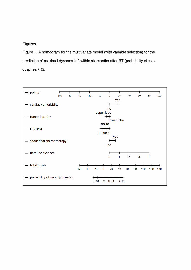
Figures
Figure 1. A nomogram for the multivariate model (with variable selection) for the
prediction of maximal dyspnea ≥ 2 within six months after RT (probability of max
dyspnea ≥ 2).

Figure 2. ROC curves for the multivariable model (with selected variables) for predicting
maximal dyspnea >=2 within 6 months after the start of RT on the training set (solid line,
cross-validated AUC = 0.72) and the validation set (dashed line, AUC = 0.67), as well
as the ROC curve for the model including only mean lung dose as a predictor (dotted
line, AUC = 0.50).

Supplementary material
Supplement Table 1. Contingency tables, odds rations (with 95% CI) and p-values for the hypotheses that (1) baseline
dyspnea ≥ 2 and cardiac comorbidity are independent, and (2) current smoking status and cardiac comorbidity are
independent. Results are shown for both the training set and the validation set.
Dataset training set validation set training set validation set
baseline dyspnea <2
baseline dyspnea ≥2
baseline dyspnea <2
baseline dyspnea ≥2
not current smoker
current smoker
not current smoker
current smoker
with CC 58 17 26 7 53 15 18 10
no CC 160 21 85 18 111 62 76 35 odds ratio (95% CI)
2.23 (1.10 – 4.53)
1.27 (0.48 – 3.39)
0.51 (0.26 – 0.97)
1.21 (0.5-2.88)
p-value 0.023 0.63 0.039 0.67 n 256 136 241 139
CC, cardiac comorbidity; CI, confidence interval; n, number.

Supplement Table 2. Contingency tables, odds rations (with 95% CI) and p-values for the hypothesis that baseline
dyspnea ≥ 2 and cardiac comorbidity are independent for the subgroup of patients who received and who did not receive
chemotherapy, respectively.
Dataset
received chemotherapy
did not received chemotherapy
training set validation set training set validation set max dyspn <2
max dyspn ≥2
max dyspn <2
max dyspn ≥2
max dyspn <2
max dyspn ≥2
max dyspn <2
max dyspn ≥2
with CC 31 22 14 14 7 7 3 3
no CC 113 31 77 34 21 9 4 2 odds ratio (95% CI)
2.59 (1.32 – 5.08)
2.26 (0.97 – 5.26)
2.33 (0.63 – 8.62)
2 (n.a.)
p-value 0.005 0.05 0.2 n.a n 197 139 44 12
CC, cardiac comorbidity; CI, confidence interval; n, number; max dyspn, maximal dyspnea (within 6 months from the start
of RT), n.a., not applicable.

Supplement Figure 1. Frequency of radiation induced lung toxicity (measured by a
CTCv3.0 dyspnea score ≥ 2 within 6 months from the beginning of radiotherapy), for
patients with/without cardiac comorbidity. The hypothesis that the odds ratio for post-
radio(chemo)therapy (RCHT) dyspnea grade ≥ 2 for the cardiac vs. no-cardiac
comorbidity patients is equal to 1 is rejected (odds ratio = 2.6, p-value 0.0006, 95% CI =
1.5-4.6), total number of patients = 259).
patie
nts
(%
)
maximal dyspnea grade < 2 after RCHT
maximal dyspnea grade ≥ 2 after RCHT
n=43
n=34
n=42
n=140

Supplement Figure 2. Boxplot for Mean Heart Dose for four groups: (1) CC (cardiac
comorbidity) = yes and maximal dyspnea <2, (2) CC = yes and maximal dyspnea ≥ 2,
(3) CC = no and maximal dyspnea <2, and (4) CC = no and maximal dyspnea ≥ 2. The
p-value = 0.71 from a Kruskal-Wallis test for equality of the medians across the four
groups indicates no evidence of inequality.
0
5
10
15
20
25
30
35
Me
an
He
art
Do
se
CC = yes, Maximal dyspnea < 2
CC = yes, Maximal dyspnea >=2
CC = no, Maximal dyspnea < 2
CC = no, Maximal dyspnea >= 2

Supplement Figure 3. Panel (a): ROC curves for the multivariable model (with selected
variables) for predicting maximal dyspnea >=2 within 6 months after the start of RT on
the training set (solid line, cross-validated AUC = 0.72) and the validation set (dashed
line, AUC = 0.67), as well as a ROC curve for the model including only mean lung dose
as a predictor (dotted line, AUC = 0.502). Panel (b): relative risk and benefit at two
example points on the solid ROC curve in Panel (a).
At point 1, 52 (out of 259) patients are predicted not to develop RILT, while 5 actually
did. Therefore, when using this point for predicting whether a patient could receive
higher radiation doses, these patients have a relative risk of 9.6% for developing RILT.
This is a significant improvement (p = 0.005, chi-square equality-of-proportions test)
from the 29.3% of patients developing RILT in the entire training dataset. Furthermore,
at point 2 in the ROC curve, out of 31 patients who are predicted to develop RILT, 20
(64.5%) indeed develop RILT, the proportion being likewise significantly different
(p<0.001) from the 29.3% in the training dataset as a whole.
a b

Supplement Figure 4. Pre- and post-radiotherapy distribution of dyspnea scores
(measured by a CTCv3.0 dyspnea score within 6 months from the beginning of
radiotherapy) for the training set of 259 patients.
0 1 2 3 4
0
1
2
3
4
0
1
0
0
0
4
1
3
2
5
Dyspnea grade pre-RT
4
3
1
1
13
9
14
19
97
1
36
29
9
4
0
Dysp
ne
a g
rad
e p
ost-
RT
0
20
40
60
80