Embed Size (px)
Citation preview

DAY 4
• Cardiac Cycle
• ECG Lab

DAY 5MARCH 7 AND 8
• Cardiac Cycle
• ECG Lab finish

HEART SOUNDS -
Heart Sounds –• opening and closing of the
valves (LUB, DUB), • flow of blood into and out of
the chambers, • vibrations in muscle
http://depts.washington.e
du/physdx/heart/demo.ht
ml

• Stethoscope- instrument to listen and measure heart sounds

CARDIAC CYCLEOne complete heartbeat.
http://www.blaufuss.org/tutorial/index1.html
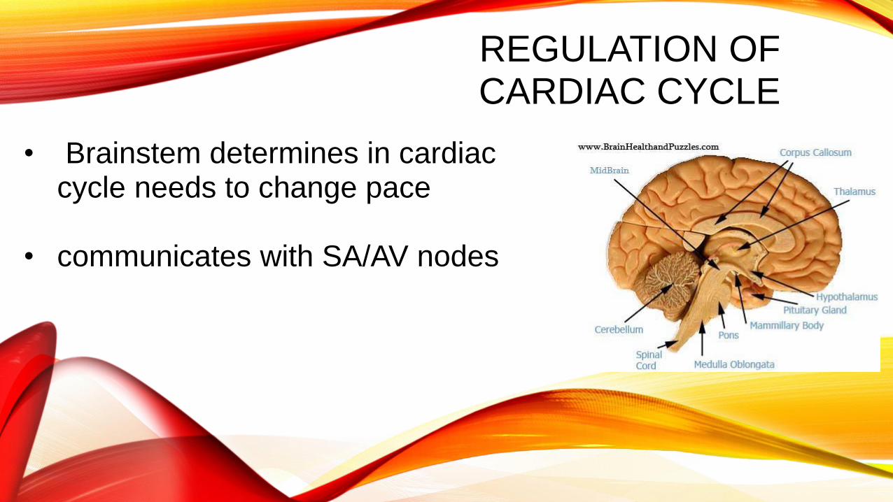
REGULATION OF CARDIAC CYCLE
• Brainstem determines in cardiac cycle needs to change pace
• communicates with SA/AV nodes

CARDIAC CONDUCTION
S-A Node
A-V Node


PARTS OF THE EKG• P Wave – Atrial contraction
• QRS Complex – Ventricular contraction
• T Wave – ventricular recovery

ECG OR EKG
recording of the electrical changes during a cardiac cycle


DAY 6
• Pulmonary and systemic circulations• Blood vessels
• Returning blood to the heart
• Differences between systemic and pulmonary

Systemic Circulation• to body• Left side of heart
Pulmonary Circulation• to lungs • Right side of heart

FUNCTION OF EACH CIRCULATION
Systemic Circulation• delivers blood to all body cells and carries away waste
Pulmonary Circulation• releases carbon dioxide and gathers oxygen (lung pathway)
Coronary circulation• Provides blood to the heart tissue
• Others: Cerebral, hepatic, renal, abdominal…
VESSELS
• ARTERIES
• Carry blood away from the heart
• VEINS
• Carry blood toward the heart
• CAPILLARIES
• Where gas exchange occurs

Arteries :StrongelasticAway from heart Smaller called arterioles
VEINS –Thinner, less muscular vessels carrying blood toward the heart.Smallest ones are called venules.Contain valves.
Capillaries:Penetrate nearly all tissues.Walls are composed of a single layer of squamous cells – very thin.Exchange of gases

RETURNING BLOOD TO YOUR HEART
Veins have 3 adaptations to help return blood to your heart
1. Valves
2. Skeletal muscles
3. Slightly muscular walls of the veins

Major Blood Vessels
Aorta - The aorta is the largest artery. (leaves left ventricle)
Pulmonary Trunk/Artery – splits into left and right, both lead to the lungs (leaves left ventricle)
Pulmonary Veins – return blood from the lungs to the heart (connects to left atrium)
Superior and Inferior Vena Cava – return blood from the head and body to the heart (connects to right atrium)

PULSE
• Every time the heart beats there is a pulse or surge of blood in your arteries.
• Average pulse rate is 70 – 80 beats per minute (bpm)
• Easiest to feel it closer to your heart.

DAY 7
• Blood Pressure
• Notes
• Lab

BLOOD PRESSURE
• CAUSED BY THE PUSH OF THE BLOOD ON THE VESSELS AFTER THE BLOOD EXITS THE HEART
• HIGHEST PRESSURE IN THE AORTA AND ARTERIES,
• LOWEST PRESSURE IN THE VENA CAVA AND OTHER VEINS

CONTROL OF BLOOD FLOW:
VASOCONSTRICTION –NARROWING BLOOD VESSEL’S DIAMETER
VASODILATION – EXPANDING BLOOD VESSEL’S DIAMETER
CONTROLLED BY MUSCLES AROUND THE VESSELS MAKING VESSEL OPENING LARGER OR SMALLER

Factors affecting blood pressure:
1. Cardiac Output2. Blood volume (5 liters for avg adult)3. Blood Viscosity

PARTS OF THE CUFF AND STETHOSCOPE
• Cuff
• Valve/pump
• Meter
• Ear buds
• bell

DAY 8
• Review
• Jeopardy

• Extra slides

CARDIAC OUTPUT
Cardiac Output = Stroke Volume x Heart Rate

SADS = (SUDDEN ARRHYTHMIA DEATH SYNDROMES OR SUDDEN ADULT DEATH SYNDROME)
Routine ECG Screening may help prevent deaths in young people

INTERPRETING EKGSAn ECG is printed on paper covered with a grid of squares.Notice that five small squares on the paper form a larger square. The width of a single small square on ECG paper represents 0.04 seconds.
A common length of an ECG printout is 6 seconds; this is known as a "six second strip."

PATH OF BLOOD FLOW

Check your labels!

The flaps of the bicuspid and tricuspid valves are anchored to the ventricle walls
This prevents the valves from being pushed up into the atria during ventricular systole (contraction)
Can you identify these parts?

1. Right Atrium 2. Right Atrioventricular Valve
(Tricuspid Valve) 3. Right Ventricle 4. Left Atrium 5. Left Atrioventricular Valve
(Mitral Valve) 6. Left Ventricle 7. Papillary Muscle 8. Chordae Tendinae
9. Mitral Valve cusps
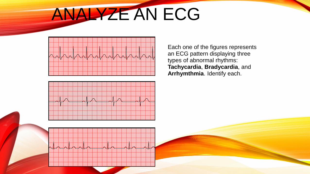
ANALYZE AN ECG
Each one of the figures represents an ECG pattern displaying three types of abnormal rhythms: Tachycardia, Bradycardia, and Arrhymthmia. Identify each.


Blood flow through veins – not very efficient. Slow, weak “pushing” by arterial blood pressure is not much of a factor at all. Important factors include:
1. Contraction of the diaphragm.
2. Pumping action of the skeletal muscles.
3. Valves in the veins.

Disorders of the Circulatory System
1. MVP - mitral valve prolapse, the mitral valve does not close all the way; this creates a clicking sound at the end of a contraction.
2. Heart Murmurs – valves do not close completely, causing an (often) harmless murmur sound. Sometimes holes can occur in the septum f the heart which can also cause a murmur
3. Myocardial Infarction (MI) - a blood clot obstructs a coronary artery, commonly called a “heart attack”

4. Atherosclerosis – deposits of fatty materials such as cholesterol form a “plaque” in the arteries which reduces blood flow. Advanced forms are called arteriosclerosis. Treatment: Angioplasty, where a catheter is inserted into the artery and a balloon is used to stretch the walls open. A bypass can also treat clogged arteries, a vein is used to replace a clogged artery. Coronary bypass refers to a procedure where the coronary artery is bypassed to supply blood to the heart. (The phrase “quadruple bypass” means that 4 arteries were bypassed.)
http://www.mayoclinic.com/health/carotid-angioplasty-and-stenting/MM00772(Mayo Clinic)
5. Hypertension – high blood pressure, the force within the arteries is too high. A sphygmomanometer can be used to diagnose hypertension