Embed Size (px)
Citation preview

Brit. Heart J., 1969, 31, 26.
Cardio-auditory SyndromesCardiac and Genetic Study of 511 Deaf-mute Children*
A. SANCHEZ CASCOS, L. SANCHEZ-HARGUINDEY, AND P. DE RABAGOFrom the Cardiac and Genetic Departments, Fundacicn Jimenez Diaz, Madrid, Spain
In 1957 Jervell and Lange-Nielsen described aNorwegian family, with 4 sibs who all had deaf-mutism, an extraordinarily long Q-T interval, andfainting attacks. Levine and Woodworth describeda new case in 1958. Fraser and colleagues (Fraser,Froggatt, and James, 1964a; Fraser, Froggatt, andMurphy, 1964b; Friedmann, Fraser, and Froggatt,1966) carried out a survey in the U.K. of almost1500 deaf-mute children, and discovered 9 morecases; with this British study it seemed clear thatthere was a recessive type of inheritance. Threemore cases have been reported from Norway(Jervell, Thingstad, and Endsj6, 1966) and one fromthe U.S.A. (Lisker and Finkelstein, 1966). Nocases were found in a survey of 369 deaf-mutechildren from Detroit (James, 1967) nor in anotherof 211 children from Rome (Puletti, Jacobellis, andBorghi, 1967).On the other hand, several families have been
described (Romano, Gemme, and Pongiglione,1963; Ward, 1964; Barlow, Bosman, and Cochrane,1964) in which a dominant pattern of inheritanceseems to account for a syndrome of a long Q-Tinterval plus fainting attacks-possibly due toventricular fibrillation-but without deaf-mutism.
In 1958, Lewis et al. reported a family withassociated deaf-mutism and congenital heart disease.This family was described again by Koroxenidis etal. in 1966, and it seems clear that the mother and4 of her 8 children had pulmonary stenosis, eithervalvular, infundibular, or both; 2 of the affectedchildren were deaf-mute, both also having an atrialseptal defect, and one of them an obstructive sub-aortic stenosis and mitral incompetence.Though the association of deaf-mutism and
congenital heart disease could be coincidental, intheory a second (Lewis) cardio-auditory syndrome
Received April 29, 1968.* Partially submitted for the Vth European Congress of
Cardiology, Athens, 1968.26
may exist because of the presence of obstruction inboth outflow tracts in one affected sib. It would beinteresting to analyse the relation of this syndrometo the obscure group of obstructive cardiomyo-pathies.There are several interesting points to be investi-
gated in relation to the cardio-auditory syndrome(or syndromes). (a) Its prevalence in SouthernEurope, i.e. a Mediterranean country, because thegene has been considered to be Nordic in origin(Fraser et al., 1964b). (b) The relation betweenthis syndrome and the Romano-Ward syndrome.(c) The mechanism of inheritance and other geneticanalysis. (d) The possibility of detection of hetero-zygotes by electrocardiogram, as suggested byFraser. (e) A complete cardiac examination ofaffected children. (f) A metabolic analysis ofaffected children.For these reasons it was decided to carry out a
cardiac and genetic survey in 3 deaf-mute schoolsin Madrid, representing all regions of Spain.
SUBJECTS AND METHODSStudies were made on 511 deaf-mute children (270
boys, 241 girls). Only those accidentally absent fromschool were not included in the survey.The first survey consisted of an auscultatory and
electrocardiographic study of all the children. In all,28 were selected according to the following criteria:(i) a Q-T interval which was long or in the upper normalrange; (ii) fainting attacks; (iii) murmurs or arrhythmias.These 28 children, as well as 15 relatives (parents and
sibs) of 7 of them, were subjected to a cardiac and geneticstudy, consisting of (a) pedigree; (b) dermatoglyphs, i.e.frequency of basic finger patterns (arch, ulnar, or radialloop, whorl), total finger ridge count, and the position ofthe axial triradius (t) defined by atd angle amplitude,were considered (t° for an angle not wider than 450; t'for a wider angle); (c) karyotype; (d) electrocardiogram;(e) cardiac examination; (f) haematological analysis, i.e.erythrocyte count, blood ions, enzymes, and bloodgroups.
on January 20, 2022 by guest. Protected by copyright.
http://heart.bmj.com
/B
r Heart J: first published as 10.1136/hrt.31.1.26 on 1 January 1969. D
ownloaded from
Cardio-auditory Syndromes. Cardiac and Genetic Study of 511 Deaf-mute Children
I OWV
4X mmg%m.4...~$bV4sU t
II oVR aVE
PIG.1.Elcroadigam 2 VIJ t Vs
Y V4
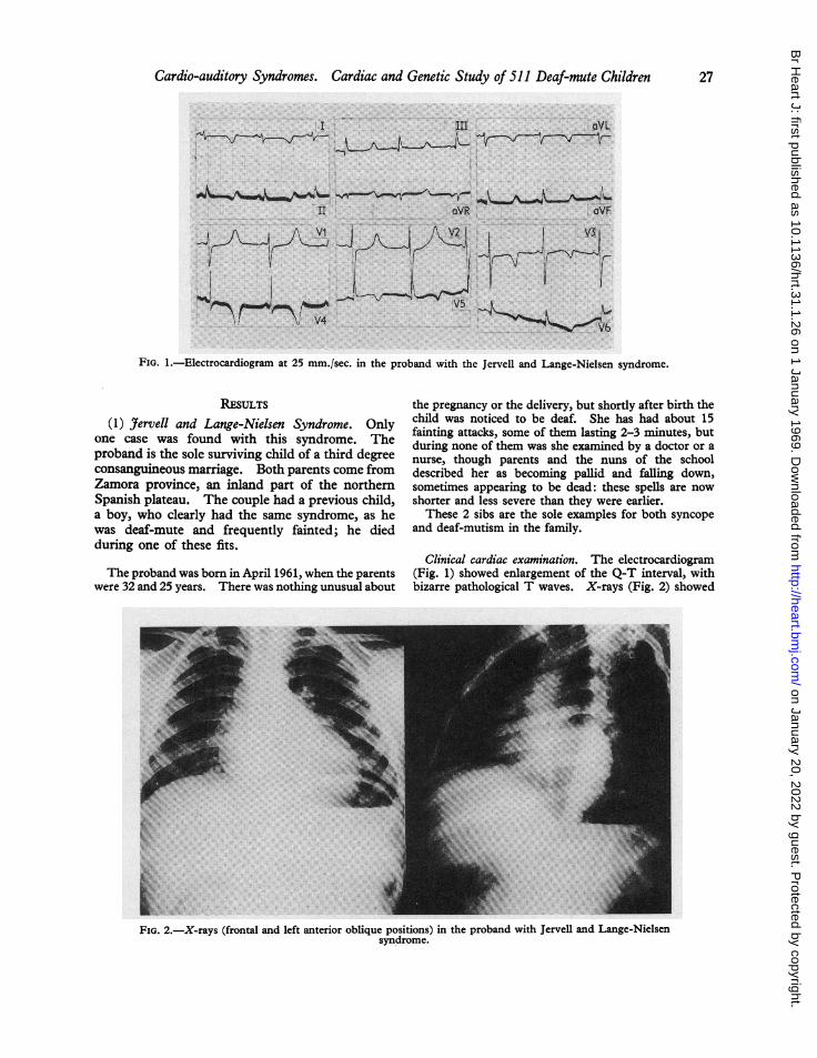
FIG. 1.-Electrocardiogram at 25 mm./sec. in the proband with the Jervell and Lange-Nielsen syndrome.
RESULTS
(1) J7ervell and Lange-Nielsen Syndrome. Onlyone case was found with this syndrome. Theproband is the sole surviving child of a third degreeconsanguineous marriage. Both parents come fromZamora province, an inland part of the northernSpanish plateau. The couple had a previous child,a boy, who clearly had the same syndrome, as hewas deaf-mute and frequently fainted; he diedduring one of these fits.
The proband was born in April 1961, when the parentswere 32 and 25 years. There was nothing unusual about
the pregnancy or the delivery, but shortly after birth thechild was noticed to be deaf. She has had about 15fainting attacks, some of them lasting 2-3 minutes, butduring none of them was she examined by a doctor or anurse, though parents and the nuns of the schooldescribed her as becoming pallid and falling down,sometimes appearing to be dead: these spells are nowshorter and less severe than they were earlier.These 2 sibs are the sole examples for both syncope
and deaf-mutism in the family.
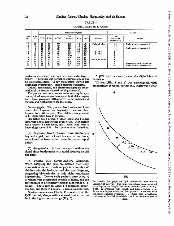
Clinical cardiac examination. The electrocardiogram(Fig. 1) showed enlargement of the Q-T interval, withbizarre pathological T waves. X-rays (Fig. 2) showed
FIG. 2.-X-rays (frontal and left anterior oblique positions) in the proband with Jervell and Lange-Nielsensyndrome.
27
on January 20, 2022 by guest. Protected by copyright.
http://heart.bmj.com
/B
r Heart J: first published as 10.1136/hrt.31.1.26 on 1 January 1969. D
ownloaded from
Sanchez Cascos, Sanchez-Harguindey, and de Rabago
TABLE ICARDIAC DATA IN 12 CASES
Electrocardiogram X-rays
Case Age VI LeftNo. (yr.) Q-T R-R AQRS pattern R/S V6 Others ventricular Others
hypertrophy
1 10 32 60 550 RS 1-2 qR Nodal rhythm ++ Right ventric. hypertrophy2 6 34 64 400 RS 1-2 qR ++3 5 32 64 300 Rs 1 3 qRs + + Right ventric. hypertrophy4 5 34 70 450 rS 0-6 qR ++5 8 36 70 700 RS 0*8 qRs T-V1-3 +6 10 32 55 850 Rs 2 qR T-V1-3 +7 12 32 60 00 RSr' 1-7 Rs +8 10 34 68 900 RS 0*8 qR +9 12 30 50 00 rS 0 5 qR Flat T in V5-6 +10 22 33 76 600 RS 0-8 qR11 10 29 46 200 RS 0 9 qRs _ Ascending aortic dilatation12 9 32 60 500 rS 0 7 qRs + Right ventric. hypertrophy
cardiomegaly, mainly due to a left ventricular hyper-trophy. The father was normal on examination, as washis electrocardiogram. X-ray examination showed leftventricular hypertrophy. Blood pressure was normal.
Clinical, radiological, and electrocardiographic exam-ination of the mother showed nothing abnormal.The proband and both parents had normal erythrocyte
counts, blood ions, transaminases, and lactic dehydrogen-ase. Blood group was 0 Rh positive for the proband andmother, and A Rh positive for the father.
Dermatoglyphs. The proband has 8 arches and 2 lowcount ulnar loops on her finger-tips; there are ulnarloops on both little fingers. The total finger-ridge countis 4. Both palms have t° triradius.Her father has 4 arches, 5 ulnar loops, and 1 radial
loop, with a total finger ridge count of 25. Her motherhas 3 arches, 6 ulnar loops, and 1 radial loop, with a
finger ridge count of 51. Both parents have t° triradius.
(2) Congenital Heart Disease. Two children, aboy and a girl, both selected because of murmurs,were found to have ostium secundum atrial septaldefect.
(3) Arrhythmias. A boy presented with occa-sional sinus bradycardia with nodal escapes; he didnot faint.
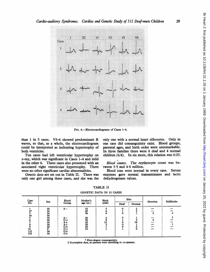
(4) Possible New Cardio-auditory Syndrome.When analysing the data, we noticed that x-rayexamination showed cardiomegaly in a number ofchildren who also had abnormal electrocardiograms,suggesting biventricular or only right ventricularhypertrophy. Twelve such patients were listed, 4of whom were ascertained because of faints, and therest because of a tendency towards high range Q-Tvalues. The x-rays in Cases 1-4 indicated abnor-malities, and those of Cases 5-12 were also abnormal.
Cardiac examination (Table I) revealed that theQ-T interval, always within normal limits, used tobe in the higher normal range (Fig. 3).
AQRS: half the cases presented a slight left axisdeviation.
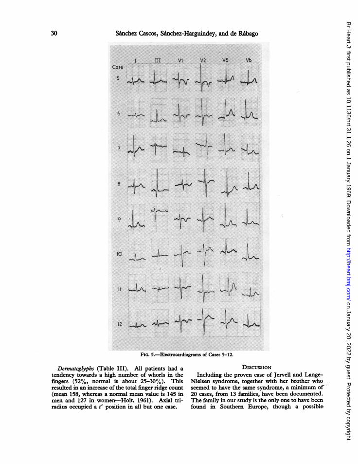
Vi lead (Fig. 4 and 5) was pathological, withpredominant R waves, so that R/S index was higher
I a
RR
FIG. 3.-In this graph the Q-T interval has been plottedagainst R-R interval. The range values have been calculatedaccording to the Heglin-Holzmann formula (0 39 VR-R±0 04). i =Proband with Jervell and Lange-Nielsen syn-drome (the higher value) and her parents. =Cases withthe cardio-auditory syndrome. x =Case with arrhythmia,both cases with atrial septal defect, and the brother of one of
these.
28
on January 20, 2022 by guest. Protected by copyright.
http://heart.bmj.com
/B
r Heart J: first published as 10.1136/hrt.31.1.26 on 1 January 1969. D
ownloaded from
Cardio-auditory Syndromes. Cardiac and Genetic Study of 511 Deaf-mute Children
I III VI V2 VS VbCase
3 _ 5, j4 .....ji_
¶jV\%4L%~~~~..4vJVA4L,,~I., 4
3~~~~~~~~~~~~~~~~~~~~~~~~~~~~~~~~~~~~~~~~~~~~~~~~~~~~~~~~~~~~~~~~~~~~~~~
44|$o X 4
FIG. 4.-Electrocardiograms of Cases 1-4.
than 1 in 5 cases. V5-6 showed predominant R
waves, so that, as a whole, the electrocardiogramcould be interpreted as indicating hypertrophy ofboth ventricles.Ten cases had left ventricular hypertrophy on
x-ray, which was significant in Cases 1-4 and mildin the other 6. Three cases also presented with an
associated right ventricular hypertrophy. Therewere no other significant cardiac abnormalities.
Genetic data are set out in Table II. There was
only one girl among these cases, and she was the
only one with a normal heart silhouette. Only inone case did consanguinity exist. Blood groups,parental ages, and birth order were unremarkable.In three families there were 6 deaf and 4 normalchildren (6/4). In six more, this relation was 6/23.
Blood counts. The erythrocyte count was be-tween 3X5 and 4X6 million.
Blood ions were normal in every case. Serumenzymes gave normal transaminases and lacticdehydrogenase values.
TABLE II
GENETIC DATA IN 12 CASES
Case Blood Mother's Birth SibsNo. Sex Group age (yr.) order Abortion Stillbirths
1 M A- 32 3 0 2 = -2* m 45 4 1 2 - -
3 M 36 8 0 1 2 4
5 M A- 26 4 0 1 - 26 M 0+ 36 8 1 2 4 -
7 M A+ 42 15 0 14 --8 M A+ 29 2 0 1 - 19 M 0+ 29 1 0 2 - -
10 F O+ 23 1 1 0 - -
lit M 0+12t M 0+
* First-degree consanguinity.t Incomplete data, as parents were unwilling to co-operate.
29
on January 20, 2022 by guest. Protected by copyright.
http://heart.bmj.com
/B
r Heart J: first published as 10.1136/hrt.31.1.26 on 1 January 1969. D
ownloaded from
Sanchez Cascos, Sanchez-Harguindey, and de Rabago
I III VI 12 Vs Y6CaSe
5 4 J
10-2l;,
12 _,J A4-- l 0FIG. 5.-Electrocardiograms of Cases 5-12.
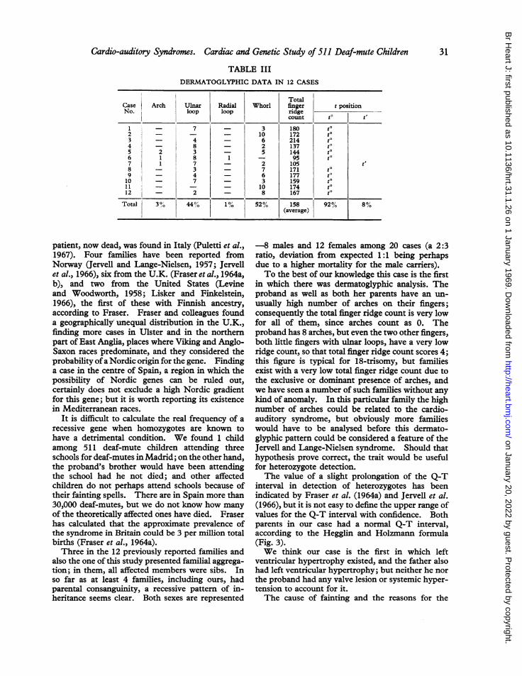
Dermnatoglyphs (Table III). All patients had a
tendency towards a high number of whorls in thefingers (52%, normal is about 25-30%). Thisresulted in an increase of the total finger ridge count(mean 158, whereas a normal mean value is 145 inmen and 127 in women-Holt, 1961). Axial tri-radius occupied a t0 position in all but one case.
DISCUSSIONIncluding the proven case of Jervell and Lange-
Nielsen syndrome, together with her brother whoseemed to have the same syndrome, a minimum of -
20 cases, from 13 families, have been documented.The family in our study is the only one to have beenfound in Southern Europe, though a possible
30
-4 '- .0-i
91
on January 20, 2022 by guest. Protected by copyright.
http://heart.bmj.com
/B
r Heart J: first published as 10.1136/hrt.31.1.26 on 1 January 1969. D
ownloaded from
Cardio-auditory Syndromes. Cardiac and Genetic Study of 511 Deaf-mute Children
TABLE IIIDERMATOGLYPHIC DATA IN 12 CASES
I ~~~~~~~~TotalCase Arch Ulnar Radial Whorl finger t positionNo. loop loop nrige
0
count t t'
1 _ 7 - 3 180 t '2 j - - - 10 172 t
3 4 - 6 214 to4 - 8 - 2 137 to5 2 3 - 5 144 t
6 1 8 1 - 95 t
7 1 7 - 2 105 t.8 - 3 - 7 171 t
9 1- 4 - 6 177 t
10 - ~~ ~~ ~~~~~~~~~~~7- 3 15911 _ - - 10 174 to12 - 2 - 8 167 to
Total 3% 44%O' 1% 52% 158 92% 8%(average)
patient, now dead, was found in Italy (Puletti et al.,1967). Four families have been reported fromNorway (Jervell and Lange-Nielsen, 1957; Jervellet al., 1966), six from the U.K. (Fraser et al., 1964a,b), and two from the United States (Levineand Woodworth, 1958; Lisker and Finkelstein,1966), the first of these with Finnish ancestry,according to Fraser. Fraser and colleagues founda geographically unequal distribution in the U.K.,finding more cases in Ulster and in the northernpart of East Anglia, places where Viking and Anglo-Saxon races predominate, and they considered theprobability ofa Nordic origin for the gene. Findinga case in the centre of Spain, a region in which thepossibility of Nordic genes can be ruled out,certainly does not exclude a high Nordic gradientfor this gene; but it is worth reporting its existencein Mediterranean races.
It is difficult to calculate the real frequency of arecessive gene when homozygotes are known tohave a detrimental condition. We found 1 childamong 511 deaf-mute children attending threeschools for deaf-mutes in Madrid; on the other hand,the proband's brother would have been attendingthe school had he not died; and other affectedchildren do not perhaps attend schools because oftheir fainting spells. There are in Spain more than30,000 deaf-mutes, but we do not know how manyof the theoretically affected ones have died. Fraserhas calculated that the approximate prevalence ofthe syndrome in Britain could be 3 per million totalbirths (Fraser et al., 1964a).Three in the 12 previously reported families and
also the one of this study presented familial aggrega-tion; in them, all affected members were sibs. Inso far as at least 4 families, including ours, hadparental consanguinity, a recessive pattern of in-heritance seems clear. Both sexes are represented
-8 males and 12 females among 20 cases (a 2:3ratio, deviation from expected 1:1 being perhapsdue to a higher mortality for the male carriers).To the best of our knowledge this case is the first
in which there was dermatoglyphic analysis. Theproband as well as both her parents have an un-
usually high number of arches on their fingers;consequently the total finger ridge count is very lowfor all of them, since arches count as 0. Theproband has 8 arches, but even the two other fingers,both little fingers with ulnar loops, have a very lowridge count, so that total finger ridge count scores 4;this figure is typical for 18-trisomy, but familiesexist with a very low total finger ridge count due tothe exclusive or dominant presence of arches, andwe have seen a number of such families without anykind of anomaly. In this particular family the highnumber of arches could be related to the cardio-auditory syndrome, but obviously more familieswould have to be analysed before this dermato-glyphic pattern could be considered a feature of theJervell and Lange-Nielsen syndrome. Should thathypothesis prove correct, the trait would be usefulfor heterozygote detection.The value of a slight prolongation of the Q-T
interval in detection of heterozygotes has beenindicated by Fraser et al. (1964a) and Jervell et al.(1966), but it is not easy to define the upper range ofvalues for the Q-T interval with confidence. Bothparents in our case had a normal Q-T interval,according to the Hegglin and Holzmann formula(Fig. 3).We think our case is the first in which left
ventricular hypertrophy existed, and the father alsohad left ventricular hypertrophy; but neither he northe proband had any valve lesion or systemic hyper-tension to account for it.The cause of fainting and the reasons for the
31
on January 20, 2022 by guest. Protected by copyright.
http://heart.bmj.com
/B
r Heart J: first published as 10.1136/hrt.31.1.26 on 1 January 1969. D
ownloaded from

Sa'nchez Cascos, Sanchez-Harguindey, and de Rabago
prolongation of the Q-T interval have been ex-haustively discussed previously: no definite expla-nation has been found, but a metabolic cause seemsto be ruled out. We have nothing to add to thisas our family's data were normal. Fraser et al.(1964a) were able to find some histological lesion inthe Purkinje system that could account for Q-Tprolongation and even for an arrhythmic nature ofsyncopes and sudden death; but whether thisfinding is primary or whether it depends on anoxiaduring spells is difficult to decide.The presence of left ventricular hypertrophy in
the proband as well as in her father suggested to usthat the Jervell and Lange-Nielsen syndrome couldbe a special type of cardiomyopathy. Suddendeath and faints caused by fright, anger, or fearpossibly relate this syndrome to some adrenergicmechanism; an excess of cardiac catecholamines isnowadays thought to account for outflow obstructionand cardiomegaly in cardiomyopathy (Goodwin,1967).
In this connexion, it is interesting to consider thefamily reported first by Lewis et al. (1958) andKoroxenidis et al. (1966). Association of congenitalheart disease with deaf-mutism in its members couldbe coincidental, but the possibility exists that itrepresents a second (Lewis) cardio-auditory syn-drome, related again to cardiomyopathies, since oneaffected member presented subaortic obstructionplus infundibular stenosis as well as atrial septaldefect. Our series presented two cases of atrialseptal defect; whether they represent an example ofthis second (Lewis) cardio-auditory syndrome isdifficult to assert, as the presence of two cases ofatrial septal defect among 500 random childrencould be coincidental.One case in our series presented an arrhythmia
without fainting. Its presence is reputed also tobe coincidental.
Finally, in the analysis of the cardiac data fromour deaf-mute series, several cases emerge that couldrepresent a new (3rd) cardio-auditory syndrome.It seems to be defined by a radiological left ventri-cular hypertrophy (on some occasions it wasassociated with a right ventricular hypertrophy),plus an electrocardiogram with a tendency to a highR in Vl (R/S quotient in that lead is always highfor a child's age) and also a high R in V5-6, togetherwith a tendency in some cases towards left axisdeviation (these electrocardiograms being typical ofDuchenne's muscular dystrophy-Perloff et al.,1967); the Q-T interval is within the range ofnormalvalues, but some cases show a tendency towardshigher than normal values.
Four cases clearly belonged to this syndrome and8 more probably belonged. The cardiomegaly of
this syndrome seems once more to represent a linkbetween cardio-auditory syndromes and cardio-myopathies. Dermatoglyphic analysis of these 12cases seems to present a pattern, with a highincidence of whorls (52%, while the incidence incontrols is about 25-30%), and consequently a hightotal finger ridge count. There was only one girlin these 12 children, and she was not a definite case;the possibility of a sex-linked gene therefore exists.
SUMMARYThe authors carried out a cardio-genetic study of
511 deaf-mute children. One girl had the Jervelland Lange-Nielsen syndrome. Her parents wereconsanguineous and an elder sib, a boy, who haddied previously, also had the syndrome. Thefamily was interesting from a dermatoglyphic pointof view: the proband had 8 arches and 2 ulnar loops,with total finger ridge counts scoring 4; both parentsalso had a high number of arches on their fingers.Twelve more children were found to present some
common features that could define a new cardio-auditory syndrome: all but two had radiologicalevidence of left ventricular hypertrophy; most ofthem had a high R/S quotient in Vl, with a sug-gestion of biventricular hypertrophy; in some casesthere was a tendency towards left axis deviation.In most cases, dermatoglyphs showed an increasein the total finger ridge count due to the high numberof whorls.The relation between these cardio-auditory
syndromes and cardiomyopathies is discussed.
REFERENCESBarlow, J. B., Bosman, C. K., and Cochrane, J. W. C. (1964).
Congenital cardiac arrhythmia. Lancet, 2, 531.Fraser, G. R., Froggatt, P., and James, T. N. (1964a).
Congenital deafness associated with electrocardiographicabnormalities, fainting attacks and sudden death. Arecessive syndrome. Quart. J. Med., 33, 361.- -, and Murphy, T. (1964b). Genetical aspects of the
cardio-auditory syndrome of Jervell and Lange-Nielsen(congenital deafness and electrocardiographic abnormal-ities). Ann. hum. Genet., 28, 133.
Friedmann, I., Fraser, G. R., and Froggatt, P. (1966).Pathology of the ear in the cardioauditory syndrome ofJervell and Lange-Nielsen (recessive deafness withelectrocardiographic abnormalities). J7. Laryng., 80,451.
Goodwin, J. F. (1967). Disorders of the outflow tract of theleft ventricle. Brit. med. J., 2, 461.
Holt, S. B. (1961). Quantitative genetics of finger-printpatterns. Brit. med. Bull., 17, 247.
James, T. N. (1967). Congenital deafness and cardiacarrhythmias. Amer. J3. Cardiol., 19, 627.
Jervell, A., and Lange-Nielsen, F. (1957). Congenital deaf-mutism, functional heart disease with prolongation ofthe Q-T interval, and sudden death. Amer. Heart J.,54, 59.
, Thingstad, R., and Endsjo, T. (1966). The surdo-cardiac syndrome. Three new cases of congenital
32
on January 20, 2022 by guest. Protected by copyright.
http://heart.bmj.com
/B
r Heart J: first published as 10.1136/hrt.31.1.26 on 1 January 1969. D
ownloaded from
Cardio-auditory Syndromes. Cardiac and Genetic Study of 511 Deaf-mute Children
deafness with syncopal attacks and Q-T prolongation inthe electrocardiogram. Amer. Heart J., 72, 582.
Koroxenidis, G. T., Webb, N. C., Moschos, C. B., and Lehan,P. H. (1966). Congenital heart disease, deaf-mutismand associated somatic malformations occurring inseveral members of one family. Amer. J. Med., 40, 149.
Levine, S. A., and Woodworth, C. R. (1958). Congenitaldeaf-mutism, prolonged Q-T interval, syncopal attacksand sudden death. New Engl. J3. Med., 259, 412.
Lewis, S. M., Sonnenblick, B. P., Gilbert, L., and Biber, D.(1958). Familial pulmonary stenosis and deaf-mutism:clinical and genetic considerations. Amer. Heart J.,
55, 458.Lisker, S. A., and Finkelstein, D. (1966). The cardio-
auditory syndrome of Jervell and Lange-Nielson:Report of an additional case with radio-electrocardio-
graphic monitoring during exercise. Amer. J3. med. Sci.,252, 458.
Perloff, J. K., Roberts, W. C., de Leon, A. C., and O'Doherty,D. (1967). The distinctive electrocardiogram ofDuchenne's progressive muscular dystrophy. Amer.3. Med., 42, 179.
Puletti, M., Jacobellis, G. F., and Borghi, F. (1967). Lasindrome di Jervell e Lange-Nielsen. Studio cardio-logico di 211 sordomuti. Cuore e Circol., 51, 251.
Romano, C., Gemme, G., and Pongiglione, R. (1963).Aritmie cardiache rare dell' etu pediatrica. Accessisincopali per fibrillazione ventricolare parossistica.(Presentazione del primo caso della letteratura pediatricaitaliana.) Clin. pediat. (Bologna), 45, 656.
Ward, 0. C. (1964). A new familial cardiac syndrome inchildren. Y. Irish med. Ass., 54, 103.
3
33
on January 20, 2022 by guest. Protected by copyright.
http://heart.bmj.com
/B
r Heart J: first published as 10.1136/hrt.31.1.26 on 1 January 1969. D
ownloaded from