Embed Size (px)
Citation preview
Cardiovascular Development
• The first three weeks• By the beginning of the third week, blood vessel
formation begins in the tissue surrounding the yolk sac.
• These primitive vessels are at first not connected to each other, but do start to make primitive blood cells.
• In the third week the heart and large vessels form in the cardiogenic area from mesoderm.
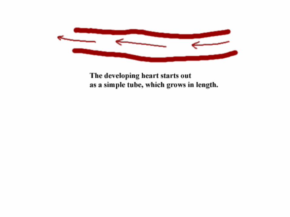
• The first step in formation of the cardiovascular system is the development of two simple tubes lying on either side of the midline in the mesoderm.
• These tubes are connected to: • umbilical arteries connected to vessels in
the chorion (primitive placenta): • vitelline arteries to the yolk sac; • dorsal intersegmental arteries to the body
of the embryo. • Umbilical veins, vitelline veins and cardinal
veins return the blood to the circulation. • At 21 days the two longitudinal tubes (the
dorsal aortae) fuse at their cranial ends to form the primitive heart.
• Heart beats can be detected by ultrasound by the fifth week.
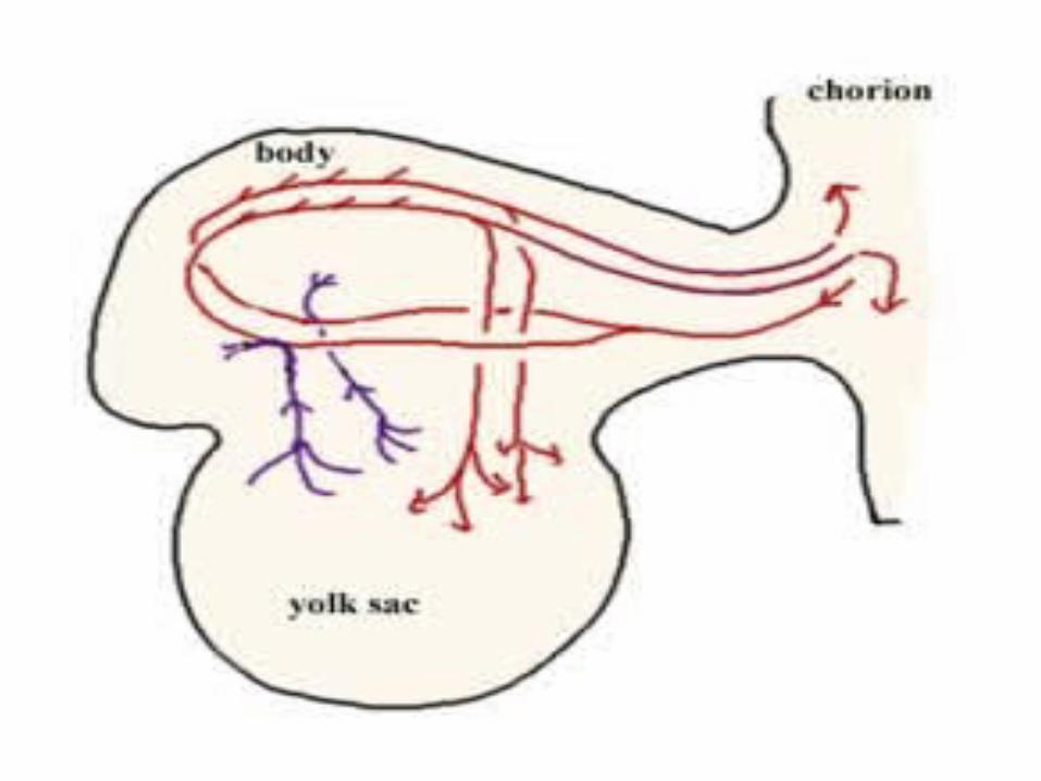
• At 20 days the circulation is primitive but the three areas to be connected are present –
• the chorion (early placenta),
• the yolk sac (vitelline arteries) • and the body (intersegmental
arteries).
• Development of the heart
• Fusion of the primitive heart tubes into a single atrioventricular tube is the initial event in development of the heart.
• Mesoderm around the endothelial tube differentiates into myocardium.
• As the head fold caries the developing heart caudally, ventral to the foregut and caudal to the oropharyngeal membrane, the heart tube develops dilatations forming the truncus arteriosus, bulbus cordis, ventricle, atrium, and sinus venosus.
• The truncus arteriosus connects the heart to the future arterial system
• and the sinus venosus receives blood from the umbilical, vitelline and common cardinal veins (i.e. from the chorion or placenta, from the yolk sac, and from the embryo).
• Growth of the heart tube is uneven, but it is fixed to adjacent tissue at its two ends - to the pharyngeal arches cranially and to the primitive diaphragm caudally.
• Accelerated growth of the bulbus cordis and ventricle causes the heart tube to bend and invaginate the pericardium.
• The next steps involve partitioning of the tube into atria and ventricles.
• Partitioning of the heart• During the fourth week the dorsal and ventral
walls bulge into the lumen of the tube to form an atrial and a ventricular chamber, connected by left and right atrioventricular canals.
• The primitive atrium is divided into right and left atria by the growth of septa.
• The septum primum grows down towards the endocardial cushions.
• As the septum grows down the space below it is termed the foramen primum.
• Eventually the septum primum reaches and fuses with the endocardial cushions closing the foramen primum.
• As the septum primum ends its descent an aperture appears in its substance, the foramen secundum.
• At this stage then the septum primum separates the atrial chamber into right and left, although there is a hole (the foramen secundum) in the septum primum, allowing communication between the atria.
• During the fifth week a second septum, the septum secundum grows down on the right side of the septum primum, to eventually overlap the hole in the septum primum.
• The septum secundum is crescent shaped and does not entirely reach the floor of the right atrium.
• The oval shaped gap,allowing blood topass through the interatrial septum, is the foramen ovale.
• While the atrium is undergoing partitioning the same process is occurring in the ventricle.
• A muscular ridge grows up from the floor of the ventricle towards the atrioventricular cushions.
• As the septum grows upwards itn leaves a communication between the two ventricles, the interventricular foramen.
• By the seventyh week the interventricular foramen is closed by the fusion of tissues from the interventricular septum and the endocardial cushions.
• During the fifth week there is partitioning of the truncus arteriosus and bulbus cordis.
• The spiral septum developing in this outlet from the heart eventually fused with the endocardial cushions so that the output from the right ventricle enters the pulmonary trunk, and the output from the left ventricle enters the aorta.
• The sinus venosus of the primitive heart opened into the common atrium.
• As development proceeds the opening of the sinus venosus moves to the right atrium.
• As the sinus venosus moves to the right the venous drainage from the rest of the body, the head, the vitelline and umbilical veins also move to the right.
• A small part of the sinus venosus still lies behind the left side of the heart, to become the coronary sinus.
• The fetal circulation
• In the fetus the four-chambered heart circulates blood through the fetal body, through the placenta and through the vessels supplying the gut (previously the vitelline vessels)
• The blood arriving at the right side of the heart through the superior and inferior venae cavae has come from the fetus (body and head), from the digestive system and liver, and from the umbilical vein.
• The blood in the umbilical vein is highly oxygenated.
• The blood mixes in the right atrium and is directed towards the interatrial septum where it passes through the foramen ovale into the left atrium.
• From the left atrium it passes into the left ventricle and so to the systemic circulation through the aorta.
• Any blood which passes into the right ventricle leaves that chamber though the pulmonary trunk to pass to the aorta through the ductus arteriosus, but also to a small extent to the lungs.
• The aorta thus distributes blood to the head, the body including the gut, and to the placenta through the paired umbilical arteries.
• At the placenta the blood is oxygenated and collected in the single umbilical vein.
• The umbilical vein travels to the liver where a connection, the ductus venosus, shunts most of the blood into the inferior vena cava.
• This circulation ensures that the head and upper limbs receive oxygenated blood, that the body receives fairly well oxygenated blood with some addition of venous blood from the head and lungs.
• Since at this stage the lungs are not inflated, the resistance in the pulmonary circulation is high and the blood is diverted back into the systemic circulation through the ductus arteriosus.
• The ductus venosus diverts blood from the umbilical vein bypassing the liver, directly into the IVC.
• Changes at birth
• At birth the lungs will inflate and the placenta will become disconnected from the source of oxygenation.
• With the first breath the resistance in the pulmonary circulation falls as the lungs inflate.
• Blood in the right ventricle is able to pass through the pulmonary circulation where oxygenation occurs.
• Oxygenated blood from the lungs returns to the left atrium.
• Cutting and tying the umbilical cord closes both the umbilical arteries and the umbilical vein.
• Other significant changes in the circulation occur over the next hours and days.
• The ductus arteriosus starts to constrict and is usually permanently closed by three months after birth.
• As pressure in the left atrium rises, the septum primum is forced against the septum secundum, closing the foramen ovale.
• Eventually the two septa fuse to permanently separate the two atria.
• Absence of flow in the umbilical vein causes it to become ligamentous - the ligamentum teres, although a narrow channel remains.
• The ductus venosus becomes the ligamentum venosum and the blood from the portal vein must pass through the liver before reaching the IVC.