Embed Size (px)
Citation preview

Special Topics in Cardiovascular Pharmacotherapy 0733-8651/87 $0.00 + .20
Cardiovascular Drugs in Pregnancy
Josef Widerhorn, MD, * J. Nathan Rubin, MD, * William H. Frishman, MD, t and Uri Elkayam, MD:j:
Pregnant women often require medical therapy for treatment of various coexisting cardiovascular conditions. The effects of the cardiovascular drugs in pregnant women are discussed in this article. .
PHARMACOLOGIC CONSIDERATIONS IN PREGNANCY
Complex physiologic changes occurring during pregnancy in the cardiovascular, pulmonary, renal, gastrointestinal, and endocrine systems may affectthe pharmacokinetics of drugs, with severalconsequences on the fetomaternal unit. 107, 150. 173. 198
Drug absorption may be influenced by thedecreased motility of the gastrointestinal tract thatoccurs during pregnancy. Reduced gastric motilitymay cause stagnation of drugs in the stomach.170Diminished acid secretion, combined with increased production of alkaline mucus, can affectgastric pH and, consequently, the degree of ionization and solubility of drugs. 150. 173 The prolongation of transit timethrough the small bowel may lead to increasedmetabolism of drugs in the gut wa1l41 or, con-versely, .may allow a more complete absorptionwith increased bioavailability. 53, 107 In addition, the administration of opiate analgesics or antacids, frequently used during labor, also may delay ordecrease drug absorption. 162. 193
During pregnancy, the distribution of
drugs is modified by increased plasma volume, total body water, and total body fat.l07, 150. 198 Serum concentration of albumin, the principal drug binding protein, decreases progressively throughout pregnancy. 46 This change, along with altered binding to alacid glycoprotein,78. 254 increase in fatty acids and lipids, and hormonal changes, may lead to an increase of the unbound drug frac-tion.107 Furthermore, the difference in fetal and 'maternal plasma binding capacity produces a complex situation that also has to be considered in the interpretation of therapeutic and toxIC drug concentrations}8. 107. 150, 198. 254
The metabolism of many drugs is altered during pregnancy; however, these changes are not necessarily clinically significant. 107 The liver h;,ls an important role in drug metabolism. Hepatic clearance is dependent on the binding affinity of drugs, hepatic blood flow, and hepatic enzymatic systems. Sex hormones may have opposite effects on the hepatic microsomal oxidase system.42. 44. 59 For example, estrogens inhibit the microsomal oxidase system for ethylmorphine and hexabarbitone44; on the other hand, progesterone enhances hepatic enzymatic acti vi ty. 42. 59
The fetoplacental unit plays an active role in metabolism of various drugs. The placenta contains many enzymatic systems that transform several drugs into toxic or nontoxic products.
*Clinical Instructor. of Medicine, University of Southern California School of Medicine; and Section of Cardiology, Department of Medicine, Los Angeles County-University of Southern California Medical Center, Los Angeles, California
tProfessor of Medicine, Albert Einstein College of Medicine; and Director of Medicine, Hospital of the Albert Einstein College of Medicine, Bronx, New York *Associate Professor of Medicine, University of Southern California School of Medicine; and Director of In-Patient Cardiology, Section of Cardiology, Department of Medicine, Los Angeles County-University of Southern California Medical Center, Los Angeles, California
Cardiology Clinics-Vol. 5, No.4, November 1987 651

652 Josef Widerhom et aI.
The fetus also participates in drug metabolism after6 to 8 weeks of gestation.l75 Placental transfer ofdrugs depends on maternal and fetal drugconcentratipq;,gegree of protein binding; placentalblood flow; anatomy, and metabolism; and acid-base equilibrium. 117. 150. 154. 198 The acidic pH of amniotic fluid, for example, can function as an "ion trap"for weak basic drugs. 150 Other important parameters are molecular weight of the drugs,degree of ionization, and lipid solubility. 206
The excretion of drugs that primarily are clearedby the kidney may be enhanced during pregnancy.Both renal blood flow and glomerular filtration rate are increased markedly. 45. 52.215 Posturalchanges that occur late in pregnancy also may influence hemodynamics and drug clearance. 150
A modem concept of teratogenesis has to embrace not only the anatomic malformations notedat birth, but also biochemical, physiologic, and behavioral modifications, which are more subtleand may manifest first during childhood or adult life. 107 Teratogenic effects depend on the chemical nature of drugs, duration of exposure, dosage, genetic susceptibility, andgestational age. During blastogenesis (first 14days), severe insults may result in abortion;however, the blastocyst is relatively resistant to teratogenic agents and there are no known viablemalformations generated during this period.I07
The most vulnerable period of gestation is that ofembryogenesis (2 to 8 weeks). Various organsystems are formed during this period and injuriesto the embryo will result in morphogenetic alterations.
During fetogenesis, the effect of drugs may besimilar to that occurring during extrauterine life;however, the systems still in differentiation (that is,the central nervous system) might be damaged. Other effects can be nonspecific, manifested bygrowth retardation or system dysfunction thatbecome evident after birth.
As a general principle, all drugs should beavoided as much as possible during pregnancy. If atherapeutic decision has to be .made, the risk-benefit ratio should be evaluated carefully. Inchoosing drugs, toxicity, metabolism, andgestational age are of crucial importance inavoiding untoward effects. If a condition requires drugs potentially harmful to the fetus, the bestsolution is to avoid pregnancy for the duration oftreatment. As previously described, thepharmacokinetics of drugs is modified inpregnancy; because of a multitude of factors, thesechanges cannot always be predicted and,
therefore, close monitoring of therapy, both clinically and by laboratory methods, is necessary.
Another source of concern is the transfer of drugs into breast milk and, consequently, to neonates. Again, this is dependent on the physicochemical characteristics of the compound. 206 Generally, only 1 to 2 per cent of maternal dose appears in milk. Except for some drugs that clearly are contraindicated, there is not enough evidence to allow or to prohibit breast feeding in mothers undergoing drug treatment.25O Most data regarding drug excretion in human milk are available from case reports. Because of the complexity of mechanisms involved in drug excretion into breast milk, various models and formulas used to estimate plasma milk ratios have limited clinical value. Close monitoring of the infant's ingested dose and plasma levels, as well as close observation for adverse effects or toxicity, th.erefore are necessary to assure safety.l50 .
CARDIAC GLYCOSIDES
Cardiac glycosides have been known to humanity since the ancient Egyptian and Roman era. Used for centuries for different purposes, digitalis emerged as one of the major drugs in the therapy of congestive heart failure, as well as for terminating paroxysmal supraventricular tachycrdia and controlling rapid ventricular rates in patients with atrial fibrillation and flutter. .
Among various digitalis preparations, digoxin is the most accepted and widely used. During pregnancy, the most common maternal and fetal indications for digoxin use are congestive heart failure and/or supraventricular tachyarrhythmias.6. 31. 68. 74. 81. 121. 130. 189. 196. 201. 211. 223 Only 60 to 80 per cent of an oral dose is absorbed, mostly in the proximal small intestine. As with other drugs, the presence of food or delayed gastric emptying that may occur in pregnancy may retard its absorption.107. 150. 170 Digoxin is only 20 to 25 per cent bound to proteins; the decreased albumin level seen during pregnancy, therefore, should not affect its serum concentration significantly. Peak level and maximal effect following oral administration are reached after 2 to 3 hours and 4 to 6 hours, respectively. The intravenous (IV) preparation has a rapid onset of action (within 5 to 20 minutes) and a maximal effect within 1.5 to 3 hours. The half life of digoxin is 36 to 40 hours. The volume of distribution (V D) is increased significantly dur

r Cardiovascular Drugs in Pregnancy 653
ing pregnancy.134 For rapid digitalization, the dosage is similar to that in the nonpregnant state-that is, 0.75 to 1.5 mg IV. Digoxin is excreted primarily by the kidneys. During pregnancy, there is a linear correlation between digoxin and creatinine clearance; the dosage, therefore, has to be adjusted when renal function is impaired. The maintenance dose has to be approximately 35 per cent of total body stores; it may need adjustments throughout pregnancy in order to obtain optimal therapeutic effect.
In 1972, Rogers and coworkersl96 reported that serum digoxin level at term was lower than 4 weeks post delivery in mothers treated with the same maintenance dose. These data could not be confirmed by other authors,6. 31, 130 however, who found digoxin concentration in the therapeutic range at the time of delivery. Luxford and colleagues130 demonstrated that despite increased digoxin and creatinine clearance and increased 24-hour urinary digoxin elimination, serum digoxin level was higher during the third trimester of pregnancy than during the postpartum period.
Several studies79. 104. 240 recently have dem-onstrated the existence of endogenous digoxinlike substances in pregnant women and neonates. These substances interfere with the radioimmunoassay for exogenous digoxin, leading to reading errors ranging from 0.1 J.Lg per L to more than 2 J.Lg per L. To complicate the situation further, the concentration of these digoxin-like substances changes with gestational age, making it extremely difficult to assses therapeutic levels.240 These data cast doubts on the utility of monitoring digoxin serum concen-tration during pregnancy and suggest the need to use electrocardiographic and clinical criteria to determine adequacy of digoxin dosing during gestation.
Treatment of fetal conditions is based on the ability of digoxin to cross the placenta. Although' this property was well documented by numerous authors, 6, 31. 68. 74. 81. 121. 130. 189, 196. 201. 211. 223 the magnitude of digoxin transfer to the fetus is somewhat conflicting. Rogers and coworkersl96 found similar concentrations of digoxin in both maternal and fetal blood at the time of delivery. Chan and colleagues31 found lower levels in fetal blood and postulated the. existence of a placental barrier or digoxin. Allonen and
. coworkers6 also noted lower digoxin concentration in fetuses but at a different gestational age (12 to 16 weeks). In light of the new reports on endogenous digoxin-like substances and their
effect on digoxin measurement, it is difficult to interpret these data.
In a study using radioisotopes, Saarikosky and associates201 demonstrated that placental transfer of digoxin occurs fairly rapidly. Five minutes following the injection of H3-digoxin in maternal blood, radioactivity was demonstrated in umbilical cord blood, and by 30 minutes, fetal and maternal blood concentrations of radioisotopes were approximately equal. They also demonstrated that the fetal heart binds digoxin much less avidly than the infant heart. Based on these reports, the fetomaternal digoxin concentration ratio probably ranges from 0.5 to 1.0.
During the past decade, digoxin has been employed with increasing frequency to treat fetal supraventricular tachycardia and congestive heart failure.68. 74. 81. 121. 189. 223 Digoxin may be adequate alone in treating fetal tachycardia.68.81 In refractory cases, it m~y be necessary . to add a seco)ld drug such as verapamiP21 or quinidine..223 As in the Jlonpregnant state, the addition of quinidine or verapamil may increase the digoxin level significantly. The same precaution regarding drug interactions therefore applies during pregnancy as in the nonpregnant state.
To date, few adverse effects have been observed in fetuses of mothers treated chronically with digoxin. Low birth weight infants occasionally have been born of mothers with cardiac conditions treated with digitalis. It has been postulated that digoxin may affect amino acid transport through the placenta, with consequent growth retardation.248 However, the duration of pregnancy and labor has been noted to be shorter in mothers on long term digoxin therapy.245 Therefore, it is conceivable that the low birth weight reported with digoxin treatment is secondary to prematurity rather than to intrauterine growth retardation.
Despite the above concerns, digoxin is con-sidered a safe drug to use in pregnancy and, to date, there are no reports of teratogenesis in humans. Caution is advised in digitalis administration, however, because overdose can be detrimental to the mother and may be lethal to the fetus. 211
Digoxin is excreted in the breast milk and the milk-plasma ratio ranged from 0.59 to 0.9.31. 124 The total amount of digoxin ingested daily by the infant has been estimated to be approximately 1/100 of the pediatric recommended dose of 12.5 J.Lg per kg per day. 124 No apparent clinical effects were demonstrated in newborns;
l II
"" 11:
654 Josef Widerhorn et ai.
digoxin therapy of the mother therefore should not affect breast feeding decisions.31. 124
ANTIARRHYTHMIC DRUGS .,.1 >,
Quinidine
Quinidine, a class Ia antiarrhythmic drug, is a dextrostereoisomer of quinine. Both alkaloids have similar properties and spectrum of action; quinidine, however, is more effective in sup-pressing both supraventricular and ventricular arrhythmias. It is used for cardioversion, as well as for prophylaxis against recurrences of paroxysmal supraventricular tachycardia, atrial flutter, atrial fibrillation, and re-entrant arrhythmia associated with Wolff-Parkinson-White syndrome. It frequently is employed in suppressing ventricular premature beats and ventricular tachycardia. .
Quinidine has a depressant effect .on the myocardium that is directly proportional to plasma concentration.77 It decreases myocardial automaticity, excitability, and conduction velocity and increases the fibrillation threshold. The diminished conduction velocity and the increased effective refractory period induced by quinidine are very useful in terminating reentry.58 Because of its vagolytic effects, the atrioventricular (A V) conduction may be enhanced in some patients. Quinidine also has (Xadrenergic blocking effects.
Approximately 70 to 80 per cent of an oral dose of quinidine is absorbed.37 Food and antacid may delay the absorption. In patients with congestive heart failure, the decreased volume of distribution (V D) may lead to higher drug levels despite diminished absorption.4o Maximum serum levels are attained within 60 to 90 minutes and the half-life is 6 to 8 hours. Eighty percent is bound to protein and the unbound fraction, therefore, increases during hypoalbuminemic conditions such as gestation.150 Quinidine is metabolized primarily in the liver and some of the metabolites are active. Ten to 20 percent is eliminated unchanged by the kidney251 and alkalinization of the urine may decrease urinary excretion.94 The therapeutic level is 2.5 to 5 !Jog per ml; levels greater than 7 to 8 !Jog per ml are associated with increased toxicity.
Unfortunately, quinidine has many adverse effects. Nausea, vomiting, and diarrhea can be disabling. Some patients may develop skin rashes, thrombocytopenia, hemolytic anemia, or fever. In 3 to 4 per cent of patients, QT
prolongation may lead to polymorphic ventricular tachycardia ("torsade de pointes"), which has a reported mortality of 10 per cent.48 Among interactions with other drugs, of particular im-portance is the potentiation of warfarin . and augmentation of digoxin concentration. In up to 30 per cent of patients, side effects are serious enough to discontinue therapy.
Quinidine has been used during pregnancy since 1930.144 Several recent reports have documented its transplacental transfer.35, 50, 223 Hill and coworkers80 reported a case of quinidine treatment throughout the pregnancy to prevent maternal ventricular tachycardia. During preg-nancy, maternal quinidine levels are approximately constant; that is, 5.1 and 7.8 !Jog per ml. At delivery, the level in the neonate was 2.3 !Jog per ml and in the amniotic fluid, 9.3 !Jog per ml. Five days postpartum the mother's level rose to 9 !Jog per ml. The authors hypothesized that the high levels found in the amniotic fluid may have b~en because of fetal voiding or a technical error in measurement of quinidine metabolites.
Spinnato and associates223 described three cases of fetal supraventricular tachycardia treated with digoxin and quinidine. In all three cases, the addition of quinidine to digoxin was successful in controlling fetal arrhythmia and, in two of these fetuses, ascites as well. At delivery, quinidine maternal levels were 0.7 to 2.1 !Jog per ml; cord blood levels, 0.5 to 1.6 !Jog per ml; and amniotic fluid level, 0.9 !Jog per ml. The low levels may be explained by the fact that quinidine was discontinued 24 hours prior to delivery. No side effects were noted; fetal development was normal and the labor was uneventful.
Quinidine has a minimal oxytocic effect144. 231 that manifests mainly after the onset of spontaneous uterine contraction.231 Fetal thrombocy topenia also was associated with quinidine treat-ment.135 Toxic doses may cause premature labor, 14 abortion, 141 or damage of the fetal eighth cranial nerve. 140
However, the large clinical experience with use of this drug in pregnancy has shown an extremely low incidence of side effects and no known teratogenic effects. Quinidine use appears to be safe for both maternal and fetal indications. Quinidine is secreted in the breast mild, and the milk-plasma ratio is 0.71. 8e
c .
Ij I I I
I, I
I tl
j I
Iqf I
I ,
Procainamide
Procainamide, synthesized from procaine by substituting an ester linkage with an amide

--;,Cardiovascular Drugs in Pregnancy
group, was introduced as an antiarrhythmic agent in 1951.133 The drug is very effective in abolishing premature ventricular contractions, ventricular tachycardia, and supraventricular tachycardia associated with Wolff-ParkinsonWhite syndrome,82 although it is less effective than quinidine for supniveritticular arrhythmia.
It has a depressant effect on the heart, similar to that of quinidine; it decreases automaticity, excitability, conduction velocity, and contractility. The effective refractory period is prolonged and the fibrillation threshold increased. Its anticholinergic activity is less than that of quinidine.
Procainamide can be given orally and paren-terally. Following oral administration, approxi-mately 75 to 95 per cent of the dose is absorbed. The absorption may be retarded with delayed gastric emptying, decreased intestinal motility, or intestinal pH changes.18 Only 10 .to 20 per cent is bound to plasma proteins. Peak levels are reached in 45 to 75 minutes after an oral capsule dose. Half-life is' about 2.5 to 5.0 hours in patients with no evidence of heart or kidney failure. Therapeutic levels are 4 to 8 J.Lg per ml. Approximately 40 to 70 per cent of a procainamide dose is eliminated unchanged by the kidneys; renal impairment, therefore, may decrease elimination markedly, with a consequent increase in serum levels. Ten to 34 percent of the drug is metabolized in the liver to N-acetylprocainamide (NAPA)64 at a rate that varies depending on acetylator status. NAPA has less antiarrhythmic potency and its efficacy is limited. The treatment with NAPA probably does not induce antinuclear antibody formation or lupus syndrome,62 however, as does procainamide. NAPA is eliminated predominantly by the kidneys; its therapeutic range is 9.4 to 19.5 J.Lg per ml. Congestive heart failure and renal impairment prolong both procainamide and NAPA half-lives.95 In order to avoid toxicity, both procainamide and NAPA levels have to be monitored closely.
Procainamide can be administered intravenously as 100 mg boluses (25 mg per minute) every 5 to 10 minutes until therapeutic effects are obtained or 1 g is given. Intravenous administration may produce hypotension, QT interval prolongation, and, serious arrhythmias.159 Nausea, vomiting, and diarrhea are noted more frequently with the oral route but are less pronounced than with quinidine. Central ner-vous system (CNS) toxicity may manifest as 'mental depression, hallucinations, and psychosis. Hypersensitivity reaction such as drug fever, agranulocytosis, and skin rashes may oc
655
cur and drug-induced lupus may develop in 20 to 40per cent of the patients. 159
Transplacental transfer of procainamide is well documented in two reports of treatment of fetal supraventricular tachycardia. 51. 66 Dumesic and coworkers51 reported successful control of fetal supraventricular tachycardia and heart failure when procainamide was administered to the mother in addition to digoxin. The fetal arrhythmia was refractory to previous treatment with digoxin a10ne or in combination with propranolol. After 4 weeks of treatment, the arrhythmia recurred and became more difficult to control, despite increased dose of digoxin and procainamide. Cesarean section was performed and, at delivery, although digoxin levels were equal in the maternal and neonate blood (0.8 J.Lg per ml), procainamide level was higher in maternal blood (15.6 J.Lg per ml versus 4.3 J.Lg per ml in fetus).
A similar therapeutic approach was reported by Given and coworkers.66 Again, fetal supra-ventricular t~chycardia resistant to the combination of digoxin and propranolol converted with digoxin and procainamide. In this case, fetal tachyarrhythmia also recurred. But the digoxin and procainamide levels at delivery were discordant with those found by Dumesic's team. Fetal levels were 30 per cent higher for procainamide and 50 per cent lower for digoxin. NAPA levels in the maternal and fetal blood were 3.0 J.Lg per ml and 3.7 J.Lg per ml, respectively. In both cases, it is unclear whether the therapeutic success was because of synergistic digoxin-procainamide action or procainamide effect alone.
At the present time, there are no data available on procainamide pharmacokinetics in the maternofetal unit. There is no evidence of teratogenic effects76. 142 and the drug appears to be safe in pregnancy. 36, 143, 144 Because of limited information and serious potential side effects, however, procainamide should be used only when drugs with a better safety record, such as quinidine, fail or cannot be used.
Translactal passage of procainamide was reported by Pittard and Glazier.lso They found a milk-plasma ratio of 4.3 :t 2.4 for procainamide and of 3.8 :t 1.8 for NAPA. Although the high ratio may suggest accumulation in the milk of both procainamide and NAPA, the amount ingested daily by the infant is not expected to produce significant plasma levels. ISO
,I.
J,II
[,
\11I /
II ,il
Disopyramide
Disopyramide, a relatively recent class Ia antiarrhythmic drug, was approved in the

656 Josef Widerhorn et al.
United States in 1977 for the treatment of ventricular arrhythmias. In suppression of pre-mature ventricular contractions, it appears equal to or better than quinidine or procainamide.75 During mYC9<;ardial infarction, it is use ful in reducing PVCs and the frequency of ventricular tachycardia; it is not clear if it can prevent ventricular fibrillation, however. 90 In Europe, it was found to be equal to quinidine in the treatment and prophylaxis of supraventricular arrhythmias.73, 128
Disopyramide has electrophysiologic properties similar to other class I antiarrhythmic agents; it decreases excitability, conduction velocity, automaticity, and contractility. It also prolongs the effective refractory period and action potential duration. It has marked negative inotropic effects and increases systemic vascular resistance.106. 157 These hemodynamic effects, while not .clinically evident in patients with normal ventricular function, may be deleterious in patients with limited cardiac function.182 Disopyramide has 10 per ceD:t of the anticholinergic effects of atropine.149 Approxi-mately 80 to 90 per cent of an oral dose is absorbed; peak plasma levels are reached by 1 to 2 hours, and the half-life is about 6 to 9 hours. Approximately 30 per cent was found to be bound to plasma proteins at a concentration of 3 f.Lg per ml; the binding varies directly with serum concentration, however, and may be as high as 90 per cent.159 Forty to 90 percent is eliminated in the urine unchanged; dosage has to be adjusted in renal failure. The remainder of the drug is metabolized in the liver via dealkylation.69 Therapeutic levels are 3 to 6 f.Lg per ml. The oral dose is 300 to 800 f.Lg per day; this dose has to be reduced in hepatic, cardiac, or renal insufficiency.
The majority of untoward effects of disopyr-amide are caused by its anticholinergic activity and include include dry mouth, constipation, blurred vision, and urinary retention. It may precipitate congestive heart failure in patients with ventricular dysfunction182 and, similar to quinidine, can induce QT prolongation, ventriculartachycardia, or "torsade de pointes. "139. 161
Animal studies have shown that disopyramide crosses the placenta. With very high doses, an increased incidence of low birth weight fetuses was observed, but there were no teratogenic effects.
There is very little information regarding disopyramide treatment in pregnant women. Shaxted and colleagues210 described a 26-week pregnant patient treated with 600 mg per day for symptomatic ventricular tachycardia. At de
livery, the maternal and fetal levels were 2.3 f.Lg per ml and 0.9 f.Lg per ml, respectively. No adverse effects were noted, and the delivery was normal. Leonard and coworkers,115 however, reported the treatment of refractory su-praventricular tachycardia in a pregnant woman with mitral valve prolapse in which the admin-istration of disopyramide initiated uterine con-tractions that resolved when the drug was dis-continued.
Disopyramide is secreted in the breast milk in concentrations similar to those in plasma; no adverse effects were noted in the infant. 11
Because of the limited information, the use of disopyramide in pregnancy should be reserved for patients refractory to quinidine.
Lidocaine
Lidocaine is a local anesthetic of the amide type that has been used as an antiarrhythmic agent since 1950. During the fifties, lidocaine was employed mainly In the cardiac catheterization laboratory; today it is one of the drugs most commonly used in intensive cardiac care units. Lidocaine is very effective in suppressing ventricular premature beats and ventricular tachyarrhythmias, particularly during acute myocardial infarction, cardiac surgery, and digitalis toxicity. 65 It is of little benefit for supraventricular arrhythmias. Lidocaine depresses automaticity in Purkinje fibers and increases the threshold for ventricular fibrillation.19 The action potential duration is decreased significantly as is, to a lesser extent, the effective refractory period (ERP) in both Purkinje fibers and ventricular muscle. The effect on the ERP of A V node is variable. Lidocaine effectively decreases conduction velocity and suppresses ventricular re-entry in ischemic myocardium. There is no apparent effect on blood pressure or contractility. 165
Lidocaine is administered parenterally; oral administration is ineffective because of extensive metabolism during first pass through the liver. It has immediate onset of action and its half-life is approximately 100 minutes. A loading dose of 1 to 1.5 f.Lg per kg of body weight is followed by continuous infusion. A second dose, usually half of the first dose, may be necessary. Lidocaine is 70 per cent bound to proteins, principally to (Xl-acid glycoprotein, and its clearance approximates hepatic blood flow. Any condition or drug that decreases hepatic blood flow, such as liver disease, congestive heart failure, propranolol,23 or cimetidine10I; therefore, it may

Cardiovascular Drugs in Pregnancy
decrease lidocaine clearance. Prolonged infusion of lidocaine for several days also may decrease itshepatic clearance; the dose therefore should be adjusted after 24, 48, and 72 hours. 13. 28
Lidocaine is metabolized in the liver to twocompounds, glycinexylidide and monoethylgly-cinexylidide. Both metabolites are less active thanlidocaine,93 but they may contribute to itsantiarrhythmic activity and CNS toxicity. 20 About 10 per cent oflidocaine undergoes kidneyexcretion unchanged.
Lidocaine in toxic doses may produce myo-cardial depression and hypotension; however, CNSside effects most commonly are observed.Paresthesias, blurred vision, dizziness, drowsiness, hallucination, tremor, and seizures may manifestwhen levels are 5 J.l.g per ml or above. 159
Lidocaine mainly has been used during preg-nancy for epidural or local anesthesia. Stokes andcolleagues227 reported the use of lidocaine as anant,iarrhythmic agent in an 18-week pregnant woman who suffered an acute _myocardialinfarction and cardiac arrest. The infant wasdelivered at 38 weeks of gestation with somegrowth retardation but had normal neurologicexamination at birth and at 17 months.
The drug crosses the placenta rapidly.17. 213Following maternal administration, it can bedetected in the umbilical cord in 2 minutes,237 andthe maternofetal plasma concentration ratio is 0.5 to0.7. The lower fetal drug concentration may beattributed to lower fetal concentration of (Xl-acid glycoprotein, which is approximately one-third of maternal levels. 254 This hypothesis is supported by Tucker,237 who has found a lower bindingcapacity of fetal plasma for lidocaine. Shnider andcoworkers213 using ultrafiltration techniques,however, demonstrated similar binding capacities inboth fetal and maternal plasma. Lidocaine'smetabolism in the fetus also is hepatic.
Shnider and colleagues,212 in a study of 57mothers treated with lidocaine, observed five infants with CNS depression at birth. Three of these infants had a lidocaine level greater than 3J.l.g per m!. No apparent evidence of CNS toxicitywas noted when the fetal .level was below 2.5 J.l.g per ml. The therapeutic range oflidocaine in the nonpregnant state is 1 to 5 J.l.g perml. If the fetal plasma concentration is 50 to 60 percent of the maternal level, it probably is safe tohave maternal levels below 4 J.l.g per ml in order to avoid maternofetal toxicity.
As a weak base, lidocaine may be trapped by theslightly acidic environment of amniotic
657
fluid. Several studies 1., 25, 178 have shown higher fetal drug concentration during fetal acidosis. In addition, acidosis may increase the unbound fraction of lidocaine, facilitating further fetal trapping. 27
Kim and coworkers97 described a case of accidental lidocaine injection of the fetal scalp during local anesthesia or episiotomy. Fifteen minutes after birth, the newborn manifested severe toxicity, demonstrated by apnea, hypotonia, fixed dilated pupils, and then at 1 hour, seizures. The fetal lidocaine level was 14 J.l.g per m!. With appropriate treatment, the neonate recovered completely, with normal neurologic and behavioral examination at 3 days and at 7 months of age.
The Collaborative Perinatal Project surveyed 293 of 50,282 mothers who had exposure to lidocaine during their first trimester.76 Lidocaine administration could not be associated with increased risk of any major group of malformations. Anomalies of the respiratory system (three cases), tumors (two cases), and inguinal herni~s (eight cases), qowever, had greater frequency than expected. .
Several studies showed that lidocaine does not have deleterious neurobehavioral effects on neonates. 2, 96 Despite the paucity of information of lidocaine's use as an antiarrhythmic agent during pregnancy, study data indicate that lidocaine is safe as long as blood levels are monitored closely.
Mexiletine
Mexiletine (see Medical Clinics of North America, Vol. 72, No.2, 1988), a class Ib antiarrhythmic drug, structurally is very similar to lidocaine. Initially studied as an anticonvulsant drug, it soon became evident that mexiletine possessed antiarrhythmic properties resembling those of lidocaine. It is efficacious in suppressing ventricular premature beats and ventricular tachycardia, but it may not prevent ventricular fibrillation during acute myocardial infarction.229 Its antiarrhythmic activity seems to be independent of the nature of underlying cardiac condition-that is, it has equal effectiveness in acute and chronic ischemic heart disease, as well as in cardiomyopathy.30 MexiIe tine has a membrane stabilizing effect, a manifestation of its local anesthetic properties. It slows the maximal rate of depolarization of the action potential and shortens the action potential duration in Purkinje fibers.3O, 151, 163. 176. 181, 229. 236, 2S5 In patients with a compromised A V

658 Josef Widerhorn et ai.
node or His-Purkinje systems, the conduction velocities are decreased and the effective refractory periods may be increased. It has no effect on the normal sinus node, but patients with diseased sinus.-ngdes may develop sinus arrest.3O Mexiletine has no apparent hemodynamic effects, 30. 181 and does not significantly affect the ejection fraction in patients with left ventricular dysfunction. 225
Mexiletine is absorbed almost completely in the proximal small bowel. The delayed gastric emptying that occurs in pregnancy may retard the absorption. The peak level is reached after 1.5 hours.255 Seventy-five percent of the drug is protein bound. First pass hepatic metabolism is only 10 per cent, with bioavailability of 90 per cent following oral administration. The drug is metabolized in the liver. The renal clearance of the unchanged drug is highly dependent on urinary pH, varying from 35 per cent in acid urine to 1 per cent'in alkaline urine.1~1 The halflife is 8 to 10 hours in the normal subject but may be prolonged to 15 hours in patients with myocardial infarction.176 The therapeutic range is 0.75 to 2 mg per ml, and it has a narrow toxic-therapeutic window. Following a loading oral dose of 400 to 600 mg, a daily dose of 600 to 120 mg should be sufficient to achieve therapeutic levels. The dosage has to be reduced in patients with chronic liver disease. 163
The adverse effects of mexiletine may occur in 30 to 70 per cent of the patients and are related to plasma concentration. Tremor, diplopia, nausea, and vomiting particularly are frequent. Ataxia, sleep disturbances, fatigue, headache, psychosis, seizures, fever, and rashes also have been reported. Thrombocytopenia and hepatitis are rare complications. Mexiletine also is arrhythmogenic; several authors reported induction of "torsade de pointes. "34. 181
In 1980, Timis and coworkers236 reported a case of ventricular tachycardia in a 32-week pregnant patient treated successfully with a daily dose of propranolol 120 mg and mexiletine 600 mg. On this regimen, the trough plasma concentration of mexiletine was 0.3 to 0.6 !J.g per L. At 39 weeks of pregnancy, the patient went into spontaneous labor and delivered a normal child. For 6 hours post delivery, however, the infant heart rate was only 90 beats per minute; thereafter, it rose to 120 beats per minute and remained stable throughout puerperium. The fetal-maternal ratio of mexiletine plasma concentration was 1. 0, Indicating free transplacental transfer. Mexiletine is secreted in breast milk; however, the daily quantity ingested by the infant is minimal and, therefore,
not detectable in plasma.236 Because of very limited experience with this drug, no recom-mendation can be made until its safety in preg-nancy is documented further.
Amiodarone
Amiodarone (see Medical Clinics of North America, Vol. 72, No.2, 1988),' a benzofuran derivative, was introduced in the late 1960s as an antianginal drug. Since introduction, however, its unusual class III antiarrhythmic properties became evident and it has been widely employed in the management of various supraventricular and ventricular arrhythmias. Amiodarone prolongs the duration of action potential and reduces the maximum rate of depolarization.218 It increases the refractory period and prolongs repolarization.217 It does not affect resting membrane potential. It depresses the
sinus. node 1md prol~ngs PR, QRS, and QTc intervals. 60. 156 In patients with Wolff-
ParkinsonWhite syndrome, it increases refractoriness in both the retrograde and antegrade pathways. 246
Amiodarone has noncompetitive a- and 13-adrenergic blocking activity, and is a potent coronary and systemic vasodilator.32 It also reduces myocardial contractility and heart rate and may lower the blood pressure.
Absorption of amiodarone following oral administration is variable and unpredictable; however, it is estimated to be approximately 40 per cent. Its oral bioavailability ranges from 22 to 86 per cent. 114. 192 Peak plasma concentration is reached within 2 to 10 hours, but a therapeutic effect may take up to 21 days to occur. The metabolism of amiodarone is not fully elucidated. After absorption, the drug is widely distributed into various tissues. Being lipophilic, it accumulates mostly in adipose tissue but also is taken up extensively by the lung, the liver, and cardiac and skeletal muscle.114 The drug undergoes hepatic metabolism and, of its metabolites, desethylamiodarone (DEA) accumulates in plasma during chronic therapy. Only 1 per cent of the dose is excreted unchanged in the urine. Biliary excretion probably plays an important role.114 Protein binding is about 96 per cent. 192 The elimination half-life ranges from 13 to 100 days, with an average of 40 to 50 days.114 To initiate oral treatment, a loading dose of 800 mg to 1600 mg per day is given for 1 to 3 weeks, then decreased to 600 to 800 mg per day for 1 month, and, thereafter, to a maintenance dose of 400 mg per day. The

Cardiova~cular Drugs in Pregnancy ,
therapeutic plasma levels are 1 to 2.5 mg perml. 114
Amiodarone has numerous adverse effects;70 themost severe is pulmonary fibrosis that carries a 10 per cent mortality and may be reversible if the drugis discontinued. Amiodarone may produce sinusbradycardia, A V block, QT prolongation, and"torsade de pointes." Anorexia, nausea, vomiting,and elevation of transaminases are common.Amiodarone has a high iodide content-that is, 75 mg of elemental iodide in a 200 mg dose of drug;both hypoand hyperthyroidism may occur. Cornealmicrodeposits may affect the patient's vision. Pho-tosensitivity and bluish gray discoloration of theskin develop not infrequently. CNS toxicity(tremor, ataxia, and dizziness) or peripheralneuropathy is uncommon, but reported. The adverseeffects may persist months after discontinuation of drug.
Amiodarone interacts with numerous drugsl31; jtpotentiates the effects of warfarin and increasesserum concentration of digoxin,
diltiazem, quinidine, procainamide, and phen ytoin. .
Transplacental transfer of amiodarone has beendocumented in several reports. 7. 129. 137. 179.195 McKenna and coworkers137 treated a 34 week pregnant patient who had paroxysms of atrial flutter-fibrillation associated with Wolff-Parkinson-White syndrome and resistant toquinidine. A loading dose of 800 mg per day for 1week was followed by a maintenance dose of 400mg per day. At 41 weeks, the patient delivered anormal child that was slightly bradycardic for 48hours (104 to 120 beats per minute). Amiodaroneand DEA plasma levels in the infant at birth wereapproximately 25 per cent of the mother's levels.
Pitcher and colleagues179 also reported a case ofa patient with atrial tachycardia resistant topropranolol, digoxin, and verapamil, treatedsuccessfully with amiodarone during the last 3 weeks of pregnancy. Transplacental transfer of amiodarone and DEA was 10 per cent and 25 per cent, respectively. Neither the mother nor the child had untoward effects.
Robson and associates195 described two addi-tional cases in which amiodarone was administered during pregnancy for longer time periods. In thefirst caSe, amiodarone. was given at a dose of 200mg per day to control atrial fibrillation associatedwith mitral stenosis. The patient became pregnantwhile on treatment, and the drug was continuedthrough the pregnancy. At 34 weeks, she delivereda healthy baby. Maternal drug levels throughpregnancy were
659
0.5 to 0.7 mg per ml. The cord drug level was 0.05 mg per ml and the amniotic fluid level was 0.02 mg per ml. Desethylamiodarone levels in maternal plasma, cord blood, and amniotic fluid were 0.8 mg per L, 0.15 mg per Land 0.11 mg per L, respectively. In the second case, amiodarone (400 mg per day) was given in addition to metoprolol (50 mg per day) to control atrial tachycardia in a 22-week pregnant patient. At 39 weeks, she delivered a healthy child. Again, amiodarone and DEA levels in cord blood were 10 and 20 per cent of maternal levels, respectively.
Treatment of fetal supraventricular tachycardia with amiodarone during pregnancy was attempted in two cases. 7. 129 Armoux and coworkers7 described a patient with fetal supraventricular tachycardia and congestive heart failure resistant to digoxin alone or in combination with either sotalol or verapamil who was successfully treated with digoxin and amiodarone. Started at 31 weeks of pregnancy, amiodarone was continued until term (38 weeks). A normal infantwas delivered with an amiodarone level at 12.7 per cent" of maternal level. The comparison of doses and fetomaternal levels shows a linear concentration-dosage relation. Maternal levels, therefore, may be used as an indicator for fetal levels.
The conclusion from the above reports is that amiodarone crosses the placenta; however, fetal levels are approximately 10 per cent of maternal levels. The concentration of amiodarone in the placenta is higher than in adipose tissue, but the significance of this finding is not clear.
In the previously described case reports, no harmful effects on the fetus were observed. Until more data are available, however, caution is recommended regarding the use of amiodarone during pregnancy.
Amiodarone is secreted in breast milk in quantities significant enough to be detected in infant blood. 137 The effect of chronic amiodarone exposure in neonates is unknown; breast feeding, therefore, is not recommended to women who are treated with amiodarone.
Verapamil
Verapamil (see Medical Clinics of North America, Vol. 72, No.1, 1988) a papaverine derivative, initially was used in the 1960s as an antianginal agent. The drug was approved in the United States in 1981 for angina pectoris and supraventricular tachyarrhythmias. It is very effective in terminating A V nodal and

660 Josef Widerhorn et aI.
bypass tract re-entrant tachycardias.244 In patients with atrial flutter or fibrillation and multifocal atrial tachycardia, verapamil slows the ventricular response :and sometimes causes conversion to' sinus rhythm.244 It generally is not used for ventricular arrhythmias unless they are precipitated by coronary spasm. Other indications are angina, hypertension, and hypertrophic cardiomyopathy.
Verapamil blocks the slow influx of calcium and probably of sodium in the sinus and A V nodes.I03 It decreases the conduction velocity and increases the refractory period in nodal tissue. The direct effect of slowing the sinus node generally is overwhelmed by sympathetic activation secondary to peripheral vasodilation.
Oral verapamil is absorbed quite completely, is 9Q per cent bound to proteins, and reaches peak levels in l.to 2 hours. During first pass through the liver, however, it is eliminated extensively, so the drug has a bioavailability of only 10 to 20 per cent. In the liver, the drug is metabolized to several compounq.s,. of which' norverapamil is most active. The elimination half-lives of verapamil and norverapamil are 3 to 7 hours and 8 to 10 hours, respectively. The hepatic metabolism of verapamil may decrease with chronic administration. In the patient with advanced liver disease (that is, cirrhosis), the half-life of verapamil may increase dramatically, up to 14 to 16 hours or more.221 The onset of action with IV preparation is 10 to 15 minutes and its duration is approximately 6 hours. 216 The usual dose employed is 0.15 mg per kg given intravenously at 1 mg per minute.
In patients with compromised sinus or A V node, verapamil may precipitate bradycardia, asystole, hypotension, or A V block. It may increase digoxin levels. The most frequent non-cardiac side effects are headache, dizziness, nausea, constipation, and peripheral edema. It may induce galactorrhea and hyperprolactinemia.57. 207
The majority of reports regarding verapamil administration during pregnancy are from Europe. The drug was used for different indications: maternal and fetal supraventricular arrhythmia,99. 252, 2S3 premature labor,IS:.or severe pre-eclampsia, 209
Klein and colleagues99 described a 38-yearold pregnant hyperthyroid patient treated with verapamil for supraventricular tachycardia re sistant to digoxin and propranolol. The arrhythmia converted to sinus rhythm after 5 mg of IV verapamil. The fetal monitoring tracing showed a transient decrease in fetal heart rate but no decelerations. One month later, the patient
delivered uneventfully. Wolff2S3 also studied six pregnant patients who were administered a single oral dose of 80 mg of verapamil 49 to 564 minutes prior to delivery. The umbilical vein levels were 17 to 26 per cent of maternal concentration. The same investigator also re ported one case of successful cardioversion of fetal supraventricular tachycardia with digoxin and verapamil without adverse effects. Successful fetal cardioversion With verapamil also was described by other authors.5. 16, 100
Despite several reports demonstrating no fetal side effects or teratogenesis, the experience with chronic verapamil therapy is limited. Further studies are therefore necessary in order to assess its safety. Verapamil is secreted in breast milk, but the total daily dose ingested by the baby is very small and no apparent neonatal effects have been noted.88. 148
t I
~-ADRENERGIC BLOCKERS
The therapeutic indications for ~-adrenergic blocking drugs are varied. These agents have proven efficacious in multiple conditions occurring during pregnancy, including dysfunctional labor,152 hypertension, 21, 22, 54, 123, 184,232 thyrotoxicosis,26, 112. 184 hypertrophic cardiomyopathy, 166, 238 migraine headache, glaucoma, and maternap9. 184. 208. 2S3 and fetal51. 66. 100, 208, 252 supraventricular tachyarrhythmias. With some exceptions,233 however, the treatment of fetal tachyarrhythmias has been rather disappointing. 153
The desire to ensure both maternal health and normal fetal growth has led to questions regarding the safety of ~-blocker use during pregnancy. In this article, the world experience with ~-adrenergic blockers in pregnancy is reviewed and recommendations regarding their clinical use are proposed. The effects of ~blockers on the fetus and neonate are reviewed elsewhere in this issue (see article by Kornbluth and colleagues) and the drugs them-selves are discussed extensively by Frishman in the Medical Clinics of North America (Vol. 72, No.1, 1988).
I t
~ b t I .
Adrenergic Influences on Maternal-Fetal Physiology62
~-Adrenergic activity during pregnancy assumes physiologic importance because of the direct effects the sympathetic nervous system has on umbilical blood flow and uterine tone and contractility (Table 1). Because of the dif
Cardiovascular Drugs in Pregnancy
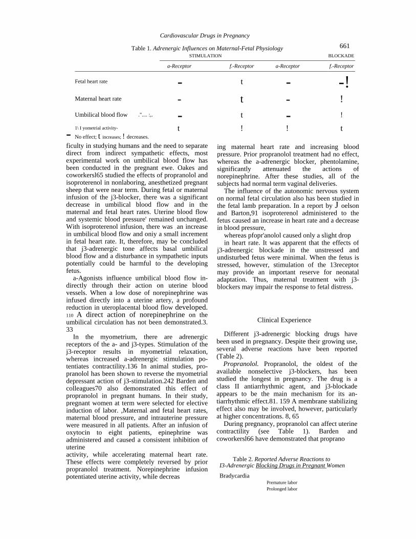
Table 1. Adrenergic Influences on Maternal-Fetal Physiology STIMULATION BLOCKADE
a-Receptor f.-Receptor a-Receptor f.-Receptor
Fetal heart rate - t - -!Maternal heart rate - t - ! Umbilical blood flow .".... :,. - t - !
1\ I yometrial activity- t ! ! t - No effect; t increases; ! decreases.
661
ficulty in studying humans and the need to separate direct from indirect sympathetic effects, most experimental work on umbilical blood flow has been conducted in the pregnant ewe. Oakes and coworkersl65 studied the effects of propranolol and isoproterenol in nonlaboring, anesthetized pregnant sheep that were near term. During fetal or maternal infusion of the j3-blocker, there was a significant decrease in umbilical blood flow and in the maternal and fetal heart rates. Uterine blood flow and systemic blood pressure' remained unchanged. With isoproterenol infusion, there was _ an increase in umbilical blood flow and oniy a small increment in fetal heart rate. It, therefore, may be concluded that j3-adrenergic tone affects basal umbilical blood flow and a disturbance in sympathetic inputs potentially could be harmful to the developing fetus.
a-Agonists influence umbilical blood flow in-directly through their action on uterine blood vessels. When a low dose of norepinephrine was infused directly into a uterine artery, a profound reduction in uteroplacental blood flow developed. 110 A direct action of norepinephrine on the umbilical circulation has not been demonstrated.3. 33
In the myometrium, there are adrenergic receptors of the a- and j3-types. Stimulation of the j3-receptor results in myometrial relaxation, whereas increased a-adrenergic stimulation po-tentiates contractility.136 In animal studies, pro-pranolol has been shown to reverse the myometrial depressant action of j3-stimulation.242 Barden and colleagues70 also demonstrated this effect of propranolol in pregnant humans. In their study, pregnant women at term were selected for elective induction of labor. ,Maternal and fetal heart rates, maternal blood pressure, and intrauterine pressure were measured in all patients. After an infusion of oxytocin to eight patients, epinephrine was administered and caused a consistent inhibition of uterine activity, while accelerating maternal heart rate. These effects were completely reversed by prior propranolol treatment. Norepinephrine infusion potentiated uterine activity, while decreas
ing maternal heart rate and increasing blood pressure. Prior propranolol treatment had no effect, whereas the a-adrenergic blocker, phentolamine, significantly attenuated the actions of norepinephrine. After these studies, all of the subjects had normal term vaginal deliveries.
The influence of the autonomic nervous system on normal fetal circulation also has been studied in the fetal lamb preparation. In a report by J oelson and Barton,91 isoproterenol administered to the fetus caused an increase in heart rate and a decrease in blood pressure,
whereas pfopr'anolol caused only a slight drop in heart rate. It was apparent that the effects of
j3-adrenergic blockade in the unstressed and undisturbed fetus were minimal. When the fetus is stressed, however, stimulation of the 13receptor may provide an important reserve for neonatal adaptation. Thus, maternal treatment with j3-blockers may impair the response to fetal distress.
Clinical Experience
Different j3-adrenergic blocking drugs have been used in pregnancy. Despite their growing use, several adverse reactions have been reported (Table 2).
Propranolol. Propranolol, the oldest of the available nonselective j3-blockers, has been studied the longest in pregnancy. The drug is a class II antiarrhythmic agent, and j3-blockade appears to be the main mechanism for its an-tiarrhythmic effect.81. 159 A membrane stabilizing effect also may be involved, however, particularly at higher concentrations. 8, 65
During pregnancy, propranolol can affect uterine contractility (see Table 1). Barden and coworkersl66 have demonstrated that proprano
Table 2. Reported Adverse Reactions to I3-Adrenergic Blocking Drugs in Pregnant Women
Bradycardia Premature labor Prolonged labor
662 Josef Widerhorn et al.
Ii,. ", i:
101, given to pregnant women, blocks the inhib-itory effects of epinephrine on myometrial ac-tivity. The administration of propranolol therefore may facilitate an increase in uterine contractility. ""
:-.
The pharmacokinetics of propranolol during pregnancy are similar to those in the nonpregnant state. O'Hare and colleagues169 administered propranolol to six healthy pregnant volunteers between 32 and 36 weeks of gestation. There were no significant changes in elimination half-life, clearance, VD, and bioavailability. Perruca and coworkers,142 however, found that the protein binding of propranolol decreased during pregnancy.
Propranolol readily crosses the placenta39. 43. 67. 112. 184. 233 and, at delivery, the fetal serum concentration is equal t039. 184 or less than233 the maternal concentration. The unbound fraction of propranolol is .higher in fetal plas~a, how ever, probably because of low concentration of (Xl-acid glycoprotein. Because of decreased he patic metabolism and altered protein binding, the serum concentration and half-life' of pro pranolol may be increased in the neonate during thefirst days of life. 67. 184
Several fetal and neonatal adverse effects were reported.21. 43. 54. 132. 166. 166. 184. 232. 239 Pruyn and associatesl84 noted that 10 to 11 neonates frommothers treated chronically with propranolol had intrauterine growth retardation. Extrapolating data from an animal study,l65 they suggested thatpropranolol may decrease umbilical blood flow and, consequently, fetal nu trition. While reported by many other authors21. 54. 132. 166. 187.232 the true incidence of intrauterinegrowth retardation is unclear. In some prospective studies,21. 54. 166. 232 the incidence of intra-uterine growth retardation was only 3 to 4 per cent(4 of 94 pregnancies). Furthermore, two of thesefour mothers who had small babies deliverednormal babies in subsequent pregnancies, despite continuous treatment with propranolol. 166 Redmond,187 in an extensive review of human andanimal literature, concluded that the evidenceimplicating propranolol in growth retardation issuggestive but inconclusive.
There are many hypothetical mechan~sms bywhich propranolol may induce growth'retardation. A decrease in cardiac output combined with plasmavolume contraction and increased systemic vascular resistance occurring duringhypertension in pregnancy may decrease uterine blood flow.187 Propranolol has been shown toreduce umbilical blood flow in pregnant ewes.165 Other postulated mechanisms may in-volve a decreased peripheral conversion of T4
,I '!H
to T3 and a theoretical effect on neurotransmitters that may affect the brain's influence on fetal growth. 187
Turnstall and colleagues239 described a delay in the onset of respiration of newborns when propranolol was administered to the mother prior to cesarean section. Another major source of concern is the effect of propranolol and of 13-blockade therapy in general on the fetal response to hypoxia. During asphyxia, several hormonal, metabolic, and circulatory adaptive mechanisms are activated. Most responses are mediated through catecholamines and l3-receptors. As has been demonstrated in experimental animal studies,85. 91 propranolol therapy may be particularly deleterious during these circumstances of fetal distress. l3-blockade also may be responsible for lack of fetal heart acceleration provoked by sound stimulationl66 and persistently negative throughout the nonstress test and contraction stress test. 132
To date, propranolol has not been demonstrated to be teratogenic. However, three cases of congenital malformatibns have been associated with propranolol therapy during preg nancy; for example, pyloric stenosis, crepitus of the hip, and tracheoesophageal fistula. 22. 29. 167 It is not clear at all if this is a casual occurrence or a true teratogenic effect of propranolol. In any case, if the last hypothesis is correct, these malformations probably are very rare.
Although the information concerning use of propranolol during pregnancy has expanded steadily, its safety is still somewhat controversial. Despite favorable reviews,l23. 199 there is a potential for side effects29. 184. 187. 205 that should be anticipated by the clinician.
Propranolol is excreted in breast milk. 12. 93. 116 Karlberg and coworkers93 showed that propranolol is secreted into the breast milk in a dosedependent manner and the milk-plasma ratio is approximately 1.0. Bauer and colleagues12 found a milk-plasma ratio of 0.4 to 0.6; they estimated that the daily dose ingested by the infant probably is 1 per cent of the recommended pediatric daily dose. No adverse effects were observed in infants breast fed by mothers treated with propranolol. However, the newborn hepatic microsomal enzyme systems are immature and propranolol may accumulate. Careful observation of these infants is therefore recommended.
Metoprolol. Metoprolol is a I3cselective blocking agent. It is similar in its effectiveness to other nonselective l3-adrenoreceptor blocking drugs used in the treatment of angina pectoris4 or essential hypertension. 15 Because of its primary 131-selective properties, the drug
i

Cardiovascular Drugs in Pregnancy 663
theoretically would not interfere with 132-me-diated peripheral vasodilatation or 132 effects on uterine tone.
In pregnancy, metoprolol has been used pri-marily to control hypertension61. 84. 202. 203 or tachyarrhythmias.24 Its metaQ.olism during preg-nancy is increased and, therefore, half-life, serum concentration, and bioavailability are de-creased.83 The most plausible explanation is that steroid hormones increase the activity of the hepatic mono-oxygenase system, which is involved in metoprolol metabolism.83 Protein binding is not altered during pregnancy.
Metoprolol crosses the placenta, and the fe tomaternal serum concentration ratio is approx-imately 1.0.122.203 Because of redistribution and! or relative immaturity of hepatic enzymatic systems in the newborn, the serum concentration of metoprolol increases four-fold during the first hours of life, but then declines over 5 to 20 hours. 122 ' .
Metoprolol alone or combined with hydralazine was studied in 101 pregnant hypertensive patients.203 The metoprolol group experienced lower perinatal mortality (2 per cent versus 8 per cent) and a lower incidence of intrauterine growth retardation (11.7 per cent versus 16.3 per cent). Theoretically, this might be explained by metoprolol's lack of action on uterine tone (132 effect) and l3-mediated vasodilatation. No differences in Apgar score, gestational age, or birth weight were noted.
Hogstedt and colleagues,84 in a recent con-trolled trial, compared the combination of me-toprolol and hydralazine with non pharmacologic management of hypertension in 161 pregnant patients. The outcome for the neonates with respect to birth weight, head Cfircumference, Apgar score, incidence of respiratory distress, bradycardia, and hypoglycemia was similar in both groups, confirming the results previously reported.220 There was one case of fetal malfor mation in each group. This may be an unrelated event because the combination metoprolol-hydralazine was started during the second and third trimesters. To date, no cases of fetal malformation induced by metoprolol have been reported, but experience during the first trimester is lacking.
Sandstrom204 has shown the combination of metoprolol and hydralazine to be superior to hydralazine and thiazide for the treatment of pregnant hypertensive patients with regard to maternal well-being, fetal intrauterine growth, 10-minute Apgar scores, and perinatal mortality.
Despite encouraging data, caution is recom
mended. Kjellmer and associates,98 in an animal study, showed that 131-adrenoreceptor blockade is potentially dangerous in cases of fetal asphyxia. These data are in agreement with previous experimental results obtained with the nonselective l3-blocker propranolol. 29.85
Metoprolol is secreted in breast milk,1I9 but the daily fetal quantity ingested by the neonate is very small. Unless hepatic function. in the newborn is markedly impaired, breast feeding should not be discouraged. Finally, metoprolol appears to be safe in
pregnancy, but the data are still limited. Pending further studies assessing metoprolol's safety in
pregnancy, caution is recommended. Atenolol. Atenolol is another relatively selective
I3cadrenergic blocking agent used to treat hypertension during pregnancy. 113. 127. 138.200,220. 23S Its fetal effects are described in the article by Kornbluth and colleagues in this issue.
Like metoprolol, atenolol has negative ino
tropic and chronotropic activity. However, it , has insignificant partial ~1 mimetic activity and weak membrane stabilizing properties. 194
Transplacental transfer of atenolol is well documented. 113. 138 Although there is a three- or six-fold individual variation in plasma atenolol concentration in each patient studied, its con-centration was found to be equal in maternal and fetal blood (ratio 1.0).138 Atenolol is secreted in breast milk and no effects were noted in atenolol-exposed babies; breast feeding, therefore, need not be discontinued. 119
Pindolol. Pindolol is a l3-adrenergic blocking agent that has partial intrinsic sympathomimetic activity. It also has some membrane stabilizing
effects, but much less than propranolol's. 8
The potential advantage of intrinsic agonist activity in a l3-blocker is that the decrease in heart rate and cardiac output at rest is not as pronounced as with nonagonistic l3-blocking drugs. The exercise-induced increase in heart rate and cardiac output still is blunted, however.204 This may be of particular benefit in patients with ventricular dysfunction or those prone to bradycardia.6S
Theoretically, pindolol may be particularly advantageous in pregnancy because the lack of effects on resting heart rate and cardiac output, combined with a direct vasodilator effect, are not expected to compromise uterine blood flOW197 or to decrease basal fetal heart rate.87
Pindolol crosses the placenta, and the feto-maternal concentration ratio is less than 1. 0.71 Elimination half-lives in fetus and mother are 1.6 hours and 2.2 hours, respectively. Dubois and coworkersso compared acebutolol, pindolol,
.1'

664 Josef Widerhom et ai.
and atenolol in 56, 38, and 31 pregnant hyper-tensive patients, respectively. In the pindolol group, the birth weight was significantly higher than in the atenoloLgrgup (3345 g versus 2745 g). Apgar score, gestational ages, fetal heart rate, and frequency of hypoglycemia were not affected by pindolol treatment. Two cases of malformation were reported in the pindolol group, however: cleft palate (pindolol started at 29 weeks of gestation) and vesicoureteral reflux (second full pregnancy on pindolol in a mother with asymmetrical segmental renal hypoplasia).
In another study, 55 pindolol was compared with methyldopa in 32 consecutive patients with pregnancy-induced hypertension. Maternal blood pressure was controlled better in the pindolol group. Furthermore, a drop in creatinine levels and an increase in creatinine clearance was noted in mothers treated With pindolol. No difference was observed between the two groups in regard to intrauterine growth, Apgar score, or fetal morbidity. The birth weight was similar in the two groups (2850 g). Nonstress tests were normal and no bradycardia was observed in the pindolol group.
Rosenfeld and coworkers197 randomly assigned 44 consecutive pregnant hypertensive patients to two treatment groups: hydralazine alone (21 patients) or hydralazine and pindolol (23 patients). These investigators found a lower incidence of maternal side effects such as dizziness or headaches in the combination therapy group. No differences were noted in the two groups concerning birth weight, gestational ages, hypothermia, hypoglycemia, Apgar score, or mode of delivery. Two major malformations were noted in the hydralazine group and neonatal thrombocytopenia was noted in the combination group.
Although preliminary reports are favorable for recommending the use of pindolol in pregnancy, the information available is still limited. Additional experience is needed to confirm the safety of pindolol during pregnancy.
Labetalol. Labetalol, like propranolol, blocks both I3c and 132-adrenergic receptors. But la-betalol is unique in that it also has both a1-adrenergic blocking properties and direct va-sodilatory activity. Labetalol' crosses the pla-centa.I64. 190 The fetomaternal concentration ra tio is 0.5. Its clearance and volume of distribution are not altered during pregnancy.
There have been a number of favorable reports describing the safe use of labetalol in pregnancy.
Lamming and coworkerslll randomized 26 patients with pregnancy-induced hypertension
in two groups: one treated with labetalol (14 patients) and one treated with methyldopa (12 patients). Improvement of renal function and a better control of blood pressure were observed in the labetalol group. The incidence of spontaneous labor and Bishop score also were higher in the labetalol group. Theoretically, the higher incidence of spontaneous labor could be caused by myometrial relaxation induced by acblocking and the 132-mimetic activity. of labetalol. Furthermore, Nylund and colleaguesl64 dem onstrated that uterine blood flow was not affected despite significant reduction in blood pressure. There also is evidence that labetalol may promote fetal lung maturation. Michaep45 observed higher than expected lecithin-sphin gomyelin ratio in amniotic fluid as early as 31 weeks of gestation in patients treated with labetalol. This effect probably is mediated by 132mimetic activity because salbutamol has similar effects.l60 Several studiesl46, 186, 230 showed that no fetal adverse effects were associated with labetalol treatment.
Labetalol also is useful in the management of severe pre-eclampsia or eclampsia. 63. 147 Michaep47 randomly assigned 90 patients with severe hypertension (diastolic blood pressure greater than 105 mm Hg) to receive IV labetalol (45 patients) or IV diazoxide (45 patients). The control of blood pressure was better in the labetalol group. A precipitous drop in blood pressure (60/40 mg Hg) was noted only in the diazoxide group (eight patients). No fetal brady-cardia, hypoglycemia, hypothermia, or malfor mations were noted. There was a higher operative delivery rate in the diazoxide group. The results obtained in several studies regarding the use of labetalol for all types of hypertension during pregnancy, including hypertensive emergencies, are very encouraging. However, more information concerning its safety is warranted and, therefore, caution is recommended.
Labetalol is secreted in breast milk and no adverse effects were noted in neonates.l25 The nursing babies should be monitored closely, however.
Other I3-Blockers. The experiences with ace-butolol, oxprenolol, and sotalol are reviewed in the article by Kornbluth and coworkers.
t Ir
Recommendations
The evidence currently available regarding the safety of l3-blocking agents in pregnancy is inconclusive. It, therefore, appears preferable to offer other effective drugs already proven
I

Cardiovascular Drugs in Pregnancy
safe during pregnancy, prior to using ~-blockers. If a ~-blocker is to be used, the following guidelines may be useful.
1. Consider the pregnant woman (and the fetus) receiving ~-blockers to be a high-risk patient, deserving special. -c:ire during both pregnancy and labor. 2. Whenever possible, avoid the use of ~ blocker therapy during the first trimester.
3. Use the lowest possible therapeutic dose; combinations of low doses of ~-blockers and other agents may be the optimal drug therapy.
4. When possible, taper drug therapy at least 2 to 3 days prior to delivery, both as a way of limiting the effects of ~-blockers on uterine contractility and of preventing neonatal compli-cations secondary to ~-blockade.
5. The use of ~-blockers with ~1-selectivity or intrinsic sympathomimetic activity, or the use of an a-~-blocker (that is, labetalol) may be preferable, in that these. drugs theoretically would be less likely to interfere with -~2mediated uterine relaxation and peripheral vasodilation.
A comprehensive recommendation regarding the use of ~-blockers is extremely difficult to state because of the absence of any large scale clinical use of these agents in pregnant patients. Further studies are needed to define drug pharmacokinetics in mother and fetus. Future trials hopefully will address these questions and further clarify the role ~-blocking agents should playas therapy in controlling maternal disease states, as well as their effects on fetal and neonatal well-being.
SODIUM NITROPRUSSIDE
Sodium nitroprusside (SN) is one of the most potent drugs available for treatment of hyper-tensive emergencies. It was approved in the United States in 1974. Chemically, sodium nitroprusside is disodium pentacyanonitrosylferrate. The hypotensive component of sodium nitroprusside is the free nitroso (NO) group which interferes with calcium influx and activation, producing relaxation of vascular smooth muscle but not of uterine smooth muscle. SN directly relaxes arteriolar and venous smooth muscle, decreasing preload and afterload. Blood pressure decreases and heart rate increases slightly. Renal blood flow and glomerular filtration rate are preserved; plasma renin activity is increased. Nitroprusside reacts rapidly with hemoglobin, yielding methemoglobin and cyanide. The latter compound undergoes metabo
665
lism in the liver or kidney to thiocyanate, which is excreted in the urine. Cyanide also may be eliminated as cyanomethemoglobin or cyano-cobalamine. In case of liver diseases, hepatic immaturity, and excessive administration, cyanide ions may poison the cytochrome oxidase system, leading to anaerobic metabolism and, clinically, metabolic acidosis.
SN is given intravenously in light-protected tubing. The initial rate of infusion should be 0.1 to 0.2 J.Lg per kg per minute and slowly increased (5 to 10 J.Lg every 5 to 10 minutes) until the desired effect is obtained or a dose of 10 J.Lg per kg per minute is reached. Cyanide, thiocyanate, methemoglobin level, and arterial pH should be monitored periodically.
Prolonged use of SN and/or renal failure may result in excessive thiocyanate formation and/or accumulation that initially manifests with CNS symptoms (tinnitus, blurred vision, confusion, psychosis). Plasma,thiocyanate above 10 mg per 100 ml is toxic ana above ~O mg per 100 ml, fatal. Other adverse effects are methemoglobinemia, increased intracranial pressure, headache, rash, nausea, abdominal pain, and muscle twitching.
During pregnancy, SN has been used to control the blood pressure during intracranial aneurysm surgery"9. 249 or severe gestational hy-petension.174 SN was demonstrated to cross the placenta in both human21. 226 and animal studies.56.
11S. 156. 191. 247
The experimental data concerning the phar-macodynamic effects of SN in pregnant animals still are conflicting. 56. l1S, 156, 191. 247 Ring and coworkers191 compared SN with hydralazine in phenylephrine-induced hypertension in nearterm pregnant ewes. Both agents were equally effective in lowering the blood pressure. However, only hydralazine counteracted the effects of phenylephrine-that is, increasing uterine blood flow, heart rate, and cardiac output. Wheeler and associates247 found that nitroglycerin and nitroprusside have similar effects on uterine flow, that is, mostly increased or, in a few cases, unchanged. Ellis and coworkersS6 found that SN increases uterine blood flow in pregnant ewes, whereas Lieb and coworkers11S noted a significant (25 to 35 per cent) decrement. Naulty and colleagues156 observed a decrease in blood pressure without any changes in uterine blood flow.
Paull and coworkers174 described four severe pre-eclamptic patients with hypertension resis tant to diazoxide and other "conventional methods" treated successfully with nitroprusside. The only fetus alive at the onset of therapy was

666 Josef Widerhorn et ai.
delivered uneventfully. Four other patients with severe pregnancy-induced hypertension and refractory congestive heart failure were treated successfully with SN. 226 One premature infant expired. The 'other infants were free of side effects or malformations. The infusion rate varied from 0.013 /Lg per kg per minute to 2.75 /Lg per kg per minute. Measurements made in one case showed equal but negligible (0.1 ILg per ml) concentrations of cyanide in maternal and fetal blood.
Shoemaker and coworkers214 described a 24-week pregnant patient with severe pre-eclampsia not controlled by hydralazine and magnesium sulfate. Blood pressure was controlled with SN and labor was induced with pitocin. Fifteen hours after the onset of SN therapy, the patient delivered a 478 g stillborn infant. The nitroprusside dose was 3.9 /Lg per kg' per ml. The level of cyanide in the fetal liver was less than 10 /Lg per ml, with toxic levels reported to be 30 to 40 /Lg per ml. The authors speculate that fetal death was caused by severe eclampsia.
In summary, nitroprusside is a very effective but toxic drug.' Until further studies clarify its pharmacodynamics, kinetics, and safety during pregnancy, caution is recommended.
HYDRALAZINE
Hydralazine has been used extensively during pregnancy since the early 1950s. It is one of the agents of choice in the management of hypertensive emergencies as well as for main-tenance therapy, alone or in combination with other antihypertensive drugs.
It has a direct relaxing effect on arteriolar vascular smooth muscle, producing a decrease in systemic vascular resistance and vasodilatation.102 The hydralazine effects appear to be mediated through guanosine 3' ,5' -monophosphate (cyclic GMP).
Peripheral vasodilatation triggers compensatory sympathetic discharge, which increases the heart .rate and cardiac OUtpUt.102 Plasma renin activity is increased and, consequently, sodium and water retention may lead to edema formation. Regional vasodilatation is not equal; splanchnic, cerebral, coronary, and renal vascular beds are more dilated than skin and muscle vascular beds. Venous dilatation is minimal and, therefore, postural hypertension is infrequent.
When given orally, the drug is absorbed almost completely from the gastrointestinal tract. The systemic bioavailability of hydralazine
after its first pass through the liver depends on acetylator type. Obviously, slow acetylators will have a higher blood level and will be more prone to toxicity. Peak plasma concentration is reached within 0.5 to 2 hours, and the hypotensive effect lasts 6 to 8 hours. Parenteral administration is less influenced by acetylator type; onset of action is within 10 to 20 minutes and may last 2 to 4 hours. For the treatment of hypertensive emergency during pregnancy, Pritchard and colleaguesl83 suggested an initial IV dose of 5 mg that can be increased by 5 to 10 /Lg every 20 minutes.
With IV administration, flushing, headache, dizziness, and palpitations may be prominent. Nasal congestion, nausea, vomiting, diarrhea, fatigue, and sleep disturbances also may be common. Approximately 5 to 10 per cent of the patients may develop a lupus erythematosuslike syndrome. The syndrome is more common in slow acetylators and when doses of more than 200 mg per ~ day are used. It may become evident after 2 months of treatment and resolve slowly (6 months to years) after withdrawal of drug. Other rare complications are blood dyscrasias, rash, and fever.
Experimental data from animal models con-cerning the effect of hydralazine on uterine blood flow have been controversial. 9. 109. 191 Ring and associates191 reported an increase in uterine blood flow in pregnant hypertensive sheep, whereas Ladner and coworkers109 showed the opposite effect in normotensive pregnant sheep. Several studies92. 126, 228 investigated the effects of drugs on intervillous blood flow in hypertensive pregnant women. In all of these studies, the administration of IV hydralazine did not change the mean intervillous blood flow significantly. Looking at individual results, however, uterine blood flow was descreased in 9 of 13 patients in the Lunell study,126 in contrast to the Suomo study,228 in which the flow decreased in only 4 of 10 patients.
Vink and coworkers241 investigated the effect of IV administration of dihydralazine on maternal blood pressure, uterine activity, fetal heart rate, and growth retardation in 33 hypertensive pregnant patients. Dihydralazine, 12.5 mg, lowered the diastolic blood pressure from more than no mm Hg to 70 to 90 mm Hg in 5 minutes in 30 of 33 patients. Nineteen fetuses had deceleration in fetal heart rate concomitantly with the decrease in blood pressure; 13 of these 19 fetuses had intrauterine growth retardation; and three fetuses were stillborn. Only one fetus from the group without deceleration had intrauterine growth retardation.

Cardiovascular Drugs in Pregnancy 667
Uterine activity was not influenced by hydralazine administration. According to the authors, fetal deceleration was caused by inability of the uteroplacental unit to compensate for the acute reduction in blood pressure.
Spinnato and colleagues27 also observed a higher incidence of cesarean section deliveries secondary to fetal distress in pre-eclamptic or eclamptic patients treated with hydralazine.
Kuzniar and coworkers108 obseved that the hemodynamic effects of hydralazine are more pronounced in patients with pregnancy-induced hypertension than in those with essential hy-pertension during pregnancy. Patients with se vere pre-eclampsia have high systemic vascular resistance, decreased cardiac output, and di-minished plasma volume. In these patients, vasodilator agents such as hydralazine, are more likely to induce marked hypotension and, con-sequently, fetal distress. In contrast, the response to hydralazine is less dramatic in patients with pregnancy-associated hypertension. These observations indicate that lower doses and a gradual decrease in blood pressure are particularly beneficial in the high risk group of patients with pre-eclampsia.
In summary, the use of IV hydralazine in hypertensive emergencies such as pre-eclampsia and eclampsia is very effective in lowering the blood pressure and preventing hypertensive encephalopathy or intracranial hemorrhages. Hydralazine therapy is associated with the risk offetal distress, however, particularly in patients with a reduced uteroplacental reserve. In these patients, aprecipitous decrease in blood pressure clearly isassociated with fetal distress and should be avoided.Some authorslSS believe that the reduction inuterine blood flow is caused by catecholamine-induced vasoconstriction rather than a reduction inperfusion pres sure alone. They suggest that the concomitantadministration of antiadrenergic agents, such asmethyldopa, as well as avoidance of unnecessary reduction in blood pressure may explain the lowincidence of fetal distress noted in their patients.
Hydralazine crosses the placenta freely and alsoappears in small amounts in milk. ~20 To date, no teratogenic effects of hydralazine have beenreported.
NITRATES
Organic nitrates, esters of nitric acid, have been used extensively for more than a century for the treatment of the symptoms of ischemic
heart disease. Their use more recently was extended to congestive heart failure. Nitrates are very potent dilators, predominantly of venous vessels; arterial dilatation increases at high doses. Nitrate effects lead to a decrease in venous return and, consequently, to lower right and left ventricular end diastolic pressures. Systemic vascular resistance is not changed significantly, heart rate is unchanged or increased, and cardiac output usually is un changed. In higher doses, nitrates may lower the blood pressure and cardiac output, which triggers a compensatory sympathetic activation with reflex tachycardia and vasoconstriction. Nitroglycerin also decreases pulmonary vascular resistance. The antianginal effects mostly are because of a decrease in myocardial oxygen requirements by reducing pre- and afterload. Nitrates are metabolized in the liver. Nitroglycerin metabolites have only 10 per cent of nitroglycerin's potency and their half-life is approximately 40 minutes. Following sublingual administration, the peak plasma concentration is reduced in 4 minutes and half-life is 1 to 3 minutes. Intravenous administration is used mostly in acute ischemic syndromes and conges-tive heart failure.89 It also is used to control systemic hypertension after and during coronary artery bypass surgery.
Continuous exposure to nitrates may lead to early development of tolerance. 1. 171. 172. 188. 234 These problems probably may be avoided by increasing the dosing interval and allowing nitrate-free intervals of at least 10 hours over 24 hours.ll. 171. 172 Most common side effects are headache, dizziness, and postural hypotension; syncopal episodes are not common. During pregnancy, IV nitroglycerin was used to control severe pregnancy-induced hypertension. 86. 219 Having a very small molecular weight and being uncharged, nitroglycerin easily crosses the pla-centa.38 Experimental data in pregnant ewes47 showed a fetomaternal arterial blood concentration ratio of 0.15. The authors advanced the hypothesis that the low fetal levels probably are caused by rapid metabolism, poor placental transfer, or widespread binding.
Snyder19 and Hood86 and their colleagues used nitroglycerin to control the elevation of blood pressure that occurs during tracheal intubation in pre-eclamptic patients. Nitroglycerin not only successfully lowered the basal blood pressure, but also blunted the hyperten sive response to intubation. No evidence of fetal compromise was noted.
Cotton and colleagues38 studied the hemo-dynamic effect of IV nitroglycerin coupled with

668 Josef Widerhorn et al.
blood volume expansion in six pre-eclamptic patients. The mean gestational age was 37.3 :!: 3.6 weeks. Nitroglycerin alone effectively reduced the mean arterial blood pressure by 27.5 per cent. The hypotepsive effect was blunted by volume expansion. In two patients in whom blood pressure was lowered suddenly, fetal deceleration and bradycardia were observed. In the other three patients, a flat fetal heart rate with an average beat-to-beat variability of less than 5 beats per minute was noted. The authors suggested that this probably is because of a loss of cerebral autoregulation and increased intracranial pressure induced by nitroglycerin.
In summary, it appears that nitrate treatment is not free of side effects, and the information available regarding their use during pregnancy is very limited. Of particular concern are the effects. on uterine blood flow, oxygen'delivery, and fetal hemodynamics. Further studies are needed to clarify their effects and safety during pregnancy; therefore, no recommendation can be made at this time.
CONCLUSION
There are many clinical situations that require the use of cardiovascular medications in pregnancy. It is hard to evaluate drugs during pregnancy, so the safety experiences with many medications are limited, but certain drugs (digoxin, quinidine, (3-blockers, a-methyldopa, hydralazine) have been used with relative safety to both mother and fetus. It always is best to avoid medications in pregnancy but, when necessary, drugs for cardiovascular disease may be used when the risk-benefit ratio is favorable.
REFERENCES
1. Abrams J: Nitrate tolerance and dependence. Am Heart J 99:113-123, 1980 2. Abboud TK, Sarkis F, Blikian A, et al: Lack of adverse neurobehavioral effects oflidocaine (abstract). Anes thesiology 57:A404, 1982 3. Adams FH, Assali NS, Cushman M, et al: Interrelationship
of maternal and fetal circulations. 1. Flow pressure responses to vasoactive drugs in sheep. Pediatrics 27:627-635, 1961
4. Adolfsson L, Areskog NH, Furberg C, et al: Effects of single doses of alprenolol and two cardioselective l3-blockers (H 87/07 and H 93/20) on exerciseinduced angina pectoris. Eur J Clin Pharmacol 7:111-118, 1974
5. Allan LD, et al: Evaluation and treatment of fetal arrhythmia. Clin Cardiol 7:467, 1984 6. Allonen H, Kanto J, Iisalo E: The foetomaternal
distribution of digoxin in early human pregnancy. Acta Pharmacol ToxicoI39:477-480, 1976
7. Arnoux P, Seyral P, Llurens M, et al: Amiodarone and digoxin for refractory fetal tachycardia. Am J CardioI59:166-167, 1987
8. Arnsdorf MF: Basic understanding of the electrophys-iologic actions of antiarrhythmic drugs. Med Clin North Am 68:1247-1280, 1984
9. Assali NS, Dasqupta K, Kolm A: Measurement of uterine blood flow and uterine metabolism. Am J Obstet Gynecol 78:313,. 1959
10. Barden TP, Stauder RW: Effects of adrenergic blocking agents and catecholamines in human pregnancy. Am J Obstet Gynecoll02:225-235: 1968
11. Barnett DB, Hudson SA, McBurney A: Disopyramide and its N-monodesalkyl metabolite in breast milk. Br J Clin PharmacoI14:310-312, 1982 12. Bauer JH, Pape B, Zajicek J, et al: Propranolol in human plasma and breast milk. Am J Cardiol 43:860-862, 1979 13. Bauer LA, Brown T, Gibaldi M, et al: Influence of long term infusions on lidocaine kinetics. Clin Phar macol Ther 31:433-437, 1982 14. Bellet S: Essentials of cardiac arrhythmias. In Diag nosis and Management. Philadelphia, WB Saun ders, 1972 15. Bengtsson C, Johnsson G, Regardth CG: Plasma levels
and effects of metoprolol on blood pressure and heart rate after acute dose and between two doses during long term treatment. Clin Pharmacol Ther
17 :400-408, 1975 16. Bergmans MGM, Jonker GJ, Kock HCLV: Fetal supraventricular tachycardia: Review of the litera ture. Obstet Gynecol SUTV 40:61-68, 1985 17. Biehl D, Shnider SM, et al: Placental transfer of lidocaine: Effects of fetal acidosis. Anesthesiology 48:409-412, 1978 18. Bigger}T, Giardina EGV: Drug interaction in antiar rhythmic therapy. Ann NY Acad Sci 427:140-162, 1984 19. Bigger }T, Mandel WJ: Effects of lidocaine on the electrophysiologic properties of ventricular muscle and Purkinje's fibers. J Clin Invest 49:63-77, 1970 20: Blumer J, Strong JM, Atkinson AJ: The convulsant potency of lidocaine and its N-dealkylated metabo
lites. J Pharmacol Exp Ther 186:31-36, 1973 21. Bott-Kanner G, Schweitzer A, Reisher SH, et al:
Propranolol and hydralazine in the management of essential hypertension in pregnancy. Br J Obstet Gynecol 87:110-114, 1980
22. Bott-Kanner G, Schweitzer A, Schoenfeld A, et al: Treatment with propranolol and hydralazine throughout pregnancy in a hypertensive patient: A case report. Isr J Med Sci 14:466-468, 1978
23. Branch RA, Shand DG, Wilkinson GR, et al: The reduction of lidocaine clearance by d-propranolol: An example of a hemodynamic drug interaction. J Pharmacol Exp Ther 184:515-519, 1973
24. Brodsky MA, Sato DA, Oster PD, et al: Paroxysmal ventricular tachycardia with syncope during pregnancy. Am J CardioI58:563-564, 1986
25. Brown Wu, Bell GC, Alper MH: Acidosis, local anesthetics, and the newborn. Obstet Gynecol 48:27-30, 1976.
26. Bullock JL, Harris RE, Young R: Treatment of thy-rotoxicosis during pregnancy and propranolol. Am J Obstet Gynecol 121:242-245. 1975
27. Burney RG, DiFazio CA, Foster JH: Effects of pH on

Cardiovascular Drugs in Pregnancy protein binding of lidocaine. Anesth Analg (Cleve) 57:478-480, 1978
28. Wong BYS, Hurwitz A: Simple method for maintaining serum lidocaine levels in therapeutic range. Arch Intern Med 145:1588-1591, 1985
29. Campbell JW: A possible teratogenic effect of pro pranolol. N Engl J Med 313:518, 1986
30. Campbell RWF: Mexiletine. N Engl J Med 316:29 34, 1987 '0.' :-' 31. Chan V, Tse TF, Wong V: Transfer of digoxin across the placenta and into breast milk. Br J Obstet Gynecol 85:605-609, 1978 32. Charlier R, Deltour G, Bandine A, et al: Pharmacol ogy of amiodarone, an antiangina drug with a new biological profile. Arzheimittelforsch 18:1408-1417,
1968 33. Chez RA, Ehrenkranz RA, Oakes GK, et al: Effects ofadrenergic agents on bovine umbilical and uterine blood flows.In Longo L, Reneau DD (eds): Fetal and NewbornCardiovascular Physiology, Vol. 2. New York, GarlandPublishing, 1978, PI' 1-16 34. Cocco G, Strozzi C, Chu D, et al: Torsades de pointes as a manifestation of mexiletine toxicity. hm Heart J 100:878-
880, 1980 35. Colin A, Lambotte R: Influence teratogene des medica~nts
adminstres a la femme enceinte. Rev Med Liege 27:39-48, 1972
36. Condemi JJ, Blomgren SE, Vaughan JH: The procainamide-induced lupus syndrome. Bull Rheum Dis 20:604-608, 1970
37. Conn HL Jr, Luchi RJ: Some cellular and metabolicconsiderations relating to the action of quinidine as aprototype antiarrhythmic agent. Am J Med 37:685-699, 1964
38. Cotton DB, Longmire S, Jones MM, et al: Cardiovas cular alterations in severe pregnancy-induced hy pertension: Effects of intravenous nitroglycerin coupled with
blood volume expansion. Am J Obstet GynecoI154:1053-1059, 1986
39. Cottrill CM, McAllister RG Jr, Gettes L, et al: Propranolol therapy during pregnancy, labor, and delivery: Evidence for transplacental drug transfer and impaired neonatal drug disposition. J Pediatr 91:812-
814, 1977 40. Crouthamel WG: The effect of congestive heart failure on
quinidine pharmacokinetics. Am Heart J 90:335339, 1975
41. Dahl SG, Strandjard RE: Pharmacokinetics of chlor-promazine after single and chronic dosage. Clin Pharmacol Ther 21:437-448, 1977
42. Dam M, Christiansen J, Munch 0, et al: Original article antiepileptic drugs: Metabolism in pregnancv. Clin Pharmacokinet 4:53-62, 1979
43. Datta S, Kitzmiller JL, Ostheimer GW: Propranolol and parturition. Am J Obstet Gynecol 101:91-99, 1968 44. Davis M, Simmons q, Dordoni B, et al: Induction of hepatic enzymes during normal human preg nancy. J Obstet Gynecol Br Commonwealth 80:690694, 1973
. 45. Davison JM, Noble MCB: Serial changes in 24 hour
creatinine clearance during normal menstrual cycles and the first trimester of pregnancy. Br J Obstet
GynecoI88:10-17, 1981 . 46. Dean M, Stock B, Patterson R], et al: Serum protein binding of drugs during and after pregnancy in humans. Clin Pharmacol Ther 28:253-261, 1980 47. DeRosayro M, Nahrwold ML, Hill AB, et al: Nitro
669glycerin: Plasma levels and effects in pregnant ewes (abstract). Anesthesiology 51:S312. 1979
48. DiSegni E, Klein HO, David D, et al: Overdrive pacing in quinidine syncope and other long QT interval syndromes. Arch Intern Med 140:10361040, 1980 49. Donchin Y, Amirav B, Sahar A, et al: Sodium nitro prusside for aneurysm surgery in pregnancy. Br J Anaesth 50:849-851, 1978 50. Dubois D, Petiteolas J, Temperville B, et al: f3-blocker therapy in 125 cases of hypertension during preg nancy. Clin Exp Hyper-Hyper Pregnancy B2(1):459, 1983 51. Dumesic DA, Silverman NH, Tobias S, et al: Trans
placental cardioversion of fetal supraventricular tachycardia with procainamide. N Engl J Med 307:1128-1131, 1982 52. Dunlop W: Serial changes in renal hemodynamics during normal human pregnancy. Br J Obstet Gy necol 83:17-23, 1981 53. Eadie MJ, Lander CM, Tyrer JH: Plasma drug level
monitoring in pregnancy. Clin Pharmacokinet 2:427-436, 1977 54. Eliahou HE, Silverberg DS, Reisin E, et al: Propran olol for the treatment of hypertension in pregnancy. Br J Obstet Gynaecol 85:431-436, 1978
55. Ellenbogen 1\, Jasehervatzky 0, Davidson A: Management of pregnancy-induced hypertension with
pindolol--comparative study with methyldopa. Int J Gynaecol Obstet 24:3-7, 1986 . 56. Ellis SC, Wheeler AS, James FM III, et al: Fetal and
maternal effects of sodium nitroprusside used to counteract hypertension in gravid ewes. Am J Obstet GynecoI143:766, 1982
57. Fearrinton EL, Rand CH Jr, Rose JD: Hyperprolactine mia-galactorrhea induced by verapamil. Am J
CardioI51:1466-1467, 1983 58. Federman J, Vliestra R: Antiarrhythmic drug therapy.
Mayo Clin Proc 54:531-542, 1979 59. Fever G: Action of pregnancy and various progester ones on hepatic microsomal activities. Drug Metab Rev 9:147-169, 1979 60. Finerman WB, Hamer A, Peter T, et al: Electrophysiologic
effects of chronic amiodarone therapy in patients with ventricular arrhythmias. Am Heart J 104:987-
996, 1982 61. Finnestrom 0, Ezitis J, Rydeu G, et al: Neonatal effects of
f3-blocking drugs in pregnancy. Acta Obstet Gynecol Scand 118:91-93, 1984
62. Frishman WH, Chesner H: f3-adrenergic blockers in pregnancy. Am Heart J 115:147-152, 1988 63. Garden A, Davey DA, Dommisse J: Intravenous labetalol and dihydralazine in severe hypertension in pregnancy. Clin Exp Hypertension Bl:371, 1981 64. Giardina EGV, Dreyfuss J, Bigger JT Jr, et al: Metab olism of procainamide in normal and cardiac sub
jects. Clin Pharmacol Ther 19:339-351, 1976 65. Gilman AG, Goodman LS, Rail TW, et al: The Phar macologic Basis of Therapeutics. Edition 7. New York, MacMillan, 1985 66. Given BD, Phillippe M, Sanders SP, et al: Procain amide cardioversion of fetal supraventricular tachy
arrhythmia. Am J Cardiol 53:1460-1461, 1984 67. Gladstone GR, Horolof A, Gersony WM: Propranolol administration during pregnancy: Effects on the fetus. J Pediatr 86:962-964, 1975 68. Golichowski AM, Caldwell R, Hartsough A. et al: Pharmacologic cardioversion of intrauterine supra

670 Josef Widerhorn et aI.
II II'
ventricular tachycardia. J Reprod Med 30:139-144, 198569. Grant AM, Marshall RJ, Ankier SI: Some effects of
disopyramide and its N-dealkylated metabolite on isolated nerve and cardiac muscle. Eur J Pharmacol 49:389-394, 1978
70. Greene HL, Graham EL, Werner JA, et al: Toxic and therapeutic effectS of amiodarone in the treatment of cardiac arrhythmias. J Am ColI Cardiol 2:11141128, 1983
71. Grun.stein S, Ellenbogen A, Anderman S, et al: Transfer of pindolol across the placenta in hypertensive pregnant women. Curr Ther Res 37:587-591, 1985
72. Habib A, McCarthy JS: Effects on the neonate of propranolol administered during pregnancy. J Pediatr 91:801-811, 1977
73. Hartel G, Louhija A, Konttinen A: Disopyramide in the prevention of recurrence of atrial fibrillation after electroconversion. Clin Pharmacol Ther 15:551-555, 1974
74. Heaton FC, Vaughan R: Intrauterine supraventricular tachycardia: Cardioversion with maternal digoxin. Obstet Gynecol60:749-752, 1982
75. Heel RC, Brogden RN, Speight TM, et al: Disopyramide: A review of its pharmacologic properties and therapeutic use in treating cardiac arrhythmias. Drugs 15:331-368, 1978
76. Heinonen OP, Slone D, Shapiro S: Birth Defects and Drugs in Pregnancy. Littleton, MA, Publishing Sciences Group, 1977
77. Hemmermeister KE, et al: The comparative inotropic effects of six clinically used antiarrhythmic agents. Ann Intern Med 91:229-238, 1979
78. Herngren L, Ehrnebo M, Boreus LO: Drug binding to plasma proteins during human pregnancy and in the perinatal period. Dev Pharmacol Ther 6:11a
124, 1983 79. Hicks JM, Brett EM: Falsely increased digoxin con centrations in samples from neonates and infants. Ther Drug Monit 6:461-464, 1984 80. Hill LM, Malkasian GD: The use of quinidine sulfate throughout pregnancy. Obstet Gynecol54:366-368, 1979 81. Hirata K, Kato "H, Yoshioka F, et al: Successful treatment of fetal atrial flutter and congestive heart failure. Arch Dis Child 60:158-160, 1985 82. Hoffman BF, Rosen MR, Wit AL: Electrophysiology and
pharmacology of cardiac arrhythmias. VII. Cardiac effects of quinidine and procainamide. Am Heart J 90:117-122, 1975
83. Hogstedt S, Lindberg B, Perig DR, et al: Pregnancyinduced increase in metoprolol metabolism. Clin Pharmacol Ther 37:688-692, 1985
84. Hogstedt S, Lindeberg S, Axelsson 0, et al: A prospectivecontrolled trial of metoprolol-hydralazine treatment in hypertension during pregnancy. Acta Obstet Gynecol Scand 64:50~1O, 1985
85. Hokegard KH, Karlsson K, Kjellmer I, et ahECG changes in the fetal lamb during asphyxia in relation to j3-adrenoreceptor stimulation and blockade. Acta Physiol Scand 105:195, 1979
86. Hood DD, Dewan DM, James FM, et al: The use of nitroglycerin in preventing the hypertensive response to tracheal intubation in severe pre-eclamptics (abstract). Anesthesiology 59:A423, 1983
87. Ingermarsson I, Liedholm H, Montan S, et al: Fetal heart rate during treatment of maternal hypertension with j3-adrenergic antagonists. Acta Obstet Gynecol Scand 118(Suppl):95-97, 1984
Iii
88. Inoue H, Unno N, Ou MC, et al: Level of verapamil in human milk. Eur J Clin Pharmacol 26:657-658, 1984
89. Jaffe AS, Roberts R: The use of intravenous nitroglycerin in cardiovascular disease. Pharmacotherapy
2:273-280, 1982 " 90. Jennings G, Jones M, Besterman EM, et al: Oral
disopyramide in prophylaxis of arrhythmias following myocardial infarction. Lancet 1:51-54, 1976
91. Joelson I, Barton MD: The effect of blockade of the j3-receptors of the sympathetc nervous system of the fetus. Acta Obstet Gynecol Scand 3(Suppl)48:75-79, 1979
92. Jouppila P, Kirkinen P, Koivula A: Effect of antihy-pertensive drugs. on placental and fetal blood flow. In Kurjak A, Kossoff G (eds): Recent Advances in Ultrasound Diagosis. Amsterdam, Elsevier Science Publishers, 1984, pp 68-74
93. Karlberg B, Luudberg D, Aberg H: Excretion of propranolol in human breast milk. Acta Pharmacol Toxicol34:222-224, 1974
94. Kelliher GJ, Kowey P, Engel T, et al: Clinical phar macology of antiarrhythmic agents. Cardiovasc Clin 16:287-305, 1985 95. Kessler KM, Lowenthal DT, Werner H, et al: Quinidine
elimination in patients with Congestive heart failure or poor renal function. N Engl J Med 290:706-709, 1974
96. Kileff MB, et al: Neonatal neurobehavioral responses after epidural anesthesia for cesarean section with lidocaine and bupivacaine. Anesthesiology 57:A403, 1983
97. Kim WY, Pomerance JJ, Miller AA: Lidocaine intoxication in a newborn following local anesthesia for episiotomy. Pediatrics 64:643-645, 1979
98. Kjellmer, Dagbjartsson A, Hrbek A, et al: Maternal j3-adrenergic blockade reduces fetal tolerance to asphyxia. Acta Obstet Gynecol Scand 118:75-80, 1984
99. Klein V, Repke JT: Supraventricular tachycardia in pregnancy: Cardioversion with verapamiJ. Obstet Gynecol 63(Suppl 3):16S-18S, 1984.
100. Kleineman CS, Copel JA, Weinstein EML, et al: Treatment of fetal supraventricular tachyarrhythmias. J Clin Ultrasound 13:265, 1985
101. Knapp AB, Maguire W, Keren G, et al: The cimetidine-lidocaine interaction. Ann Intern Med 98:174, 1983
102. Koch-Wesser J: Drug therapy: Hydralazine. N Engl J Med 295:320-323, 1976 103. Kohlhardt M, Bauer B, Krause H: New selective inhibitors of the Ca conductivity in mammalian myocardial fibers. Experientia 15:288-289, 1972 104. Koren G, Farine D, et al: Significance of the endog enous digoxin-like substance in infants and mothers. Clin Pharmacol Ther 36:759-764, 1984 IOS. Kostis JB, Frishman W, Hosler MH, et al: Treatment of
angina pectoris with pindolol: The significance of intrinsic sympathomimetic activity of j3-blockers. Am Heart J 104:496, 1982
106. Kotter V, Linderer T, Schroder R: Effects of disopyramide on systemic and coronary hemodynamics and myocardial metabolism in patients with coronary p.rtery disease: Comparison with lidocaine. Am J Cardiol 46:469-475, 1980
107. Krauer B, Krauer F, Hytten F: Drug prescribing in pregnancy. In Lind T (ed): Current Reviews in Obstetrics and Gynecology. Edinburgh, Churchill Livingstone, 1984

Cardiovascular Drugs in Pregnancy
108. Kuzniar J, Skret A, Piela A, et al: Hemodynamic effects of intravenous hydralazine in pregnant women with severe hypertension. Obstet Gynecol 66:453-458, 1985
109. Ladner CN, Weston PV, Brinkman CR, et al: Effects of hydralazine on uteroplacental and fetal circulations. Am J Obstet Gynecol 108:375-381, 1970
110. Ladner E, Brinkman CR, Weston P, et al: Dynamics of uterine circulation in'pr~gnant and nonpregnant sheep. Am J Physiol 218:257-263, 1970
111. Lamming GD, Broughton, Pipkin, et al: Comparison of the a- and l3-blocking drug, labetalol, and methyldopa in the treatment of moderate and severe pregnancy-induced hypertension. Clin Exp Hypertension 865:895, 1980
112. Langer A, Hung CT, McA'Nulty JA, et aI: Adrenergic blockade: A new approach to hyperthyroidism in pregnancy. Obstet Gynecol44:181-186, 1974
113. Lardoux H, Gerard J, Blazquez G, et al: Hypertension in pregnancy: Evaluation of two l3-blockers, atenolol and labetalol. Eur Heart J 4:(Suppl)G:35-40, 1983
114. Latini R, Tognoni G, Kates RE: Clinical pharmacokinetics of amiodarone. Clin Pharmacokinet 9:136156, 1984
115. Leonard RF, Braun TE, Levy AM: Imitation of uterine contractions by disopyramide during pregnancy. N Engl J Med 299:84-85, 1978
116. Levitan A, Manion J: Propranolol therapy during pregnancy and lactation. Am J CardioI32:247, 1973 117. Levy G: Pharmacokinetics of fetal and neonatal ex posure to drugs. Obstet GynecoI58(Suppl):9S-16S, 1981 118. Lieb SM, Zugain M, Huwauhid B, et al: Nitroprusside
induced hemodynamic alteration in normotensive and hypertensive pregnant sheep. Am J Obstet Gynecol 139:925, 1981
119. Liedholm H, Melander A, Bitzen PO, et al: Accumulation of atenolol and metaprolol in human breast milk. Eur J Clin Pharmacol 20:229-231, 1981
120. Liedholm H, Wahlin-Boll E, Hanson, et al: Transplacenta passage and breast milk concentration of hydralazine. Eur J Clin PharmacoI21:417-419, 1982
121. Lilja H, Karlsson K, Lindecrantz K, et al: Treatment of intrauterine. supraventricular tachycardia with digoxin and verapamil. J Perinat Med 12:151-153, 1984
122. Lindeberg S, Sandstrom B, Lundborg P, et al: Disposition of the adrenergic blocker metoprolol in the late pregnant woman, the amniotic fluid, the cord blood, and the neonate. Acta Obstet Gynecol Scand 118:61-64, 1984
123. Livingstone I, Braswell PW, Bevan EB, et al: Propranolol in pregnancy: Three year prospective study. Clin Exp Hyper-Hyper Pregnancy B2(2):341-350, 1983
124. Loughnan PM: Digoxin excretion in human breast milk. J Pediatr 92:1019-1020, 1978 125. Lunnel NO, Kulas J, Rane A: Transfer of labetolol into amniotic fluid and breast milk in lactating women. Eur J Clin Pharmacol 28:597-599, 1985
126. Lunell NO, Levander R, Nylund L, et al: Acute effect of dihydralazine on uteroplacental blood flow in hypertension during pregnancy. Gynecol Obstet Invest 16:274-282, 1983 127. Lunell NO, Persson B, Aragon G, et al: Circulatory and
metabolic effects of acute l3-blockade in severe pre-eclampsia. Acta Obstet Gynecol Scand 58:443445, 1979
128. Luoma PV, et al: Efficacy ofintravenous disopyramide
671
in the termination of supraventricular arrhythmias. J Clin PharmacoI18:293-301, 1978
129. Lusson JR, Bey tout M, Jacquetin B, et al: Traitement d'une tachycardie supraventriculaire foetale: Asso-ciation digoxine-amiodarone. Coeur 15:315-317, 1985
130. Luxford AME, Kellaway GSM: Pharmacokinetics of digoxin in pregnancy. Eur J Clin PharmacoI25:117121, 1983
131. Marcus FI: Drug interactions with amiodarone. Am Heart J 106:924-930, 1983 132. Margulis E, Binder D, Cohen AW: The effects of propranolol on the nonstress test. Am J Obstet Gynecol 148:340-341, 1984 133. Mark LC, Kayden HJ, Steele JM, et al: The physio logic disposition and cardiac effects of procainamide. J Pharmacol Exp Ther 102:5-15, 1951 134. Marzo A, et al: Dati preliminari sulla farmacocinetica della digossina in gravidanza. Bollettino Societa Italiana Biologia Sperimentale 56:219-223, 1980 135. Mauer AM, Devaux LO, Lahey ME: Neonatal and maternal thrombocytopenic purpura due to quinine. Pediatrics 19:84-87, 1957 136. Maughan GB, Shabanah EH, Toth A: Experiments with pharmacologic sympatholysis in the gravid. Am J Obstet Gynecol 97:764-776, 1967
137. McKenna WJ,~-Harris L, et al: Amiodarone therapy during pregnancy. Am J CardioI51:1231-1233, 1983 138. Melander A, Niklasson B, Ingemarsson I, et al: Trans placental passage of atenolol in man. Eur J Clin PharmacoI14:93-94, 1978 139. Meltzer RS, McMorrow M, Robert EW, et al: Atypical ventricular tachycardia as a manifestation of diso pyramide toxicity. Am J CardioI42:1049-1052, 1978
140. Mendelson CL: Disorders of heart beat during preg nancy. Am J Obstet Gynecol 72:1268-1301, 1956
141. Merz W: Herzrhythmusstorungen in der schwanger shaft. Dtsch Med Wochenschr 97:1987-1988, 1972 142. Merz W, Effert S, Heinrich KW: Heart disease in pregnancy, intra- and postpartum. Z Geburtschilfe Perinatal 178:317-336, 1974 143. Metcalf J, Ueland K: The heart and pregnancy. In Hurst JW (ed): The Heart. Edition 4. New York, McGraw-Hill, 1978, p 1721 144. Meyer J, Lackner JE, Schochet SS: Paroxysmal tachy cardia in pregnancy. JAMA 94:1901-1904, 1930 145. Michael CA: Use of labetalol in the treatment of severe hypertension during pregnancy. Br J Clin Pharmacol 8(Suppl 2):2115-2153, 1979 146. Michael CA: The evaluation of labetalol in the treat ment of hypertension complicating pregnancy. Br J Clin Pharmacol B(Suppll):127S, 1982 147. Michael CA: Intravenous labetalol and intravenous diazoxide in severe hypertension complicating preg nancy. Aust NZ J Ohstet Gynecol 26:26, 1986 148. Miller MR, Withers R, Bhamra R, et al: Verapamil and breast feeding. Eur J Clin Pharmacol 30:125 126, 1986 149. Mirro MJ, Watanabe AN, Bailey JC: Electrophysiologic
effects of disopyramide and quinidine on guinea-pig atria and canine cardiac Purkinje fibers: Dependence on underlying cholinergic tone. Circ Res 46:660-668, 1980
150. Mitani GM, Steinberg I, Lien E, et al: The pharma-cokinetics of antiarrhythmic agents in pregnancy and lactation. Clin Pharmacokinet 12:253-291, 1987
151. Mitchell BG, Clements JA, Pottage A, et al: Mexile tine disposition: Individual variation in response to

672 Josef Widerhorn et ai.
urine acidification and alkalinisation. Br J Clin PharmacoI16:281-284, 1983
152. Mitrani A, Oettinger M, Abinader EG, et al: Use of propranolol in dysfunctional labor. Br J Obstet Gynecol 82:651-6q5, 1975
153. Mosler KH, Jung' Hi" Methoden der pharmakolo-gischenGeburtserleichterung und Uterus-Relaxation. Internationales Symposion, Bad-Aachen. Stuttgart, Germany. Thieme, 1972, p 170 154. Moya F, Thorndike V: Passage of drugs across the
placenta. Am J Obstet Gynecol84:1778-1798, 1962 155. Naden PR, Redmna WGC: Antihypertensive drugs in
pregnancy. Clin PerinatoI12:521-538, 1985 156. Nademanee K, et al: Antiarrhythmic efficacy and
electrophysiologic actions of amiodarone in patients with life-threatening ventricular arrhythmias: Potent suppression of spontaneously occurring tachy-arrhythmias versus inconsistent abolition of induced ventricular tachycardia. Am Heart J 103:950-959, 1982
157. Naqvi N, Thompson DS, Morgan WE, et al: Hemo-dynamic effects of disopyramide in patients after open-heart surgery. Br Heart J 42:587-594, 1979
158. Naulty J, Cefalo RC, Levis PE: Fetal toxicity of nitroprusside in the pregnant ewe. Am J Obstet GynecoI139:708, 1981
159. Nestico PF, DePace NL, Morganroth J: Therapy with conventional antiarrhythmic drugs for ventricular arrhythmias. Med Clin North Am 58:1295-1319, 1984
160. Nicholas TE, Lugg MA, Johnson RG: Maternal admin-istration of salbutamol and labetalol increases the amount of alveolar surfactant in lung of the dog, 27 foetal rabbit. Proc Aust Physiol Pharmacol Soc 9:146P, 1978
161. Nicholson WJ, Martin CE, Gracey JG, et al: Disopyramide-induced ventricular fibrillation. Am J CardioI43:1053-1055, 1979
162. Nimmo WS, Wilson J, Prescott LF: Narcotic analgesics and delayed gastric emptying during labor. Lancet 1:890-893, 1975
163. Nitsch J, Steinbeck G, Luderitz B: Increase of mexiletine plasma levels due to delayed hepatic metabolism in patients with chronic liver disease. Eur Heart J 4:810-814, 1983
164. Nylund L, Lunell NO, Lewander R, et al: Labetalol for the treatment of hypertension in pregnancy. Acta Obstet Gvnecol Scand 118:71-73, 1984
165. Oakes GK, Walker AD, Ehrenkranz RA, et al: Effect of propranolol infusion on the umbilical and uterine circulations of pregnant sheep. Am J Obstet Gyne-coI126:1038-1042, 1976
166. Oakley GDG, McGarry K, Limb DG, et al: Management of pregnancy in patients with hypertrophic cardomyopathy. Br Med J 1:1749-1750, 1979
167. O'Connor PC, Jick H, Hunter JR, et al: Propranolol and pregnancy outcome. Lancet 2:1168, 1981 168. Jensen OH: Fetal heart rate response to a controlled sound
stimulus after propranolol administration to the mother. Acta Obstet Gynecol Scand 63:199202, 1984
169. O'Hare MF, Kinney CD, Murnaghan GA, et al: Pharmacokinetics of propranolol during pregnancy. Eur J Clin PharmacoI27:583-587, 1984
170. Parbhoo SP, Johnston IDS: Effect of estrogens and progestogens on gastric secretion in patients with duodenal ulcer. Gut 7:612-618, 1966
171. Parker OJ, Farrell B. Lahey AK, et al: Effect of intervals between doses on the development of
tolerance to isosorbide dinitrate. N Engl J Med 316:1440-1444, 1987
172. Parker OJ, Vankoughnett K, Farrell B: Comparison of buccal nitroglycerin and oral isosorbide dinitrate for nitrate tolerance in stable angina pectoris. Am J CardioI56:724-728, 1985
173. Parker W A: Effects of pregnancy on pharmacokinetics. In Benet LZ, et al (eds): Pharmacokinetic Basis for Drug Treatment. New York, Raven Press, 1984
174. Paull J: Clinical report of the use of sodium nitroprusside in severe preeclampsia. Anaesth Intensive Care 3:72, 1975
175. Pelkonen 0: Detoxification and toxification processes in the human fetoplacental unit. In Krauer B, Krauer F, Hytten FE, et al (eds): Drugs and Pregnancy: Maternal drug handling; fetal drug exposure. London, Academic Press, 1984, pp 63-71
176. Pentikainen PJ, Halinen MO, Helin MJ: Pharmacokinetics of oral mexiletine in patients with acute myocardial infarction. Eur J Clin Pharmacol25:773777, 1983
177. Perucca E, Ruprah M, Richeus A: Altered drug binding to serum proteins in pregnant women: Therapeutic relevance. R Soc Health J 74:422-426, 1981
178. Petrie RH, et al: Placental transfer of lidocaine following paracervical block. Obstet' Gynecol 120:791801, 1974:
179. Pitcher D, Leather HM, Storey GCA, et al: Amio darone in pregnancy. Lancet 1:597-598, 1983 180. Pittard WB III, Glazier H: Procainamide excretion in
human milk. J Pediatr 102:631-633, 1983 181. Podrid PJ, Lown B: Mexiletine for ventricular arrhyth
mias. Am J Cardiol 47:895-902, 1981 182. Podrid PJ, Schoeneberger A, Lown B: Congestive heart failure caused by oral disopyramide. N Engl J Med 302:614-617, 1980 183. Pritchard JA, Pritchard SA: Standardized treatment of 154 consecutive cases of eclampsia. Am J Obstet Gynecol 123:543-551, 1975
184. Pruyn SC, Phelan JP, Buchanan GC: Long term propranolol therapy in pregnancy: Maternal and fetal outcome. Am J Obstet Gynecol 135:485-489, 1979
185. Rahimtoola SH, Sinno MZ, Loeb HS, et al: Lidocaine infusion in acute myocardial infarction. Arch Intern Med 128:416-418, 1971
186. Redman CWG: A controlled trial of the treatment of hypertension in pregnancy: Labetalol compared with methyldopa. In Riley A, Symonds EM (eds): The Investigation of Labetalol in the Management of Hypertension in Pregnancy. Princeton, Excerpta Medica, 1982, p 101
187. Redmond GP: Propranolol and fetal growth retarda tion. Semin Perinatal 6:142-147, 1982 188. Reichek N, Priest C, Zimrin D, et al: Antianginal effects of nitroglycerine patches. Am J Cardiol54:1 7, 1984 . 189. Repke IT, Steibach G: Fetal supraventricular tachy cardia refractory to digoxin cardioversion. J Reprod Med 31:195-197, 1986 190. Riley AJ: Clinical pharmacology of labetalol in preg nancy. J Cardiovasc Pharmacol 3:S53-S59, 1981 191. Ring G, Krawes E, Shnider S, et al: Comparison of nitroprusside and hydralazine in hypertensive preg nant ewes. Obstet Gynecol 50:598-602, 1977 192. Riva E, Gerna M, Latini R, et al: Pharmacokinetics of amiodarone in man. J Cardiovasc Pharmacol 4:270-275, 1982
193. Roberts RB, Shirley MA: The obstetrician's role in

Cardiovascular Drugs in Pregnancy 673
reducing the risk of aspiration pneumonitis. Am J Obstet GvnecoI124:611-617, 1976
194. Robertson jIS, Kaplan NM, Caldwell ADS, et al: 13-blockade in the 1980s: Focus on atenolol. Drugs 25:1-340, 1983
195. Robson D, Jeeva Raj MV, Storey GCA, et al: Use of amiodarone during pregnancy. Postgrad Med J
61:75-77, 1985 ' , :" 196. Rogers MC, Willerson JT, Goldblatt A, et al: Serum
digoxin concentrations in the human fetus, neonate, and infant. N Engl J Med 287:1010-1013, 1972
19i. Rosenfeld J, Bott-Kauner G, Boner G, et aI: Treatment of hypertension during pregnancy with hydralazine monotherapy or with combined therapy with hydalazine and pindolol. Eur J Obstet Gynecol
Reprod Bioi 22:197-204, 1986 198. Rotmensch HH, Elkayam U, Frishman W: Antiar rhythmic drug therapy during pregnancy. Ann In tern Med 98:487-497, 1983 199. Rubin PC: l3-blockers in pregnancy. N Engl J Med 305:22, 1323-1326, 1981 200. Rubin PC, Bulters L, Clark DM, et aI: Placebo controlled trial of atenolol in treatment qf preg nancy-associated hypertension. Lancet 1:431-434, 1983 201. Saarikoski S: Placental transfer and fetal uptake of3H digoxin in humans. Br J Obstet Gynecol 83:879 884, 1976 202. Sandstrom B: Adrenergic l3-receptor blockers in hy pertension of pregnancy. Clin Exp Hypertension B1:127-141, 1982 203. Sandstrom B: Antihypertensive treatment with the adrenergic l3-receptor blocker, metaprolol, during pregnancy. Gynecol Obstet Invest 9:195-204, 1978
204. Sandstrom B: Adrenergic l3-receptor blockers in hy pertension of pregnancy. Clin Exp Hypertension 1:127-141, 1982 205. Sandstrom B: Clinical trials of adrenergic antagonists in pregnancy hypertension. Acta Obstet Gynecol Scand 118(Suppl):57-60, 1984 206. Schanker LS: Passage of drugs across body mem branes. Pharmacol Rev 14:501-530, 1962 207. Schoen R, Frishman WH, Shamoon H: Hormonal and metabolic effects of calcium-channel antagonists in man. Am J Med 1988 (in press)
208. Schroeder JS, Harrison DC: Repeated cardioversion during pregnancy: Treatment of refractory paroxysmal atrial tachycardia during three successive pregnancies. Am J Cardiol 27:445-446, 1971
209. Serafini PC, Petracco AL, Vicosa HN Jr, et aI: Arterial hyposensitive effect of verapamil in severe pre
eclampsia: Preliminary study. Arq Bras Cardiol 32:57-61, 1979 210. Shaxted EJ, Mitton PJ: Disopyramide in pregnancy:
A case report. Curr Med Res Opin 6:70-72, 1979 211. Sherman JL, Locke RV: Transplacental neonatal dig
italis intoxication. Am J CardioI6:834-837, 1960 212. Shnider SM, Way EL: Plasma levels of lidocaine in mother
and newborn following obstetrical Conduction anesthesia: Clinical applications. Anesthesiology 29:951-958, 1968
213. Shnider SM, Way EL: The kinetics of transfer of lidocaine across the human placenta. Anesthesiology 29:944-950, 1968
214. Shoemaker CT, Meyers M: Sodium nitroprusside for control of severe hypertensive disease of pregnancy: A case report and discussion of potential toxicity. Am J Obstet GynecoI149:171-173, 1984
215. Sims EAH, Krantz KE: Serial studies of renal function
during pregnancy and the puerperium in normal women. J Clin Invest 3i:1764-1774, 1958
216. Singh BN, Collett JT, Chew CYC: New perspectives in the pharmacologic therapy of cardiac arrhythmias. Prog Cardiovasc Dis 22:243-301, 1980
217. Singh Bi':, Vaughan Williams EM: The effects of amiodarone, a new antianginal drug on cardiac muscle. Br J Pharmacol 39:657-667, 1970
218. Sioskey GE: Amiodarone: A unique antiarrhythmic agent. Clin Pharmacokinet 2:330-340, 1983
219. Snyder SW, Wheeler AS, James FM: The use of nitroglycerin to control severe hypertension of pregnancy
during cesarean section. Anesthesiology 51:563-566, 1979
220. Solum T, Montan S, Sjoberg NO: Treatment of preg-nancy-induced hypertension with a l3-specific blocker. Clin Exp Hypertension B1(2-3):259, 1982
221. Somogyi A, Albrecht M, Kliems G, et aI: Pharmaco-kinetics, bioavailability, and ECG response of verapamil in patients with liver cirrhosis. Br J Clin Pharmacol 12:51-60, 1981
222. Spinnato J, Sibai BM, Anderson GD: Fetal distress after hydralazine therapy for severe pregnancyinduced hypertension. South Med J 79:559-562, 1986
223. Spinnato JA, et al: Fetal supraventri~ular tachycardia: In utero therapy with digoxin and quinidine. Obstet
Gynecol 64:730-735, 1984 ' 224. Stec GP, 'Lertora JJL, Atkinson AJJ, et al: Remission of
procainamide-induced lupus erythematosus with N-acetylprocainamide therapy. Ann Intern Med 90:799-800, 1979
225. Stein J, Podrid P, Lown B: Effects of oral mexiletine on left and right ventricular function. Am J Cardiol 54:575-578, 1984
226. Sterupel JE, O'Grady JP, Morton MJ, et al: Use of sodium nitroprusside in complications of gestational hypertension. Obstet Gynecol 60:533-538, 1982
227. Stokes 1M, Evans J, Stone M: Myocardial infarction and cardiac arrest in the second trimester followed by assisted vaginal delivery under epidural analgesia at 38 weeks gestation. Br J Obstet Gynecol 91:197 198, 1983
228. Suomo S, Saankoski S, Tahvanainem K, et al: Acute effects of dihydralazine mesylate, furosemide, and metoprolol on maternal hemodynamics in pregnancy-induced hypertension. Am J Obstet Gynecol 155:122-125, 1986
229. Symllie HC, Doar JWH, Head CD, et al: A trial of intravenous and oral mexiletine in acute myocardial infarction. Eur J Clin Pharmacol 26:537-542, 1984
230. Symonds EM, Lamming GD, Jadoul F, et al: Clinical and biochemical aspects of the use of labetalol in the treatment of hypertension in pregnancy: Comparison with methyldopa. In Riley A, Symonds EM (eds): The Investigation of Labetalol in the Manage ment of Hypertension in Pregnancy. Princeton, Excerpta Medica, 1982, p 62
231. Szekeley P, Snaith L: Heart Disease and Pregnancy. Edinburgh/London, Churchill Livingstone, 1974 232. Tcherdakoff PH, Colliard M, Berrard E, et al: Pro pranolol in hypertension during pregnancy. Br Med J 2:670, 1978 233. Teuscher A, Bossi E, Imhof P, et al: Effect of pro pranolol on fetal tachycardia in diabetic pregnancy. Am J CardioI42:304-307, 1978 234. Thadani U, Fung HL, Darke CA, et al: Oral isosorbide dinitrate in angina pectoris: Comparison of duration
of action and dose-response relation during acute
------------------------------------------------------------------------------------------------------------------------------------..-------.---------.

674 Josef Widerhorn et at. and sustained therapy. Am J Cardiol 49:411-419, 1982
235. Thorley KJ, McAinsh J, Cruickshank JM: Atenolol in the treatment of pregnancy-induced hypertension. Br J Clin PharmacoI12:725-730, 1981
236. Timis AD, Jackson G, Holt DW: Mexiletine for control of ventricular dysi-hythmias in pregnancy. Lancet 2:647-648, 1980
237. Tucker GT, Boyes RN, Bridenbaugh PO, et al: Binding of anilide-type local anesthetic in human plasma. Anesthesiology 33:304-314, 1970
238. Turner GM, Oakley CM, Dixon HG: Management of pregnancy complicated by hypertrophic obstructive cardiomyopathy. Br Med J 4:281-284, 1968
239. Turnstall MB: The effects of propranolol on the onset of breathing at birth. Br J Anesthesiol 41:792, 1969 240. Valdes R Jr: Endogenous digoxin-like immunoreactive factors:
Impact on digoxin measurements and potential physiologic implications. Clin Chern 31:1525-1531, 1985
241. Vink GS, Moodley J, Philpott RH: Effects of hydralazine on the fetus in the treatment of maternal hypertension. Obstet Gynecol 50:519, 1970
242. Wa!1sbrough H, Nakanishi H, Wood C: Effect of epinephrine on h)lman uterine activity in vitro and in vivo. Obstet Gynecol 30:779-789, 1967
243. Warang PK, Crouthamel WG, Carliner NH, Lidocaine and its active metabolites. Clin Pharmacol
Ther 24:654-662, 1978 - . 244. Waxman HL, Myerburg RJ, Appel R, et al: Verapamil for control of
ventricular rate in paroxysmal supraventricular tachycardia and atrial fibrillation of flutter. Ann Intern Med 94:1-6, 1981
245. Weaver JB, Pearson JF: Influence of digitalis on time of onset and duration oflabor in women with cardiac disease. Br Med J 3:519-520, 1973
246. Wellens HJJ, Lie KI, Bar FW, et al: Effect of amio
darone in the Wolff-Parkinson-White syndrome. Am J CardioI38:189-194, 1976
247. Wheeler AS, James FM, Greiss FC, et al: Effects of nitroglycerin to control severe hypertension of pregnancy during cesarean section (abstract). Anesthesiology 51:563-564, 1979
248. Whitsett JA, Wallick ET: Ouabain binding with Na +_ K + -ATPase activity in human placenta. Am J Physiol 238:E38-E45, 1980
249. Willoughby JS: Sodium nitroprusside, pregnancy, and multiple intracranial aneurysms. Anaesth Intensive Care 12:351-357, 1984
250. Wilson ]T, Brown RD, Cherek DR,. et al: Drug excretion in human breast milk: Principles, pharmacokinetics, and projected consequences. Clin Pharmacokinet 5:1-66, 1980
251. Winkle RA, Glantz SA, Harrison DC: Pharmacologic therapy for ventricular arrhythmia. Am J Cardiol 36:629-647, 1975
252. Wladimiroff JW, Stewart PA: Fetal therapy: Treatment of fetal cardiac arrhythmias. Br J Hosp Med 34:134-140, 1985
253. Wolff F, Breuker KH, Schlensker KH, et al: Prenatal diagnosis and therapy of fetal heart rate anomalies: With a contribution on the placental transfer of verapamiJ. J Perinat Med 8:203-208, 1980
254. Wood M, Wood AJJ: Changes in plasma drug binding and a1-actd glycoprotein in mother and newborn infant. Clin Pharmacol Ther 29:522-526, 1981
255. Wo~sley RL, Wang T, Ston'e W, et al: Pharmacology, electrophysiology, and pharmacokinetics of mexiletine. Am Heart J
107:1058-1065, 1984
Section of Cardiology USC School of Medicine
2025 Zonal Avenue Los Angeles, CA 90033 (Uri Elkayam, MD) I
i r