Embed Size (px)
Citation preview
3/7/2019
1
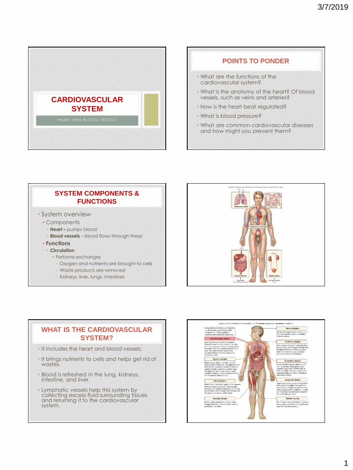
H E AR T AN D B L O O D V E S S E L S
CARDIOVASCULAR
SYSTEM
POINTS TO PONDER
• What are the functions of the cardiovascular system?
• What is the anatomy of the heart? Of blood vessels, such as veins and arteries?
• How is the heart beat regulated?
• What is blood pressure?
• What are common cardiovascular diseases and how might you prevent them?
SYSTEM COMPONENTS &
FUNCTIONS
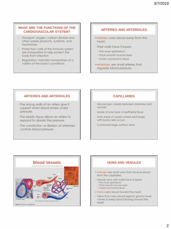
• System overview
• Components
• Heart – pumps blood
• Blood vessels – blood flows through these
• Functions
• Circulation
• Performs exchanges
• Oxygen and nutrients are brought to cells
• Waste products are removed
• Kidneys, liver, lungs, intestines
WHAT IS THE CARDIOVASCULAR
SYSTEM?
• It includes the heart and blood vessels.
• It brings nutrients to cells and helps get rid of wastes.
• Blood is refreshed in the lung, kidneys, intestine, and liver.
• Lymphatic vessels help this system by collecting excess fluid surrounding tissues and returning it to the cardiovascular system.
3/7/2019
2
WHAT ARE THE FUNCTIONS OF THE
CARDIOVASCULAR SYSTEM?
1. Transport: oxygen, carbon dioxide and
other wastes products, nutrients, and
horomones
2. Protection: cells of the immune system
are transported to help protect the
body from infection
3. Regulation: maintain homeostasis of a
variety of the body’s conditions
ARTERIES AND ARTERIOLES
• Arteries carry blood away from the heart.
• Their walls have 3 layers.
• Thin inner epithelium
• Thick smooth muscle layer
• Outer connective tissue
• Arterioles are small arteries that
regulate blood pressure.
ARTERIES AND ARTERIOLES
• The strong walls of an artery give it support when blood enters under
pressure;
• The elastic tissue allows an artery to
expand to absorb the pressure.
• The constriction or dilation of arterioles
controls blood pressure.
CAPILLARIES
• Microscopic vessels between arterioles and
venules
• Made of one layer of epithelial tissue
• Form beds of vessels where exchange
with body cells occurs
• Combined large surface area
Figure 5.2 Structure of a capillary bed.
Copyright © The McGraw-Hill Companies, Inc. Permission required for reproduction or display.
a.
v.
v. = vein; a. = artery
valve
blood
flow
blood
flow
connective
tissue
artery
arteriole
capillary bed
arteriovenous
shunt
vein
venule
smooth muscle
endothelium
elastic tissue
precapillary
sphincter
(left): © Ed Reschke; (right): © Biophoto Associates/Photo Researchers
Blood Vessels VEINS AND VENULES
• Venules are small veins that receive blood
from the capillaries.
• Venule and vein walls have 3 layers. • Thin inner epithelium • Thick smooth muscle layer
• Outer connective tissue
• Veins carry blood toward the heart.
• Veins that carry blood against gravity have
valves to keep blood flowing toward the
heart.
3/7/2019
3
TYPES OF BLOOD VESSELS
vein artery
capillary
Copyright © The McGraw-Hill Companies, Inc. Permission required for reproduction or display.
arteriovenous
shunt
vein
venule
arteriole
artery
connective
tissue
v.=vein; a.=artery
blood
flow
valve
blood
flow
smooth muscle
endothelium
elastic tissue
precapillary
sphincter
v.
a.
(left): © Ed Reschke
Figure 5.2 Structure of a capillary bed.
HOW CAN YOU TELL THE DIFFERENCE BETWEEN AN ARTERY AND VEIN?
TYPES OF BLOOD VESSELS
• The Capillaries: exchange • Arterioles branch into capillaries.
• The capillaries have walls that are only one cell
thick and allow exchange of substances with tissue fluid.
• Not all capillary beds are open at the same time.
• Contraction of a precapillary sphincter muscle
closes off the bed and then blood flows through an arteriovenous shunt, bypassing the capillary
bed, going directly into a venule.
arteriovenous
shunt
TYPES OF BLOOD VESSELS
• The Veins: To the Heart
• Venules drain into veins that return blood to the heart.
• Veins have much less smooth
muscle and connective tissue than arteries.
• Veins often have valves that prevent the backward flow of
blood due to gravity when closed.
3/7/2019
4
THE HEART
• Located behind and
slightly left of the
breastbone(sternum)
• Cone-shaped • Points slightly left
• Has 4 chambers • 2 atria (sing. = atrium)
• 2 ventricles
THE HEART
• Muscular
• Myocardium = cardiac muscle tissue
• Lies inside a fibrous
pericardium
ANATOMY OF THE HEART
• Large, muscular organ consisting of mostly cardiac tissue called the myocardium
• Surrounded by a sac called the pericardium
• Consists of 2 sides, right and left, separated by a septum
• Consists of 4 chambers: 2 atria and 2 ventricles
• 2 sets of valves: semilunar valves and atrioventricular valves (AV valves)
• Valves produce the “lub” and “dub” sounds of the heartbeat
EXTERNAL ANATOMY OF THE HEART
left subclavian artery
left common carotid artery
brachiocephalic artery
superior vena cava
aorta
left pulmonary artery
pulmonary trunk
left pulmonary veins
right pulmonary artery
right pulmonary veins
left atrium
left cardiac vein
right atrium
right coronary artery
left ventricle
right ventricle
left anterior descending
coronary artery
inferior vena cava
apex
3/7/2019
5
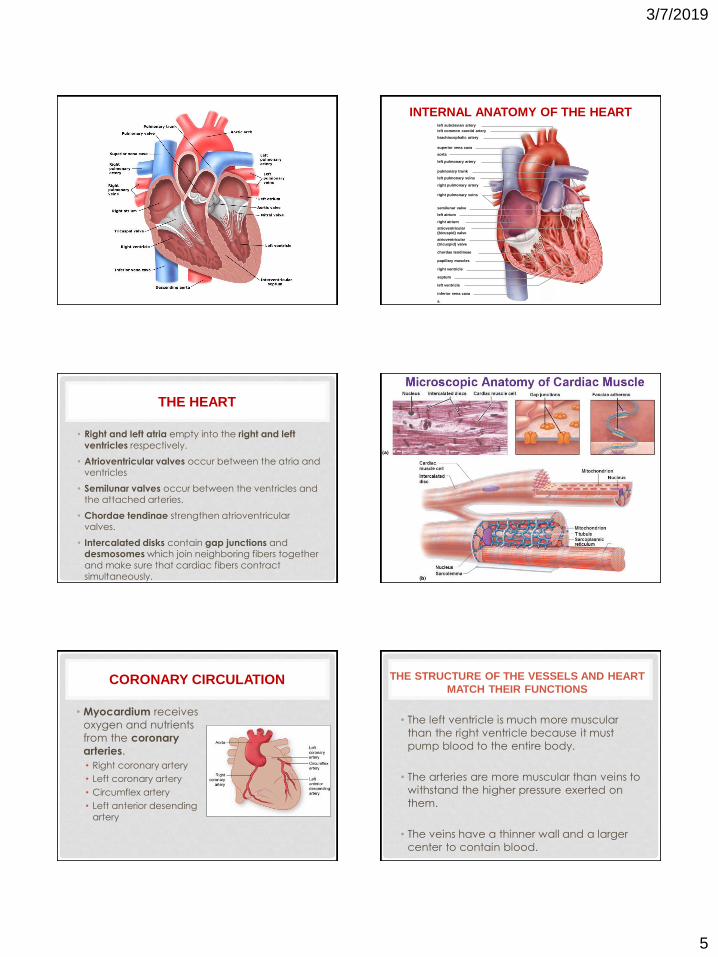
INTERNAL ANATOMY OF THE HEART left subclavian artery
left common carotid artery
brachiocephalic artery
superior vena cava
aorta
left pulmonary artery
pulmonary trunk
left pulmonary veins
right pulmonary artery
right pulmonary veins
semilunar valve
left atrium
right atrium
atrioventricular
(bicuspid) valve
atrioventricular
(tricuspid) valve
chordae tendineae
papillary muscles
right ventricle
septum
left ventricle
inferior vena cava
a.
THE HEART
• Right and left atria empty into the right and left
ventricles respectively.
• Atrioventricular valves occur between the atria and ventricles
• Semilunar valves occur between the ventricles and the attached arteries.
• Chordae tendinae strengthen atrioventricular valves.
• Intercalated disks contain gap junctions and desmosomes which join neighboring fibers together
and make sure that cardiac fibers contract simultaneously.
THE HEART
CORONARY CIRCULATION
• Myocardium receives
oxygen and nutrients
from the coronary
arteries.
• Right coronary artery
• Left coronary artery
• Circumflex artery
• Left anterior desending
artery
THE STRUCTURE OF THE VESSELS AND HEART
MATCH THEIR FUNCTIONS
• The left ventricle is much more muscular
than the right ventricle because it must
pump blood to the entire body.
• The arteries are more muscular than veins to
withstand the higher pressure exerted on
them.
• The veins have a thinner wall and a larger
center to contain blood.
3/7/2019
6
VISUALIZING BLOOD FLOW THROUGH THE HEART
Figure 5.4 The heart is a double pump.
PASSAGE OF BLOOD THROUGH
THE HEART
• From the body through the heart
• Superior and inferior vena cava, right atrium, right ventricle, pulmonary arteries to the lungs.
• From the lungs
• Blood goes back to the heart via pulmonary veins, left atrium, left ventricle, and then to the
body through the aorta.
• Oxygen-poor blood never mixes with
oxygen-rich blood!
HOW DOES THE HEARTBEAT OCCUR?
• During systole, the atria contract together followed by the ventricles contracting together.
• This is followed by diastole, a rest phase, when the chambers relax.
• This cardiac cycle, or heartbeat, occurs 70 times/minute on average.
THE HEART
• Each heartbeat is called a cardiac cycle.
• Systole refers to the contraction of heart
chambers
• Diastole refers to their relaxation.
• Heart sounds, lub-dub
• Due to the closing of the atrioventricular valves
• Followed by the closing of the semilunar valves.
WHAT IS THE CARDIAC CYCLE?
Figure 5.5 The stages of the cardiac cycle.
bicuspid valve aortic semilunar valve semilunar
valves close
(“dup”)
superior
vena cava semilunar
valves
pulmonary
trunk
aorta
right
atrium
right
ventricle
a.
aorta
b.
atrioventricular (AV)
valves close
(“lub”)
d. pulmonary
trunk
c.
right
atrium
inferior
vena cava
left
atrium
left
ventricle
represents
contraction
d: © Biophoto Associates/ Photo Researchers
HEARTBEAT IS CONTROLLED
• Internal Control of Heartbeat
• Sinoatrial (SA) node (pacemaker) initiates the beat and causes the atria to contract.
• Atrioventricular (AV) node conveys the stimulus
and initiates contraction of the ventricles.
• The signal for the ventricles to contract travels
from the AV node through the atrioventricular bundle to the smaller Purkinje fibers.
• These impulses travel between gap junctions at
intercalated disks.
3/7/2019
7
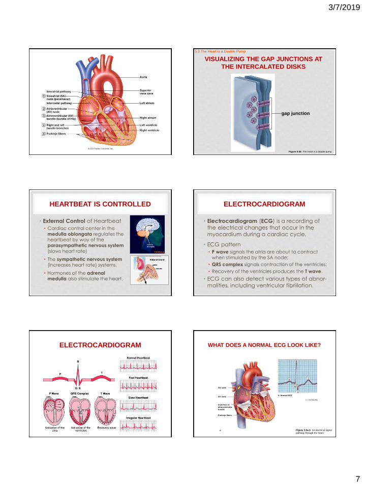
VISUALIZING THE GAP JUNCTIONS AT
THE INTERCALATED DISKS
gap junction
Figure 5.4b The heart is a double pump.
5.3 The Heart is a Double Pump
HEARTBEAT IS CONTROLLED
• External Control of Heartbeat
• Cardiac control center in the
medulla oblongata regulates the heartbeat by way of the parasympathetic nervous system
(slows heart rate)
• The sympathetic nervous system (increases heart rate) systems.
• Hormones of the adrenal medulla also stimulate the heart.
ELECTROCARDIOGRAM
• Electrocardiogram (ECG) is a recording of
the electrical changes that occur in the
myocardium during a cardiac cycle.
• ECG pattern
• P wave signals the atria are about to contract when stimulated by the SA node;
• QRS complex signals contraction of the ventricles;
• Recovery of the ventricles produces the T wave.
• ECG can also detect various types of abnor-
malities, including ventricular fibrillation.
ELECTROCARDIOGRAM WHAT DOES A NORMAL ECG LOOK LIKE?
SA node
AV node
branches of
atrioventricular
bundle
Purkinje fibers
a.
b. Normal ECG
P
Q
T
S
R
b: © Ed Reschke
Figure 5.6a-b An electrical signal
pathway through the heart.
3/7/2019
8
FEATURES OF THE
CARDIOVASCULAR SYSTEM
• Pulse Rate Equals Heart Rate
• The surge of blood entering the arteries following a heartbeat causes their elastic walls to stretch
and then recoil. This is felt as a pulse.
• The pulse rate of the radial or carotid artery
indicates the heart rate.
• Blood Flow Is Regulated
• Beating of the heart is necessary for homeostasis because it creates the pressure that propels
blood in the arteries and the arterioles
FEATURES OF THE
CARDIOVASCULAR SYSTEM
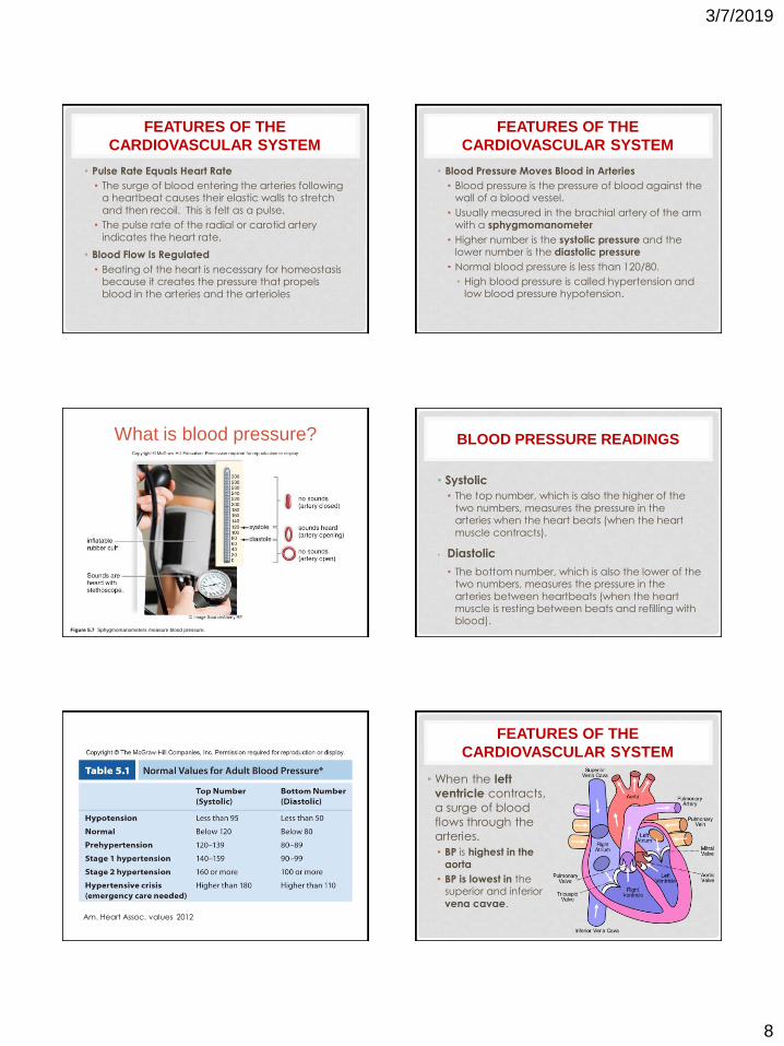
• Blood Pressure Moves Blood in Arteries
• Blood pressure is the pressure of blood against the wall of a blood vessel.
• Usually measured in the brachial artery of the arm with a sphygmomanometer
• Higher number is the systolic pressure and the lower number is the diastolic pressure
• Normal blood pressure is less than 120/80.
• High blood pressure is called hypertension and low blood pressure hypotension.
Figure 5.7 Sphygmomanometers measure blood pressure.
What is blood pressure? BLOOD PRESSURE READINGS
• Systolic
• The top number, which is also the higher of the
two numbers, measures the pressure in the arteries when the heart beats (when the heart
muscle contracts).
• Diastolic
• The bottom number, which is also the lower of the two numbers, measures the pressure in the
arteries between heartbeats (when the heart muscle is resting between beats and refilling with blood).
Am. Heart Assoc. values 2012
FEATURES OF THE
CARDIOVASCULAR SYSTEM
• When the left
ventricle contracts,
a surge of blood
flows through the
arteries.
• BP is highest in the
aorta
• BP is lowest in the superior and inferior
vena cavae.

3/7/2019
9
49
BLOOD FLOW
• Blood Flow Is Slow in the
Capillaries
• Blood moves slowly in capillaries because there
are more capillaries than arterioles.
• Slow pace allows time for exchanges between capillary blood and tissue
cells.
What is important about blood flow?
Figure 5.8 Blood velocity and
pressure in the blood vessels.
Rela
tiv
e m
ag
nitu
de
total
cross-sectional
area of
vessels
velocity
blood
pressure
arteries arterioles capillaries venules veins
Blood flow (starting from heart)
BLOOD FLOW
• Blood Flow in Veins
Returns Blood to Heart
• Venous return is dependent upon skeletal
muscle contraction, the presence of valves in veins, and respiratory
movements.
• Once blood has
moved past a valve it closes preventing backward return.
TWO CARDIOVASCULAR PATHWAYS
• Blood flows in two circuits:
• Pulmonary
• Systemic circuits
• The Pulmonary Circuit: Exchange of Gases
• Pulmonary arteries take blood from the right
ventricle to the lungs
• Carbon dioxide is given off
• Oxygen is picked up
• Pulmonary veins return it to the left atrium.
• Pulmonary arteries carry oxygen-poor blood while pulmonary veins carry oxygen-rich blood.
TWO CARDIOVASCULAR PATHWAYS
• The Systemic Circuit: Exchanges with Tissue
Fluid
• Aorta is largest artery in the systemic circuit
• Receives blood from the heart
• Superior and inferior venae cavae
• Trace the Path of Blood – see following figure
Ascending Aorta
Descending Aorta
3/7/2019
10
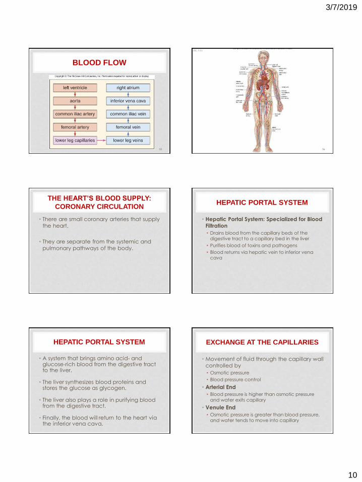
BLOOD FLOW
55 56
FIG. 5.11
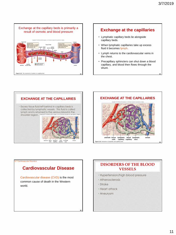
THE HEART’S BLOOD SUPPLY:
CORONARY CIRCULATION
• There are small coronary arteries that supply
the heart.
• They are separate from the systemic and
pulmonary pathways of the body.
HEPATIC PORTAL SYSTEM
• Hepatic Portal System: Specialized for Blood
Filtration
• Drains blood from the capillary beds of the digestive tract to a capillary bed in the liver
• Purifies blood of toxins and pathogens
• Blood returns via hepatic vein to inferior vena cava
HEPATIC PORTAL SYSTEM
• A system that brings amino acid- and glucose-rich blood from the digestive tract to the liver.
• The liver synthesizes blood proteins and stores the glucose as glycogen.
• The liver also plays a role in purifying blood from the digestive tract.
• Finally, the blood will return to the heart via the inferior vena cava.
EXCHANGE AT THE CAPILLARIES
• Movement of fluid through the capillary wall
controlled by
• Osmotic pressure
• Blood pressure control
• Arterial End
• Blood pressure is higher than osmotic pressure
and water exits capillary
• Venule End
• Osmotic pressure is greater than blood pressure, and water tends to move into capillary
3/7/2019
11
61
Exchange at the capillary beds is primarily a
result of osmotic and blood pressure
Copyright © The McGraw-Hill Companies, Inc. Permission required for reproduction or display.
from heart
Arterial End
Blood pressure is higher
than osmotic pressure.
Net pressure out.
water
oxygen amino
acids
Tissue Fluid
glucose carbon
dioxide
wastes
arteriole smooth
muscle fiber blood pressure
osmotic pressure
plasma
protein
Venous End
Osmotic pressure is higher
than blood pressure.
Net pressure in.
venule
to heart
salt
water
Figure 5.12 The movement of solutes in a capillary bed. 62
Exchange at the capillaries
• Lymphatic capillary beds lie alongside
capillary beds.
• When lymphatic capillaries take up excess
fluid it becomes lymph.
• Lymph returns to the cardiovascular veins in
the chest.
• Precapillary sphincters can shut down a blood
capillary, and blood then flows through the
shunt.
EXCHANGE AT THE CAPILLARIES
• Excess tissue fluid left behind in capillary beds is
collected by lymphatic vessels. This fluid is called lymph and is returned to the venous blood in the
shoulder region.
64
EXCHANGE AT THE CAPILLARIES
Figure 5.13 Interaction of lymphatic and capillary beds.
Copyright © The McGraw-Hill Companies, Inc. Permission required for reproduction or display.
venule lymphatic
duct
blood
capillary
lymphatic
capillary
tissue
cells
arteriole
65
Cardiovascular Disease
Cardiovascular disease (CVD) is the most
common cause of death in the Western
world.
5.7 Cardiovascular Disorders
DISORDERS OF THE BLOOD VESSELS
• Hypertension/high blood pressure
• Atherosclerosis
• Stroke
• Heart attack
• Aneurysm
3/7/2019
12
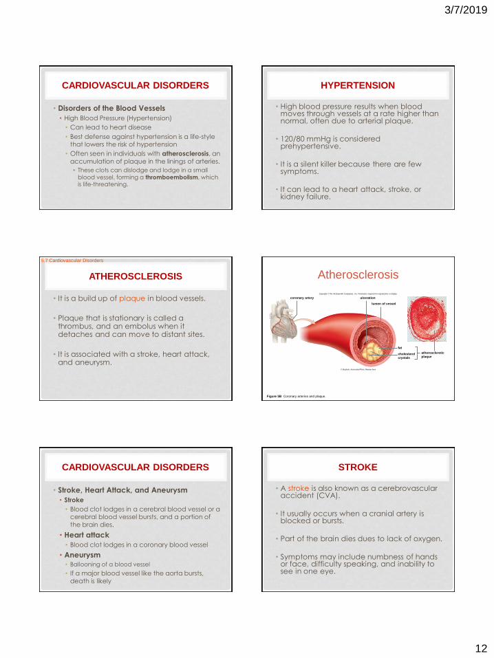
CARDIOVASCULAR DISORDERS
• Disorders of the Blood Vessels
• High Blood Pressure (Hypertension)
• Can lead to heart disease
• Best defense against hypertension is a life-style that lowers the risk of hypertension
• Often seen in individuals with atherosclerosis, an
accumulation of plaque in the linings of arteries.
• These clots can dislodge and lodge in a small
blood vessel, forming a thromboembolism, which is life-threatening.
HYPERTENSION
• High blood pressure results when blood moves through vessels at a rate higher than normal, often due to arterial plaque.
• 120/80 mmHg is considered prehypertensive.
• It is a silent killer because there are few symptoms.
• It can lead to a heart attack, stroke, or kidney failure.
ATHEROSCLEROSIS
• It is a build up of plaque in blood vessels.
• Plaque that is stationary is called a thrombus, and an embolus when it detaches and can move to distant sites.
• It is associated with a stroke, heart attack, and aneurysm.
5.7 Cardiovascular Disorders
Figure 5B Coronary arteries and plaque.
Atherosclerosis
coronary artery ulceration
lumen of vessel
atherosclerotic
plaque cholesterol
crystals
fat
Copyright © The McGraw-Hill Companies, Inc. Permission required for reproduction or display.
© Biophoto Associates/Photo Researchers
CARDIOVASCULAR DISORDERS
• Stroke, Heart Attack, and Aneurysm
• Stroke
• Blood clot lodges in a cerebral blood vessel or a cerebral blood vessel bursts, and a portion of
the brain dies.
• Heart attack
• Blood clot lodges in a coronary blood vessel
• Aneurysm • Ballooning of a blood vessel
• If a major blood vessel like the aorta bursts,
death is likely
STROKE
• A stroke is also known as a cerebrovascular accident (CVA).
• It usually occurs when a cranial artery is blocked or bursts.
• Part of the brain dies dues to lack of oxygen.
• Symptoms may include numbness of hands or face, difficulty speaking, and inability to see in one eye.
3/7/2019
13
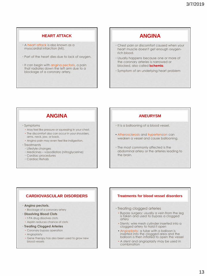
HEART ATTACK
• A heart attack is also known as a myocardial infarction (MI).
• Part of the heart dies due to lack of oxygen.
• It can begin with angina pectoris, a pain that radiates down the left arm due to a blockage of a coronary artery.
ANGINA
• Chest pain or discomfort caused when your
heart muscle doesn't get enough oxygen-
rich blood.
• Usually happens because one or more of
the coronary arteries is narrowed or
blocked, also called ischemia.
• Symptom of an underlying heart problem
ANGINA
• Symptoms
• May feel like pressure or squeezing in your chest.
• The discomfort also can occur in your shoulders, arms, neck, jaw, or back.
• Angina pain may even feel like indigestion.
• Treatments • Lifestyle changes • Medicines – vasodilators (nitroglycerine) • Cardiac procedures
• Cardiac Rehab
ANEURYSM
• It is a ballooning of a blood vessel.
• Atherosclerosis and hypertension can
weaken a vessel and cause ballooning.
• The most commonly affected is the
abdominal artery or the arteries leading to
the brain.
CARDIOVASCULAR DISORDERS
• Angina pectoris.
• Blockage of a coronary artery
• Dissolving Blood Clots
• t-PA drug dissolves clots
• Aspirin reduces chance of clots
• Treating Clogged Arteries
• Coronary bypass operation
• Angioplasty
• Gene therapy has also been used to grow new
blood vessels
Treatments for blood vessel disorders
• Treating clogged arteries
• Bypass surgery: usually a vein from the leg is taken and used to bypass a clogged artery
• Stents: wire mesh cylinder inserted into a clogged artery to hold it open
• Angioplasty: a tube with a balloon is inserted into the clogged area and the balloon is then inflated to open the vessel
• A stent and angioplasty may be used in combination
3/7/2019
14
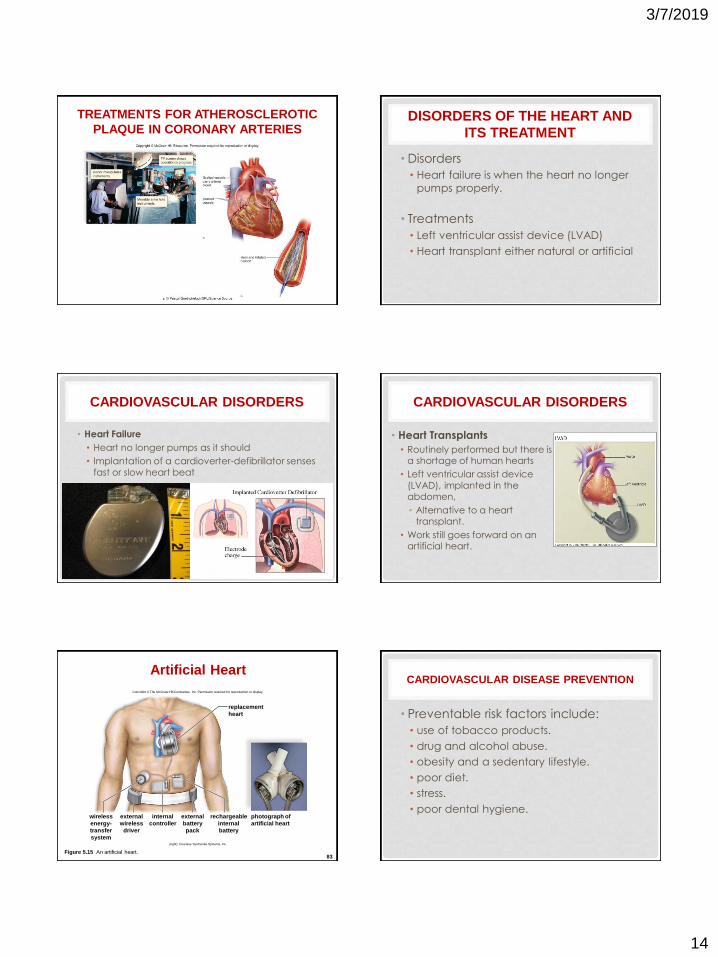
TREATMENTS FOR ATHEROSCLEROTIC
PLAQUE IN CORONARY ARTERIES DISORDERS OF THE HEART AND
ITS TREATMENT
• Disorders
• Heart failure is when the heart no longer
pumps properly.
• Treatments
• Left ventricular assist device (LVAD)
• Heart transplant either natural or artificial
CARDIOVASCULAR DISORDERS
• Heart Failure
• Heart no longer pumps as it should
• Implantation of a cardioverter-defibrillator senses
fast or slow heart beat
CARDIOVASCULAR DISORDERS
• Heart Transplants
• Routinely performed but there is a shortage of human hearts
• Left ventricular assist device
(LVAD), implanted in the abdomen,
• Alternative to a heart transplant.
• Work still goes forward on an artificial heart.
83 Figure 5.15 An artificial heart.
Artificial Heart
Copyright © The McGraw-Hill Companies, Inc. Permission required for reproduction or display.
replacement
heart
photograph of
artificial heart
wireless
energy-
transfer
system
rechargeable
internal
battery
external
battery
pack
internal
controller
external
wireless
driver
(right): Courtesy SynCardia Systems, Inc.
CARDIOVASCULAR DISEASE PREVENTION
• Preventable risk factors include:
• use of tobacco products.
• drug and alcohol abuse.
• obesity and a sedentary lifestyle.
• poor diet.
• stress.
• poor dental hygiene.
3/7/2019
15
HOMEOSTASIS
• All body systems work together to maintain homeostasis.
• How Body Systems Work Together
• The cardiovascular system delivers oxygen and nutrients
to, and takes away metabolic wastes from, the tissue fluid that surrounds cells.
• The lymphatic system returns tissue fluid to the
bloodstream.
• The muscular system makes essential contributions to body movement.
• In the skeletal system, bones contribute calcium ions, which are important to blood clotting.
• The urinary system regulates acid-base and water-salt
balance of blood and tissue fluid.