Embed Size (px)
Citation preview
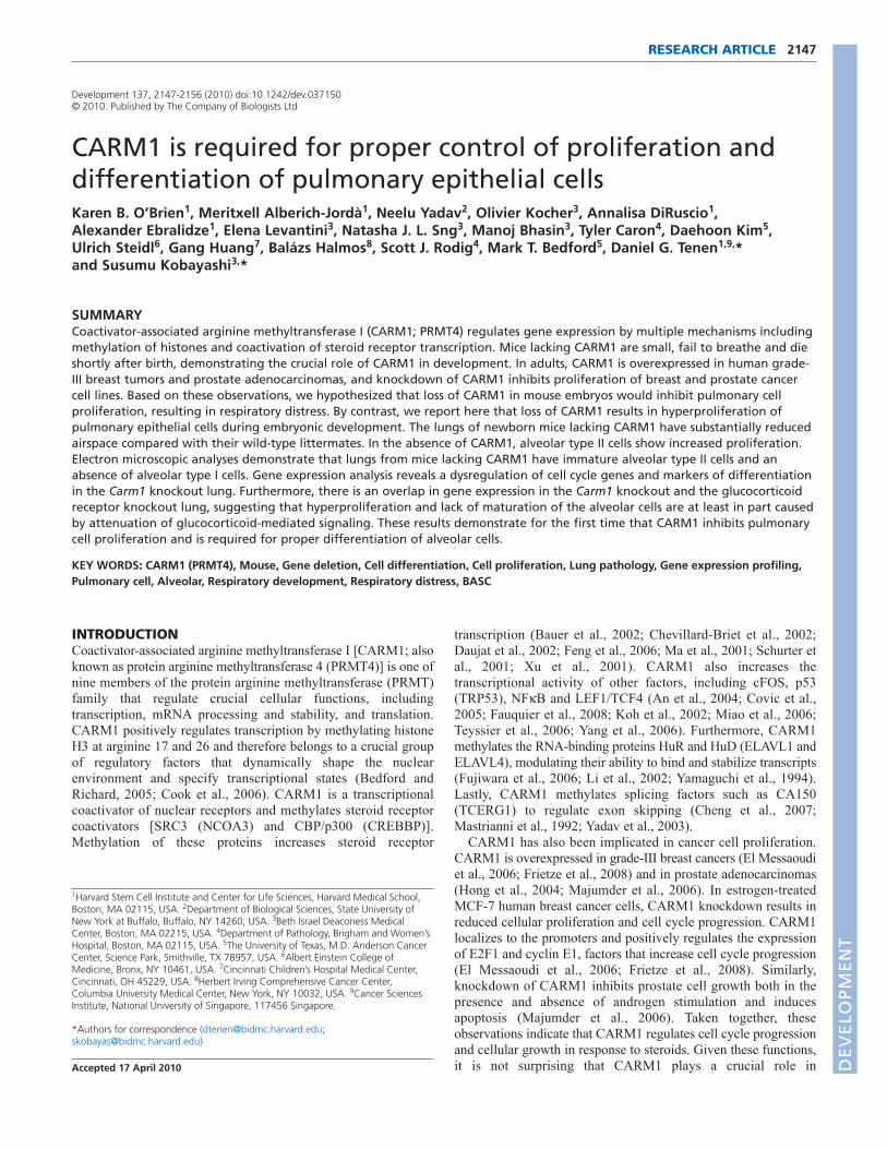
2147RESEARCH ARTICLE
INTRODUCTIONCoactivator-associated arginine methyltransferase I [CARM1; alsoknown as protein arginine methyltransferase 4 (PRMT4)] is one ofnine members of the protein arginine methyltransferase (PRMT)family that regulate crucial cellular functions, includingtranscription, mRNA processing and stability, and translation.CARM1 positively regulates transcription by methylating histoneH3 at arginine 17 and 26 and therefore belongs to a crucial groupof regulatory factors that dynamically shape the nuclearenvironment and specify transcriptional states (Bedford andRichard, 2005; Cook et al., 2006). CARM1 is a transcriptionalcoactivator of nuclear receptors and methylates steroid receptorcoactivators [SRC3 (NCOA3) and CBP/p300 (CREBBP)].Methylation of these proteins increases steroid receptor
transcription (Bauer et al., 2002; Chevillard-Briet et al., 2002;Daujat et al., 2002; Feng et al., 2006; Ma et al., 2001; Schurter etal., 2001; Xu et al., 2001). CARM1 also increases thetranscriptional activity of other factors, including cFOS, p53(TRP53), NFB and LEF1/TCF4 (An et al., 2004; Covic et al.,2005; Fauquier et al., 2008; Koh et al., 2002; Miao et al., 2006;Teyssier et al., 2006; Yang et al., 2006). Furthermore, CARM1methylates the RNA-binding proteins HuR and HuD (ELAVL1 andELAVL4), modulating their ability to bind and stabilize transcripts(Fujiwara et al., 2006; Li et al., 2002; Yamaguchi et al., 1994).Lastly, CARM1 methylates splicing factors such as CA150(TCERG1) to regulate exon skipping (Cheng et al., 2007;Mastrianni et al., 1992; Yadav et al., 2003).
CARM1 has also been implicated in cancer cell proliferation.CARM1 is overexpressed in grade-III breast cancers (El Messaoudiet al., 2006; Frietze et al., 2008) and in prostate adenocarcinomas(Hong et al., 2004; Majumder et al., 2006). In estrogen-treatedMCF-7 human breast cancer cells, CARM1 knockdown results inreduced cellular proliferation and cell cycle progression. CARM1localizes to the promoters and positively regulates the expressionof E2F1 and cyclin E1, factors that increase cell cycle progression(El Messaoudi et al., 2006; Frietze et al., 2008). Similarly,knockdown of CARM1 inhibits prostate cell growth both in thepresence and absence of androgen stimulation and inducesapoptosis (Majumder et al., 2006). Taken together, theseobservations indicate that CARM1 regulates cell cycle progressionand cellular growth in response to steroids. Given these functions,it is not surprising that CARM1 plays a crucial role in
Development 137, 2147-2156 (2010) doi:10.1242/dev.037150© 2010. Published by The Company of Biologists Ltd
1Harvard Stem Cell Institute and Center for Life Sciences, Harvard Medical School,Boston, MA 02115, USA. 2Department of Biological Sciences, State University ofNew York at Buffalo, Buffalo, NY 14260, USA. 3Beth Israel Deaconess MedicalCenter, Boston, MA 02215, USA. 4Department of Pathology, Brigham and Women’sHospital, Boston, MA 02115, USA. 5The University of Texas, M.D. Anderson CancerCenter, Science Park, Smithville, TX 78957, USA. 6Albert Einstein College ofMedicine, Bronx, NY 10461, USA. 7Cincinnati Children’s Hospital Medical Center,Cincinnati, OH 45229, USA. 8Herbert Irving Comprehensive Cancer Center,Columbia University Medical Center, New York, NY 10032, USA. 9Cancer SciencesInstitute, National University of Singapore, 117456 Singapore.
*Authors for correspondence ([email protected];[email protected])
Accepted 17 April 2010
SUMMARYCoactivator-associated arginine methyltransferase I (CARM1; PRMT4) regulates gene expression by multiple mechanisms includingmethylation of histones and coactivation of steroid receptor transcription. Mice lacking CARM1 are small, fail to breathe and dieshortly after birth, demonstrating the crucial role of CARM1 in development. In adults, CARM1 is overexpressed in human grade-III breast tumors and prostate adenocarcinomas, and knockdown of CARM1 inhibits proliferation of breast and prostate cancercell lines. Based on these observations, we hypothesized that loss of CARM1 in mouse embryos would inhibit pulmonary cellproliferation, resulting in respiratory distress. By contrast, we report here that loss of CARM1 results in hyperproliferation ofpulmonary epithelial cells during embryonic development. The lungs of newborn mice lacking CARM1 have substantially reducedairspace compared with their wild-type littermates. In the absence of CARM1, alveolar type II cells show increased proliferation.Electron microscopic analyses demonstrate that lungs from mice lacking CARM1 have immature alveolar type II cells and anabsence of alveolar type I cells. Gene expression analysis reveals a dysregulation of cell cycle genes and markers of differentiationin the Carm1 knockout lung. Furthermore, there is an overlap in gene expression in the Carm1 knockout and the glucocorticoidreceptor knockout lung, suggesting that hyperproliferation and lack of maturation of the alveolar cells are at least in part causedby attenuation of glucocorticoid-mediated signaling. These results demonstrate for the first time that CARM1 inhibits pulmonarycell proliferation and is required for proper differentiation of alveolar cells.
KEY WORDS: CARM1 (PRMT4), Mouse, Gene deletion, Cell differentiation, Cell proliferation, Lung pathology, Gene expression profiling,Pulmonary cell, Alveolar, Respiratory development, Respiratory distress, BASC
CARM1 is required for proper control of proliferation anddifferentiation of pulmonary epithelial cellsKaren B. O’Brien1, Meritxell Alberich-Jordà1, Neelu Yadav2, Olivier Kocher3, Annalisa DiRuscio1,Alexander Ebralidze1, Elena Levantini3, Natasha J. L. Sng3, Manoj Bhasin3, Tyler Caron4, Daehoon Kim5,Ulrich Steidl6, Gang Huang7, Balázs Halmos8, Scott J. Rodig4, Mark T. Bedford5, Daniel G. Tenen1,9,*and Susumu Kobayashi3,*
DEVELO
PMENT

2148
development. Mice with a targeted deletion of Carm1 (Carm1/)are small and have defects in the differentiation of multiple celltypes including T cells and adipocytes (Kim et al., 2004; Yadav etal., 2008; Yadav et al., 2003). Recently, it has been shown that micecarrying the enzyme-dead form of CARM1 phenocopy the Carm1knockout, suggesting that CARM1 requires enzymatic activity forits known cellular functions (Kim et al., 2009). Carm1 knockoutanimals die shortly after birth and suffer from respiratory distress.Carm1/ animals fail to inflate their lungs after birth, and havereduced alveolar air space compared with wild-type littermates.These observations suggest that CARM1 is an important regulatorof lung development. However, detailed studies of CARM1expression and function in lung have not been described.
Development of the distal lung and alveolar sacculation aretightly regulated by a myriad of hormone signals and a cascade ofinteracting transcription factor pathways that are just beginning tobe elucidated (Cardoso and Lu, 2006; Maeda et al., 2007).Progenitor cells in the distal lung differentiate to multiple typesincluding Clara bronchiole epithelial cells and alveolar type II(AT2) cells. AT2 cells are cuboidal and located in the alveolar sacsthat produce the surfactant required to reduce surface tension forthese sacs to fill with air. AT2 cells differentiate to alveolar type I(AT1) epithelial cells that coordinate air exchange to capillaries inthe distal lung. Given that CARM1 functions to enhance thegrowth and proliferation of breast and prostate cancer cells, wehypothesized that the reduced airspace seen in Carm1/ lungsresults from inhibited alveolar cell proliferation, resulting in loss ofsurfactant protein and collapsed alveolar sacs.
In this study, we demonstrate that CARM1 is expressed inpulmonary epithelial cells. Contrary to our expectation that loss ofCARM1 would result in reduced cellular growth, we observedhyperproliferation of AT2 cells in the lung of Carm1/ mice. Wefurther demonstrate a block in differentiation from AT2 to AT1 cellsin the absence of CARM1. Microarray analysis reveals a loss ofexpression of genes crucial for cell cycle regulation and AT1differentiation. Together, these data demonstrate for the first time thatCARM1 is expressed in the lung and is crucial for development,growth and pulmonary epithelial cell function. The data furtherdemonstrate that pulmonary cell proliferation increases in theabsence of CARM1, in direct contrast to findings in other tissues.
MATERIALS AND METHODSMouse generation and genotypingThe generation of mice with targeted disruption of Carm1 (Carm1/ mice)has been described (Yadav et al., 2003). The targeting strategy resulted ina disruption of transcription at exon 2, and a predicted null phenotype thatwas confirmed by immunofluorescence and western blot analysis. Thegenotyping of Carm1/ mice has been described previously (Yadav et al.,2003).
Antibodies and immunohistochemistry (IHC)Lung tissues were fixed in 10% paraformaldehyde overnight at 4°C. Thefollowing antibodies and dilutions were used for IHC: anti-CARM1(1:1000; IHC-00045, Bethyl Laboratories); anti-SPC (1:1000; SC-7705,Santa Cruz Biotechnology); anti-CCSP (1:1000; SC-9772, Santa CruzBiotechnology); anti-AQP5 (1:500; ab78486, Abcam); anti-Ki-67 (1:250;clone SP6, Vector Laboratories); and anti-vWF (1:1000; A0082, DAKO).Fixed, paraffin-embedded tissue sections were deparaffinized andendogenous peroxidase activity was quenched in 1% phosphate-bufferedH2O2 for 15 minutes at room temperature. Antigen retrieval was achievedby steaming slides in 10 mM citric acid (anti-CARM1, -SPC) or EDTA(anti-CCSP, -AQP5, -Ki-67, -vWF). Antibody staining was detected usingthe appropriate mouse or rabbit Envision Kit (DAKO) with DAB andcounterstained with Hematoxylin according to standard protocols. For the
double-marker IHC, slides were incubated with normal horse serumblocking solution for 30 minutes. Subsequently, the slides were incubatedwith the first primary antibody for 1 hour (anti-SPC, -CCSP or -vWF)using a similar DAB detection system as described above for the single-marker IHC. Then, slides were incubated with the second primary antibody(anti-CARM1 and -Ki-67) for 1 hour, followed by detection with thealkaline phosphatase-Fast Red system.
Transmission electron microscopy (TEM)Embryonic lungs were isolated, dissected to 0.5 cm cubes and placed infixing buffer (2.5% glutaraldehyde in 0.1 M sodium cacodylate buffer)overnight at 4°C and subsequently stored at 4°C in 0.1 M sodiumcacodylate buffer. The samples were subsequently dehydrated in ascendingalcohols, cleared with propylene oxide, and infiltrated with a mixture ofEpon resin and propylene oxide overnight. They were then infiltrated withpure Epon resin and polymerized at 60°C for 48 hours. The hardenedblocks were sectioned at 70 nm on a Reichert-Jung Ultracut Eultramicrotome. The sections were placed on nickel grids and stained forcontrast with uranyl acetate and lead citrate. They were viewed andphotographed on a JEOL JEM-1011 electron microscope.
Quantitative real-time (qRT) PCR analysisqRT-PCR analysis was performed in a Rotor Gene 6000 SequenceDetection System (Corbett Life Science). RNA was isolated, DNase Itreated, reverse transcribed, and ~10 ng of the resulting cDNA was used inamplification reactions with SYBR Green PCR Master Mix (AppliedBiosystems) and 500 nM of each gene-specific forward or reverse primer(see Table S1 in the supplementary material). For each gene, at least fourwild-type and three Carm1/ littermates were tested. qRT-PCR reactionsconsisted of one cycle of 95°C for 10 minutes, followed by 40 cycles of95°C for 20 seconds and 60°C for 1 minute.
Western blot analysisTwenty-five micrograms of total lung protein were separated by SDS-PAGE, transferred to nitrocellulose and blocked in 5% non-fat milk.Proteins were immunoblotted with rabbit polyclonal anti-CARM1 (1:2000;ab51742, Abcam), which detects a region that is predicted to be deleted inthe targeted allele.
Flow cytometry and isolation of RNA from sorted populationsPulmonary cells from 12-week-old mice were isolated as described (Kimet al., 2005). Sca-1-FITC, CD45.1- and CD45.2-biotin, pecam1-biotin, andstreptavidin-TC were from BD Pharmingen and viable cells were isolatedbased on the exclusion of propidium iodide. Cell sorting was performedwith a high-speed cell sorter (MoFlo, Beckman Coulter). Cells werecollected in RLT buffer containing 1% -mercaptoethanol and 20 ngbacterial carrier RNA (Roche Diagnostics) per sample according to theRNeasy Micro protocol (Qiagen) optimized for small amounts of RNA.RNA was then reverse transcribed and subjected to qRT-PCR analysis.
Chromatin immunoprecipitation (ChIP) assayCross-linking, nuclei isolation and ChIP assay were performed aspreviously described (Ebralidze et al., 2008). Briefly, lung cells from E18.5wild-type mice were cross-linked with 1% formaldehyde for 10 minutes atroom temperature and nuclei were collected as follows. Approximately1�106 cells were washed three times with ice-cold PBS supplemented with1 mM PMSF. The cell pellet was resuspended in lysis buffer [10 mM Tris-HCl pH 8.0, 10 mM NaCl, 0.5% NP40, freshly supplemented with proteaseinhibitors (Roche Applied Science)], homogenized, and incubated for 15minutes on ice. Nuclei were recovered by centrifugation at 600 g for 10minutes, resuspended in 500 l of storage buffer (1.75 ml water, 2 mlglycerol, 0.2 ml 20� Buffer A) supplemented with protease inhibitors, andstored at –80°C. ChIP was performed using the ChIP-IT Kit according tothe manufacturer’s recommendations (Active Motif) using antibodies toCARM1 (ab51742, Abcam), p53 (sc-6243, Santa Cruz) and glucocorticoidreceptor (ab3579, Abcam) and rabbit IgG (53007, Active Motif). Primersused for Scn3b were 5�-CTAGAGAACAGGAGAAAAGGGCCT-3� and5�-CGAGCTTCGGATAAGCTTTAGGGT-3�. Promoter analysis wasperformed with MatInspector V2.2 software (Quandt et al., 1995).
RESEARCH ARTICLE Development 137 (13)
DEVELO
PMENT

RNA interferenceCarm1-specific (ON-TARGET plus SMART pool) and non-targetingcontrol (ON-TARGET plus Non-Target) small interfering RNAs (siRNA)were purchased from Dharmacon. BEAS-2B cells were transfected usingDharmaFECT transfection reagents (Dharmacon) according to themanufacturer’s protocol. At 48 hours post-transfection, cells wereincubated with RPMI containing 10% FBS in the presence of 0.1% ethanol(control) or 500 nM dexamethasone (Sigma-Aldrich) for an additional 72hours. RNA isolation and qRT-PCR analysis were performed as describedabove.
Statistical analysisThe paired Student’s t-test was used to determine statistical significance.
RESULTSCARM1 is expressed in alveolar type II, Claraepithelial and endothelial cellsSince we observed respiratory distress and reduced airspace in thedistal lung of Carm1/ mice, we first asked which pulmonary cellsexpress CARM1. We stained lung tissue from E18.5 mouseembryos and observed CARM1 expression throughout the lung butnot in all cells (Fig. 1A). CARM1 was found predominantly in thenucleus, with some cytoplasmic staining (Fig. 1B), and expressionwas high in cuboidal alveolar cells and in cells lining the terminalbronchioles (Fig. 1B). As expected, no CARM1 staining wasdetected in lungs from Carm1/ mice (Fig. 1C). To determinewhether CARM1 expression is limited to embryogenesis, westained lung tissue from adult mice with anti-CARM1 andobserved a similar expression pattern to that in embryonic lungs(Fig. 1D). In the adult lung, CARM1 was expressed in both thenucleus and cytoplasm, primarily in rounded epithelial cells (Fig.1E).
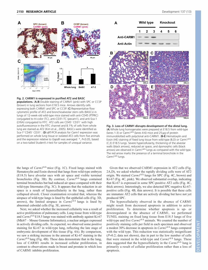
To identify which cells express CARM1, we performed doublestaining of CARM1 with surfactant-associated protein C (SPC;SFTPC – Mouse Genome Informatics), which is expressed by AT2cells. As expected, we observed cytoplasmic staining of SPCthroughout the lung, particularly in rounded cells between airspaces and near the bronchial termini (Fig. 2A, brown stain). WhenSPC staining was combined with that of CARM1 (Fig. 2A, pinkstain), we observed substantial overlap, indicating that CARM1 isexpressed in SPC-positive AT2 cells (Fig. 2A, arrows). In additionto cuboidal cells, we observed CARM1 staining in cellssurrounding the terminal bronchi. Thus, we examined whetherCARM1 is expressed in the CCSP-positive Clara epithelial cellsthat line airways. CCSP (SCGB1A1 – Mouse GenomeInformatics), a crucial secretory protein produced by Clara cells,was observed exclusively in the cells lining the bronchial airwaysof the embryonic lung (Fig. 2B, brown). When combined with anti-CARM1 (Fig. 2B, pink), we observed CCSP staining in thecytoplasm (brown) and CARM1 staining predominantly in thenuclei (pink), indicating that CARM1 is expressed in Clara cells(Fig. 2B). In addition, we observed co-expression of vonWillebrand factor (vWF) and CARM1 (see Fig. S1 in thesupplementary material), demonstrating that CARM1 is expressedin endothelial cells.
It has been proposed that AT2 and Clara cells aredifferentiated from bronchioalveolar stem cells (BASCs), whichare located at the bronchioalveolar junction and are doublepositive for SPC and CCSP (Kim et al., 2005). To determinewhether CARM1 is expressed in BACS, we isolated BASCs andAT2 cells by flow cytometry and measured Carm1 expression inthese populations by qRT-PCR. Fig. 2C is a representativesorting analysis from 8- to 12-week-old mice. We observed that
the BASC population constituted 0.3-0.8% of total lung cellsfrom each animal, whereas the AT2 population ranged from 5 to10%. As shown in Fig. 2D, Carm1 mRNA was expressed inwhole lung and in AT2 and BASCs. Carm1 expression in wholelung constituted 0.48-1.5% of that of Gapdh, reflecting themultiple cell types observed by IHC (Fig. 1). Carm1 expressionin AT2 cells was consistent between animals at 0.8-1.2% ofGapdh. These data support the IHC data demonstrating CARM1protein in AT2 cells. Since the BASC population is so small, wepooled isolated BASCs from two and three animals to obtainenough RNA to accurately measure Carm1 expression. Weobserved Carm1 expression in BASCs at 0.25% and 0.5% ofGapdh, respectively, in each of the pooled populations,demonstrating that Carm1 expression in AT2 cells was 67%higher than in BASCs (P0.02; Fig. 2D).
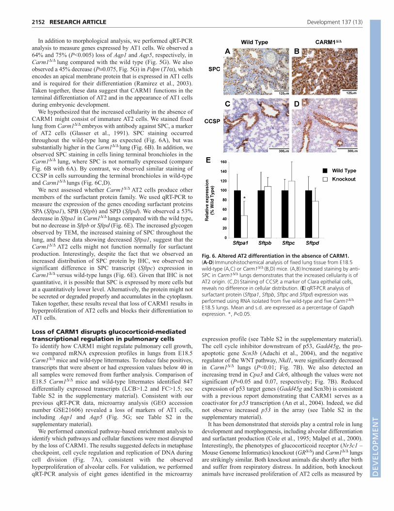
Loss of CARM1 leads to hyperproliferation ofpulmonary cellsWe next sought to identify the role of CARM1 in pulmonarydevelopment. Carm1/ mouse embryos survive to birth andrespond to stimulus but suffer from respiratory distress, fail to turnpink, and die within 20 minutes (Yadav et al., 2003). Western blotanalysis of lung protein (Fig. 3A) confirmed the loss of CARM1 in
2149RESEARCH ARTICLECARM1 regulates lung development
Fig. 1. Immunohistochemical analysis of CARM1 in embryonicand adult murine lung. (A-E)Lung sections from E18.5 (A-C) and 12-week-old (D,E) mice were stained with anti-CARM1. (A)Staining ofCARM1 is observed throughout the embryonic lung. (B)High-magnification image showing that CARM1 is expressed in both thenucleus (left red arrow) and cytoplasm (right red arrow) of cuboidal AT2cells in the spaces between alveolar sacs as well as in epithelial cellslining the terminal bronchioles (black arrows). (C)CARM1 staining isabsent in mice with a targeted deletion of Carm1. (D,E)CARM1expression is observed in cuboidal AT2 cells (D) and in the nucleus (E,left arrow) and cytoplasm (E, right arrow) of adult lung cells.
DEVELO
PMENT
2150
the lungs of Carm1/ mice (Fig. 1C). Fixed lungs stained withHematoxylin and Eosin showed that lungs from wild-type embryos(E18.5) have alveolar sacs with air space and visible terminalbronchioles (Fig. 3B). By contrast, Carm1/ lungs containedterminal bronchioles but had reduced air space compared with theirwild-type littermates (Fig. 3C). It appears that the reduction in airspace is a result of hypercellularity in the lung, rather thancollapsed alveoli. Closer examination revealed that, whereas theairspace of wild-type lungs is lined by flat epithelial cells (Fig. 3D,arrows), the limited airspace in Carm1/ lungs is lined byabnormal cuboidal cells (Fig. 3E, arrows).
Next, we asked whether the increased cellularity was a result ofactive proliferation of pulmonary cells. Lung tissue from wild-typeand Carm1/ E18.5 lungs was stained with antibody against Ki-67(Mki67 – Mouse Genome Informatics), a nuclear antigen expressedin actively dividing cells. As expected, we observed some positivestaining for Ki-67 in wild-type lung, reflecting the late stage ofembryonic development of this tissue (Fig. 4A). By comparison,we saw a striking increase in the amount of Ki-67 staining in theCarm1/ lung (Fig. 4B). These data demonstrate that in the lung,loss of CARM1 results in increased cellular proliferation, incontrast to observations made in breast and prostate in which lossof CARM1 inhibits proliferation.
Given that we observed CARM1 expression in AT2 cells (Fig.2A,D), we asked whether the rapidly dividing cells were of AT2origin. We stained Carm1/ lungs for SPC (Fig. 4C, brown) andKi-67 (Fig. 4C, pink). We observed substantial overlap, indicatingthat Ki-67 is expressed in some SPC-positive AT2 cells (Fig. 4C,thick arrows). Interestingly, we also detected SPC-negative Ki-67-positive cells (Fig. 4B, thin arrows). It is possible that these cellsare immature AT2 cells that are actively dividing but have not yetproduced SPC.
The hypercellularity observed in the absence of CARM1might result from decreased apoptosis in addition to activeproliferation. To determine whether apoptosis is up- ordownregulated in the absence of CARM1, we performedTUNEL staining on fixed lung tissue from E18.5 lungs of fivewild-type and five Carm1/ animals. We counted the number ofpositively staining cells per field in each specimen and observeda modest 30% decrease in apoptosis in Carm1/ lungs comparedwith the wild type. This reduction was statistically insignificant(P0.2; data not shown), due in part to the low number of cellsthat were stained in the wild-type and Carm1/ lungs. Thesedata suggested that the hypercellularity in the Carm1/ lung isprimarily a result of cellular proliferation rather than a loss ofapoptosis.
RESEARCH ARTICLE Development 137 (13)
Fig. 2. CARM1 is expressed in purified AT2 and BASCpopulations. (A,B)Double staining of CARM1 (pink) with SPC or CCSP(brown) in lung sections from E18.5 mice. Arrows identify cellsexpressing both CARM1 and SPC or CCSP. (C)Representative flowcytometric profile of AT2 and bronchioalveolar stem cells (BASCs) inlungs of 12-week-old wild-type mice stained with anti-CD45 (PTPRC)conjugated to tri-color (TC), anti-CD31-TC (pecam1), and anti-Sca-1(LY6A) conjugated to FITC. AT2 cells are CD45– CD31– with highautofluorescence in the FITC channel and 8.1% of cells from wholelung are stained as AT2 (Kim et al., 2005). BASCs were identified asSca-1+ CD45– CD31–. (D)qRT-PCR analysis for Carm1 expression wasperformed on whole lung tissue or isolated AT2 cells from five animalsand the expression relative to Gapdh was averaged. *, P<0.05, basedon a two-tailed Student’s t-test for samples of unequal variance.
Fig. 3. Loss of CARM1 disrupts development of the distal lung.(A)Whole lung homogenates were prepared at E18.5 from wild-type(lanes 1-3) or Carm1/ (lanes 4-6) mice and 25g of proteinimmunoblotted with polyclonal anti-CARM1. (B-E)Hematoxylin andEosin (HE) staining of fixed lung tissue from wild-type (B,D) or Carm1/
(C,E) E18.5 lungs. Severe hypercellularity, thickening of the alveolarwalls (black arrows), reduced air space, and dysmorphic cells (blackarrows) are observed in Carm1/ lungs as compared with the wild type.The red arrow marks the presence of a terminal bronchiole in theCarm1/ lungs.
DEVELO
PMENT

CARM1 is required for differentiation of AT2 cellsNext, we sought to gauge the level of cellular differentiation inCarm1 knockout lungs. During pulmonary development,cytoplasmic glycogen is abundant in immature AT2 cells anddecreases as it is utilized to produce surfactant protein thataccumulates in the cytoplasm in the form of lamellar bodies thatare then secreted into the alveolar space. In addition to their role inproducing surfactant, AT2 cells serve as the precursors of AT1epithelial cells that are required for gas exchange in the distal lung.We used transmission electron microscopy (TEM) to determine thelevel of cellular differentiation in wild-type and Carm1/ lungs. AtE18.5, AT2 cells in wild-type lungs contain some glycogen andvisible lamellar bodies. Furthermore, the pulmonary air space islined by a flat layer of AT1 cells (Fig. 5A). In Carm1/ lungs, AT2cells contained abundant glycogen in the cytoplasm, consistentwith incomplete cellular maturation, and lamellar bodies. Althoughthe presence of lamellar bodies and the production of somesurfactant suggest a degree of maturation of AT2 cells in theabsence of CARM1, we did not observe any AT1 cells in TEMimages of lungs from Carm1/ embryos after analyzing threeknockout embryonic lungs alongside three lungs from wild-typelittermates (Fig. 5B).
To verify the striking loss of AT1 cells in Carm1/ lungs, weperformed IHC with antibody against aquaporin 5 (AQP5), a waterchannel protein that is expressed in, and required for, the function ofAT1 cells (Verkman et al., 2000). We observed AQP5 stainingthroughout wild-type lung (Fig. 5C,E). As expected, staining wasrestricted to cells lining the air space, consistent with the role ofAQP5 in the function of AT1 cells. In the knockout lung, AQP5
expression was observed in an aberrant pattern and was substantiallyreduced (Fig. 5D,F). Many AQP5 positively stained cells wererounded, misshapen and within regions of high cellularity. Inaddition, some of the air spaces in the Carm1/ lungs lacked AQP5-positive cells altogether. These observations indicated a severedepletion of AT1 cells, consistent with observations made by TEM.
2151RESEARCH ARTICLECARM1 regulates lung development
Fig. 4. Increased proliferation of pulmonary cells in the absenceof CARM1. (A,B)Wild-type (A) and Carm1/ (B) E18.5 mouse lungswere fixed and stained for Ki-67, a marker of cell division. Increased Ki-67 staining is observed in Carm1/ lungs as compared with the wildtype. (C)Double staining of Ki-67 (pink) and SPC (brown) in lungsections from E18.5 Carm1/ mice. Thick arrows identify cellsexpressing both Ki-67 and SPC. Thin arrows identify cells expressing Ki-67 but not SPC.
Fig. 5. CARM1 is required for differentiation to AT1 cells in thedeveloping lung. (A,B)Lungs from wild-type (A) and Carm1/ (B)E18.5 mice were fixed and processed for TEM (see Materials andmethods). (A)Lamellar bodies, small patches of cytoplasmic glycogenand AT1 cells are visible in wild-type lung. By contrast, Carm1/ lungs(B) contain an overabundance of glycogen (G) and visible lamellarbodies (L), but no AT1 cells. The presence of lamellar bodies indicatessome AT2 development that is blocked before differentiation to AT1cells. Scale bars: 1m. (C-F)IHC of lungs from wild-type (C,E) andCarm1/ (D,F) E18.5 mice with anti-AQP5 shows reduced staining andabnormal morphology of AT1 cells in Carm1/ lungs. (G)qRT-PCRanalysis of markers of AT1 cells indicates loss of this cell type inCarm1/ lungs. qRT-PCR was performed using RNA isolated from fivewild-type and five Carm1/ E18.5 lungs. Mean and s.d. are expressedas a percentage of Gapdh expression. **, P <0.01; #, P0.075.
DEVELO
PMENT
2152
In addition to morphological analysis, we performed qRT-PCRanalysis to measure genes expressed by AT1 cells. We observed a64% and 75% (P<0.005) loss of Aqp1 and Aqp5, respectively, inCarm1/ lung compared with the wild type (Fig. 5G). We alsoobserved a 45% decrease (P0.075, Fig. 5G) in Pdpn (T1), whichencodes an apical membrane protein that is expressed in AT1 cellsand is required for their differentiation (Ramirez et al., 2003).Taken together, these data suggest that CARM1 functions in theterminal differentiation of AT2 and in the appearance of AT1 cellsduring embryonic development.
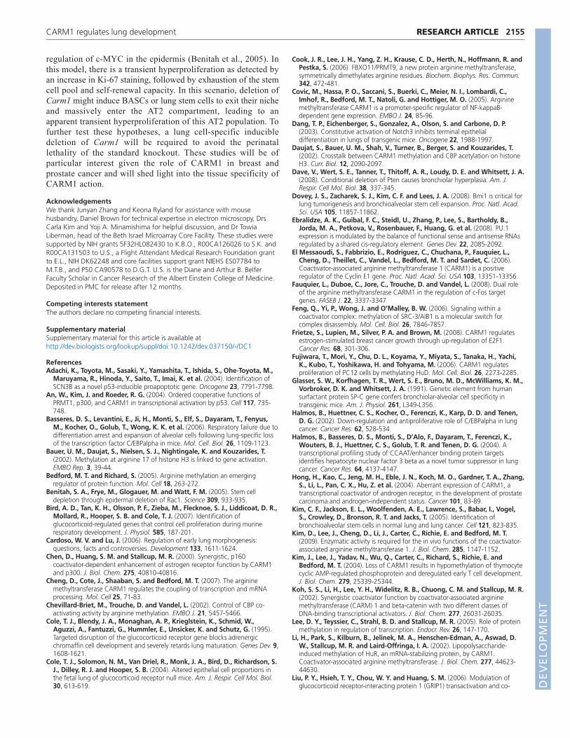
We hypothesized that the increased cellularity in the absence ofCARM1 might consist of immature AT2 cells. We stained fixedlung from Carm1/ embryos with antibody against SPC, a markerof AT2 cells (Glasser et al., 1991). SPC staining occurredthroughout the wild-type lung as expected (Fig. 6A), but wassubstantially higher in the Carm1/ lung (Fig. 6B). In addition, weobserved SPC staining in cells lining terminal bronchioles in theCarm1/ lung, where SPC is not normally expressed (compareFig. 6B with 6A). By contrast, we observed similar staining ofCCSP in cells surrounding the terminal bronchioles in wild-typeand Carm1/ lungs (Fig. 6C,D).
We next assessed whether Carm1/ AT2 cells produce othermembers of the surfactant protein family. We used qRT-PCR tomeasure the expression of the genes encoding surfactant proteinsSPA (Sftpa1), SPB (Sftpb) and SPD (Sftpd). We observed a 53%decrease in Sftpa1 in Carm1/ lungs compared with the wild type,but no decrease in Sftpb or Sftpd (Fig. 6E). The increased glycogenobserved by TEM, the increased staining of SPC throughout thelung, and these data showing decreased Sftpa1, suggest that theCarm1/ AT2 cells might not function normally for surfactantproduction. Interestingly, despite the fact that we observed anincreased distribution of SPC protein by IHC, we observed nosignificant difference in SPC transcript (Sftpc) expression inCarm1/ versus wild-type lungs (Fig. 6E). Given that IHC is notquantitative, it is possible that SPC is expressed by more cells butat a quantitatively lower level. Alternatively, the protein might notbe secreted or degraded properly and accumulates in the cytoplasm.Taken together, these results reveal that loss of CARM1 results inhyperproliferation of AT2 cells and blocks their differentiation toAT1 cells.
Loss of CARM1 disrupts glucocorticoid-mediatedtranscriptional regulation in pulmonary cellsTo identify how CARM1 might regulate pulmonary cell growth,we compared mRNA expression profiles in lungs from E18.5Carm1/ mice and wild-type littermates. To reduce false positives,transcripts that were absent or had expression values below 40 inall samples were removed from further analysis. Comparison ofE18.5 Carm1/ mice and wild-type littermates identified 847differentially expressed transcripts (LCB>1.2 and FC>1.5; seeTable S2 in the supplementary material). Consistent with ourprevious qRT-PCR data, microarray analysis (GEO accessionnumber GSE21606) revealed a loss of markers of AT1 cells,including Aqp1 and Aqp5 (Fig. 5G; see Table S2 in thesupplementary material).
We performed canonical pathway-based enrichment analysis toidentify which pathways and cellular functions were most disruptedby the loss of CARM1. The results suggested defects in metaphasecheckpoint, cell cycle regulation and replication of DNA duringcell division (Fig. 7A), consistent with the observedhyperproliferation of alveolar cells. For validation, we performedqRT-PCR analysis of eight genes identified in the microarray
expression profile (see Table S2 in the supplementary material).The cell cycle inhibitor downstream of p53, Gadd45g, the pro-apoptotic gene Scn3b (Adachi et al., 2004), and the negativeregulator of the WNT pathway, Nkd1, were significantly decreasedin Carm1/ lungs (P<0.01; Fig. 7B). We also detected anincreasing trend in Cpa3 and Cdc6, although the values were notsignificant (P0.05 and 0.07, respectively; Fig. 7B). Reducedexpression of p53 target genes (Gadd45g and Scn3b) is consistentwith a previous report demonstrating that CARM1 serves as acoactivator for p53 transcription (An et al., 2004). Indeed, we didnot observe increased p53 in the array (see Table S2 in thesupplementary material).
It has been demonstrated that steroids play a central role in lungdevelopment and morphogenesis, including alveolar differentiationand surfactant production (Cole et al., 1995; Malpel et al., 2000).Interestingly, the phenotypes of glucocorticoid receptor (Nr3c1 –Mouse Genome Informatics) knockout (GR/) and Carm1/ lungsare strikingly similar. Both knockout animals die shortly after birthand suffer from respiratory distress. In addition, both knockoutanimals have increased proliferation of AT2 cells as measured by
RESEARCH ARTICLE Development 137 (13)
Fig. 6. Altered AT2 differentiation in the absence of CARM1.(A-D)Immunohistochemical analysis of fixed lung tissue from E18.5wild-type (A,C) or Carm1/ (B,D) mice. (A,B)Increased staining by anti-SPC in Carm1/ lungs demonstrates that the increased cellularity is ofAT2 origin. (C,D)Staining of CCSP, a marker of Clara epithelial cells,reveals no difference in cellular distribution. (E)qRT-PCR analysis ofsurfactant protein (Sftpa1, Sftpb, Sftpc and Sftpd) expression wasperformed using RNA isolated from five wild-type and five Carm1/
E18.5 lungs. Mean and s.d. are expressed as a percentage of Gapdhexpression. *, P<0.05.
DEVELO
PMENT

Ki-67 IHC and display a block in differentiation of AT2 cells, withincreased cytoplasmic glycogen while still producing lamellarbodies and expressing some surfactant proteins (Bird et al., 2007;Cole et al., 2004). Previous reports show that CARM1 functions asa coactivator for steroid receptor transcription, including GRactivity (Chen et al., 2000). Therefore, we hypothesized thatCARM1 could be regulating GR transcriptional activity in the lung.We first examined whether CARM1 regulates expression of GR,such that loss of GR might account for the phenotype observed inthe Carm1/ lung. However, we did not see any changes in GR(Nr3c1) mRNA expression by microarray or qRT-PCR (see Fig. S2in the supplementary material).
Recently, gene expression analysis was performed on whole lungtissue from E18.5 wild-type and GR/ lungs (Bird et al., 2007). Todetermine whether CARM1 and GR regulate similar patterns ofgene expression, we performed a comparative analysis of the geneexpression arrays from GR/ and Carm1/ lungs (see Table S3 inthe supplementary material). A significant overlap (P<0.05) of 154genes was identified between GR/ and Carm1/ differentiallyexpressed genes (see Table S3 in the supplementary material). The
overlapping genes account for more than 22% of all genesidentified in the Carm1/ expression profile, and includeGadd45g, Scn3b, Nkd1, Klf9, Ace, Sphk1 and Cdc6 (Fig. 7B).
Next, we investigated whether CARM1 cooperates with GR toinduce target genes in vivo. We investigated Scn3b, which isinduced by DNA damage in a p53-dependent manner (Adachi etal., 2004) and is substantially downregulated in both Carm1/ andGR/ lungs. In addition to a p53 binding site, we identified a GRbinding site in the promoter region of Scn3b. We designed primersto amplify the fragment containing the binding sites for p53 andGR (Fig. 8A), and performed a chromatin immunoprecipitation(ChIP) assay. As shown in Fig. 8B, both p53 and GR bound to thepromoter region. The strong band was also detected in CARM1antibody-treated extracts, suggesting that CARM1 forms acomplex with p53 and/or GR and mediates their transcriptionalactivity.
Finally, we sought to determine whether knockdown of CARM1perturbs glucocorticoid activity in bronchial epithelial cells. To thisend, we used BEAS-2B cells, a human immortalized bronchialepithelial cell line. Cells transiently transfected with siRNA against
2153RESEARCH ARTICLECARM1 regulates lung development
Fig. 7. Gene expression analysis reveals dysregulation of cellcycle-related genes in Carm1/ lungs. (A)Gene expression analysisdemonstrates increases in pathways regulating the metaphasecheckpoint, the cell cycle, cellular division and cytoskeletal remodeling.(B)qRT-PCR analysis validates the microarray results, showing decreasedexpression of Gadd45g, Scn3b, Nkd1, Klf9, Ace, and Sphk1, andincreased expression of Cpa3 and Cdc6. Mean and s.d. are expressed asa percentage of Gapdh expression. *, P<0.05.
Fig. 8. CARM1, GR and p53 interact with the proximal promoter ofthe Scn3b gene. (A)Putative p53 and glucocorticoid receptor (GR)binding sites in the proximal promoter of Scn3b, shown forming acomplex with CARM1. Horizontal arrows indicate the positions of theprimers used in PCR in the ChIP assays. (B)ChIP assay (Southern blotshown) demonstrating binding of GR and p53 to the proximal promoterof Scn3b (gi:149259969) in lung cell isolates from wild-type mice. Notethat CARM1 immunoprecipitation detects Scn3b, suggesting that acomplex forms between CARM1, p53 and GR. (C)Knockdown ofCARM1 by siRNA. Human BEAS-2B cells were transfected with Carm1siRNA (siCarm1), non-target siRNA (non-target), or left untransfected(control) for 48 hours. RNA was isolated and expression of Carm1analyzed by qRT-PCR. Error bars indicate s.d. (n5). (Inset) Expression ofCARM1 as detected by western blot analysis. (D)Upregulation of SCN3Bis suppressed by CARM1 knockdown. BEAS-2B cells transfected withsiRNAs were incubated in the presence of ethanol (vehicle control) or500 nM dexamethasone for 72 hours. RNA was isolated and expressionof SCN3B mRNA was analyzed by qRT-PCR. The fold change in SCN3Bexpression was calculated by comparing dexamethasone-treated andvehicle control-treated cells. Error bars indicate s.d. (n5). ***, P <0.001;NS, not significant.
DEVELO
PMENT

2154
Carm1 showed substantially reduced CARM1 expression at boththe mRNA and protein levels, in comparison to cells transfectedwith non-targeting siRNA or non-transfected control cells (Fig.8C). Next, we examined whether glucocorticoid (dexamethasone)upregulates SCN3B expression in BEAS-2B cells. Indexamethasone-treated control cells or those transfected with non-targeting siRNA, SCN3B expression increased by 1.7±0.25-foldor 1.6±0.22-fold, respectively, compared with vehicle-treatedcells. However, SCN3B expression was downregulated withdexamethasone treatment in CARM1 knockdown cells (0.5±0.07-fold; Fig. 8D), suggesting not only that CARM1 is required forinduction of SCN3B upon glucocorticoid stimulation, but also thatglucocorticoid might negatively regulate SCN3B expression in theabsence of CARM1. By contrast, dexamethasone treatmentupregulated CDC6 in CARM1 knockdown cells, whereas thetreatment had a minimal effect on CDC6 expression in control cells(2.0±0.2-fold; see Fig. S3 in the supplementary material). Theseresults are consistent with our observation that Cdc6 wasupregulated in Carm1/ mouse lung, and indicate that CARM1may suppress CDC6 upregulation induced by dexamethasone (Fig.7B).
Taken together, these data suggest that CARM1 and GR regulatea similar array of genes in embryonic lung, and that the effects ofglucocorticoid hormone on gene transcription are at least in partdependent on CARM1.
DISCUSSIONThe results of this study demonstrate a crucial role for CARM1 inpulmonary development. We show CARM1 expression in airwayand alveolar epithelial cells, BASCs and endothelial cells. Wedemonstrate that AT2 cells fail to complete differentiation andhyperproliferate in the absence of CARM1. We also show a blockin AT1 cell differentiation in Carm1/ lungs. Lastly, geneexpression analysis reveals a dysregulation of genes that regulatethe cell cycle and proliferation, and a striking overlap betweenCARM1 and GR gene expression signatures, indicating that loss ofCARM1 disrupts GR signaling.
The finding that AT2 cells hyperproliferate in the absence ofCARM1 had not been anticipated, given that CARM1 expressionis increased in breast and prostate tumors, and knockdown ofCARM1 in these cell types results in decreased proliferation inresponse to hormone stimulation (El Messaoudi et al., 2006; Frietzeet al., 2008; Hong et al., 2004; Majumder et al., 2006). In thesestudies, CARM1 was shown to positively regulate cyclin E1 andE2F1, and it was hypothesized that loss of these factors contributesto the loss of proliferation in the CARM1 knockdown cells. We didnot see changes in the expression of these factors in our geneexpression analysis or in qRT-PCR of RNA from Carm1/ lungs(see Fig. S2 in the supplementary material). These results suggestthat the role of CARM1 is cell-type specific.
The overlap in gene expression in the Carm1/ lung and GR/
lung is striking. Eleven out of 23 genes that we identified asregulating the cell cycle, cell proliferation and apoptosis in theCarm1/ lung are also dysregulated in GR/ lungs. These datasupport the hypothesis that, at least in part, CARM1 functions withGR to regulate the expression of genes that regulate cellularproliferation and are consistent with previous reports that CARM1functions as a coactivator of GR transcription. CARM1 has beenshown to bind glucocorticoid-interacting protein (GRIP1) and tosynergistically function to increase GR transcriptional activity inresponse to the synthetic corticosteroid, dexamethasone (Lee et al.,2005; Liu et al., 2006; Teyssier et al., 2006). Consistent with these
reports, we demonstrated that knockdown of CARM1 disruptschanges in gene expression induced by dexamethasone in bronchialepithelial cells (Fig. 8D; see Fig. S3 in the supplementary material).
Although the lung phenotypes of Carm1/ and GR/ aresimilar, CARM1 is likely to function in additional capacities duringlung development, other than solely as a coactivator for GR.Although the overlap in dysregulated genes observed in theexpression arrays is substantial, it represents only a fraction of thegenes dysregulated in each of the knockout animals. Severalother mouse models display similar phenotypes to Carm1/
animals. Loss of the transcription factor C/EBP results inhyperproliferation of AT2 cells, a block in AT2 differentiation andloss of AT1 cells (Basseres et al., 2006; Martis et al., 2006). In thecase of C/EBP, the block in AT2 differentiation is earlier than forCARM1, with no lamellar bodies and more substantial loss ofsurfactant proteins. We performed western blot analysis and qRT-PCR and observed no loss of C/EBP in Carm1/ lungs (data notshown). Similarly, gene expression profiling reveals no loss ofCarm1 expression in lungs with a targeted deletion of Cebpa inSPC-producing pulmonary cells (Basseres et al., 2006). In addition,qRT-PCR of RNA from Carm1/ lungs shows normal expressionof Foxa2 (Hnf3), a transcriptional target of C/EBP, the loss ofwhich results in a similar phenotype to Cebpa knockouts (Halmoset al., 2004; Wan et al., 2004). Overexpression of NOTCH3 alsoleads to a block in AT2 and AT1 differentiation, although withouthyperproliferation of AT2 cells (Dang et al., 2003), and loss ofKLF5 results in a similar phenotype (Wan et al., 2008). Targeteddeletion of Pdpn is also perinatal lethal, with loss of AT1 cells andhyperproliferation of alveolar epithelial cells at a laterdevelopmental stage than for Carm1/ lungs (perinatal) (Ramirezet al., 2003). Loss of Pdpn in Carm1/ lungs is likely to contributeto the block in AT1 differentiation. However, given that CARM1methylates histones as well as transcriptional regulators, furtherstudy will be required to determine which genes are directlyregulated by CARM1 and whether this regulation is throughhistone methylation or interactions with transcription factors suchas C/EBP and FOXA2.
Notably, several of the factors, the loss of which indevelopment leads to hyperproliferation of alveolar cells, arealso downregulated in lung cancer. C/EBP and FOXA2 aredownregulated in lung cancer and function as tumor suppressors(Halmos et al., 2004; Halmos et al., 2002; Tada et al., 2006).Thus, this phenotype might be predictive of tumorigenesisresulting from loss of function in adulthood. As such, loss ofCARM1 expression might lead to increased cell growth andtumor formation. This hypothesis is particularly attractive giventhat CARM1 is expressed in the BASC population. Indeed, lossof growth regulation in BASCs has been shown to contribute totumorigenesis in several animal models (Dave et al., 2008;Dovey et al., 2008; Kim et al., 2005). To test this hypothesis, onewould ideally first determine whether the BASC populationexists in the absence of CARM1. However, we were unable toisolate BASCs by FACS from Carm1/ or wild-type embryoniclung. Indeed, the BASC population increases with age and isundetectable in embryonic lung (our unpublished observations).Given that the role of BASCs in the self-renewal of lung cellsremains ambiguous (Rawlins et al., 2009), further studies will benecessary to confirm how or if CARM1 functions in lung stemcells. Other molecular models might provide explanations as tohow the loss of CARM1 induces hyperproliferation of alveolarcells. It has been show that deletion of Rac1 stimulatesepidermal stem cells to exit their niche and proliferate through
RESEARCH ARTICLE Development 137 (13)
DEVELO
PMENT
regulation of c-MYC in the epidermis (Benitah et al., 2005). Inthis model, there is a transient hyperproliferation as detected byan increase in Ki-67 staining, followed by exhaustion of the stemcell pool and self-renewal capacity. In this scenario, deletion ofCarm1 might induce BASCs or lung stem cells to exit their nicheand massively enter the AT2 compartment, leading to anapparent transient hyperproliferation of this AT2 population. Tofurther test these hypotheses, a lung cell-specific inducibledeletion of Carm1 will be required to avoid the perinatallethality of the standard knockout. These studies will be ofparticular interest given the role of CARM1 in breast andprostate cancer and will shed light into the tissue specificity ofCARM1 action.
AcknowledgementsWe thank Junyan Zhang and Keona Ryland for assistance with mousehusbandry, Daniel Brown for technical expertise in electron microscopy, DrsCarla Kim and Yoji A. Minamishima for helpful discussion, and Dr TowiaLiberman, head of the Beth Israel Microarray Core Facility. These studies weresupported by NIH grants 5F32HL082430 to K.B.O., R00CA126026 to S.K. andR00CA131503 to U.S., a Flight Attendant Medical Research Foundation grantto E.L., NIH DK62248 and core facilities support grant NIEHS ES07784 toM.T.B., and P50 CA90578 to D.G.T. U.S. is the Diane and Arthur B. BelferFaculty Scholar in Cancer Research of the Albert Einstein College of Medicine.Deposited in PMC for release after 12 months.
Competing interests statementThe authors declare no competing financial interests.
Supplementary materialSupplementary material for this article is available athttp://dev.biologists.org/lookup/suppl/doi:10.1242/dev.037150/-/DC1
ReferencesAdachi, K., Toyota, M., Sasaki, Y., Yamashita, T., Ishida, S., Ohe-Toyota, M.,
Maruyama, R., Hinoda, Y., Saito, T., Imai, K. et al. (2004). Identification ofSCN3B as a novel p53-inducible proapoptotic gene. Oncogene 23, 7791-7798.
An, W., Kim, J. and Roeder, R. G. (2004). Ordered cooperative functions ofPRMT1, p300, and CARM1 in transcriptional activation by p53. Cell 117, 735-748.
Basseres, D. S., Levantini, E., Ji, H., Monti, S., Elf, S., Dayaram, T., Fenyus,M., Kocher, O., Golub, T., Wong, K. K. et al. (2006). Respiratory failure due todifferentiation arrest and expansion of alveolar cells following lung-specific lossof the transcription factor C/EBPalpha in mice. Mol. Cell. Biol. 26, 1109-1123.
Bauer, U. M., Daujat, S., Nielsen, S. J., Nightingale, K. and Kouzarides, T.(2002). Methylation at arginine 17 of histone H3 is linked to gene activation.EMBO Rep. 3, 39-44.
Bedford, M. T. and Richard, S. (2005). Arginine methylation an emergingregulator of protein function. Mol. Cell 18, 263-272.
Benitah, S. A., Frye, M., Glogauer, M. and Watt, F. M. (2005). Stem celldepletion through epidermal deletion of Rac1. Science 309, 933-935.
Bird, A. D., Tan, K. H., Olsson, P. F., Zieba, M., Flecknoe, S. J., Liddicoat, D. R.,Mollard, R., Hooper, S. B. and Cole, T. J. (2007). Identification ofglucocorticoid-regulated genes that control cell proliferation during murinerespiratory development. J. Physiol. 585, 187-201.
Cardoso, W. V. and Lu, J. (2006). Regulation of early lung morphogenesis:questions, facts and controversies. Development 133, 1611-1624.
Chen, D., Huang, S. M. and Stallcup, M. R. (2000). Synergistic, p160coactivator-dependent enhancement of estrogen receptor function by CARM1and p300. J. Biol. Chem. 275, 40810-40816.
Cheng, D., Cote, J., Shaaban, S. and Bedford, M. T. (2007). The argininemethyltransferase CARM1 regulates the coupling of transcription and mRNAprocessing. Mol. Cell 25, 71-83.
Chevillard-Briet, M., Trouche, D. and Vandel, L. (2002). Control of CBP co-activating activity by arginine methylation. EMBO J. 21, 5457-5466.
Cole, T. J., Blendy, J. A., Monaghan, A. P., Krieglstein, K., Schmid, W.,Aguzzi, A., Fantuzzi, G., Hummler, E., Unsicker, K. and Schutz, G. (1995).Targeted disruption of the glucocorticoid receptor gene blocks adrenergicchromaffin cell development and severely retards lung maturation. Genes Dev. 9,1608-1621.
Cole, T. J., Solomon, N. M., Van Driel, R., Monk, J. A., Bird, D., Richardson, S.J., Dilley, R. J. and Hooper, S. B. (2004). Altered epithelial cell proportions inthe fetal lung of glucocorticoid receptor null mice. Am. J. Respir. Cell Mol. Biol.30, 613-619.
Cook, J. R., Lee, J. H., Yang, Z. H., Krause, C. D., Herth, N., Hoffmann, R. andPestka, S. (2006). FBXO11/PRMT9, a new protein arginine methyltransferase,symmetrically dimethylates arginine residues. Biochem. Biophys. Res. Commun.342, 472-481.
Covic, M., Hassa, P. O., Saccani, S., Buerki, C., Meier, N. I., Lombardi, C.,Imhof, R., Bedford, M. T., Natoli, G. and Hottiger, M. O. (2005). Argininemethyltransferase CARM1 is a promoter-specific regulator of NF-kappaB-dependent gene expression. EMBO J. 24, 85-96.
Dang, T. P., Eichenberger, S., Gonzalez, A., Olson, S. and Carbone, D. P.(2003). Constitutive activation of Notch3 inhibits terminal epithelialdifferentiation in lungs of transgenic mice. Oncogene 22, 1988-1997.
Daujat, S., Bauer, U. M., Shah, V., Turner, B., Berger, S. and Kouzarides, T.(2002). Crosstalk between CARM1 methylation and CBP acetylation on histoneH3. Curr. Biol. 12, 2090-2097.
Dave, V., Wert, S. E., Tanner, T., Thitoff, A. R., Loudy, D. E. and Whitsett, J. A.(2008). Conditional deletion of Pten causes bronchiolar hyperplasia. Am. J.Respir. Cell Mol. Biol. 38, 337-345.
Dovey, J. S., Zacharek, S. J., Kim, C. F. and Lees, J. A. (2008). Bmi1 is critical forlung tumorigenesis and bronchioalveolar stem cell expansion. Proc. Natl. Acad.Sci. USA 105, 11857-11862.
Ebralidze, A. K., Guibal, F. C., Steidl, U., Zhang, P., Lee, S., Bartholdy, B.,Jorda, M. A., Petkova, V., Rosenbauer, F., Huang, G. et al. (2008). PU.1expression is modulated by the balance of functional sense and antisense RNAsregulated by a shared cis-regulatory element. Genes Dev. 22, 2085-2092.
El Messaoudi, S., Fabbrizio, E., Rodriguez, C., Chuchana, P., Fauquier, L.,Cheng, D., Theillet, C., Vandel, L., Bedford, M. T. and Sardet, C. (2006).Coactivator-associated arginine methyltransferase 1 (CARM1) is a positiveregulator of the Cyclin E1 gene. Proc. Natl. Acad. Sci. USA 103, 13351-13356.
Fauquier, L., Duboe, C., Jore, C., Trouche, D. and Vandel, L. (2008). Dual roleof the arginine methyltransferase CARM1 in the regulation of c-Fos targetgenes. FASEB J. 22, 3337-3347
Feng, Q., Yi, P., Wong, J. and O’Malley, B. W. (2006). Signaling within acoactivator complex: methylation of SRC-3/AIB1 is a molecular switch forcomplex disassembly. Mol. Cell. Biol. 26, 7846-7857.
Frietze, S., Lupien, M., Silver, P. A. and Brown, M. (2008). CARM1 regulatesestrogen-stimulated breast cancer growth through up-regulation of E2F1.Cancer Res. 68, 301-306.
Fujiwara, T., Mori, Y., Chu, D. L., Koyama, Y., Miyata, S., Tanaka, H., Yachi,K., Kubo, T., Yoshikawa, H. and Tohyama, M. (2006). CARM1 regulatesproliferation of PC12 cells by methylating HuD. Mol. Cell. Biol. 26, 2273-2285.
Glasser, S. W., Korfhagen, T. R., Wert, S. E., Bruno, M. D., McWilliams, K. M.,Vorbroker, D. K. and Whitsett, J. A. (1991). Genetic element from humansurfactant protein SP-C gene confers bronchiolar-alveolar cell specificity intransgenic mice. Am. J. Physiol. 261, L349-L356.
Halmos, B., Huettner, C. S., Kocher, O., Ferenczi, K., Karp, D. D. and Tenen,D. G. (2002). Down-regulation and antiproliferative role of C/EBPalpha in lungcancer. Cancer Res. 62, 528-534.
Halmos, B., Basseres, D. S., Monti, S., D’Alo, F., Dayaram, T., Ferenczi, K.,Wouters, B. J., Huettner, C. S., Golub, T. R. and Tenen, D. G. (2004). Atranscriptional profiling study of CCAAT/enhancer binding protein targetsidentifies hepatocyte nuclear factor 3 beta as a novel tumor suppressor in lungcancer. Cancer Res. 64, 4137-4147.
Hong, H., Kao, C., Jeng, M. H., Eble, J. N., Koch, M. O., Gardner, T. A., Zhang,S., Li, L., Pan, C. X., Hu, Z. et al. (2004). Aberrant expression of CARM1, atranscriptional coactivator of androgen receptor, in the development of prostatecarcinoma and androgen-independent status. Cancer 101, 83-89.
Kim, C. F., Jackson, E. L., Woolfenden, A. E., Lawrence, S., Babar, I., Vogel,S., Crowley, D., Bronson, R. T. and Jacks, T. (2005). Identification ofbronchioalveolar stem cells in normal lung and lung cancer. Cell 121, 823-835.
Kim, D., Lee, J., Cheng, D., Li, J., Carter, C., Richie, E. and Bedford, M. T.(2009). Enzymatic activity is required for the in vivo functions of the coactivator-associated arginine methyltransferase 1. J. Biol. Chem. 285, 1147-1152.
Kim, J., Lee, J., Yadav, N., Wu, Q., Carter, C., Richard, S., Richie, E. andBedford, M. T. (2004). Loss of CARM1 results in hypomethylation of thymocytecyclic AMP-regulated phosphoprotein and deregulated early T cell development.J. Biol. Chem. 279, 25339-25344.
Koh, S. S., Li, H., Lee, Y. H., Widelitz, R. B., Chuong, C. M. and Stallcup, M. R.(2002). Synergistic coactivator function by coactivator-associated argininemethyltransferase (CARM) 1 and beta-catenin with two different classes ofDNA-binding transcriptional activators. J. Biol. Chem. 277, 26031-26035.
Lee, D. Y., Teyssier, C., Strahl, B. D. and Stallcup, M. R. (2005). Role of proteinmethylation in regulation of transcription. Endocr. Rev. 26, 147-170.
Li, H., Park, S., Kilburn, B., Jelinek, M. A., Henschen-Edman, A., Aswad, D.W., Stallcup, M. R. and Laird-Offringa, I. A. (2002). Lipopolysaccharide-induced methylation of HuR, an mRNA-stabilizing protein, by CARM1.Coactivator-associated arginine methyltransferase. J. Biol. Chem. 277, 44623-44630.
Liu, P. Y., Hsieh, T. Y., Chou, W. Y. and Huang, S. M. (2006). Modulation ofglucocorticoid receptor-interacting protein 1 (GRIP1) transactivation and co-
2155RESEARCH ARTICLECARM1 regulates lung development
DEVELO
PMENT
2156
activation activities through its C-terminal repression and self-associationdomains. FEBS J. 273, 2172-2183.
Ma, H., Baumann, C. T., Li, H., Strahl, B. D., Rice, R., Jelinek, M. A., Aswad,D. W., Allis, C. D., Hager, G. L. and Stallcup, M. R. (2001). Hormone-dependent, CARM1-directed, arginine-specific methylation of histone H3 on asteroid-regulated promoter. Curr. Biol. 11, 1981-1985.
Maeda, Y., Dave, V. and Whitsett, J. A. (2007). Transcriptional control of lungmorphogenesis. Physiol. Rev. 87, 219-244.
Majumder, S., Liu, Y., Ford, O. H., 3rd, Mohler, J. L. and Whang, Y. E. (2006).Involvement of arginine methyltransferase CARM1 in androgen receptorfunction and prostate cancer cell viability. Prostate 66, 1292-1301.
Malpel, S., Mendelsohn, C. and Cardoso, W. V. (2000). Regulation of retinoicacid signaling during lung morphogenesis. Development 127, 3057-3067.
Martis, P. C., Whitsett, J. A., Xu, Y., Perl, A. K., Wan, H. and Ikegami, M.(2006). C/EBPalpha is required for lung maturation at birth. Development 133,1155-1164.
Mastrianni, D. M., Eddy, R. L., Rosenberg, H. F., Corrette, S. E., Shows, T. B.,Tenen, D. G. and Ackerman, S. J. (1992). Localization of the human eosinophilCharcot-Leyden crystal protein (lysophospholipase) gene (CLC) to chromosome19 and the human ribonuclease 2 (eosinophil-derived neurotoxin) andribonuclease 3 (eosinophil cationic protein) genes (RNS2 and RNS3) tochromosome 14. Genomics 13, 240-242.
Miao, F., Li, S., Chavez, V., Lanting, L. and Natarajan, R. (2006). Coactivator-associated arginine methyltransferase-1 enhances nuclear factor-kappaB-mediated gene transcription through methylation of histone H3 at arginine 17.Mol. Endocrinol. 20, 1562-1573.
Quandt, K., Frech, K., Karas, H., Wingender, E. and Werner, T. (1995). MatIndand MatInspector: new fast and versatile tools for detection of consensusmatches in nucleotide sequence data. Nucleic Acids Res. 23, 4878-4884.
Ramirez, M. I., Millien, G., Hinds, A., Cao, Y., Seldin, D. C. and Williams, M.C. (2003). T1alpha, a lung type I cell differentiation gene, is required for normallung cell proliferation and alveolus formation at birth. Dev. Biol. 256, 61-72.
Rawlins, E. L., Okubo, T., Xue, Y., Brass, D. M., Auten, R. L., Hasegawa, H.,Wang, F. and Hogan, B. L. (2009). The role of Scgb1a1+ Clara cells in thelong-term maintenance and repair of lung airway, but not alveolar, epithelium.Cell Stem Cell 4, 525-534.
Schurter, B. T., Koh, S. S., Chen, D., Bunick, G. J., Harp, J. M., Hanson, B. L.,Henschen-Edman, A., Mackay, D. R., Stallcup, M. R. and Aswad, D. W.(2001). Methylation of histone H3 by coactivator-associated argininemethyltransferase 1. Biochemistry 40, 5747-5756.
Tada, Y., Brena, R. M., Hackanson, B., Morrison, C., Otterson, G. A. andPlass, C. (2006). Epigenetic modulation of tumor suppressor CCAAT/enhancerbinding protein alpha activity in lung cancer. J. Natl. Cancer Inst. 98, 396-406.
Teyssier, C., Ou, C. Y., Khetchoumian, K., Losson, R. and Stallcup, M. R.(2006). Transcriptional intermediary factor 1alpha mediates physical interactionand functional synergy between the coactivator-associated argininemethyltransferase 1 and glucocorticoid receptor-interacting protein 1 nuclearreceptor coactivators. Mol. Endocrinol. 20, 1276-1286.
Verkman, A. S., Matthay, M. A. and Song, Y. (2000). Aquaporin water channelsand lung physiology. Am. J. Physiol. Lung Cell Mol. Physiol. 278, L867-L879.
Wan, H., Xu, Y., Ikegami, M., Stahlman, M. T., Kaestner, K. H., Ang, S. L. andWhitsett, J. A. (2004). Foxa2 is required for transition to air breathing at birth.Proc. Natl. Acad. Sci. USA 101, 14449-14454.
Wan, H., Luo, F., Wert, S. E., Zhang, L., Xu, Y., Ikegami, M., Maeda, Y., Bell,S. M. and Whitsett, J. A. (2008). Kruppel-like factor 5 is required for perinatallung morphogenesis and function. Development 135, 2563-2572.
Xu, W., Chen, H., Du, K., Asahara, H., Tini, M., Emerson, B. M., Montminy,M. and Evans, R. M. (2001). A transcriptional switch mediated by cofactormethylation. Science 294, 2507-2511.
Yadav, N., Lee, J., Kim, J., Shen, J., Hu, M. C., Aldaz, C. M. and Bedford, M. T.(2003). Specific protein methylation defects and gene expression perturbationsin coactivator-associated arginine methyltransferase 1-deficient mice. Proc. Natl.Acad. Sci. USA 100, 6464-6468.
Yadav, N., Cheng, D., Richard, S., Morel, M., Iyer, V. R., Aldaz, C. M. andBedford, M. T. (2008). CARM1 promotes adipocyte differentiation bycoactivating PPARgamma. EMBO Rep. 9, 193-198.
Yamaguchi, Y., Tenen, D. G. and Ackerman, S. J. (1994). Transcriptionalregulation of the human eosinophil peroxidase genes: characterization of aperoxidase promoter. Int. Arch. Allergy Immunol. 104, 30-31.
Yang, C. K., Kim, J. H., Li, H. and Stallcup, M. R. (2006). Differential use offunctional domains by coiled-coil coactivator in its synergistic coactivatorfunction with beta-catenin or GRIP1. J. Biol. Chem. 281, 3389-3397.
RESEARCH ARTICLE Development 137 (13)
DEVELO
PMENT