Embed Size (px)
Citation preview
ORIGINAL RESEARCHpublished: 11 May 2016
doi: 10.3389/fmicb.2016.00684
Frontiers in Microbiology | www.frontiersin.org 1 May 2016 | Volume 7 | Article 684
Edited by:
Octavio Luiz Franco,
Universidade Catolica de Brasilia,
Brazil
Reviewed by:
Jack Wong,
The Chinese University of Hong Kong,
Hong Kong
Sónia Gonçalves,
Universidade de Lisboa, Portugal
*Correspondence:
Michael Reck
Specialty section:
This article was submitted to
Antimicrobials, Resistance and
Chemotherapy,
a section of the journal
Frontiers in Microbiology
Received: 15 December 2015
Accepted: 26 April 2016
Published: 11 May 2016
Citation:
Reck M and Wagner-Döbler I (2016)
Carolacton Treatment Causes
Delocalization of the Cell Division
Proteins PknB and DivIVa in
Streptococcus mutans in vivo.
Front. Microbiol. 7:684.
doi: 10.3389/fmicb.2016.00684
Carolacton Treatment CausesDelocalization of the Cell DivisionProteins PknB and DivIVa inStreptococcus mutans in vivo
Michael Reck* and Irene Wagner-Döbler
Department of Microbiology, Microbial Communication, Helmholtz Centre for Infection Research, Braunschweig, Germany
The small inhibitory molecule Carolacton has been shown to cause chain formation
and bulging in Streptococci, suggesting a defect in cell division, but it is not known
how cell division is impaired on a molecular level. Fluorescent fusion proteins have
successfully been applied to visualize protein localization and dynamics in vivo and have
revolutionized our understanding of cell wall growth, cell division, chromosome replication
and segregation. However, in Streptococci the required vectors are largely lacking. We
constructed vectors for chromosomal integration and inducible expression of fluorescent
fusion proteins based on GFP+ in S. mutans. Their applicability was verified using four
proteins with known localization in the cell. We then determined the effect of Carolacton
on the subcellular localization of GFP+ fusions of the cell division protein DivIVa and
the serine-threonine protein kinase PknB. Carolacton caused a significant delocalization
of these proteins from midcell, in accordance with a previous study demonstrating the
Carolacton insensitive phenotype of a pknB deletion strain. Carolacton treated cells
displayed an elongated phenotype, increased septum formation and a severe defect
in daughter cell separation. GFP+ fusions of two hypothetical proteins (SMU_503 and
SMU_609), that had previously been shown to be the most strongly upregulated genes
after Carolacton treatment, were found to be localized at the septum in midcell, indicating
their role in cell division. These findings highlight the importance of PknB as a key
regulator of cell division in streptococci and indicate a profound impact of Carolacton on
the coordination between peripheral and septal cell wall growth. The established vector
system represents a novel tool to study essential steps of cellular metabolism.
Keywords: protein localization, fluorescent fusion protein, inducible expression vectors, streptococci, single cell
analysis, Carolacton, cell division, serine/threonine protein kinase
INTRODUCTION
Understanding the biological function of a protein requires knowledge of its cellular localizationas exemplified, e.g., by the polar localization of chemotaxis protein clusters and the midcelllocalization of the divisome complex (Rudner and Losick, 2010; Nevo-Dinur et al., 2012).The correct spatio-temporal distribution of a protein is essential for its biological function(Govindarajan et al., 2012; Nevo-Dinur et al., 2012). Disturbing the localization pattern ofessential bacterial proteins is thus a promising strategy for the development of novel antimicrobialsubstances (Govindarajan et al., 2012).
Reck and Wagner-Döbler Analyzing Antimicrobials Using Fluorescent Proteins
Carolacton is a small inhibitorymolecule active against cells ofthe caries pathogen Streptococcus mutans growing under acidicconditions (Kunze et al., 2010). The strict stereospecific activityat nanomolar concentrations suggests that the substance actsvia a completely novel mode of action and has a moleculartarget which is present in only a few copies per cell (Stumppet al., 2015). Cell elongation, chain formation and bulging hasbeen observed in cultures treated with Carolacton and suggestdefects in cell division and a weakened cell wall (Kunze et al.,2010; Reck et al., 2011; Stumpp et al., 2015). A transcriptomeanalysis of Carolacton treated cells of S. mutans revealeddifferential expression of genes encoding proteins involvedin cell division and the down-regulation of the VicKR twocomponent system controlling cell wall metabolism (Reck et al.,2011). The influence of Carolacton on cell wall metabolismand cell division was further substantiated in a proteomeanalysis of Carolacton treated biofilms and planktonic cells(Li et al., 2013). Changes in the morphology and defects indaughter cell separation after Carolacton treatment were alsoobserved in other oral bacteria, namely S. oralis, S. gordonii andAggregatibacter actinomycetemcomitans (Stumpp et al., 2015).Finally it was shown that the serine/threonine protein kinasePknB is essential for the damage of S. mutans cells by Carolacton(Reck et al., 2011). Serine/threonine protein kinases (STPKs)represent master regulators of cell division in streptococci andare thought to mediate the switch from peripheral to septalcell wall growth and vice versa (Beilharz et al., 2012). STPKscontaining extracellular C-terminal PASTA (Penicillin bindingAnd Serine/Threonine Associated) domains sense unboundpeptidoglycan precursors and ß-lactam antibiotics (Maestroet al., 2011). Upon activation STPKs exert their regulatoryfunction through phosphorylation of target proteins by theintracellular membrane anchored kinase domain. Typical targetsof STPKs are proteins involved in translation, peptidoglycanbiosynthesis, cell division, control of virulence factors, andresistance against antibiotics and the innate immune system(Pereira et al., 2011). Recently cross-talk between STPKs andtwo component systems (TCS) has been demonstrated, sinceresponse regulators were found to be phosphorylated by STPKs(Pereira et al., 2011).
Despite the obvious influence of Carolacton on cell divisionit has never been analyzed how it interferes with the divisomeand how PknB mediates the deleterious effect of Carolacton onS. mutans. Studies evaluating the mode of action of Carolactonwere until now entirely based on global approaches and did nottake into account differences between individual cells (Reck et al.,2011; Li et al., 2013; Sudhakar et al., 2014). However, Carolactonkills only a subpopulation of biofilm cells in S. mutans andphenotypic pleomorphism was also observed in other Carolactontreated bacteria (Kunze et al., 2010; Reck et al., 2011). Thereforewe were curious to study the mechanism of Carolacton activityon the single cell level. Disturbing cell wall metabolism and celldivision is the mode of action of many known antibiotics, e.g.,ß-lactams.
Most of the early studies on the subcellular localization patternof bacterial proteins used immunostaining which required fixedcells. With the establishment of bright, fast folding and stable
GFP variants, fluorescent fusion proteins were successfullyapplied to track protein dynamics in eukaryotic and bacterialcells on the single cell level in vivo (Rizzo et al., 2009a,b).These tools also enable the application of advanced imagingtechnologies, e.g., time-lapse microscopy (Young et al., 2012).Thus, a high degree of structural organization was detectedwithin the bacterial cell and the previous concept that bacteriacontain almost no subcellular structural elements turned out tobe false (Rudner and Losick, 2010; Nevo-Dinur et al., 2012).One of the best studied examples of subcellular organizationin bacteria is the divisome protein complex, consisting of atleast 10 different proteins which are localized at midcell (Typaset al., 2012) and are required for cytokinesis and daughtercell separation. The activity of the divisome is tightly spatio-temporally controlled and linked to chromosome replication andsegregation. The PknB homolog STPK and two of its targets,DivIVa and FtsA, were already shown to be part of the divisomecomplex in S. pneumoniae and loss of STPK was demonstratedto completely alter the mechanism how the pneumococcal celldivides (Giefing et al., 2010; Beilharz et al., 2012). The DivIVaprotein is highly conserved in Gram-positive bacteria and it isbelieved to be essential for chromosome segregation and divisionsite selection in cocci (Pinho et al., 2013).
The oral pathogen S. mutans represents one of the majorcontributors to dental caries and it is associated with severediseases, e.g., infective endocarditis (Nakano et al., 2010). Whilein other Gram positive and Gram negative bacteria proteinlocalization studies are frequently found in the literature, instreptococci such reports are rare. The study of Eberhardt et al.(2009) is one of the first reports in which the authors establisheda versatile vector system for the generation of chromosomal GFPreporter strains in S. pneumoniae. In S. mutans Guo et al. (2013)used the pFW5 suicide vector (Podbielski et al., 1996) to integratea pH sensitive (pHLuorin) gfp-spaP fusion construct into theS. mutans chromosome by single homologous recombination.An inducible expression system which allows stable integrationof fusion constructs into the S. mutans chromosome via doublehomologous recombination has not yet been established.
Here the cellular response of Carolacton treatment on celldivision is analyzed for first time in individual cells. To thisend we constructed fluorescent fusion protein vectors forprotein localization studies in S. mutans. In these constructs,the expression of the fusion proteins containing an N-terminalGFP+ tag is under the control of various constitutive andinducible promoters. We verified the system by determiningthe cellular localization of S. mutans proteins with knownlocalization in the cell. We compared the different induciblepromoters in terms of expression strength and basal transcriptionand quantified the dose response behavior of the best constructs.Finally we used the reporter system to determine the effect ofCarolacton treatment on the localization of the key cell divisionproteins PknB and DivIVa. Additionally we determined thelocalization pattern of the unknown proteins SMU_503 andSMU_609. The genes encoding these proteins represent themost strongly upregulated genes upon Carolacton treatment ofS. mutans biofilms; their expression increases already 5 minafter addition of Carolacton (Reck et al., 2011). Using the new
Frontiers in Microbiology | www.frontiersin.org 2 May 2016 | Volume 7 | Article 684
Reck and Wagner-Döbler Analyzing Antimicrobials Using Fluorescent Proteins
molecular tools we gained deeper insights into themode of actionof Carolacton and determined how the viability and cell divisionof S. mutans is disturbed by the substance.
MATERIALS AND METHODS
Vector ConstructionSequences of primers used for vector construction are depositedin Table S1. Maps of the generated vectors are shown inFigure S1 and a list of the plasmids can be found in Table S2. Alist of the resulting S. mutans strains after vector transformationis presented in Table 1.
Exchange of S. pneumoniae Flanks of pJWV25
against Homologous Flanks of S. mutansS. pneumoniae flanks of vector pJWV25 were exchanged againsthomologous flanks of S. mutans in two successive steps toallow chromosomal insertion of reporter plasmids into theS. mutans genome via double homologous recombination.For vector construction a recombinase mediated cloningapproach was used (Clone EZ kit; Genscript, USA). In thefirst step vector pJWV25 was PCR amplified using primersP1_EX1/P2_EX1 thereby excising the bgaA homologous flankof S. pneumoniae from the vector sequence. Downstream flanksof S. mutans genes agaL (SMU_877), bacA1 (SMU_1342),SMU_1405, lacE (SMU_1491) and SMU_1577 were PCR-amplified using S. mutans genomic DNA as template and
TABLE 1 | Strains used in this study.
Strain Genotype Reference
MR25 UA159::1agaL::�tetM PczcD-gfp+ This study
MR26 UA159::1bacA1::�tetM PczcD-gfp+ This study
MR27 UA159::1smu_1405::�tetM PczcD-gfp+ This study
MR28 UA159::1lacE::�tetM PczcD-gfp+ This study
MR29 UA159::1smu_1577�tetM PczcD-gfp+ This study
MR30 UA159::1agaL�tetM PgtfB-gfp+ This study
MR31 UA159::1bacA1�tetM PgtfB-gfp+ This study
MR32 UA159::1smu_1405�tetM PgtfB-gfp+ This study
MR33 UA159::1lacE�tetM PgtfB-gfp+ This study
MR34 UA159::1smu_1577�tetM PgtfB-gfp+ This study
MR35 UA159::1bacA1�tetM PgtfB-gfp+-divIVa This study
MR36 UA159::1bacA1�tetM PgtfB-gfp+-pknB This study
MR37 UA159::1bacA1�tetM PgtfB-gfp+-vicR This study
MR38 UA159::1bacA1�tetM PgtfB-gfp+-atpC This study
MR39 UA159::1bacA1�tetM PXylS1-gfp+-divIVa This study
MR40 UA159::1bacA1�tetM PXylS2-gfp+-divIVa This study
MR41 UA159::1bacA1�tetM PmutIV-gfp+-divIVa This study
MR42 UA159::1bacA1�tetM PmutVI-gfp+-divIVa This study
MR43 UA159::1bacA1�tetM PXylS1-gfp+-pknB This study
MR44 UA159::1bacA1�tetM PXylS2-gfp+-pknB This study
MR45 UA159::1bacA1�tetM PmutIV-gfp+-pknB This study
MR46 UA159::1bacA1�tetM PmutVI-gfp+-pknB This study
MR47 UA159::1bacA1�tetM PXylS1-gfp+-smu_503 This study
MR48 UA159::1bacA1�tetM PXylS1-gfp+-smu_609 This study
primers containing 15 bp sequence homology to the PCRamplified pJWV25 vector fragment at their 5′ end. Used primerswere D_agaL_For/Rev; D_bacA_For/Rev; D_1405_For/Rev;D_lacA_For/Rev; D_1577_For/Rev. The PCR amplified pJWV25vector fragment and the respective S. mutans flanks werejoined using the Clone EZ kit (Genscript, USA) as describedpreviously. Recombinase reactions were transformed in chemicalcompetent E. coli DH5α and positive clones were picked andplasmids isolated using the Miniprep Kit (Qiagen, Germany).The correct sequence of the derived plasmids (pMR20-24)was verified by sequencing. Plasmids pMR20-pMR24 wereeach used as PCR- templates for the second cloning stepexchanging the second S. pneumoniae flank of pJWV25(spr0564′) against the S. mutans flanks. Primers P1_EX2/P2_EX2were used to amplifiy vectors pMR20-24. Upstream flankswith S. mutans genes were again PCR amplified using theprimers listed in Table S1 (U_agaL_For/Rev; U_bacA_For/Rev;U_1405_For/Rev; U_lacA_For/Rev; U_1577_For/Rev). Thesecond cloning steps results in plasmids pMR25-pMR29 carryingupstream and downstream flanks of the genetic loci intendedfor double homologous insertion into the chromosome (agaL,bacA1, SMU_1405, lacE, SMU_1577). The plasmid sequence wasfinally verified by sequencing and plasmids were transformed inS. mutans as described before.
Exchange of Pczcd Promoter against the S. mutans
GtfB PromoterTo construct strains expressing GFP+ fusion proteins underthe control of the S. mutans gtfB promoter, the gtfB promotersequence was fused to the gfp+ gene in a PCR driven overlapextension approach. In the first part of the PCR approachthe GtfB promoter and the GFP+ encoding sequence wereamplified individually using primer pairs PGtfB_For/Rev andGFP+_For/Rev. Overlapping sequences between both targetswere introduced via the 5′ termini of primers PGftB_Rev andGFP+_For. In a second PCR the gtfB promoter was fused togfp+ using the amplified products from step 1 as templates (each10 ng) and primers PGtfB_For /GFP+_Rev for amplification.To finally introduce the GtfB-gfp+ fusion into pMR25-pMR29,the vectors were amplified with primers P1_EX_3/P2_EX_3. Theinsert was amplified with the primers I_GtfB_GFP+_For/Revand insert and vector were ligated using the CloenEZ kit asdescribed above. The resulting plasmids pMR30-pMR34 weretransformed in S. mutans.
Construction of pMR31 Derived Vectors Encoding
GFP+ Fusion Proteins under the Control of the GtfB
PromoterVector pMR31 (homologous flanks to bacA1, GFP+ under thecontrol of PgtfB) was used as cloning vector for the generationof strains expressing GFP+ fusion proteins. Coding sequences ofDivIVa (primers DivIVa_F/R), PknB (primer PknB_F/R), VicR(primer VicR_F/R), atpC (primer atpC_F/R) SMU_503 (primerSMU_503_F/R) and SMU 609 (primer SMU_609_F/R) werethus PCR amplified. Vector pMR31 was amplified with primerpair P1_MR31 and P2_MR31 and the vector was ligated withthe purified PCR products of above listed genes. The resulting
Frontiers in Microbiology | www.frontiersin.org 3 May 2016 | Volume 7 | Article 684
Reck and Wagner-Döbler Analyzing Antimicrobials Using Fluorescent Proteins
vectors (pMR35-pMR40) were sequenced and transformed intoS. mutans UA159.
Construction of Reporter Plasmids with Inducible
PromotersFor the construction of inducible S. mutans strains plasmidpMR31 was amplified with primer pair P1Promo/P2Promothus releasing the GtfB promoter from the plasmid sequence.The promoter sequences of mutacin IV and mutacin VI wereamplified from 50 ng of genomic DNA of S. mutansUA159 usingprimer pairs PmutIV_F/R and PmutVI_F/R respectively. For theamplification of the xylose inducible XylS1 and XylS2 cassettes 5pg of plasmids pZX9 and pZX10 were used as template. Primerpairs XylS1_F/R and XylS2_F/R were used in the PCR reaction toamplify the cassettes. PCR products of the different promotersand the linearized pMR31 vector were purified using the PCRPurification Kit (Qiagen, Germany) and ligated using the CloneEZ approach. The resulting plasmids were verified by sequencing.The coding sequences of the different genes (see above) weresubsequently cloned 3′ of gfp+ and the linker sequence, asdescribed above. After transformation of the plasmids intoS. mutans the generated strains were tested for functionality.Xylose concentrations were 1% andMIP concentration was 2µMto fully induce gene expression from the promoters.
Strains and Cultivation ConditionsAll S. mutans strains were routinely propagated in in ToddHewitt broth supplemented with 0.5% (wt/vol.) yeast extract(THBY; Becton Dickinson, Heidelberg, Germany) in anincubator (5% CO2, 37
◦C) without agitation. When indicated,antibiotics were added to the medium (tetracycline 12.5 µg/mland erythromycin 10 µg/ml). To study the effects of Carolactona buffered THBY medium was used (75 mM phosphate buffer;pH 6.5).
Determination of Cell Length andCalculation of Fluorescence Intensity fromMicroscopic ImagesThe length of Carolacton treated and untreated cells wasdetermined using the Cell Sense Standard Software (Olympus,Germany) measuring 200 individual cells per phase contrastimage. Images derived from 3 different regions of eachmicroscopic sample were analyzed and finally the mean of thesewas calculated.
For the line plots of fluorescence microscopic images theImage J software (http://imagej.nih.gov/ij/) was used. Thefluorescence intensity of 5 individual and randomly chosen cellswas measured along the main cell axis. The mean of these cellswas calculated and the line plots were generated using the Origin9.0 software (www.originlab.com).
Flow CytometryOvernight cultures of the strains grown in THBY were dilutedto an OD of 0.1 and grown at 37◦C and 5% CO2. When thebacteria had reached an OD of 0.2 the culture was divided intotwo equal fractions. One fraction was induced with either 2µM synthetic MIP or 1.5% (D)-xylose, the second fraction was
used as an uninduced control. Aliquots (0.5 ml) were sampledafter 30, 60, 90, 120, 150, and 180 min post induction. Sampleswere centrifuged (5 min and 7000 rpm) and washed once withPBS. Subsequently the samples were resuspended in 1 ml ofice-cold PBS and sonicated using a MS72 sonotrode with theSonoplus HD2200 device (Bandelin, Germany) for at least 20 sat 10% power. Settings were a 0.5 s impulse which was followedby a 0.5 s break. Live/Dead staining before and after sonicationwas performed to exclude that sonication significantly interferedwith membrane integrity. For flow cytometry the LSR FortessaCell Analyser (BD, Germany) was used. 0.22 µM filtered PBSwas applied as sheath fluid. Cytometer settings were chosenas previously reported (Lemme et al., 2011). 50000 cells wereanalyzed and the resulting data processed with a self-writtenR-Script.
Vancomycin Bodipy Fl StainingO/N S. mutans WT strains were diluted to an OD of 0.1 inbuffered THBY (75 mM phosphate buffer; pH 6.5). Cells weredivided into two parts, one part was treated with 0.25 µg/mlCarolacton while the other part was used as untreated control.The cultures were grown at 37◦C and 5% CO2 until an OD600
of 0.5 was reached. Aliquots of 100 µl culture were treated withBodipy Fl vancomycin (Life Technologies, Germany) at a finalconcentration of 1 µg/ml. Subsequently cells were grown foradditional 30 min at 37◦C and 5% CO2 and then centrifuged at7000 rpm for 5 min. The supernatant was removed carefully andthe cell pellet was washed with 500 µl 1xPBS. Collected cells wereanalyzed under the fluorescence microscope.
Fluorescence MicroscopyS. mutans cultures grown in THBY were centrifuged (7000rpm; 5 min) and washed two times with 0.85% of NaCl. Thebacterial pellet was resuspended in 50-100 µl of NaCl. 3 µl ofthe re-suspended cells were transferred to a microscopic slideand covered with a cover slide. Fluorescence microscopy wasconducted using an Olympus BX60 microscope, equipped with acolorview II camera and a 100x/1.3 oil immersion objective. Thefilter U-MWIBA3 (excitation, 460–495 nm; emission, 510–550nm; dichromatic filter, 505 nm) fromOlympus (Seelze, Germany)was used to visualize GFP+. Overlay images were generatedusing the Cell Sense Standard Software (Olympus, Germany). Forbetter visualizations on printouts, brightness and contrast weremodified equally for all images using Adobe Photoshop.
RESULTS
Construction of Vectors for Expression ofGFP-Tagged Fusion Proteins in S. mutansWe modified the system of Eberhardt et al. (2009) for utilizationin S. mutans. To this end the homologous S. pneumoniae flankswere replaced with flanks that allow integration of the vector intothe S. mutans chromosome. 5 non-essential loci were tested forthis purpose; agaL (SMU_877), bacA1(SMU_1342), SMU_1405,lacE (SMU_1491), and SMU_1577. Homologous flanks werecloned into pJWV25 and the resulting plasmids (pMR25-29;Table S2 and Figure S1) were transformed into S. mutans.
Frontiers in Microbiology | www.frontiersin.org 4 May 2016 | Volume 7 | Article 684
Reck and Wagner-Döbler Analyzing Antimicrobials Using Fluorescent Proteins
Although a PCR analysis of the genomic DNA of the establishedstrains (MR25-MR29, Table 1) verified the correct integrationof the plasmids into the S. mutans chromosome via doublehomologous recombination in all cases, the corresponding zincinducible strains did not show detectable fluorescence, regardlessof the used zinc concentration. The zinc inducible promoter of S.pneumoniaemight be nonfunctional under the tested conditions,e.g., due to a low import or strong efflux of Zn2+ in S. mutans.Alternatively, the ortholgoue of the pneumococcal SczAtranscriptional regulator (Kloosterman et al., 2007) in S. mutans(SMU_439) might not recognize the S. pneumoniae promotersequence.
Thus, the Pczcd promoter was replaced with the strongpromoter of the glycosyltransferase B gene (gtfB) of S. mutans(Biswas et al., 2008) in all constructs (plasmids pMR30-34,Table S2). Strikingly all resulting strains (MR30-34, Table 1)showed green fluorescence upon growth in complex medium(Figure S2), indicating that indeed the Pczcd promoter is notfunctional in S. mutans under the tested conditions. Nosignificant difference in the growth characteristics or the specificfluorescence was detected between the different strains. Even inprokaryotes the genomic location can have a profound influenceon the expression of target proteins (Govindarajan et al., 2012).However, all genomic loci tested here were equally well suited forthe expression of GFP+ tagged fusion proteins.
Subcellular Localization of Fusion Proteinsin S. mutansTo test whether GFP+ tagged fusion proteins can be successfullyexpressed with the established vector system and localized inthe correct cellular compartment we used pMR31 (integrationinto the bacA1 locus) and cloned the coding sequences ofdifferent S. mutans proteins with cytoplasmic, membraneand midcell/divisome localization into the plasmid (plasmidspMR35-38) and transformed them into S. mutans. The resultingS. mutans strains MR35-38 were tested for the localization ofGFP+ tagged fusion proteins.
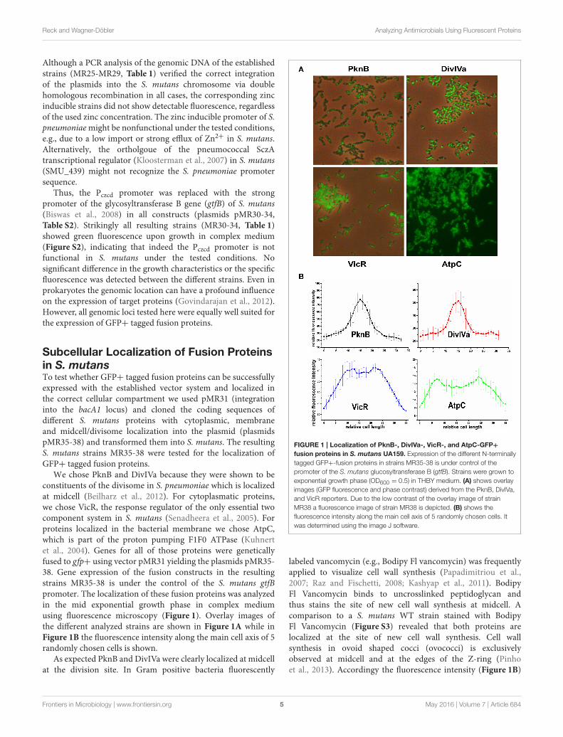
We chose PknB and DivIVa because they were shown to beconstituents of the divisome in S. pneumoniae which is localizedat midcell (Beilharz et al., 2012). For cytoplasmatic proteins,we chose VicR, the response regulator of the only essential twocomponent system in S. mutans (Senadheera et al., 2005). Forproteins localized in the bacterial membrane we chose AtpC,which is part of the proton pumping F1F0 ATPase (Kuhnertet al., 2004). Genes for all of those proteins were geneticallyfused to gfp+ using vector pMR31 yielding the plasmids pMR35-38. Gene expression of the fusion constructs in the resultingstrains MR35-38 is under the control of the S. mutans gtfBpromoter. The localization of these fusion proteins was analyzedin the mid exponential growth phase in complex mediumusing fluorescence microscopy (Figure 1). Overlay images ofthe different analyzed strains are shown in Figure 1A while inFigure 1B the fluorescence intensity along the main cell axis of 5randomly chosen cells is shown.
As expected PknB and DivIVa were clearly localized at midcellat the division site. In Gram positive bacteria fluorescently
FIGURE 1 | Localization of PknB-, DivIVa-, VicR-, and AtpC-GFP+
fusion proteins in S. mutans UA159. Expression of the different N-terminally
tagged GFP+-fusion proteins in strains MR35-38 is under control of the
promoter of the S. mutans glucosyltransferase B (gtfB). Strains were grown to
exponential growth phase (OD600 = 0.5) in THBY medium. (A) shows overlay
images (GFP fluorescence and phase contrast) derived from the PknB, DivIVa,
and VicR reporters. Due to the low contrast of the overlay image of strain
MR38 a fluorescence image of strain MR38 is depicted. (B) shows the
fluorescence intensity along the main cell axis of 5 randomly chosen cells. It
was determined using the image J software.
labeled vancomycin (e.g., Bodipy Fl vancomycin) was frequentlyapplied to visualize cell wall synthesis (Papadimitriou et al.,2007; Raz and Fischetti, 2008; Kashyap et al., 2011). BodipyFl Vancomycin binds to uncrosslinked peptidoglycan andthus stains the site of new cell wall synthesis at midcell. Acomparison to a S. mutans WT strain stained with BodipyFl Vancomycin (Figure S3) revealed that both proteins arelocalized at the site of new cell wall synthesis. Cell wallsynthesis in ovoid shaped cocci (ovococci) is exclusivelyobserved at midcell and at the edges of the Z-ring (Pinhoet al., 2013). Accordingy the fluorescence intensity (Figure 1B)
Frontiers in Microbiology | www.frontiersin.org 5 May 2016 | Volume 7 | Article 684
Reck and Wagner-Döbler Analyzing Antimicrobials Using Fluorescent Proteins
showed at peak at midcell and resembled a bell-shaped normaldistribution.
For the VicR reporter strain MR37 the bacterial cellswere found to be entirely fluorescing green, confirming thecytoplasmic localization of GFP+-VicR. Fluorescence intensityof the VicR fusion protein showed a steep increase from thecell poles toward a broad plateau along the main cell axis. Thisbehavior is consistent with the cytoplasmic localization of VicR.
AtpC was localized in the bacterial membrane as expected,too. The GFP fluorescence of strain MR38 was concentratedin a halo surrounding the cytoplasm of the cell. This is clearlyreflected in the intensity of fluorescence across the cell axis.Two peaks were observed at the cell poles while the fluorescenceintensity in the central region between the cell poles was lower.
To conclude, all studied proteins showed their expectedsubcellular localization. The established system is thereforeapplicable for single cell analysis of protein localization inS. mutans. In the next step we introduced inducible promoters tobe able to investigate timed fusion protein expression in differentcellular compartments.
Establishment of an Inducible FluorescentProtein Expression System in S. mutansSince most native proteins are only transiently expressed,constitutive intracellular overexpression of fluorescent fusionproteins often interferes with host cell metabolism and mightcause misfolding of the target protein and/or the fluorescenttag. This is particularly important when studying toxic proteinsin vivo whose toxicity often depends strongly on theirconcentration. Thus, utilization of a linearly inducible andtightly controllable expression system is required for studyinglocalization and dynamics of folding-sensitive and toxic proteins.In S. mutans only very few inducible expression systems arepresently available as genetic tools and most of them show eitherlow induction ratios and/or high basal transcription. Xie et al.developed two fine-tunable xylose inducible promoter cassettesand demonstrated their application by studying toxin/antitoxinsystems in S. mutans (Xie et al., 2013). The XylS1cassette isspecifically designed for high expression levels at the cost ofhigher basal transcription, while the XylS2 cassette ensures verylow basal transcription but only medium expression strength(Xie et al., 2013). Thus, reporter strains expressing the GFP+-DivIVa and GFP+-PknB proteins under the control of either theXylS1or the XylS2 promoter cassettes were generated. To thisend plasmids pMR35 and pMR36 were modified and the gtfBpromoter was replaced by either the XylS1 or the XylS2 promotercassette yielding plasmids pMR39-40 (DivIVa) and pMR43-44 (PknB). In addition we constructed strains whose GFP+fusion protein expression is inducible by the mutacin inducingpeptide (MIP), formerly termed competence stimulating peptide(CSP). We previously demonstrated that MIP highly inducesexpression of bacteriocin encoding genes independent fromthe growth medium (Perry et al., 2009; Reck et al., 2015),including the two peptide bacteriocinMutacin IV (SMU_150 andSMU_151) and Mutacin VI (SMU_423). Bacteriocin productionwas shown to be tightly regulated in S. mutans, thus making
MIP responsive promoters potentially effective gene inductionsystems (Perry et al., 2009; Reck et al., 2015). The promoters ofmutacin IV and mutacin VI were cloned 5′ of the GFP+ codingregion of plasmids pMR35 and pMR36, thereby replacing thegtfB promoter. After transformation of the resulting plasmidspMR41-42 and pMR45-46 in S. mutans the MIP inducible strainsMR41-42 and MR45-46 expressing DivIVa and PknB GFP+fusion proteins were obtained.
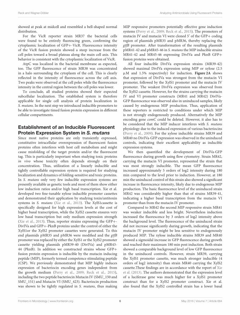
All four inducible DivIVa expression strains (MR39-42)showed maximal DivIVa expression using MIP or xylose (2.5µM and 1.5% respectively) for induction. Figure 2A showsthat expression of DivIVa was strongest from the mutacin VIpromoter, followed by the XylS1 promoter and the mutacin IVpromoter. The weakest DivIVa expression was observed fromthe XylS2 cassette. However, for the strains carrying the mutacinIV and VI promoter constructs (MR41 and MR42) DivIVa-GFP fluorescence was observed also in uninduced samples, likelycaused by endogenous MIP production. Thus, application ofthese reporters is restricted to conditions under which MIPis not strongly endogenously produced. Alternatively the MIPencoding gene comC could be deleted. However, it also has tobe considered that the MIP inducer interferes with S. mutansphysiology due to the induced expression of various bacteriocins(Perry et al., 2009). For the xylose inducible strains MR39 andMR40 no DivIVa-GFP expression was observed in the uninducedcontrols, indicating their excellent applicability as inducibleexpression systems.
We then quantified the development of DivIVa-GFPfluorescence during growth using flow cytometry. Strain MR42,carrying the mutacin VI promoter, represented the strain thatwas most strongly inducible. The mean GFP fluorescenceincreased approximately 5 orders of log2 intensity during 180min compared to the level prior to induction. However, at 180min the uninduced control of this strain also showed a significantincrease in fluorescence intensity, likely due to endogenous MIPproduction. The basic fluorescence level of the uninduced strainMR42 was considerably higher than that of strains MR39-41,indicating a higher basal transcription from the mutacin VIpromoter than from the mutacin IV promoter.
Compared to MR42 the second MIP responsive strain MR41was weaker inducible and less bright. Nevertheless inductionincreased the fluorescence by 3 orders of log2 intensity abovethe background level. The fluorescence of the uninduced straindid not increase significantly during growth, indicating that themutacin IV promoter might be less sensitive to endogenouslyproduced MIP. The xylose inducible strains MR39 and MR40showed a sigmoidal increase in GFP fluorescence during growthand reached their maximum 180 min post induction. Both strainshowed a comparable background level of low GFP fluorescencein the uninduced controls. However, strain MR39, carryingthe XylS1 promoter cassette, was much stronger inducible (4orders of log2 intensity) than strain MR40 carrying the XylS2cassette.These findings are in accordance with the report of Xieet al. (2013). The authors demonstrated that the expression levelof a luciferase gene was much higher for a XylS1 promoterconstruct than for a XylS2 promoter construct. Xie et al.also found that the XylS2 controlled strain has a lower basal
Frontiers in Microbiology | www.frontiersin.org 6 May 2016 | Volume 7 | Article 684
Reck and Wagner-Döbler Analyzing Antimicrobials Using Fluorescent Proteins
FIGURE 2 | Comparison of the expression strength and basal transcription of different inducible GFP+-DivIVa reporter strains in S. mutans UA159.
D-xylose inducible strains carrying a chromosomal gfp+-divIVa fusion construct under the control of the XylS1 (MR39) and XylS2 (MR40) promoter cassettes,
respectively, were constructed in S. mutans. In addition DivIVa reporter strains inducible by the competence stimulating peptide (MIP) and carrying the mutacin IV
(PmutIV, MR41) and mutacin VI promoter (PmutVI, MR42) were generated. Strains MR39-42 were grown in complex THBY medium to early exponential growth phase
(OD600 = 0.2) and GFP+-DivIVa expression was fully induced with either 2 µM MIP (pMR41-42) or 1.5% (D)-Xylose (MR39-40). 0, 30, 60, 90, 120, 150, and 180 min
post induction cells were collected, washed and analyzed using fluorescence microscopy. In (A) overlay microscopic images (green fluorescence/phase contrast) of
the corresponding induced (upper panel) and uninduced strains (lower panel) 3 h post induction are presented. In part (B) line plots of the relative fluorescence
intensity in course of time of strains MR39-42 are shown. For the generation of line plots the GFP fluorescence of 50,000 individual cells was recorded using flow
cytometry. Red line plots indicate induced strains, while in the black line plots the fluorescence intensity of the corresponding uninduced strains is shown. The mean
and the standard deviation of three independent biological replicates are presented.
expression level than the XylS1 carrying strain (Xie et al., 2013).This was not observed in our analysis and might be due to thehigher detection limit of GFP compared to luciferase.
To compare the applicability of the constitutive reporterstrains for protein localization studies with that of induciblestrains we quantified the GFP fluorescence of two strains (MR35and MR49) constitutively expressing a DivIVa-GFP+ fusionduring growth (Figure S4). DivIVa expression in the strainMR49
is under control of the strong lactococcal P23 promoter (Biswaset al., 2008) while strain MR35 carries the gtfB promoter. Thenon-fluorescent S. mutans WT strain was used to determinethe autofluorescence (Figure S4). In comparison to the inducibleexpression strains MR39 and MR41-42 (see Figure 2) thefluorescence of strains MR35 and MR49 was relatively weak(Figure S4). Thus, the inducible expression strains are superiorfor protein localization studies. To conclude, the high basal
Frontiers in Microbiology | www.frontiersin.org 7 May 2016 | Volume 7 | Article 684
Reck and Wagner-Döbler Analyzing Antimicrobials Using Fluorescent Proteins
transcription of the mutacin VI promoter (MR 42) and its strongresponse to endogenously produced MIP make it unsuitable forexpression studies. The promoter of mutacin IV is too weaklyinduced to generate clear signals. The XylS1 promoter (MR39)is well suitable, although it is slightly less inducible than theMutacin VI promoter. Thus, we subsequently used XylS1 basedexpression constructs which combine a low basal transcriptionwith a high level of induction.
For this construct (strain MR39) we then quantified the dose-response behavior to the inducer D-xylose for concentrationsranging from 0.005% (3.33∗103 µM) to 4% (2.66 ∗105 µM). GFPexpression was determined 3 h after induction which representsthe peak of fluorescence (Figure 2B). The dose response curvehad a sigmoid shape (Figure S5A), concentrations above 0.1%(6.66∗103 µM) strongly induced GFP fluorescence, while theresponse was saturated at concentrations exceeding 2% (1.33∗105
µM) D-xylose. Between 0.2 and 2% the response was linearand thus these concentrations are suitable to obtain a rangeof expression levels of the fusion proteins. The EC50 valuewas calculated to be 2.86∗104 (±0.51∗104) µM D-xylose(Figure S5B).
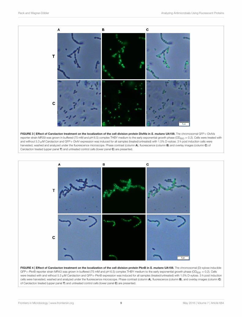
Effect of Carolacton on the LocalizationPattern of Cell Division ProteinsWe analyzed whether Carolacton alters the localization patternof the cell division proteins PknB and DivIVa. Thus, the xylose-induced (1.5%) reporter strains MR39 and MR43, expressingGFP fusions of DivIVa and PknB respectively, were grown tomid-exponential phase under buffered acidic conditions (pH6.5, 75 mM phosphate buffer) in the presence of Carolacton.The acidic pH is a prerequisite for the membrane damageand reduction in viability caused by Carolacton in S. mutans(Reck et al., 2011). Carolacton treatment caused aberrant cellmorphologies with strongly elongated and enlarged cells andthe formation of long chains (Figure 3). The mean length ofS. mutans single cells increased from 1.02 µm (±0.21 µm) to1.32 µm (±0.47 µm) and was more variable, as indicated by thehigher standard deviation. However, “normal” cells remained inthe Carolacton treated samples, demonstrating that phenotypicheterogeneity makes some cells more sensitive than others. Forboth strains Carolacton treatment caused an increase in septumformation (Figures 3, 4). Some cells contained 3 septa per cell,indicating impaired septal closure and thus lack of correctdaughter cell separation. In the Carolacton treated samplesof strain MR39 (Figure 3, upper panel) DivIVa was localizedsimultaneously at midcell and at the new cell poles, while itwas found mainly at midcell in the untreated controls, againsuggesting that septal closure is proceeding too slow to properlyconstrict the dividing cell. The appearance of extremely largeand elongated cells might be caused by inhibition of the septalcell wall synthesis machinery. Thus, peripheral cell wall synthesiselongated the cell, while the correct onset of septal cell wallsynthesis and thus septum closure to constrict the daughter cellsfailed. Thus, the occurrence of elongated cells and cell chainscould both be explained by this mechanism. As PknB is known tocontrol switching between peripheral to septal cell wall synthesis
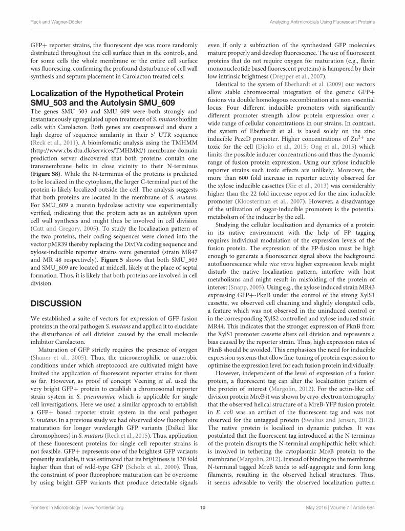
(Beilharz et al., 2012), we analyzed the effect of Carolactonon PknB localization using strain MR43. A significantly alteredlocalization pattern of PknB in comparison to the untreatedcontrol was observed (Figure 4). While in the controls PknB waslocated exclusively at midcell as expected, it was dispersed overa much larger part of the cell in Carolacton treated cultures. Insome cells PknB even seemed to be randomly distributed in thebacterial membrane, indicated by a halo of green fluorescencearound the cells. The observed influence of Carolacton treatmenton PknB localization is in accordance with the proposed modeof action of Carolacton via PknB (Reck et al., 2011). Theseresults demonstrate that Carolacton has a profound impacton cell division in S. mutans and causes partial delocalizationof the key regulator of cell division PknB. The inhibition ofseptal cell wall synthesis might be a mechanistic consequenceof this delocalization. Moreover these results demonstrate theimportance of PknB as a main regulator of cell division instreptococci.
In the xylose-induced but non-Carolacton treated strainMR43 slight cell chaining was observed (Figure 3), which was notobserved in the corresponding uninduced controls. To excludethat the Carolacton caused delocalization of cell division proteinsis an artifact of xylose induction by the XylS1 promoter, werepeated the experiment using strains MR40 and MR44 whichcarry the XylS2 promoter. In these strains the expression levelof the PknB and DivIVa fusion proteins is significantly lower,thus the probability that the fusion protein expression interfereswith the S. mutans metabolism is lower. We found the samealtered patterns of cell division and delocalization of the celldivision proteins PknB and DivIVa for the Carolacton treatedsamples as with the XylS1 promoter construct (Figures S6, S7).Moreover, no chaining was seen in the xylose induced but non-Carolacton treated strainMR44. Thus, higher expression levels ofPknB should be avoided as they interfere with the physiology andcell division of S. mutans.
It has to be taken into account that the fluorescent tag can alterthe localization pattern of the studied protein (Margolin, 2012).Therefore we used Bodipy-FL vancomycin staining to verify theCarolacton-mediated alterations in cell division observed in thereporter strain analysis. Fluorescent vancomycin staining targetsthe sites of nascent cell wall synthesis and was frequently appliedto study cell division in Gram-positive bacteria (Papadimitriouet al., 2007; Raz and Fischetti, 2008). In the untreated controlsmost of the stained cells had a single equatorial fluorescent band,thus new cell wall was synthesized at the division site at midcell,in accordance with the proposed model of cell wall synthesis inovoid cocci (Pinho et al., 2013). Some cells synthesized new cellwalls simultaneously at the old and the new division site, thusboth the equator and the future poles of the dividing cell werefluorescing green. This might indicate that peripheral cell wallsynthesis exceeded the septal cell wall synthesis in these cells.Thus, although the constriction of the two daughter cells had notbeen finished properly the peripheral cell wall machinery alreadystarted elongating the new cells.
Carolacton treated cells showed increased septum formationand a significantly altered pattern of cell wall synthesis. Infull accordance with the findings using the DivIVa and PknB
Frontiers in Microbiology | www.frontiersin.org 8 May 2016 | Volume 7 | Article 684
Reck and Wagner-Döbler Analyzing Antimicrobials Using Fluorescent Proteins
FIGURE 3 | Effect of Carolacton treatment on the localization of the cell division protein DivIVa in S. mutans UA159. The chromosomal GFP+-DivIVa
reporter strain MR39 was grown in buffered (75 mM and pH 6.5) complex THBY medium to the early exponential growth phase (OD600 = 0.2). Cells were treated with
and without 5.3 µM Carolacton and GFP+-DivIV expression was induced for all samples (treated/untreated) with 1.5% D-xylose. 3 h post induction cells were
harvested, washed and analyzed under the fluorescence microscope. Phase contrast (column A), fluorescence (column B) and overlay images (column C) of
Carolacton treated (upper panel T) and untreated control cells (lower panel C) are presented.
FIGURE 4 | Effect of Carolacton treatment on the localization of the cell division protein PknB in S. mutans UA159. The chromosomal (D)-xylose inducible
GFP+-PknB reporter strain MR43 was grown in buffered (75 mM and pH 6.5) complex THBY medium to the early exponential growth phase (OD600 = 0.2). Cells
were treated with and without 5.3 µM Carolacton and GFP+-PknB expression was induced for all samples (treated/untreated) with 1.5% D-xylose. 3 h post induction
cells were harvested, washed and analyzed under the fluorescence microscope. Phase contrast (column A), fluorescence (column B), and overlay images (column C)
of Carolacton treated (upper panel T) and untreated control cells (lower panel C) are presented.
Frontiers in Microbiology | www.frontiersin.org 9 May 2016 | Volume 7 | Article 684
Reck and Wagner-Döbler Analyzing Antimicrobials Using Fluorescent Proteins
GFP+ reporter strains, the fluorescent dye was more randomlydistributed throughout the cell surface than in the controls, andfor some cells the whole membrane or the entire cell surfacewas fluorescing, confirming the profound disturbance of cell wallsynthesis and septum placement in Carolacton treated cells.
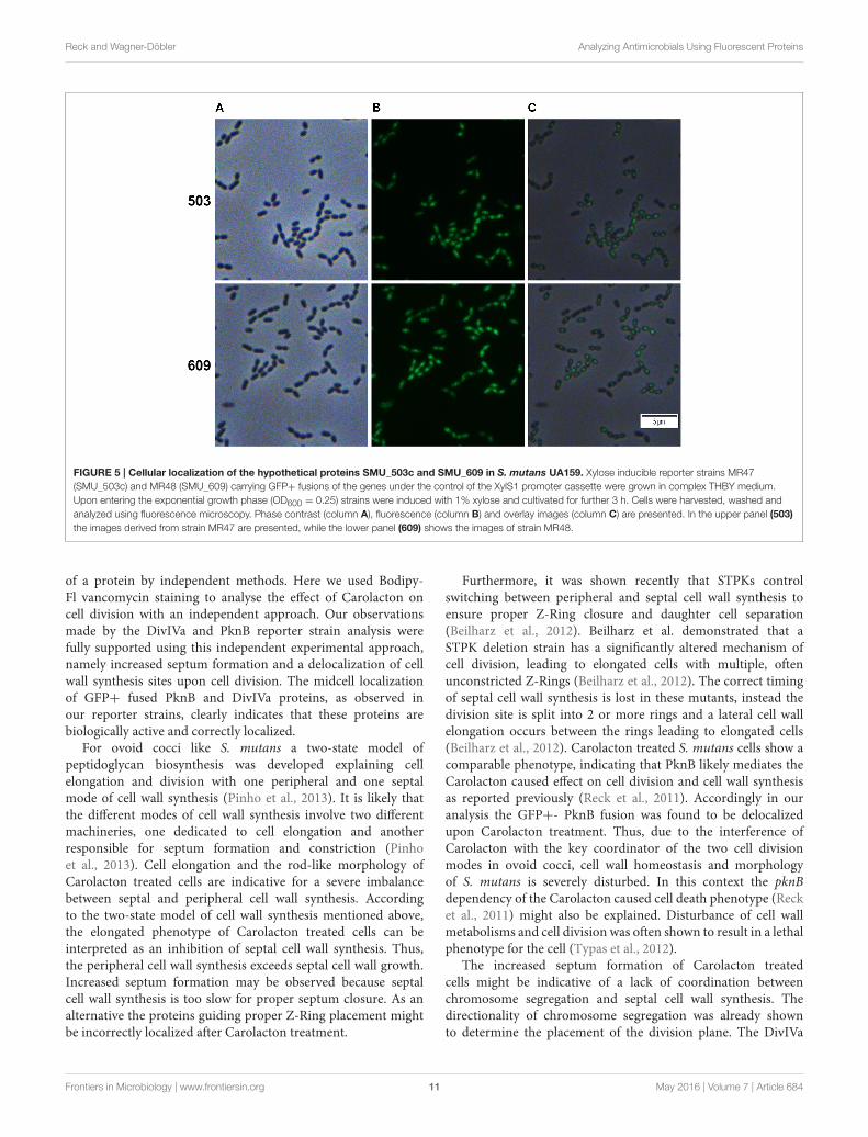
Localization of the Hypothetical ProteinSMU_503 and the Autolysin SMU_609The genes SMU_503 and SMU_609 were both strongly andinstantaneously upregulated upon treatment of S. mutans biofilmcells with Carolacton. Both genes are coexpressed and share ahigh degree of sequence similarity in their 5′ UTR sequence(Reck et al., 2011). A bioinfomatic analysis using the TMHMM(http://www.cbs.dtu.dk/services/TMHMM/) membrane domainprediction server discovered that both proteins contain onetransmembrane helix in close vicinity to their N-terminus(Figure S8). While the N-terminus of the proteins is predictedto be localized in the cytoplasm, the larger C-terminal part of theprotein is likely localized outside the cell. The analysis suggeststhat both proteins are located in the membrane of S. mutans.For SMU_609 a murein hydrolase activity was experimentallyverified, indicating that the protein acts as an autolysin uponcell wall synthesis and might thus be involved in cell division(Catt and Gregory, 2005). To study the localization pattern ofthe two proteins, their coding sequences were cloned into thevector pMR39 thereby replacing the DivIVa coding sequence andxylose-inducible reporter strains were generated (strain MR47and MR 48 respectively). Figure 5 shows that both SMU_503and SMU_609 are located at midcell, likely at the place of septalformation. Thus, it is likely that both proteins are involved in celldivision.
DISCUSSION
We established a suite of vectors for expression of GFP-fusionproteins in the oral pathogen S. mutans and applied it to elucidatethe disturbance of cell division caused by the small moleculeinhibitor Carolacton.
Maturation of GFP strictly requires the presence of oxygen(Shaner et al., 2005). Thus, the microaerophilic or anaerobicconditions under which streptococci are cultivated might havelimited the application of fluorescent reporter strains for themso far. However, as proof of concept Veening et al. used thevery bright GFP+ protein to establish a chromosomal reporterstrain system in S. pneumoniae which is applicable for singlecell investigations. Here we used a similar approach to establisha GFP+ based reporter strain system in the oral pathogenS. mutans. In a previous study we had observed slow fluorophorematuration for longer wavelength GFP variants (DsRed likechromophores) in S. mutans (Reck et al., 2015). Thus, applicationof these fluorescent proteins for single cell reporter strains isnot feasible. GFP+ represents one of the brightest GFP variantspresently available, it was estimated that its brightness is 130 foldhigher than that of wild-type GFP (Scholz et al., 2000). Thus,the constraint of poor fluorophore maturation can be overcomeby using bright GFP variants that produce detectable signals
even if only a subfraction of the synthesized GFP moleculesmature properly and develop fluorescence. The use of fluorescentproteins that do not require oxygen for maturation (e.g., flavinmononucleotide based fluorescent proteins) is hampered by theirlow intrinsic brightness (Drepper et al., 2007).
Identical to the system of Eberhardt et al. (2009) our vectorsallow stable chromosomal integration of the genetic GFP+fusions via double homologous recombination at a non-essentiallocus. Four different inducible promoters with significantlydifferent promoter strength allow protein expression over awide range of cellular concentrations in our strains. In contrast,the system of Eberhardt et al. is based solely on the zincinducible PczcD promoter. Higher concentrations of Zn2+ aretoxic for the cell (Djoko et al., 2015; Ong et al., 2015) whichlimits the possible inducer concentrations and thus the dynamicrange of fusion protein expression. Using our xylose induciblereporter strains such toxic effects are unlikely. Moreover, themore than 600 fold increase in reporter activity observed forthe xylose inducible cassettes (Xie et al., 2013) was considerablyhigher than the 22 fold increase reported for the zinc induciblepromoter (Kloosterman et al., 2007). However, a disadvantageof the utilization of sugar-inducible promoters is the potentialmetabolism of the inducer by the cell.
Studying the cellular localization and dynamics of a proteinin its native environment with the help of FP taggingrequires individual modulation of the expression levels of thefusion protein. The expression of the FP-fusion must be highenough to generate a fluorescence signal above the backgroundautofluorescence while vice versa higher expression levels mightdisturb the native localization pattern, interfere with hostmetabolisms and might result in misfolding of the protein ofinterest (Snapp, 2005). Using e.g., the xylose induced strainMR43expressing GFP+-PknB under the control of the strong XylS1cassette, we observed cell chaining and slightly elongated cells,a feature which was not observed in the uninduced control orin the corresponding XylS2 controlled and xylose induced strainMR44. This indicates that the stronger expression of PknB fromthe XylS1 promoter cassette alters cell division and represents abias caused by the reporter strain. Thus, high expression rates ofPknB should be avoided. This emphasizes the need for inducibleexpression systems that allow fine-tuning of protein expression tooptimize the expression level for each fusion protein individually.
However, independent of the level of expression of a fusionprotein, a fluorescent tag can alter the localization pattern ofthe protein of interest (Margolin, 2012). For the actin-like celldivision proteinMreB it was shown by cryo-electron tomographythat the observed helical structure of a MreB-YFP fusion proteinin E. coli was an artifact of the fluorescent tag and was notobserved for the untagged protein (Swulius and Jensen, 2012).The native protein is localized in dynamic patches. It waspostulated that the fluorescent tag introduced at the N terminusof the protein disrupts the N-terminal amphipathic helix whichis involved in tethering the cytoplasmic MreB protein to themembrane (Margolin, 2012). Instead of binding to themembraneN-terminal tagged MreB tends to self-aggregate and form longfilaments, resulting in the observed helical structures. Thus,it seems advisable to verify the observed localization pattern
Frontiers in Microbiology | www.frontiersin.org 10 May 2016 | Volume 7 | Article 684
Reck and Wagner-Döbler Analyzing Antimicrobials Using Fluorescent Proteins
FIGURE 5 | Cellular localization of the hypothetical proteins SMU_503c and SMU_609 in S. mutans UA159. Xylose inducible reporter strains MR47
(SMU_503c) and MR48 (SMU_609) carrying GFP+ fusions of the genes under the control of the XylS1 promoter cassette were grown in complex THBY medium.
Upon entering the exponential growth phase (OD600 = 0.25) strains were induced with 1% xylose and cultivated for further 3 h. Cells were harvested, washed and
analyzed using fluorescence microscopy. Phase contrast (column A), fluorescence (column B) and overlay images (column C) are presented. In the upper panel (503)
the images derived from strain MR47 are presented, while the lower panel (609) shows the images of strain MR48.
of a protein by independent methods. Here we used Bodipy-Fl vancomycin staining to analyse the effect of Carolacton oncell division with an independent approach. Our observationsmade by the DivIVa and PknB reporter strain analysis werefully supported using this independent experimental approach,namely increased septum formation and a delocalization of cellwall synthesis sites upon cell division. The midcell localizationof GFP+ fused PknB and DivIVa proteins, as observed inour reporter strains, clearly indicates that these proteins arebiologically active and correctly localized.
For ovoid cocci like S. mutans a two-state model ofpeptidoglycan biosynthesis was developed explaining cellelongation and division with one peripheral and one septalmode of cell wall synthesis (Pinho et al., 2013). It is likely thatthe different modes of cell wall synthesis involve two differentmachineries, one dedicated to cell elongation and anotherresponsible for septum formation and constriction (Pinhoet al., 2013). Cell elongation and the rod-like morphology ofCarolacton treated cells are indicative for a severe imbalancebetween septal and peripheral cell wall synthesis. Accordingto the two-state model of cell wall synthesis mentioned above,the elongated phenotype of Carolacton treated cells can beinterpreted as an inhibition of septal cell wall synthesis. Thus,the peripheral cell wall synthesis exceeds septal cell wall growth.Increased septum formation may be observed because septalcell wall synthesis is too slow for proper septum closure. As analternative the proteins guiding proper Z-Ring placement mightbe incorrectly localized after Carolacton treatment.
Furthermore, it was shown recently that STPKs controlswitching between peripheral and septal cell wall synthesis toensure proper Z-Ring closure and daughter cell separation(Beilharz et al., 2012). Beilharz et al. demonstrated that aSTPK deletion strain has a significantly altered mechanism ofcell division, leading to elongated cells with multiple, oftenunconstricted Z-Rings (Beilharz et al., 2012). The correct timingof septal cell wall synthesis is lost in these mutants, instead thedivision site is split into 2 or more rings and a lateral cell wallelongation occurs between the rings leading to elongated cells(Beilharz et al., 2012). Carolacton treated S. mutans cells show acomparable phenotype, indicating that PknB likely mediates theCarolacton caused effect on cell division and cell wall synthesisas reported previously (Reck et al., 2011). Accordingly in ouranalysis the GFP+- PknB fusion was found to be delocalizedupon Carolacton treatment. Thus, due to the interference ofCarolacton with the key coordinator of the two cell divisionmodes in ovoid cocci, cell wall homeostasis and morphologyof S. mutans is severely disturbed. In this context the pknBdependency of the Carolacton caused cell death phenotype (Recket al., 2011) might also be explained. Disturbance of cell wallmetabolisms and cell division was often shown to result in a lethalphenotype for the cell (Typas et al., 2012).
The increased septum formation of Carolacton treatedcells might be indicative of a lack of coordination betweenchromosome segregation and septal cell wall synthesis. Thedirectionality of chromosome segregation was already shownto determine the placement of the division plane. The DivIVa
Frontiers in Microbiology | www.frontiersin.org 11 May 2016 | Volume 7 | Article 684
Reck and Wagner-Döbler Analyzing Antimicrobials Using Fluorescent Proteins
protein is a direct target of PKnB and is thought to be mainlyresponsible for the correct septum placement after chromosomesegregation (Pinho et al., 2013). Thus, the failed septumplacement and closure observed upon Carolacton treatment isin full accordance with the PknB dependent mode of action ofCarolacton and the altered DivIVa localization pattern observedupon Carolacton treatment. However, the DivIVa delocalizationwas less pronounced than that observed for the PknB protein.Again this is indicative that PknB rather than DivIVa localizationis disturbed by the substance. It might be speculated that DivIVais correctly localized but not properly phosphorylated due tothe Carolacton mediated disturbance of PknB activity. Thus,the protein is present at it intended cellular site but might lackits biological activity. It remains to be elucidated whether thecorrect localization of DivIVa is dependent on the presence ofPknB. Recently it was discovered in S. pneumonaie that theprotein MapZ represents another STPK target and might alsofunction in division site selection (Fleurie et al., 2014). WhetherCarolacton treatment also interferes with the localization of theMapZ ortholog in S. mutans is another interesting questionwhichneeds to be studied in the future.
Here we identified the so far largely uncharacterized proteinsSMU_503 and SMU_609 to be localized at midcell and thusbeing likely components of the divisome machinery. The genesencoding these proteins were instantaneously upregulated uponCarolacton treatment and thus belong to the primary cellularresponse (Sudhakar et al., 2014). Deletion of these genesdid not alter the sensitivity of S. mutans toward Carolacton,demonstrating that they are not essential for the mode ofaction of Carolacton (Reck et al., 2011; Sudhakar et al.,2014). Therefore, their upregulation most likely represents acompensation mechanism of the cell to cope with the deleteriouseffects of Carolacton.
For SMU_609 an autolysin activity was verified (Catt andGregory, 2005). Thus, it is highly likely that the proteinis involved in cell wall remodeling upon cell division. Forboth, lateral and septal cell wall synthesis, a tightly controlledand balanced interplay between new cell wall synthesis andbreakdown of the old cell wall to insert new peptidoglycanprecursorsmust exist (Typas et al., 2012). Disturbing homeostasisbetween cell wall synthesis and cell wall breakdown is likelylethal for the cell. As both the peripheral and septal cell wallmachineries are localized at midcell in ovoid cocci (Pinhoet al., 2013), it is hard to predict whether SMU_609 is involvedin the hydrolysis of lateral or septal cell wall. Based on theobservation that mainly septal cell wall synthesis is inhibited byCarolacton treatment (elongated cells), it can be speculated thatthe upregulation of SMU_609 found after Carolacton treatment(Reck et al., 2011; Li et al., 2013) is a compensation reaction of thecell. Consequently SMU_609 might belong to the septal cell wallmachinery.
For SMU_503 a Pfam database analysis showed that theprotein contains several eukaryotic-like domains with unknownfunctions. Due to the presence of a typical folate carrierdomain the protein may function in the import of folateinto the cell and thus might be particularly involved inpurine, pyrimidine and methionine metabolism. Accordingly
the key genes of pyrimindine metabolism were instantaneouslyupregulated together with SMU_503 in Carolacton treated cells(Sudhakar et al., 2014). Bacteria are often dependent on a denovo biosynthesis pathway for folate. Bacterial enzymes of thispathway are attractive drug targets due to their complete absencein mammals (Bermingham and Derrick, 2002). Firmicutes areable to both synthesize and import folate. Genes encoding highaffinity folate binding proteins were found in many Firmicutesand are sometimes localized adjacent to folate salvage genes(Eudes et al., 2008). Folate is particularly important in periodsof rapid cell division, thus the co-localization of SMU_503 withthe divisome might be explained in this context. The SID-1RNA_chan and the Serinc domain indicate that SMU_503 islocalized in the membrane and may also function as a dsRNAchannel.
Taken together, the observations made in this studydemonstrate the versatile nature of GFP based localizationreporter strains to study the effect of antimicrobials on cellularmetabolism. The utilization of single cell fluorescent reporterstrains to elucidate the mode of action of antimicrobials wase.g., applied by Beilharz et al. (2012). The authors foundthat antibiotics targeting the last steps of cell wall synthesiscause a GFP+-STPK delocalization which further indicates aregulatory role for STPKs in cell wall metabolism. Using aMinD-GFP fusion Wenzel et al. elucidate that small cationicantimicrobial peptides delocalize peripheral membrane proteinswith strong implications on the membrane potential andthe respiration in B. subtilis (Wenzel et al., 2014). Thesame authors demonstrated earlier that the delocalizationof GFP-MinD fusion proteins represents an indicator forantimicrobials disturbing the membrane potential. With thehelp of FP reporter strains it thus might also be promisingto screen compound libraries for substances that alter thelocalization pattern of essential virulence proteins but do notkill the bacteria. Compounds that attenuate virulence areunlikely to cause resistance development and might representa fruitful future alternative for antibiotics (Keyser et al.,2008).
Moreover our study shows that STPK represent attractivedrug targets due to their central regulatory role in virulence,host-cell interaction, cell division and cell wall synthesis (Pereiraet al., 2011). Thus, approaches targeting STPKs or their cognatephosphatases gain increasing attention in drug discovery.
Fluorescent fusions proteins dramatically changed ourperspective on bacteria which were previously considered assimple, unstructured vessels filled with nutrients and enzymes.The surprisingly high degree of structural organization withinthe well-studied organisms E. coli and B. subtilis implies that awealth of novel mechanisms of protein localization waits to bediscovered also in streptococci. Our established system can beapplied to study cell division in S. mutans in general. Determiningthe localization of key cell division proteins in differentgene deletion background can provide valuable informationwhich factors are responsible for the correct localization andassembly of the cell division machinery. Understanding proteinlocalization and dynamics in more detail will provide new toolsand strategies to fight back bacterial infections.
Frontiers in Microbiology | www.frontiersin.org 12 May 2016 | Volume 7 | Article 684
Reck and Wagner-Döbler Analyzing Antimicrobials Using Fluorescent Proteins
AUTHOR CONTRIBUTIONS
MR designed and conducted the experiments, MR and IWdesigned the study and wrote the manuscript.
FUNDING
MR was funded by the BMBF (program e:bio; grant number 031A299).
ACKNOWLEDGMENTS
We thank Rolf Müller for providing Carolacton and Jan-WillemVeening for sending us plasmid pJWV25.
SUPPLEMENTARY MATERIAL
The Supplementary Material for this article can be foundonline at: http://journal.frontiersin.org/article/10.3389/fmicb.2016.00684
Figure S1 | Plasmid maps of chromosomal integrative plasmids used in
this study. Resistance cassettes are shown in red color while the genetic
GFP+fusion constructs are shown in green. Homologous flanks allowing
integration of the plasmids into the S. mutans chromosome are shown in gray
color.
Figure S2 | Fluorescence microscopic images of strains MR30-34 growing
in complex THBY medium. Strains MR30-34, carrying gfp+ in different genomic
loci (agaL, bacA1, SMU_1405, lacE, SMU_1577) and under control of the
glycosyltransferase B (gtfB) promoter were grown to an OD600 of 0.8. Cells were
collected, washed and analyzed using florescence microscopy. Fluorescence
images of the strains and a non-fluorescing wild-type strain (control) are presented.
Figure S3 | Visualization of de novo cell wall synthesis in Carolacton
treated and untreated S. mutans wild-type cells. Untreated and Carolacton
treated (5.3 µM) S. mutans cells were grown in complex THBY medium to an
OD600 of 0.5. Subsequently cells were stained with 1 µM Bodipy-Fl vancomycin
for 30 min. Cells were harvested, washed and analyzed using fluorescence
microscopy. The fluorescence microscopic images of Carolacton treated (A) and
untreated (B) cells are shown.
Figure S4 | Temporal development of fluorescence intensity for two
chromosomal reporter strains constitutively expressing GFP+DivIVa.
Reporter strains MR35 and MR49 were grown in complex THBY medium to early
exponential growth phase (OD600 = 0.2) and GFP+-DivIVa expression was
recorded every 30 min for 3 h. Cells were collected, washed and analyzed using
flow cytometry. Line plots of the relative fluorescence intensity in course of time of
strains MR35 (red), MR49 (blue) and the non-fluorescing S. mutans WT cells
(black) are shown. For the generation of line plots the GFP fluorescence of 50,000
individual cells was recorded. The mean and the standard deviation of 3
independent biological replicates are presented.
Figure S5 | Dose response curve of the XylS1 promoter cassette to
inducer D-xylose in strain MR39. The Gfp+-DivIVa reporter strain MR39 was
grown in complex THBY medium to an OD600 of 0.2. Cells were split in several
identical aliquots and treated with different concentrations of D-xylose ranging
from 0 to 2.66∗105 µM. 3 h post induction cells were collected, washed,
sonicated and analyzed using flow cytometry. The relative GFP fluorescence
intensity of 50000 individual cells was measured for each sample. The
corresponding dose response curve is shown in part (A). In part (B) the dose
response is plotted in the logarithmic scale for the inducer concentration (black
curve) and the fitted dose response curve assuming sigmoidal dose-response
behavior is shown (red line). Based on the fitted curve the EC50 value was
calculated using the software Origin 9.0.
Figure S6 | Effect of Carolacton treatment on the localization of cell
division protein DivIVa in S. mutans UA159. The chromosomal GFP+-DivIVa
reporter strain MR40 carrying the xylose inducible XylS2 promoter cassette was
grown in buffered (75 mM and pH 6.5) complex THBY medium to the early
exponential growth phase (OD600 = 0.2). Cells were treated with and without 5.3
µM Carolacton and GFP+-DivIV expression was induced for all samples
(treated/untreated) with 1.5% D-xylose. 3 h post induction cells were harvested,
washed and analyzed under the fluorescence microscope. Phase contrast
(column A), fluorescence (column B) and overlay images (column C) of Carolacton
treated (upper panel T) and untreated control cells (lower panel C) are presented.
Figure S7 | Effect of Carolacton treatment on the localization of cell
division protein PknB in S. mutans UA159. The chromosomal GFP+-PknB
reporter strain carrying the xylose inducible XylS2 promoter cassette (MR43) was
grown in buffered (75 mM and pH 6.5) complex THBY medium to the early
exponential growth phase (OD600 = 0.2). Cells were treated with and without 5.3
µM Carolacton and GFP+-PknB expression was induced for all samples
(treated/untreated) with 1.5% D-xylose. 3 h post induction cells were harvested,
washed and analyzed under the fluorescence microscope. Phase contrast
(column A), fluorescence (column B) and overlay images (column C) of Carolacton
treated (upper panel T) and untreated control cells (lower panel C) are presented.
Figure S8 | THMM prediction of transmembrane helices for the
hypothetical proteins SMU_503 and SMU_609. The amino acid sequence of
the proteins was analyzed using the TMHMM server version 2.0. The plot shows
the posterior probabilities of inside/outside/transmembrane helix for each residue.
Table S1 | Plasmids used in this study.
REFERENCES
Beilharz, K., Novakova, L., Fadda, D., Branny, P., Massidda, O., and Veening, J. W.
(2012). Control of cell division in Streptococcus pneumoniae by the conserved
Ser/Thr protein kinase StkP. Proc. Natl. Acad. Sci. U.S.A. 109, E905–E913. doi:
10.1073/pnas.1119172109
Bermingham, A., and Derrick, J. P. (2002). The folic acid biosynthesis pathway in
bacteria: evaluation of potential for antibacterial drug discovery. Bioessays 24,
637–648. doi: 10.1002/bies.10114
Biswas, I., Jha, J. K., and Fromm, N. (2008). Shuttle expression plasmids for
genetic studies in Streptococcus mutans. Microbiology 154, 2275–2282. doi:
10.1099/mic.0.2008/019265-0
Catt, D. M., and Gregory, R. L. (2005). Streptococcus mutans murein
hydrolase. J. Bacteriol. 187, 7863–7865. doi: 10.1128/JB.187.22.7863-786
5.2005
Djoko, K. Y., Ong, C. L., Walker, M. J., and McEwan, A. G. (2015). The role of
copper and zinc toxicity in innate immune defense against bacterial pathogens.
J. Biol. Chem. 290, 18954–18961. doi: 10.1074/jbc.R115.647099
Drepper, T., Eggert, T., Circolone, F., Heck, A., Krauss, U., Guterl, J. K.,
et al. (2007). Reporter proteins for in vivo fluorescence without oxygen. Nat.
Biotechnol. 25, 443–445. doi: 10.1038/nbt1293
Eberhardt, A., Wu, L. J., Errington, J., Vollmer, W., and Veening, J. W.
(2009). Cellular localization of choline-utilization proteins in Streptococcus
pneumoniae using novel fluorescent reporter systems. Mol. Microbiol. 74,
395–408. doi: 10.1111/j.1365-2958.2009.06872.x
Eudes, A., Erkens, G. B., Slotboom, D. J., Rodionov, D. A., Naponelli, V.,
and Hanson, A. D. (2008). Identification of genes encoding the folate-
and thiamine-binding membrane proteins in Firmicutes. J. Bacteriol. 190,
7591–7594. doi: 10.1128/JB.01070-08
Fleurie, A., Lesterlin, C., Manuse, S., Zhao, C., Cluzel, C., Lavergne, J. P., et al.
(2014). MapZmarks the division sites and positions FtsZ rings in Streptococcus
pneumoniae. Nature 516, 259–262. doi: 10.1038/nature13966
Giefing, C., Jelencsics, K. E., Gelbmann, D., Senn, B. M., and Nagy, E. (2010).
The pneumococcal eukaryotic-type serine/threonine protein kinase StkP co-
localizes with the cell division apparatus and interacts with FtsZ in vitro.
Microbiology 156, 1697–1707. doi: 10.1099/mic.0.036335-0
Frontiers in Microbiology | www.frontiersin.org 13 May 2016 | Volume 7 | Article 684
Reck and Wagner-Döbler Analyzing Antimicrobials Using Fluorescent Proteins
Govindarajan, S., Nevo-Dinur, K., and Amster-Choder, O. (2012).
Compartmentalization and spatiotemporal organization of macromolecules
in bacteria. FEMS Microbiol. Rev. 36, 1005–1022. doi: 10.1111/j.1574-
6976.2012.00348.x
Guo, L., Hu, W., He, X., Lux, R., McLean, J., and Shi, W. (2013).
investigating acid production by Streptococcus mutans with a surface-
displayed pH-sensitive green fluorescent protein. PLoS ONE 8:e57182. doi:
10.1371/journal.pone.0057182
Kashyap, D. R., Wang, M., Liu, L. H., Boons, G. J., Gupta, D., and Dziarski,
R. (2011). Peptidoglycan recognition proteins kill bacteria by activating
protein-sensing two-component systems. Nat. Med. 17, 676–683. doi: 10.1038/
nm.2357
Keyser, P., Elofsson, M., Rosell, S., and Wolf-Watz, H. (2008). Virulence blockers
as alternatives to antibiotics: type III secretion inhibitors against Gram-
negative bacteria. J. Intern. Med. 264, 17–29. doi: 10.1111/j.1365-2796.2008.01
941.x
Kloosterman, T. G., van der Kooi-Pol, M. M., Bijlsma, J. J., and Kuipers,
O. P. (2007). The novel transcriptional regulator SczA mediates protection
against Zn2+ stress by activation of the Zn2+-resistance gene czcD in
Streptococcus pneumoniae.Mol.Microbiol. 65, 1049–1063. doi: 10.1111/j.1365-
2958.2007.05849.x
Kuhnert, W. L., Zheng, G., Faustoferri, R. C., and Quivey, R. G. Jr. (2004).
The F-ATPase operon promoter of Streptococcus mutans is transcriptionally
regulated in response to external pH. J. Bacteriol. 186, 8524–8528. doi:
10.1128/JB.186.24.8524-8528.2004
Kunze, B., Reck, M., Dotsch, A., Lemme, A., Schummer, D., Irschik, H., et al.
(2010). Damage of Streptococcus mutans biofilms by Carolacton, a secondary
metabolite from the myxobacterium Sorangium cellulosum. BMC Microbiol.
10:199. doi: 10.1186/1471-2180-10-199
Lemme, A., Gröbe, L., Reck, M., Tomasch, J., and Wagner-Döbler, I. (2011).
Subpopulation-specific transcriptome analysis of competence-stimulating-
peptide-induced Streptococcus mutans. J. Bacteriol. 193, 1863–1877. doi:
10.1128/JB.01363-10
Li, J., Wang, W., Wang, Y., and Zeng, A. P. (2013). Two-dimensional gel-based
proteomic of the caries causative bacterium Streptococcus mutans UA159 and
insight into the inhibitory effect of Carolacton. Proteomics 13, 3470–3477. doi:
10.1002/pmic.201300077
Maestro, B., Novaková, L., Hesek, D., Lee, M., Leyva, E., Mobashery, S., et al.
(2011). Recognition of peptidoglycan and beta-lactam antibiotics by the
extracellular domain of the Ser/Thr protein kinase StkP from Streptococcus
pneumoniae. FEBS Lett. 585, 357–363. doi: 10.1016/j.febslet.2010.12.016
Margolin, W. (2012). The price of tags in protein localization studies. J. Bacteriol.
194, 6369–6371. doi: 10.1128/JB.01640-12
Nakano, K., Nomura, R., Matsumoto, M., and Ooshima, T. (2010). Roles of oral
bacteria in cardiovascular diseases–from molecular mechanisms to clinical
cases: Cell-surface structures of novel serotype k Streptococcus mutans strains
and their correlation to virulence. J. Pharmacol. Sci. 113, 120–125. doi:
10.1254/jphs.09R24FM
Nevo-Dinur, K., Govindarajan, S., and Amster-Choder, O. (2012). Subcellular
localization of RNA and proteins in prokaryotes. Trends Genet. 28, 314–322.
doi: 10.1016/j.tig.2012.03.008
Ong, C. L., Walker, M. J., and McEwan, A. G. (2015). Zinc disrupts central
carbon metabolism and capsule biosynthesis in Streptococcus pyogenes. Sci.
Rep. 5:10799. doi: 10.1038/srep10799
Papadimitriou, K., Pratsinis, H., Nebe-von-Caron, G., Kletsas, D., and Tsakalidou,
E. (2007). Acid tolerance of Streptococcus macedonicus as assessed by flow
cytometry and single-cell sorting. Appl. Environ. Microbiol. 73, 465–476. doi:
10.1128/AEM.01244-06
Pereira, S. F., Goss, L., and Dworkin, J. (2011). Eukaryote-like serine/threonine
kinases and phosphatases in bacteria. Microbiol. Mol. Biol. Rev. 75, 192–212.
doi: 10.1128/MMBR.00042-10
Perry, J. A., Jones, M. B., Peterson, S. N., Cvitkovitch, D. G., and Lévesque,
C. M. (2009). Peptide alarmone signalling triggers an auto-active bacteriocin
necessary for genetic competence. Mol. Microbiol. 72, 905–917. doi:
10.1111/j.1365-2958.2009.06693.x
Pinho, M. G., Kjos, M., and Veening, J. W. (2013). How to get (a)round:
mechanisms controlling growth and division of coccoid bacteria. Nat. Rev.
Microbiol. 11, 601–614. doi: 10.1038/nrmicro3088
Podbielski, A., Spellerberg, B., Woischnik, M., Pohl, B., and Lütticken, R. (1996).
Novel series of plasmid vectors for gene inactivation and expression analysis
in group A streptococci (GAS). Gene 177, 137–147. doi: 10.1016/0378-
1119(96)84178-3
Raz, A., and Fischetti, V. A. (2008). Sortase A localizes to distinct foci on
the Streptococcus pyogenes membrane. Proc. Natl. Acad. Sci. U.S.A. 105,
18549–18554. doi: 10.1073/pnas.0808301105
Reck, M., Rutz, K., Kunze, B., Tomasch, J., Surapaneni, S. K., Schulz, S., et al.
(2011). The biofilm inhibitor Carolacton disturbs membrane integrity and cell
division of Streptococcus mutans through the serine/threonine protein kinase
PknB. J. Bacteriol. 193, 5692–5706. doi: 10.1128/JB.05424-11
Reck, M., Tomasch, J., and Wagner-Döbler, I. (2015). The Alternative Sigma
Factor SigX Controls Bacteriocin Synthesis and Competence, the Two Quorum
Sensing Regulated Traits in Streptococcus mutans. PLoS Genet. 11:e1005353.
doi: 10.1371/journal.pgen.1005353
Rizzo, M. A., Davidson, M. W., and Piston, D. W. (2009a). Fluorescent protein
tracking and detection: applications using fluorescent proteins in living cells.
Cold Spring Harb. Protoc. 2009:pdb.top64. doi: 10.1101/pdb.top64
Rizzo, M. A., Davidson, M. W., and Piston, D. W. (2009b). Fluorescent protein
tracking and detection: fluorescent protein structure and color variants. Cold
Spring Harb. Protoc. 2009:pdb.top63. doi: 10.1101/pdb.top63
Rudner, D. Z., and Losick, R. (2010). Protein subcellular localization in bacteria.
Cold Spring Harb. Perspect. Biol. 2:a000307. doi: 10.1101/cshperspect.a000307
Scholz, O., Thiel, A., Hillen, W., and Niederweis, M. (2000). Quantitative analysis
of gene expression with an improved green fluorescent protein. p6. Eur. J.
Biochem. 267, 1565–1570. doi: 10.1046/j.1432-1327.2000.01170.x
Senadheera, M. D., Guggenheim, B., Spatafora, G. A., Huang, Y. C., Choi,
J., Hung, D. C., et al. (2005). A VicRK signal transduction system in
Streptococcus mutans affects gtfBCD, gbpB, and ftf expression, biofilm
formation, and genetic competence development. J. Bacteriol. 187, 4064–4076.
doi: 10.1128/JB.187.12.4064-4076.2005
Shaner, N. C., Steinbach, P. A., and Tsien, R. Y. (2005). A guide to choosing
fluorescent proteins. Nat. Methods 2, 905–909. doi: 10.1038/nmeth819
Snapp, E. (2005). Design and use of fluorescent fusion proteins in
cell biology. Curr. Protoc. Cell Biol. Chapter 21:Unit 21.4. doi:
10.1002/0471143030.cb2104s27
Stumpp, N., Premnath, P., Schmidt, T., Ammermann, J., Dräger, G., Reck,
M., et al. (2015). Synthesis of new Carolacton derivatives and their activity
against biofilms of oral bacteria. Org. Biomol. Chem. 13, 5765–5774. doi:
10.1039/C5OB00460H
Sudhakar, P., Reck, M., Wang, W., He, F. Q., Wagner-Döbler, I., and Zeng, A. P.
(2014). Construction and verification of the transcriptional regulatory response
network of Streptococcus mutans upon treatment with the biofilm inhibitor
Carolacton. BMC Genomics 15:362. doi: 10.1186/1471-2164-15-362
Swulius, M. T., and Jensen, G. J. (2012). The helical MreB cytoskeleton in
Escherichia coli MC1000/pLE7 is an artifact of the N-Terminal yellow
fluorescent protein tag. J. Bacteriol. 194, 6382–6386. doi: 10.1128/JB.00505-12
Typas, A., Banzhaf, M., Gross, C. A., and Vollmer, W. (2012). From the regulation
of peptidoglycan synthesis to bacterial growth and morphology. Nat. Rev.
Microbiol. 10, 123–136. doi: 10.1038/nrmicro2677
Wenzel, M., Chiriac, A. I., Otto, A., Zweytick, D., May, C., Schumacher, C.,
et al. (2014). Small cationic antimicrobial peptides delocalize peripheral
membrane proteins. Proc. Natl. Acad. Sci. U.S.A. 111, E1409–E1418. doi:
10.1073/pnas.1319900111
Xie, Z., Qi, F., and Merritt, J. (2013). Development of a tunable wide-range gene
induction system useful for the study of streptococcal toxin-antitoxin systems.
Appl. Environ. Microbiol. 79, 6375–6384. doi: 10.1128/AEM.02320-13
Young, J. W., Locke, J. C., Altinok, A., Rosenfeld, N., Bacarian, T., Swain,
P. S., et al. (2012). Measuring single-cell gene expression dynamics in
bacteria using fluorescence time-lapse microscopy. Nat. Protoc. 7, 80–88. doi:
10.1038/nprot.2011.432
Conflict of Interest Statement: The authors declare that the research was
conducted in the absence of any commercial or financial relationships that could
be construed as a potential conflict of interest.
Copyright © 2016 Reck andWagner-Döbler. This is an open-access article distributed
under the terms of the Creative Commons Attribution License (CC BY). The use,
distribution or reproduction in other forums is permitted, provided the original
author(s) or licensor are credited and that the original publication in this journal
is cited, in accordance with accepted academic practice. No use, distribution or
reproduction is permitted which does not comply with these terms.
Frontiers in Microbiology | www.frontiersin.org 14 May 2016 | Volume 7 | Article 684