Embed Size (px)
Citation preview

1
Virginia HsuGillian Lieberman, MD
Carotid Body TumorsCarotid Body Tumors
Virginia Hsu, Harvard Medical School Year IVGillian Lieberman, MD
Virginia Hsu, Harvard Medical School Year IVGillian Lieberman, MD
July 2002

2
Virginia HsuGillian Lieberman, MD
Our PatientOur Patient
•
AD is a 48 year old male with 7-8 year history of a left neck mass
•
No previous imaging studies•
On exam:
“Firm, well-circumscribed, round L submandibular mass. No
tenderness to palpation, no bruit.” •
CT scheduled and ENT consulted
•
AD is a 48 year old male with 7-8 year history of a left neck mass
•
No previous imaging studies•
On exam:
“Firm, well-circumscribed, round L submandibular mass. No
tenderness to palpation, no bruit.”•
CT scheduled and ENT consulted

3
Virginia HsuGillian Lieberman, MD
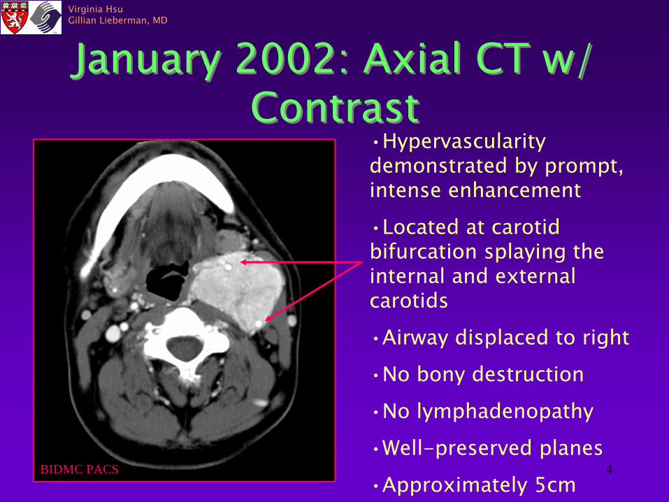
January 2002: Axial CT w/ Contrast
January 2002: Axial CT w/ Contrast
Mandible
External Carotid a.
Internal Carotid a.
Internal Jugular v.
Mass
Submandi bular
g.
SCMExternal Jugular v.
Vertebral a.
BIDMC PACS
4
Virginia HsuGillian Lieberman, MD
January 2002: Axial CT w/ Contrast
January 2002: Axial CT w/ Contrast
•Hypervascularity demonstrated by prompt,
intense enhancement
•Located at carotid bifurcation splaying the internal and external carotids
•Airway displaced to right
•No bony destruction
•No lymphadenopathy
•Well-preserved planes
•Approximately 5cmBIDMC PACS

5
Virginia HsuGillian Lieberman, MD
CT January 2002CT January 2002
Extends from carotid bifurcation superiorly to C1
BIDMC PACS
BIDMC PACS

6
Virginia HsuGillian Lieberman, MD
Carotid Space AnatomyCarotid Space Anatomy•Internal carotid artery
•Internal jugular vein
•Sympathetic chain
•Cranial nerves IX, X, XI, XII
•Lymph nodeshttp://www.bartleby.com/107/143.html
7
Virginia HsuGillian Lieberman, MD
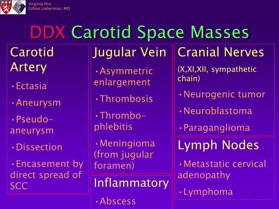
DDX
Carotid Space MassesDDX
Carotid Space MassesCarotid Artery•Ectasia•Aneurysm
•Pseudo- aneurysm
•Dissection
•Encasement by direct spread of SCC
Jugular Vein•Asymmetric enlargement
•Thrombosis
•Thrombo- phlebitis
•Meningioma (from jugular
foramen)
Cranial Nerves(X,XI,XII, sympathetic chain)
•Neurogenic
tumor
•Neuroblastoma
•Paraganglioma
Lymph Nodes•Metastatic cervical adenopathy
•LymphomaInflammatory•Abscess
8
Virginia HsuGillian Lieberman, MD
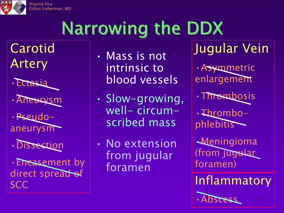
Narrowing the DDXNarrowing the DDX•
Mass is not intrinsic to blood vessels
Carotid Artery•Ectasia
•Aneurysm
•Pseudo- aneurysm
•Dissection
•Encasement by direct spread of SCC
Jugular Vein•Asymmetric enlargement
•Thrombosis
•Thrombo- phlebitis
•Meningioma (from jugular
foramen)
•
Slow-growing, well-
circum-
scribed mass
•
No extension from jugular foramen
Inflammatory•Abscess

9
Virginia HsuGillian Lieberman, MD
Narrowing the DDX con’tCranial Nerves(X,XI,XII, sympathetic chain)
•Neurogenic
tumor
•Neuroblastoma
•Paraganglioma
Lymph Nodes•Metastatic cervical adenopathy
•Lymphoma
•Hypervascularity
•No lymphadenopathy
•No calcification or necrosis
It must be a paraganglioma-
a
carotid body tumor!
It must be a paraganglioma-
a
carotid body tumor!
•Paraganglioma

10
Virginia HsuGillian Lieberman, MD
ParagangliomasParagangliomas•Rare tumors that arise from specialized neural crest cells associated with autonomic ganglia.
•4 extradrenal
locations
Group I: Great vessels of chest and neck
GroupII: Vagus
nerve
Group III: Aorticosympathetic
chain
Group IV: Visceral organs

11
Virginia HsuGillian Lieberman, MD
ParagangliomasParagangliomasHead and Neck
•
Carotid body paraganglioma
•
Vagal
paraganglioma (nodose
ganglia)
•
Glomus
tympanicum- middle ear along
tympanic plexus•
Glomus
jugulare-
jugular bulb
Glenner, GG and Grimley PM. Tumors of the Extra-Adrenal Paraganglion System. Bethesda, MD: Armed Forces Institute of Pathology, 1974
12
Virginia HsuGillian Lieberman, MD
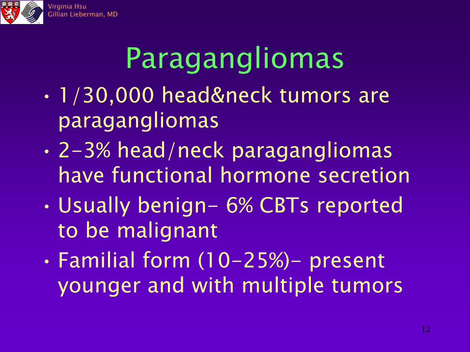
Paragangliomas•
1/30,000 head&neck
tumors are
paragangliomas•
2-3% head/neck paragangliomas
have functional hormone secretion•
Usually benign-
6% CBTs
reported
to be malignant•
Familial form (10-25%)-
present
younger and with multiple tumors

13
Virginia HsuGillian Lieberman, MD
Carotid Body Tumors•
CB sits in adventitia at bifurcation of common carotid a.
•
Regulates respiration and maintains arterial gases (chemoreception)
•
Hyperplasia seen in chronic hypoxic states-
altitude, COPD, cyanotic heart
disease•
Presentation: avg. age= 45, slow-
growing, asymptomatic or mass-related effects, 10% present with CN palsy
14
Virginia HsuGillian Lieberman, MD
Imaging StudiesImaging Studies
• CT•
MRI/MRA
•
Ultrasound•
Angiography
•
Radionuclide imaging
•
CT•
MRI/MRA
•
Ultrasound•
Angiography
•
Radionuclide imaging

15
Virginia HsuGillian Lieberman, MD
CTCT
•
Thin section scanning from thoracic inlet to skull base in patients with CB or vagal
paragangliomas
or other palpable neck mass
•
Examines integrity of associated soft tissues
•
Detection of multiple lesions•
3D reconstruction visualizes associated vasculature.
•
Thin section scanning from thoracic inlet to skull base in patients with CB or vagal
paragangliomas
or other palpable neck mass
•
Examines integrity of associated soft tissues
•
Detection of multiple lesions•
3D reconstruction visualizes associated vasculature.

16
Virginia HsuGillian Lieberman, MD
CT May 2002CT May 2002
•
No significant interval change to large enhancing mass
•
Displacement of airway to right
BIDMC PACS

17
Virginia HsuGillian Lieberman, MD
3-D CT Reconstruction3-D CT Reconstruction
Lustrin ES, PalestroC, Kirubahara V: Radiographic evaluation and assessment of paragangliomas. Otolaryngologic Clinics of North America 34(5) Oct 2001

18
Virginia HsuGillian Lieberman, MD
MRIMRI•
Aids in lesion diagnosis and localization
•
Differentiates mass from surrounding inflammatory changes, fluid or vascular structures
•
More sensitive for delineating encroachment and encasement of vessels
•
Images middle ear structures and bony erosions
•
Coronal sequences

19
Virginia HsuGillian Lieberman, MD
•Paraganglioma
•Carotid arteries
•Trachea
BIDMC PACS
T1 MRI w/o ContrastT1 MRI w/o Contrast

20
Virginia HsuGillian Lieberman, MD
MRI: T2MRI: T2•Well-defined mass•heterogeneous hyperintensity•Punctate flow voids. •“Salt and pepper” pattern: due to high vascularity with associated areas of hemorrhage, slow-
flowing blood, and tumor cells.BIDMC PACS
21
Virginia HsuGillian Lieberman, MD
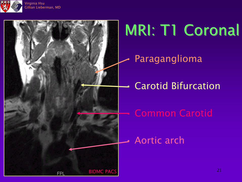
MRI: T1 CoronalMRI: T1 Coronal
•
Paraganglioma
•
Carotid Bifurcation
•
Common Carotid
•
Aortic arch
BIDMC PACS

22
Virginia HsuGillian Lieberman, MD
MRI: Time of FlightMRI: Time of Flight
•
Splaying of internal and external carotid arteries
•
No aneurysm or stenosis
BIDMC PACS

23
Virginia HsuGillian Lieberman, MD
MRAMRA
•
Noninvasive •
Delineates displacement of vasculature
•
Demonstrates tumor vascular supply
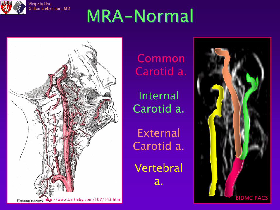
24
Virginia HsuGillian Lieberman, MD
Common Carotid a.
Internal Carotid a.
External Carotid a.
Vertebral a.
MRA-NormalMRA-Normal
http://www.bartleby.com/107/143.html BIDMC PACS

25
Virginia HsuGillian Lieberman, MD
MRA MRA
R L
BIDMC PACS BIDMC PACS
26
Virginia HsuGillian Lieberman, MD
Ultrasound
•
Delineates tumor margins, size and location
•
Doppler: demonstrates hypervascularity of paragangliomas
•
Surveys neck for other lesions•
Differentiates CBTs
from vascular
anomalies and pseudoaneurysms •
Can obtain US guided fine needle aspiration
•
Delineates tumor margins, size and location
•
Doppler: demonstrates hypervascularity of paragangliomas
•
Surveys neck for other lesions•
Differentiates CBTs
from vascular
anomalies and pseudoaneurysms•
Can obtain US guided fine needle aspiration

27
Virginia HsuGillian Lieberman, MD
UltrasoundUltrasound
Well-defined, hypoechoic heterogeneous mass at carotid bifurcation measuring 5.7x4.2x4.1 cm
BIDMC PACS
BIDMC PACS

28
Virginia HsuGillian Lieberman, MD
Doppler Ultrasound
Doppler Ultrasound
Hypervascular mass, splaying of
internal and external carotid arteries
BIDMC PACS
BIDMC PACS

29
Virginia HsuGillian Lieberman, MD
Ultrasound-Guided Fine Needle Aspiration
Ultrasound-Guided Fine Needle Aspiration
BIDMC PACS
30
Virginia HsuGillian Lieberman, MD
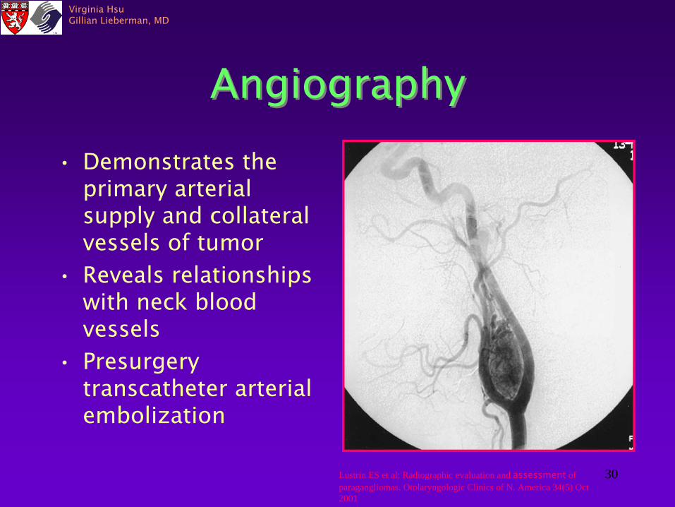
AngiographyAngiography•
Demonstrates the primary arterial supply and collateral vessels of tumor
•
Reveals relationships with neck blood vessels
•
Presurgery transcatheter
arterial
embolization
Lustrin ES et al: Radiographic evaluation and assessment
of paragangliomas. Otolaryngologic Clinics of N. America 34(5) Oct 2001

31
Virginia HsuGillian Lieberman, MD
Radionuclide ImagingRadionuclide Imaging•
Pentetreotide= octreotide
radiolabelled
with 111
indium-DTPA binds somatostatin
type 2
receptors in paragangliomas
•
Uses: follow recurrent disease, locates multiple lesions, detects familial paragangliomas
Lustrin ES et al: Radiographic evaluation and assessment
of paragangliomas. Otolaryngologic Clinics of N. America 34(5) Oct 2001
32
Virginia HsuGillian Lieberman, MD
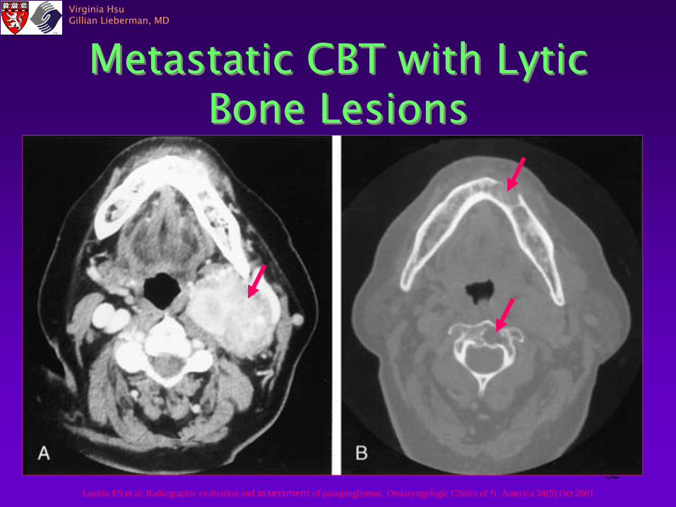
Metastatic CBT with Lytic Bone Lesions
Metastatic CBT with Lytic Bone Lesions
Lustrin ES et al: Radiographic evaluation and assessment
of paragangliomas. Otolaryngologic Clinics of N. America 34(5) Oct 2001
33
Virginia HsuGillian Lieberman, MD
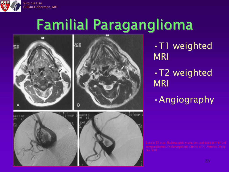
Familial ParagangliomaFamilial Paraganglioma•T1 weighted MRI
•T2 weighted MRI
•Angiography
Lustrin ES et al: Radiographic evaluation and assessment
of paragangliomas. Otolaryngologic Clinics of N. America 34(5) Oct 2001
34
Virginia HsuGillian Lieberman, MD
Vagal
ParagangliomaVagal
Paraganglioma
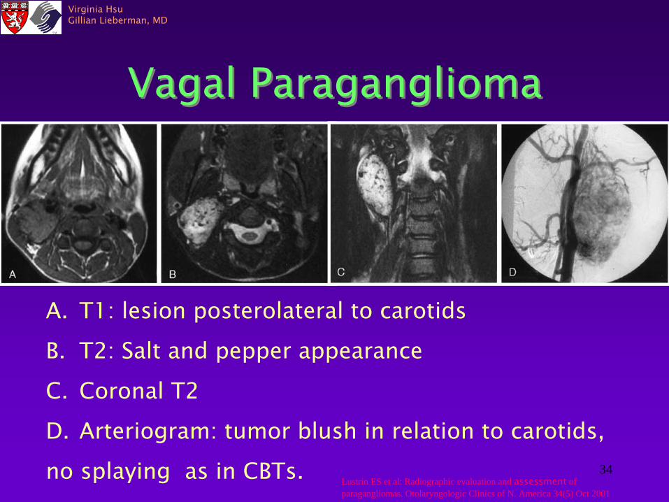
A.
T1: lesion posterolateral
to carotidsB.
T2: Salt and pepper appearance
C.
Coronal T2
D.
Arteriogram: tumor blush in relation to carotids,
no splaying as in CBTs. Lustrin ES et al: Radiographic evaluation and assessment
of paragangliomas. Otolaryngologic Clinics of N. America 34(5) Oct 2001

35
Virginia HsuGillian Lieberman, MD
Glomus
JugulareGlomus
Jugulare
•
Destructive lesion in skull base involving middle ear and hypoglossal canal
A.T1 w/o contrastB,C. T1 w/ contrast
Lustrin ES et al: Radiographic evaluation and assessment
of paragangliomas. Otolaryngologic Clinics of N. America 34(5) Oct 2001

36
Virginia HsuGillian Lieberman, MD
Glomus
TympanicumGlomus
Tympanicum
•
High resolution axial CT of right temporal bone
Lustrin ES et al: Radiographic evaluation and assessment
of paragangliomas. Otolaryngologic Clinics of N. America 34(5) Oct 2001

37
Virginia HsuGillian Lieberman, MD
Gross PathologyGross PathologyWell-defined neoplasm with a pseudocapsule
Well-defined neoplasm with a pseudocapsule
Glenner, GG and Grimley PM. Tumors of the Extra-Adrenal Paraganglion System. Bethesda, MD: Armed Forces Institute of Pathology, 1974

38
Virginia HsuGillian Lieberman, MD
HistologyHistologyOur Patient:
no malignant cells
•Small nests of bland cells with centrally placed hyperchromic
nuclei that form clusters called Zellballen
(cell balls).
Glenner, GG and Grimley PM. Tumors of the Extra-Adrenal Paraganglion System. Bethesda, MD: Armed Forces Institute of Pathology, 1974

39
Virginia HsuGillian Lieberman, MD
Our Patient…
•
Followed by ENT•
Will have surgical resection of CBT
•
Followed by ENT•
Will have surgical resection of CBT
40
Virginia HsuGillian Lieberman, MD
ReferencesReferencesGlenner GG and Grimley PM. Tumors of the Extra-Adrenal Paraganglion System. Bethesda, MD: Armed
Forces Institute of Pathology, 1974Lustrin ES et al: Radiographic evaluation and assessment
of paragangliomas. Otolaryngologic Clinics of N. America 34(5) Oct 2001
Som PM, Bergeron RT. Head and Neck Imaging. St. Louis: Mosby Year Book, 1991.
http://brighamrad.harvard.edu/Cases/bwh/hcache/74/full/htmlhttp://www.bartleby.com/107/143.htmlhttp://individual.uptodateonline.com/application/topic/topicText.asp?file=brain_ca/10730
Mafee MF, Raofi B, Kumar A, Muscato C. Glomus faciale, glomus jugulare, glomus tympanicum, glomus vagale, carotid body tumors and simulating lesion. Radiologic Clinics of North America 38(5) Sept 2000
Novelline RA, Squire LF. Living Anatomy. Philadelphia: Hanley & Belfus, Inc., 1987
Myssiorek D. Head and Neck Paragangliomas: An Overview. Otolaryngologic Clinics of N. America 34(5) Oct 2001
Van der Mey AGL, Jansen JC, van Baalen, JM. Paragangliomas of the Head and Neck:Management of carotid body tumors. Otolaryngologic Clinics of N. America 34(5) Oct 2001
Wasserman PG, Savargaonkar P. Paragangliomas of the Head and Neck: Classification, patholgy, differential diagnosis. Otolaryngologic Clinics of N. America 34(5) Oct 2001
McCaffrey TV, Myssiorek D, Marrinana M. Paragangliomas of the Head and Neck: Physiology and biochemistry. Otolaryngologic Clinics of N. America 34(5) Oct 2001
Richardson MS.Skull Base Tumor Surgery: Pathology of skull base tumors. Otolaryngologic Clinics of N. America 34(6) Dec 2001
41
Virginia HsuGillian Lieberman, MD
AcknowledgementsAcknowledgements
•
Gwendolyn Dole, MD•
Pamela Lepkowski
•
Larry Barbaras and Cara Lyn D’Amour
•
Gillian Lieberman, MD
•
Gwendolyn Dole, MD•
Pamela Lepkowski
•
Larry Barbaras and Cara Lyn D’Amour
•
Gillian Lieberman, MD