Embed Size (px)
Citation preview
CARS 2002 Computer Assisted Radiology and Surgery

Springer-Verlag Berlin Heidelberg GmbH
CARS 2002 Computer Assisted Radiology and Surgery
Proceedings of the 16th International Congress and Exhibition Paris, June 26- 29,2002
Edited by H. U. Lemke, M. W. Vannier, K. Inamura, A. G. Farman, K. Doi, and J. H. C. Reiber
, Springer
PROFESSOR HEINZ U. LEMKE, PHD
Technical University Berlin Computer Graphics and Computer Assisted Medicine Secr. FR 3-3 Franklinstrasse 28-29 10587 Berlin, Germany
PROFESSOR KIYONARI INAMURA, PHD
Osaka University Faculty of Medicine School of Allied Health Sciences Department of Radiological Technology & Medical Engineering 1-7 Yamadaoka, Suita City Osaka, 565-0871, Japan
PROFESSOR KUNIO DOl, PHD
University of Chicago Hospitals Department of Radiology Kurt Rossmann Laboratories 5841 S. Maryland Avenue, Mailcode 2026 Chicago, IL 60637, U.S.A.
PROFESSOR MICHAEL W. VANNIER, MD
The University of Iowa College of Medicine Department of Radiology 200 Hawkins Drive Room 3966 JPp Iowa City, IA 552242-1077, U.S.A.
PROFESSOR ALLAN G. FARMAN, PHD, DSc
University of Louisville School of Dentistry Department of Diagnosis and General Dentistry 501 South Preston, Room 222E Louisville, KY 40292, U.S.A.
PROFESSOR JOHAN H. C. REIBER, PHD
Leiden University Medical Center Division of Image Processing Department of Radiology P. O. Box 9600 Albinusdreef 2 2300 RC Leiden, The Netherlands
This volume of the CARS proceedings is published simultaneously in print and on the web. The web edition contains colour images.
ISBN 978-3-642-62844-3 ISBN 978-3-642-56168-9 (eBook) DOI 10.1007/978-3-642-56168-9
Library of Congress Cataloging-in-Publication Data
This work is subject to copyright. All rights are reserved, whether the whole or part of the material is concerned, specifically the rights of translation, reprinting, reuse of illustrations, recitation, broadcasting, reproduction on microfilm or in any other way, and storage in data banks. Duplication of this publication or parts thereof is permitted only under the provisions of the German Copyright Law of September 9, 1965, in its current version, and permissions for use must always be obtained from Springer. Verlag or CARS. Violations are liable for prosecution under the German Copyright Law.
http://www.springer.de http://www.cars·int.de
© Springer· Verlag Berlin Heidelberg 2002 and CARS Originally published by Springer-Verlag Berlin Heidelberg New York in 2002
Softcover reprint of the hardcover 1St edition 2002
The use of general descriptive names, registered names, trademarks, etc. in this publication does not imply, even in the absence of a specific statement, that such names are exempt from the relevant protective laws and regulations and therefore free for general use.
SPIN 10755372 3113130 - 5 4 3 2 1 0-
Honorary President Kiyonari Inamura, PhD, Osaka University, Faculty of Medicine, Osaka (J)
Congress Organizing Committee
Ulrich Dick, MD University of Chicago Hospitals (USA)
Kees de Wilde Philips Medical Systems, Best (NL)
Kunio Doi, PhD University of Chicago Hospitals (USA)
Allan G. Farman, PhD, DSc University of Louisville (USA)
Guy Frija, MD Societe Francaise de Radiologie Medicale, Paris (F)
J. Thomas Lambrecht, MD, DMD, PhD University of Basle (CH)
Heinz U. Lemke, PhD Technical University Berlin (D)
Roberto Passariello, MD University "La Sapienza", Rome (I)
Pierre Rabischong, MD, PhD Centre Propara, Montpellier (F)
Johan H.C. Reiber, PhD Leiden University Medical Center (NL)
Hans G. Ringertz, MD, PhD Karolinska Hospital, Stockholm (S)
Ramin Shahidi, PhD
v
Toyomi Fujino, MD, PhD, FACS International Med. Information Center, Tokyo (J) Stanford University Medical Center (USA) Gary M. Glazer, MD Stanford Univ. School of Med., Palo Alto, CA (USA)
Kiyonari Inamura, PhD Osaka University (J)
Takahiro Kozuka, MD Kaizuka Municipal Hospital, Osaka (J)
Industrial Advisory Board
Nicola H. Strickland, MD Hammersmith Hospital, London (UK)
Kintomo Takakura, MD, PhD Tokyo Women's Medical University (J)
Michael W. Vannier, MD The University of Iowa (USA)
Chairman: Kees de Wilde, Philips Medical Systems, Best (NL)
Heinz-Christoph Blied Siemens AG, MED, Erlangen (D)
Karel H.P. Cromzigt IEARC, Den Hague (NL)
Gerhard Kacmaczyk Image Devices GmbH, Taunusstein (D)
Annelies Kin Agfa Europe NV, Mechelen (B)
Kenneth A. Marks, MBA DOME Imag. Syst., Inc., Tiburon, (USA)
Willem Overlaet, PhD Toshiba Medical Sys., Zoetermeer (NL) Jiirgen Reyinger GE Medical Systems, Dornstadt (D)
Saeid Mitchell Seyedin CBYON, Palo Alto, CA (USA)
Kurt R. Smith, DSc Medtronics, Broomfield, CO (USA)
Chris Varian Eastman Kodak Company, Hemel (UK)
Stefan Vilsmeier BrainLAB GmbH, Heimstetten (D)
VI
Program Committee
Andreas Adam, MB, RCP, FRCR Guy's Hospital, London (UK)
Paul R. Algra, MD, PhD Medisch Centrum Alkmaar (NL)
David J. Allison, MD Hammersmith Hospital, London (UK)
Mostafa Analoui, PhD Indiana University, Indianapolis, IN (USA)
Yutaka Ando, MD Keio University, Tokyo (1)
Licinio Angelini, MD, FACS Universita "La Sapienza", Rome (I)
Takehide Asano, MD, PhD Chiba University School of Medicine (1)
Hanna Bachtiar Iskandar, DDS University of Indonesia, Jakarta (RI)
Frits H. Barneveld Binkhuysen, MD, PhD Hospital Eemland, Amersfoort (NL)
Elizabeth Beckmann, BSc Lanmark, Beaconsfield (UK)
Leonard Berliner, MD Swissray Int., Staten Island, NY (USA)
Silvio Diego Bianchi, MD Universita degli Studi di Torino (I)
Josip S. Bill, MD, DDS Julius-Maximillians-University, WUrzburg (D)
Uldis Bite, MD Mayo Clinic, Rochester, MN (USA)
Michel Biery, MD H6pital de Bicetre, Le Kremlin-Bicetre (F)
Siegfried Bocionek, PhD Siemens Health Services GmbH, Erlangen (D)
Hugo G. Bogren, MD, PhD UC Davis Med. Center, Sacramento, CA (USA)
Nicolaas Bom, PhD Erasmus Universiteit Rotterdam (NL)
Hans G. Bosch, MSc Leiden University Medical Center (NL)
Carlos A. Bruguera, MD Inst. de Ensenanza Audiovis., Buenos Aires (RA)
Jean Noel Bruneton, MD Centre Antoine-Lacassagne, Nice (F)
Richard D. Bucholz, MD, FACS Saint Louis University (USA)
Gerhard Buess, MD Nova Med Klinik, MUnchen (D)
Jean-Marie Caille, MD Groupe Hospitalier Pellegrin, Bordeaux (F)
Davide Caramella, MD University of Pis a (I)
Ernest V. Carcia, PhD Emory Univ .. , Atlanta, GA (USA)
Robert Cavezian, MD Cabinet de Radiologie, Paris (F)
Curtis Ssu-Kuang Chen, DDS, MSD, PhD National Taiwan University, Taipei (CHN)
S. James Chen, PhD University of Colorado, Denver, CO (USA)
Kiyoyuki Chinzei, PhD Mechanical Engineering Lab. AIST, Ibaraki (1)
John C. Chiu, MD California Spine Inst., Thousand Oaks (USA)
Hiroaki Chiyokura, PhD Keio University, Kanagawa (1)
Gary E. Christensen, PhD University of Iowa (USA)
Philippe Cinquin, MD, PhD CHU Grenoble, La Tronche (F)
Michel Claudon, MD H6pitaux de Brabois, Vandoeuvre (F)
Claus D. Claussen, MD Eberhard-Karls-Univ. TUbingen (D)
Kevin Cleary, PhD Georgetown Univ., Washington, DC (USA)
Alan C.F. Colchester, MD, PhD University of Kent, Canterbury (UK)
Bernard L. Crowe, BA, MPH Health Inf. Soc. of Australia, Canberra (AUS)
Jack T. Cusma, PhD Mayo Clinic, Rochester, MN (USA)
Paolo Dario, PhD Scuola Superiore S. Anna, Pisa (I)
Albert de Roos, MD Leiden University Medical Center (NL)
Carlo del Favero, MD Ospedale "Valduce", Como (I)
Anthony M. DiGioia III, MD Shadyside Hospital, Pittsburgh, PA (USA)
Jouke Dijkstra, PhD Leiden University Medical Center (NL)
Takeyoshi Dohi, PhD The University of Tokyo (1)
Huilong Duan, PhD Zhejiang University Hangzhou (RC)
Andre J. Duerinckx, MD, PhD VA North Texas HC System, Dallas, TX (USA)
Masahiro Endo, PhD National lost. of Radiological Scie., Chiba (J)
Rolf Ewers, MD, DMD Allg. Krankenhaus der Stadt Wien (A) Aly A. Farag, PhD University of Louisville (USA)
Taeko T. Farman, PhD, DMD, RT University of Louisville (USA)
Allan G. Farman, PhD, DSc University of Louisville (USA)
Eckhard Fleck, MD Deutsches Herzzentrum Berlin (D)
Kevin T. Foley, MD Image-Guided Surg. Res. Ctr.,Memphis, (USA)
Erik Fosse, MD, PhD University of Oslo (N)
Bernard Fraysse, MD H6pital Purpan, Toulouse (F)
Hiroshi Fujita, PhD Gifu University (1)
Giinther Gell, PhD Universitat Graz - Landeskrankenhaus (A)
Bernard Gibaud Universite de Rennes I (F)
Maryellen L. Giger, PhD The University of Chicago (USA)
Stephen Golding, FRCR University of Oxford, Headington (UK)
Dietrich H.W. Gronemeyer, MD Universitat Witten-Herdecke (D)
Chiaki Hamanishi, MD Kinki University School of Medicine, Osaka (1)
Daijo Hashimoto, MD, PhD Tokyo Metropolitan Police Hospital (1)
Makoto Hashizume, MD, PhD, FACS Kyushu University, Fukuoka (1)
Stefan Hallfeld, MD, MDS Ruprecht-Karls-Universitat, Heidelberg (D)
VII
David Hatcher Univ. of California, San Francisco, CA (USA)
David J. Hawkes, PhD Guy's, Hospital, London (UK)
J.H.C. Hendriks, MD University Hospital Nijmegen (NL)
Atsuko Heshiki, MD Saitama Medical School (1)
Kenneth R. Hoffmann, PhD University at Buffalo (USA)
Karl-Heinz Hohne, PhD Universitat Hamburg (D)
Steven C. Horii, MD Univ. of Pennsylvania, Philadelphia, PA (USA)
Alexander Horsch, PhD University of Munich (D)
Walter Hruby, MD Sozialmedizinisches Zentrum Ost, Wien (A)
H.K. Huang, DSc, FRCR (Hon.) Univ. of South. California, Los Angeles, CA (USA)
Junpei Ikezoe, MD Ehime University (1)
Herwig Imhof, MD Allgemeines Krankenhaus der Stadt Wien (A)
Hiroshi Iseki, MD, PhD Tokyo Women's Medical College (1)
Takeo Ishigaki, MD, PhD Nagoya University (1)
Akira Ito, PhD Japanese Foundation for Cancer Res., Tokyo (1)
Ian T. Jackson, MD lost. f.Craniofacial a.R.Surg., Southfield (USA)
C. Carl Jaffe, MD Yale Univ. School of Me d., New Haven (USA)
Pierre Jannin, PhD Universite de Rennes I (F)
Werner Jaschke, MD Univ-Klinik fUr Radiodiagnostik, Innsbruck (A)
Jack Jellins, PhD Intern. Breast Ultrasound Sch., Sydney (AUS)
Peter Jensch, PhD OFFIS e.V., Oldenburg (D)
Ferenc A. Jolesz, MD Harvard Medical School, Boston, MA (USA)
Leo Joskowicz, PhD The Hebrew University of Jerusalem (IL)
VIII
Willi A. Kalender, PhD Friedrich-Alexander-Universitat, Erlangen (D)
Shigenobu Kanda, DDS, PhD Kyushu University, Fukuoka (1)
Kazuhiro Katada, MD Fujita Health University, Aichi (J)
Amami Kato, MD, PhD Osaka University Medical School (1)
Erwin Keeve, PhD CAESAR, Bonn (D)
Ron Kikinis, MD Harvard Medical School, Boston, MA (USA)
Reinhard Klette, PhD University of Auckland (NZ)
Klaus Jochen Klose, MD Universitats-Klinikum, Marburg (D)
Goran Knezevic, DDS, PhD University of Zagreb (HR)
Hidefumi Kobatake, PhD Tokyo Univ. of Agriculture & Technology (J)
Masahiro Kobayashi, MD Keio University, Tokyo (1)
Gerhard Koning, MSc Leiden University Medical Center (NL)
Martti Kormano, MD, PhD Turku University Central Hospital (FIN)
Uwe G. Kuhnapfel, PhD Forschungszentrum Karlsruhe GmbH (D)
Chikazumi Kuroda, MD Osaka Medical Center for Cancer (1)
Axel Kuttner, MD Klinik. der Eberhard-Karls-Univ. TUbingen (D)
Frode Laerum, MD, PhD, MHA University of Oslo (N)
J. Thomas Lambrecht, MD, DMD, PhD University of Basle (CH)
Alexandra Lansky, MD Cardiov.r Research Found., New York (USA)
Tore A. Larheim, PhD, DDS University of Oslo (N)
Stephane Lavallee, PhD PRAXIM, La Tronche (F)
Swamy Laxminarayan, PhD New Jersey Inst.ofTechn., Newark, NJ (USA)
Lilian L.Y. Leong, MBBS Queen Mary Hospital, Hong Kong (RC)
Yves Ligier, PhD CareON S.A., Grand Saconnex (CH)
Jae Hoon Lim, MD Sung Kyun Kwan University, Seoul (ROK)
Martin J. Lipton, MD The University of Chicago Hospitals (USA)
Yu-Qing Liu, MD FuWai Hosp. & Cardiovascular Inst.. Beijing (RC)
Tim C. Lueth, PhD Campus Yirchow-Klinikum. Berlin (D)
Xiu-chen Ma, PhD Peking Univ. Sch.ofStomatology, Beijing (RC)
Riley H. Lunn, DDS Chattanoooga, TN (USA)
Heber MacMahon, MD The University of Chicago (USA)
Sumio Makino, PhD Yokohama-City (1)
Borut Marincek, MD Universitatsspital ZUrich (CH)
Steffen Markle, PhD Technical University Berlin (D)
Tom H. Marwick, MD University of Queensland, Brisbane (AUS)
Herbert K Matthies, PhD Hannover Medical School (D)
Hans-Peter Meinzer, PhD Deutsches Krebsforschungsz., Heidelberg (D)
Andreas Melzer, MD MUhlheimer Radiologie Inst., MUlheim (D)
Reto A. Meuli, MD CHUY. Lausanne (CH)
Kazuo Miyasaka, MD Hokkaido University, Sapporo (1)
Kensaku Mori, PhD Nagoya University (1)
Seong K Mun, PhD Georgetown University, Washington, DC (USA)
Eike Nagel, MD Deutsches Herzzentrum Berlin (D)
KS. Nagesh R.Y. Dental College, Bangalore (IND)
Hironobu Nakamura, MD, PhD Osaka University Medical School (1)
Wolfgang Niederlang, PhD Krankenhaus Dresden-Friedrichstadt (D)
Robert M. Nishikawa, PhD The University of Chicago (USA)
Hiromu Nishitani, MD, PhD The University of Tokushima (1)
Lutz-P. Nolte, PhD Maurice E. MUlier Institute, Bern (CH)
FridtjofNiisslin, PhD Eberhard-Karls-Universitat, TUbingen (0)
Takahiro Ochi Osaka University (1)
NagaakiOhyama,PhD Tokyo Institute of Technology, Yokohama (1)
Silas Olsson, MSc Telia Research AB, Farsta (S)
Dietrich Onnasch, PhD University of Kiel (0)
Stelios Orphanoudakis, PhD Institute of Computer Science, Heraklion (GR)
Michel Osteaux, MD, PhD Vrije Universiteit Brussels (B)
Helmut Oswald, PhD T-Systems HCS AG, Bern (CH)
Hiroshi Oyama, MD Kyoto University Hospital (1)
Paolo Pavone, MD Universita "La Sapienza", Rome (I)
Heinz-Otto Peitgen, PhD University of Bremen (0)
Prem Pillay, MD Asian Brain Spine Nerve Ctr., Singapore (SGP)
Gabriel E. Pislaru Charlotte, NC (USA)
E. James Potchen, MD Michigan State Univ., East Lansing, MI (USA)
Henri Primo Siemens Medical Syst., Inc., Iselin. N1 (USA)
Osman M. Ratib, MD, PhD Univ. of California, Los Angeles, CA (USA)
Hans F. Reinhardt, MD Bethesda Spital. Baste (CH)
Maximilian Reiser, MD Klinikum GroBhadern, MUnchen (0)
Stephen J. Riederer, PhD Mayo Clinic, Rochester, MN (USA)
Otto Rienhoff, MD Georg-August-Universitat, Gottingen (0)
Rainer K. Rienmiiller, MD Universitatskliniken Graz (A)
Richard A. Robb, PhD Mayo Foundation. Rochester, MN (USA)
Ichiro Sakuma, PhD The University of Tokyo (1)
Georges Salamon, MD NW Univ. Med. School, Chicago, IL (USA)
Richard M. Satava, MD Yale University. New Haven, CT (USA)
Ronald B. Schilling, PhD PGI Corporation, Los Altos Hills, CA (USA)
Peter M. Schlag, MD, PhD Robert-Rossle-Klinik, Berlin (0)
Wolfgang Schlegel, PhD Oeutsches Krebsforschungsz., Heidelberg (0)
Rainer M.M. Seibel, MD Univ. WitteniHerdecke, MUlheimIRuhr (0)
Wolfhard Semmler, MD Oeutsches Krebsforschungsz., Heidelberg (0)
Jean Sequeira, PhD Laboratoire d'lnformatique de Marseille (F)
lekado Shibata, MD, PhD Toho University. Tokyo (1)
Faina Shtern, MD Harvard Medical School, Boston, MA (USA)
Robert Sigal, MD, PhD Institut Gustave-Roussy, Villejuif (F)
Peter Sloot, PhD University of Amsterdam (NL)
Milan Sonka, PhD University of Iowa (USA)
Edward V. Staab, MD National Cancer Inst.. Rockville, MO (USA)
Gero Strauss, MD University of Leipzig (0)
Nobuhiko Sugano, MD Osaka University Med. School (1)
Predrag Sukovic, MS
IX
University of Michigan, Ann Arbor, MI (USA)
Naoki Suzuki, MD, PhD 1ikei University School of Medicine, Tokyo (1)
Takashi Takahashi, PhD Kyoto University Hospital (1)
Hiroshi Takeda, MD, PhD Osaka University Medical School (1)
x
Shin-ichi Tamura, PhD Osaka University Medical School (1)
Russell H. Taylor, PhD Johns Hopkins Univ., Baltimore, MD (USA)
Bart M. ter Haar Romeny, PhD University Hospital Utrecht (NL)
Andrew E. Todd-Pokropek, PhD University College of London (UK)
Thomas Tolxdorff, PhD Freie Universitat Berlin (D)
Jun-Ichiro Toriwaki, PhD Nagoya University (1)
Hikmet Umar, DMD, MSIS, DlC, PhD Temple University, Philadelphia, PA (USA)
Vlastimil Valek, MD, PhD University Hospital Bmo (CZ)
Rob J. van der Geest, MSc Leiden University Medical Center (NL)
Ernst E. van der Wall Leiden University Medical Center (NL)
Rob van Geuns Philips Medical Systems, Best (NL)
Paul P.F.G.M. van Waes, MD, PhD University Hospital Utrecht (NL)
Past Honorary Presidents
CAR '89 Heinz Oeser, MD Berlin (D)
CAR '91 Auguste Wackenheim, MD Strasbourg (F)
CAR '93 J. Oscar M.e. Craig, MD London (UK)
Jos Vander Sioten, PhD Katholieke Universiteit Leuven, Heverlee (B)
Robert H. Vandre, DDS, MS US Army, Maryland, MD (USA)
Max A. Viergever, PhD Utrecht University (NL)
Clemens von Birgelen, MD Universitatsklinikum Essen (D)
Robert F. Wagner, PhD Food and Drug Admin., Rockville, MD (USA)
Andreas Wahle, PhD University ofIowa (USA)
Mamoru Wakoh, DDS, PhD Tokyo Dental College, Chiba (1)
Thomas Wendler, PhD Philips Research Laboratories, Hamburg (D)
Karl-Jiirgen Wolf, MD Univ.-Klinikum Benjamin Franklin, Berlin (D)
Richard Wootton, PhD, DSc The Univ. of Queensland, St. Lucia (AUS)
Hirouki Yoshida, PhD The University of Chicago (USA) Frans W. Zonneveld, PhD University Hospital Utrecht (NL)
CAR '95 Alexander R. Margulis, MD San Francisco (USA)
CAR '97 Takahiro Kozuka, MD Osaka (J)
CAR '98 Herbert Kaufmann, MD Berlin (D)
CARS '99 Raffaella de Dominicis, MD Florence (I)
CARS 2000 Pierre Rabischong, MD, PhD Montpellier (F)
CARS 2001 Toyomi Fujino, MD, PhD Tokyo (J)
CARS 2002 in cooperation with
AAOMR
BAR
BIR
CRS
CURAC
DGBMT
DRG
ESCR
ESEM
American Academy of Oral and Maxillofacial Radiology
Bulgarian Association of Radiology
The British Institute of Radiology
Czech Radiological Society
Deutsche Gesellschaft flir Computer- und Roboterassistierte Chirurgie e.V.
Deutsche Gesellschaft flir Biomedizinische Technik e.V.
Deutsche Rontgengesellschaft
European Society of Cardiac Radiology
European Society for Engineering and Medicine
EuroPACS European Association for Promotion ofinformation Exchange on PACS Research
GI Gesellschaft flir Informatik
XI
GMDS Deutsche Gesellschaft flir Medizinische Informatik, Biometrie und Epidemiologie e.V.
Hungarian Society for Computer Assisted Radiology
IADMFR The International Association of Dento-Maxillo-Facial Radiology
IEARC International Exhibitors Association on Radiological Congresses
IEEE
JCR
JRS
JSOMR
The Institute of Electrical and Electronics Engineers
Japanese College of Radiology
Japan Radiological Society
Japanese Society for Oral and Maxillofacial Radiology
NVvR Nederlandse Vereniging voor Radiologie
ORG Osterreichische Rontgengesellschaft
OWGTM Osterreichische Wissenschaftliche Gesellschaft flir Telemedizin
Polish Section of the Polish-German Radiological Society
PMSR Polish Medical Society of Radiology
RCR The Royal College of Radiologists
EFSUMB European Federation for Ultrasound in Medicine and Biology
SFR Societe Franc;aise de Radiologie Medicale
SGR-SSR Schweizerische Gesellschaft flir Radiologie
SICTECA Societa Italiana di Chirurgia TecnoIogica e Computer-Assistita
SIRM Societa Italiana di Radiologia Medica
Chinese Society for Computer Assisted Radiology
SPIE The International Society for Optical Engineering
SRS Slovak Radiological Society
TU Technische Universitat Berlin
WABT World Academy of Biomedical Technologies
XII
ISCAS - International Society for Computer Aided Surgery
Administrative Council
President: Kintomo Takakura, MD, PhD Tokyo Women's Medical University, Tokyo (J)
Past President: Michael W. Vannier, MD The University of Iowa (USA)
Vice Presidents: J. Thomas Lambrecht, MD, DMD, PhD University of Basle (CH)
Jeffrey L. Marsh, MD St. Louis Children's Hospital (USA)
Board Members
Richard D. Bucholz, MD, FACS Saint Louis University (USA)
Alan C.F. Colchester, MD, PhD University of Kent, Canterbury (UK)
Daijo Hashimoto, MD, PhD Tokyo Metropolitan Police Hospital (J)
Honorary Board Member: Pierre Rabischong, MD, PhD Centre Propara, Montpellier (F)
General Secretary: Heinz U. Lemke, PhD Technical University Berlin (D)
Adjoint Secretary: Takeyoshi Dohi, PhD The University of Tokyo (J)
Treasurer: Toyomi Fujino, MD, PhD International Med. Information Center, Tokyo (J)
Adjoint Treasurer: Rolf Ewers, MD, DMD Allg. Krankenhaus der Stadt Wien (A)
Richard A. Robb, PhD Mayo Foundation, Rochester, MN (USA)
Russell H. Taylor, PhD Johns Hopkins University, Baltimore, MD (USA)
Jun-Ichiro Toriwaki, PhD Nagoya University (J)
xv
Preface
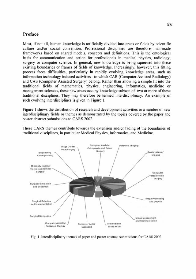
Most, if not all, human knowledge is artificially divided into areas or fields by scientific culture and/or social convention. Professional disciplines are therefore man-made frameworks based on shared models, concepts and definitions. This is the ontological basis for communication and action for professionals in medical physics, radiology, surgery or computer science. In general, new knowledge is being squeezed into these existing boundaries or frames of fields of knowledge. Increasingly, however, this fitting process faces difficulties, particularly in rapidly evolving knowledge areas, such as information technology induced activities - to which CAR (Computer Assisted Radiology) and CAS (Computer Assisted Surgery) belong. Rather than allowing a simple fit into the traditional fields of mathematics, physics, engineering, informatics, medicine or management sciences, these new areas occupy knowledge subsets of two or more of these traditional disciplines. They may therefore be termed interdisciplinary. An example of such evolving interdisciplines is given in Figure I.
Figure 1 shows the distribution of research and development activities in a number of new interdisciplinary fields or themes as demonstrated by the topics covered by the paper and poster abstract submissions to CARS 2002.
These CARS themes contribute towards the extension and/or fading of the boundaries of traditional disciplines, in particular Medical Physics, Informatics, and Medicine.
Engineering Anth ropometty
Minimally Invasive Thoracic Abdominal
Surgery
Surgical Simulatjon and Education
Surgicill Robotics and Instrumentation
Surgical Navigation
Computer Assisted Radiation Therapy
Cardiovascular
Imaging
Computed I
Imaging
Image Processing and Display
Image Management and CommunIcation
Fig. I Interdisciplinary themes of paper and poster abstract submissions for CARS 2002

XVI
The following brief defmitions of the most important interdisciplines may serve as a guideline regarding the structure on which the CARS 2002 program has been compiled:
Medical Imaging (MI) The science and engineering needed for the sourcing, acqUisition, and processing of signals from the human body to generate multi-dimensional digital images.
Cardiovascular Imaging (CVI) MI applied to the special requirements of the cardiovascular system and extended with IPD methods to allow quantitatively and qualitatively enhanced medical diagnosis.
Computed Maxillofacial Imaging (CMI) MI applied to the special requirements of maxillofacial structures and extended with IPD methods to allow quantitatively and qualitatively enhanced medical diagnosis in this area.
Image Processing and Display (IPD) Processing of elements/objects of images, e.g. pixels, voxels, regions and contours to enhance the quality, understanding, and representation of images. Image processing steps may include enhancement, restoration, registration, segmentation, analysis, interpretation, and multi-modality image fusion. Display steps may include various visualisation algorithms to represent data interrelationships or to enhance reality.
Image Management and CommunicationlPACS Systems for storage, transmission, manipulation and display of digital images and the management of these images to enable an effective and hardcopyless (e.g. film less) healthcare infrastructure.
Telemedicine and E-health (TMEH) The use of information and communication technology to enable the transfer of medical data and programs to support healthcare activities between geographically separate locations.
Computer Aided Diagnosis (CAD) MI and IPD methods applied to specific body parts to enable an image-guided diagnosis which enhances sensitivity and/or specificity of a diagnostic procedure carried out by humans.
Engineering Anthropometry (EA) Acquisition, modelling, management and analysis of anatomic/physical landmarks and 3D surface data of different human populations. EA methods are based on data models, software tools and theoretical constructs and principles to support, for example, the searching and visualisation of 3D objects (human body scans). In healthcare, EA tools may also, for example, support anatomic atlas-based segmentation as well as the design of endoprosthetic devices and implants.
Surgical Navigation (SN) IT-based systems to assist surgical procedures by guiding instruments within a surgical target volume to an operated volume with maximum accuracy and safety.
XVII
Surgical Robotics and Instrumentation (SRI) IT and MEMS-based systems which use SN or SS to passively assist surgical activities semi-automatically or fully automatically.
Surgical Simulation (SS) and Education IPD methods and simulation software applied to an surgical target volume to optimize surgical activities for the purpose of training and preoperative planning.
Image Guided Neurosurgery (lGNS) The use of one or more methods derived from SN, SS or SRI, with a strong bias towards using images, to achieve minimally invasive interventions directed towards surgical target structures including the brain or spinal cord.
Computer Assisted Orthopaedic and Spinal Surgery (CADS) The use of one or more methods derived from SN, SS or SRI to achieve minimally invasive interventions directed towards surgical target volume including orthopaedic structures, such as the spinal vertebrae, hip, knee, ankle, or long bones. In a wider definition it may also include rapid prototyping and the design and manufacturing of endoprosthetic devices and implants.
Minimally Invasive Thoracic Abdominal Surgery (MIT AS) The use of one or more methods derived from SN, SS or SRI to achieve minimally invasive interventions directed towards surgical target volume within the thorax (excluding the cardiovascular structures) and the abdomen.
Computer Assisted Radiation Therapy (CART) Modelling of radiation target structures with IPD methods and the application of radiation dose planning programs to optimize target volume (tumor) radiation treatment.
The subset of knowledge from the traditional fields of mathematics, physics, engineering, informatics, medicine, and management science required to advance the CARS interdisciplines given above is also subject of change. A proper understanding of this may illuminate which traditional profession comes closest to pursuing the respective activities within the CARS themes.
Results derived from CARS methods and applications provide valuable data, information, knowledge and even wisdom, and are changing medicine and associated professions. The impact this will have on current educational curricula as well as on changing the professional profiles of medical physicists, radiologists, surgeons, and computer scientists is substantial and requires further examination. As yet, few teaching institutions have responded to this challenge and are ready to offer appropriate educational courses and training. CARS 2002 and future CARS congresses are aimed at showing the direction into which a significant part of medicine will evolve in research, development and practice.
Heinz U. Lemke Paris, June 2002
XVIII
Acknowledgements
Once upon a time, back in the 1960s, when I first had to program a computer it was done in an assembler language. The good thing about this was that this language or interface to the computer, had a finite set of instructions and a finite set of combining these. The bad things were the lack of computational power of the workstation and the user interface to the assembler. On execution, however, the computer did more or less what one wanted it to do. Now, in 2002, times have changed. The good thing now is, that some twelve levels of hardware and software sophistication separate the assembler from the modem "easy to use" graphical user interfaces. Super computational power takes care of everything. The bad thing is, that the computer does not always do what one wants it to do.
Anyone who does not believe this, I should like to invite to do the electronic editing of the next CARS proceedings. The non-believers probably belong to the approx. 5% of authors who submitted their papers according to the author instruction for electronic submission for the CARS proceedings. These authors focused not only on providing excellent content but also on the structure and visual appearance of their papers. The other 95% obviously preferred to focus on content only and have worked, as I like to do, that is, leaving styling and layout of the paper to others.
The move into the electronic age was necessary in order to improve the quality and ensure the timely appearance of the CARS proceedings as a hardbound copy and also on the web. It has meant, however, a considerable amount of extra workload for Franziska Schweikert and her team, in particular Dagmar Harrison, in addition to the "usual" organizational and management work. During a two week period before and at the deadline of paper submission (i.e. 15th March, 2002) approx. 170 papers and 110 posters were received at the CARS Conference Office and electronically prepared and edited. The four weeks in March meant double-shift work for Franziska Schweikert and this is my written promise that next for year's CARS, we will engage additional assistance.
I also should like to acknowledge and thank for the continuing help received from the personnel of my department at the Technical University of Berlin, in particular from Helga Kallan, Steffen Markle, Kai Kochy and Rene Tschirley. This is carried out in an environment marked by an increasing number of students in courses and for supervision, against a background of further cuts in teaching staff positions.
We are all delighted to have Prof. Kiyonari Inamura as the Honorary President of CARS 2002. I have known Kiyo Inamura for more than 10 years and had the pleasure to work closely together with him on a number of projects and many congress events. His contributions to research and development in many fields relating to CARS are documented in numerous publications. In particular, I treasure him for his engagement in advancing interdisciplinary and international cooperation. Through his remarkable selflessness, he is a great benefit not only for Japan but also for the rest of the world.
Heinz U. Lemke Paris, June 2002
XIX
Prelude
CARS, Quo Vadis?
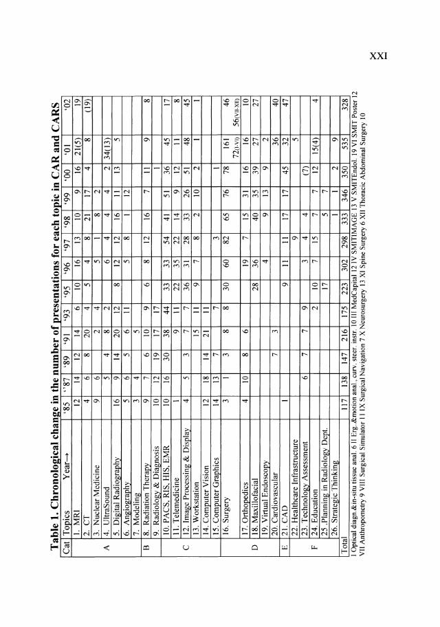
It is generally known that CARS involves a very wide spectrum of topics based on medicine, physics, computer science and even sociology. Since the first CAR Conference in 1985 up to the 16th CARS in 2002, there have been more than 60 topics. The terminology of these topics mainly came from the title of the sessions. However, I found that these topics could be grouped into 6 main categories, A to F, as shown below:
A. Modalities and their related topics 1 MRI, 2. CT, 3. Nuclear medicine, 4. Ultrasound, 5. Digital radiography involving a flat panel detector, 6. Angiography involving DSA, 7. Mathematical modeling for reconstruction etc., Multi-modality imaging and Medical imaging
B. Application of modalities 8. Radiation therapy and minimal invasive therapy, 9. Computer assisted radiology, Radiology diagnosis and Cardiology, Pediatric radiology, Neurology, IVR, Dosimetry in diagnosis and Computer assisted stereotaxy
C. Image processing, communication, display, interaction and related systems 10. PACS, RIS, HIS, Electronic med. record, Workflow, Standards such as DICOM, IHE (Integrating Healthcare Enterprise), 11. Telemedicine, 12. Image processing & display, Human-computer interaction and Virtual reality, 13. Workstation and Voice recognition applic., 14. Computer vision, 15. Computer graphics
D. Surgery, orthopaedics, maxillofacial and other related invasive topics 16. Surgery, Anthropometry, Surgical simulator, Surgical navigation, Neurosurgery, Spinal surgery, Thoracic and abdominal surgery, Standards in information-guided therapy, Surgical robotics, Optical diagnosis & in-situ tissue analysis, Ergonomics & motion analysis, Curved and steerable instr. and Endoluminal, 17. Orthopaedics, 18. Maxillofacial and Implantology, 19. Virtual endoscopy, 20. Cardio-vascular, Coronary and Medical innovation and technology
E. 21. Computer Aided Diagnosis
F. Surrounding and supporting infrastructure 22. Healthcare infrastructure, Interface between medicine and computer sciences, 23. Technology assessment and/or social implications, 24. Education, Knowledge based systems, Expert systems, Learning systems and Training systems, 25. Planning in the radiology department, 26. Strategic thinking, Decision making and Biointelligence
The chronological change in the number of presentations is shown in Table 1. The bracketed number indicates a double counted number. Example: (5) means that 5 presentations are double counted somewhere else in the same year. 16. Surgery in 2001 has 161 presentations and 72, the latter are broken down into I -VI. Also 16. Surgery in 2002 has 46 presentations and 56, the latter are composed of VII - XII. The features of CARS compared with those of other international conferences would be as follows:
xx
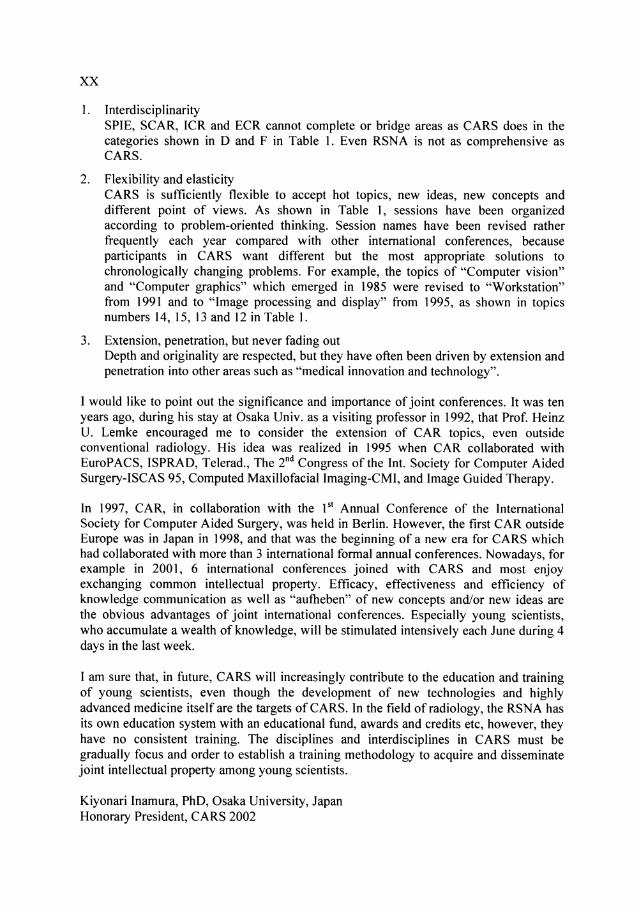
1. Interdisciplinarity SPIE, SCAR, ICR and ECR cannot complete or bridge areas as CARS does in the categories shown in 0 and F in Table 1. Even RSNA is not as comprehensive as CARS.
2. Flexibility and elasticity CARS is sufficiently flexible to accept hot topics, new ideas, new concepts and different point of views. As shown in Table 1, sessions have been organized according to problem-oriented thinking. Session names have been revised rather frequently each year compared with other international conferences, because participants in CARS want different but the most appropriate solutions to chronologically changing problems. For example, the topics of "Computer vision" and "Computer graphics" which emerged in 1985 were revised to "Workstation" from 1991 and to "Image processing and display" from 1995, as shown in topics numbers 14, 15, 13 and 12 in Table 1.
3. Extension, penetration, but never fading out Depth and originality are respected, but they have often been driven by extension and penetration into other areas such as "medical innovation and technology".
I would like to point out the significance and importance of joint conferences. It was ten years ago, during his stay at Osaka Univ. as a visiting professor in 1992, that Prof. Heinz U. Lemke encouraged me to consider the extension of CAR topics, even outside conventional radiology. His idea was realized in 1995 when CAR collaborated with EuroP ACS, ISPRAD, Telerad., The 2nd Congress of the Int. Society for Computer Aided Surgery-ISCAS 95, Computed Maxillofacial Imaging-CMI, and Image Guided Therapy.
In 1997, CAR, in collaboration with the 151 Annual Conference of the International Society for Computer Aided Surgery, was held in Berlin. However, the first CAR outside Europe was in Japan in 1998, and that was the beginning of a new era for CARS which had collaborated with more than 3 international formal annual conferences. Nowadays, for example in 2001, 6 international conferences joined with CARS and most enjoy exchanging common intellectual property. Efficacy, effectiveness and efficiency of knowledge communication as well as "autbeben" of new concepts and/or new ideas are the obvious advantages of joint international conferences. Especially young scientists, who accumulate a wealth of knowledge, will be stimulated intensively each June during 4 days in the last week.
I am sure that, in future, CARS will increasingly contribute to the education and training of young scientists, even though the development of new technologies and highly advanced medicine itself are the targets of CARS. In the field of radiology, the RSNA has its own education system with an educational fund, awards and credits etc, however, they have no consistent training. The disciplines and interdisciplines in CARS must be gradually focus and order to establish a training methodology to acquire and disseminate joint intellectual property among young scientists.
Kiyonari Inamura, PhD, Osaka University, Japan Honorary President, CARS 2002
Tab
le 1
. Ch
I -
---
---
--~-I c
h .
th
b f
-------
----
_._
-----------
--
--
--
----
----
----
--fl
h .
. C
AR
and
CA
RS
C
at
Top
ics
Yea
r .......
'8
5 "8
7
'89
'91
'93
'95
'96
'97
'98
'99
'00
'01
'02
I.
MR
I 12
14
12
14
6
10
16
13
10
9 16
21
(5)
19
2.
CT
4
6 8
20
4 5
4 8
21
17
4 8
( 19)
3.
Nuc
lear
Med
icin
e 9
6 2
4 5
I 8
2 A
4.
U
ltra
Sou
nd
5 4
8 2
6 4
4 4
2 34
(13)
5.
D
igit
al R
adio
Efa
phy
16
9 14
20
12
8
12
12
16
II
I3
5 6.
A
ngio
grap
hy
5 6
5 6
II
5 8
I 12
7.
M
odel
ing
3 4
5 B
8.
R
adia
tion
Ther~
9 7
6 10
9
6 8
12
16
7 I I
9
8 9.
R
adio
logy
& D
iagn
osis
10
12
19
17
17
I
10.
PA
CS
, R
IS,
HIS
, E
MR
10
16
30
38
44
33
33
54
41
51
36
45
17
II
. T
elem
edic
ine
I 9
II
22
35
22
14
9 12
I I
8
C
12. I
mag
e P
roce
ssin
g &
Dis
play
4
5 3
7 7
36
31
28
33
26
51
48
45
13. W
orks
tati
on
15
II
9 7
8 2
10
2 I
I 14
. Com
pute
r V
isio
n 12
18
14
21
II
15
. Com
pute
r G
raph
ics
14
13
7 7
3 I
16. S
urge
ry
3 I
3 8
8 30
60
82
65
76
78
16
1 46
n(
l-VI)
56(V
II-XI
I) 17
. Ort
hope
dics
4
10
8 6
19
7 15
31
16
16
10
D
18
. Max
illo
faci
al
28
36
40
35
39
27
27
19. V
irtu
al E
ndos
copy
4
9 13
9
2 20
. Car
diov
ascu
lar
7 3
36
40
E
21. C
AD
I
9 II
II
17
17
45
32
47
22
. Hea
lthc
are
Infr
astr
uctu
re
9 5
23. T
echn
olog
y A
sses
smen
t 6
7 7
9 3
4 4
(7)
F 24
. E
duca
tion
2
10
7 15
7
7 12
15
(4)
4 25
.P
lann
ing
in R
adio
logy
Dep
t. 17
5
7 26
. S
trat
egic
Thi
nkin
g I
I 2
9 T
otal
11
7 13
8 14
7 21
6 17
5 22
3 30
2 29
8 33
3 34
6 35
0 53
5 32
8 I
Opt
ical
dia
gn.&
in-s
itu
tiss
ue a
nal.
6 II
Erg
.. &m
otio
n an
al.,
curv
. st
eer.
ins
tr.
10 I
II M
edC
apit
al 1
2 IV
SM
ITIM
AG
E 1
3 V
SM
ITE
ndol
. 19
VI
SM
IT P
oste
r 12
V
II A
nthr
opom
etry
9 V
III
Sur
gica
l S
imul
ator
I I
IX S
urgi
cal
Nav
igat
ion
7 X
Neu
rosu
rger
y 13
XI
Spi
ne S
urge
ry 6
XII
Tho
raci
c A
bdom
inal
Sur
gery
10
x X
......
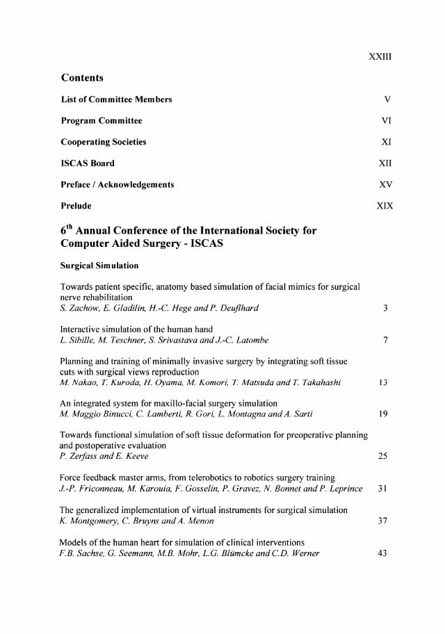
Contents
List of Committee Members
Program Committee
Cooperating Societies
ISCAS Board
Preface / Acknowledgements
Prelude
6th Annual Conference of the International Society for Computer Aided Surgery - ISCAS
Surgical Simulation
Towards patient specific, anatomy based simulation of facial mimics for surgical nerve rehabilitation
XXIII
V
VI
XI
XII
XV
XIX
S. Zachow, E. Gladilin, H.-C Hege and P. Deuflhard 3
Interactive simulation of the human hand L. Sibille, M Teschner, S. Srivastava and J.-C Latombe 7
Planning and training of minimally invasive surgery by integrating soft tissue cuts with surgical views reproduction M Nakao, T. Kuroda, H. Oyama, M Komori, T. Matsuda and T. Takahashi 13
An integrated system for maxillo-facial surgery simulation M Maggio Binllcci, C Lamberti, R. Gori, L. Montagna and A. Sarti 19
Towards functional simulation of soft tissue deformation for preoperative planning and postoperative evaluation P. ZerJass and E. Keeve 25
Force feedback master arms, from telerobotics to robotics surgery training J.-P. Friconneau, M Karouia, F. Gosselin, P. Gravez, N. Bonnet and P. Leprince 31
The generalized implementation of virtual instruments for surgical simulation K. Montgomery, C Bruyns and A. Menon 37
Models of the human heart for simulation of clinical interventions F.B. Sachse, G. Seemann, MB. Mohr, L.G. Bliimcke and CD. Werner 43
XXIV
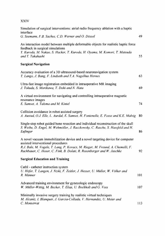
Simulation of surgical interventions: atrial radio frequency ablation with a haptic interface G. Seemann, F.B. Sachse, CD. Werner and 0. Dassel 49
An interaction model between multiple deformable objects for realistic haptic force feedback in surgical simulations Y. Kuroda, M Nakao, S. Hacker, T Kuroda, H. Oyama, M Komori, T Matsuda and T Takahashi 55
Surgical Navigation
Accuracy evaluation of a 3D ultrasound-based neuronavigation system T Lango, J. Bang, F. Lindseth and TA. Nagelhus Hernes
Ultra-fast image registration embedded in intraoperative MR imaging J. Tokuda, S. Morikawa, T Dohi and N. Hata
A virtual environment for navigating and controlling intraoperative magnetic resonance images E. Samset, A. Talsma and M Kintel
Collision avoidance in robot assisted surgery
63
69
74
A. Austad, 0.J. Elle, L. Aurdal, E. Samset, H. Fontenelle, E. Fosse and K.E. Malvig 80
Single-step robot guided bone resection and individual reconstruction of the skull S. Weihe, D. Engel, M Wehmoller, J. Raczkowsky, C Rasche, S. Hassfeld and H. Eufinger 86
A novel vacuum immobilization device and a novel targeting device for computer assisted interventional procedures R.J. Bale, M Vogele, T Lang, P. Kovacs, M Rieger, M Freund, A. Chemelli, F. Rachbauer, C Hoser, C Fink, B. Dolali, R. Rosenberger and W. Jaschke 92
Surgical Education and Training
CathI - catheter instruction system U. Hofer, T Langen, J. Nziki, F. Zeitler, J. Hesser, U. Miiller, W. Volker and R. Manner 101
Advanced training environment for gynecologic endoscopy W. Miiller-Wittig, M Becker, T Elias, U. Bockholt and G. Voss 107
Minimally invasive surgery training by realistic virtual techniques M Alcaniz, 1. Blanquer, J. Garcia-Collada, V. Hernandez, U. Meier and C Monserrat 113
xxv
Image Guided Neurosurgery
Acquisition of 3D ultrasound images during neuronavigation M MJ. Letteboer, P. WA. Willems, P. Hellier and WJ. Niessen 121 Neuronavigation with intraoperative 3D ultrasound; multimodal 2D and 3D display techniques and interactive stereoscopic visualisation for guiding surgical procedures TA. Nagelhus Hernes, F. Lindseth, T Lango, S Ommedal and G. Unsgard 127
Neurosurgical biopsies guided by 3D ultrasound - comparison of image evaluations and histopathological results T Selbekk, G. Unsgard, S Om medal, T Muller, S. Torp, G. Myhr, J. Bang and T Nagelhus Hernes 133
How to implement high-field intraoperative magnetic resonance imaging C Nimsky, 0. Ganslandt and R. Fahlbusch 139
Development of Hitchcock stereotactic frame for intraoperative open MRI H. Taniguchi, H. lseki, T Taira, H. Shirakawa, H. lwano, Y. Muragaki, M Sugiura, E. Kobayashi, K. Naemura, T Hori and K. Takakura 144
Intra-operative brain deformation using non-rigid image registration on a shared-memory multiprocessor computer T Rohlfing, CR. Maurer Jr., D.L.G. Hill, T Hartkens, WA. Hall, CL. Truwit, H. Liu, A.J. Martin and R. Shahidi 150
Real time three-dimensional image rendering in suboccipital approaches to the skull base SK. Rosahl, A. Gharabaghi, T Liebig, C Dalle-Feste and M Samii 156
The NASA smart probe project for real-time multiple microsensor tissue recognition: automating stereotactic brain biopsy and other procedures R.J. Andrews, R. Mah, S. Jeffrey, K. Freitas, M Guerrero, R. Papasin and C. Reed 161
The application accuracy of the NeuroMate robot - a quantitative comparison with frameless and frame-based surgical localization systems Q.H. Li, L. Zamorano, A. Pandya, MS Gong and F. Diaz 167
Robotics and Telesurgery
Interactive robots for medical applications J. Troccaz, P. Berkelman, P. Cinquin and A. Vilchis-Gonzales 175
XXVI
Surgical Robotics and Instrumentation
A dual-view endoscope with image shift Y. Yamauchi, J. Yamashita, Y. Fukui, K. Yokoyama, T. Sekiya, E. Ito, M. Kanai, T. Fukuyo, D. Hashimoto, H Iseki and K. Takakura 183
Head tracking of a surgical robotic scopeholder - a user involvement test of the system 0.J. £lIe, MG. Gundersen, E. Samset, G. ten Cate, L. Aurdal, A. Austad, T.K. Lien and E. Fosse 188
Application of image tracking for positioning scope in the robotic assisted laparoscopic surgery S. Payandeh and A. Zhang 194
The implementation of an intuitive man-machine interface in robot-aided endoscopic laser surgery H-W Tang, H Van Brussel, P. Koninckx and J. Vander Sloten 200
Motion estimation in minimally invasive beating heart surgery T. Ortmaier, M Grager and G. Hirzinger 206
Sensor-aided milling with a surgical robot system D. Engel, J. Raczkowsky and H. Warn 212
Software architecture for robotically assisted and image-guided minimally invasive interventions K. Cleary, A. Patriciu, S. Xu, M Mocanu and D. Stoianovici 218
Effect of video streaming delay on telemedicine based on the surgical cockpit system K. Hori, T. Kuroda, H Oyama, Y. Ozaki, T. Nakamura and T. Takahashi 224
Minimally Invasive Spine Surgery
The new frontier in minimally invasive spine surgery through computer assisted technology (CAT) J.e. Chiu, T.J. Clifford, R.A. Princenthal and R.B. Sion
Image-guided magnetic surgery M W Vannier
Computer Assisted Orthopaedic Surgery
Validation of fluoroscopy based navigation in the hip region. What you see is what you get?
233
238
N. WL. Schep, T. Walsum, J.s. de Graaf, I.A.MJ. Broeders and e. van der Werken 247
XXVII
Realistic haptic volume interaction for petro us bone surgery simulation A. Petersik, B. Pflesser, U. Tiede, KH Hahne and R. Leuwer 252
Non-invasive osteotomy using focused ultrasound S. Ishida, N. Hata, T. Azuma, S. Umemura and T. Dohi 258 Computer-assisted arthroscopic anterior cruciate ligament reconstruction J Sabczynski, E. Hille, S. Dries, W. Zylka, L. Tafler, P. Haaker and T. Istel 263
Fast generation of 3D bone models for craniofacial surgical planning: an interactive approach L. Ritter, M Lievin, R. Sader, H-F. ZeilhoJer and £. Keeve 269
A virtual fluoroscopy system based on a small and unobtrusive registration / calibration phantom R. Phillips, V Peter, G.-£. Faure, Q. Li, KP. Sherman, w.J Viant, M Bielby and A.MMA. Mohsen 275
Clinical applications of a laser guidance system with dual laser beam rays as augmented reality of surgical navigation N. Sugano, T. Sasama, S. Nishihara, H Nakase, T. Nishii, H Miki, Y. Momoi, I. Sakuma, M Fujie, S. Yoshinobu, Y. Nakajima, S. Tamura, Y. Yonenobu and T. Ochi 281
Robust computational osteotomy planning tools for autologous bone grafts in reconstructive surgery Z. Krol, M Chlebiej, P. ZerJass, H-F. ZeilhoJer, R. Sader, P. Mikolajczak and £. Keeve 285
3D assessment and simulation of surgical correction of spine deformities by in situ contouring technique R. Dumas, V LaJage, JP. Steib, D. Mitton, JA. de Guise and W. Skalli 291
Special Session on Validation of Medical Image Processing in the Context of Image-Guided Therapy
White paper: validation of medical image processing in the context of imageguided therapy P. Jannin, M Fitzpatrick, D.J Hawkes, X Pennec, R. Shahidi and M W. Vannier 299
Minimal Invasive Thoracic Abdominal Surgery
What is new in robotic surgery? M Hashizume, M Shimada, K Konishi, T. Akahoshi, M Tomikawa, S. Maehara and K Sugimachi 308
XXVIII
A new compact robot for manipulating forceps using friction wheel and gimbals mechanism T. Suzuki, E. Kobayashi, D. Kim, H Inada, T. Tsuji, T. Dohi and I. Sakuma 314
Merits of a newly-developed laparoscope manipulator: experiences with 4 cases M Tomikawa, M Hashizume, E. Kobayashi, S. Yamaguchi, I. Sakuma, M Fujie, F. Nakamura, T. Dohi, M Shimada and K Sugimachi 320 Feasibility of robot-assisted laparoscopic intestinal anastomosis; an experimental study in pigs JP. Ruurda and I.A.MJ Broeders 324
Evaluation of a magnetic tracking-guided needle placement system featuring respiratory gating in an in vitro liver model E.B. Levy, K Cleary, F. Banovac, D. Tanaka, S. Xu, D. Lindisch and N. Glossop 329
Evaluation of time-loss in robot-assisted surgery JP. Ruurda and I.A.MJ Broeders 335
HepaVision2 - a software assistant for preoperative planning in living-related liver transplantation and oncologic liver surgery H. Bourquain, A. Schenk, F. Link, B. Preim, G. Prause and H-O. Peitgen 341
Preoperative prostate cancer volume estimation based on clinically correlated needle biopsy simulation J Zeng, R. Conelly, J Bauer, W. Zhang, I.A. Sesterhenn, J W. Moul and S.K Mun 347
Resection proposals for oncologic liver surgery based on vascular territories B. Preim, H Bourquain, D. Selle, KJ Oldhafer and H.-a. Peitgen 353
16tb International Congress and Exhibition on Computer Assisted Radiology - CAR
Medical Imaging
Perfusion weighted MRl: a new tool for epilepsy surgery J-M Scarab in, B. Broche, C. Grova, C. Argaud, M Schaefer and P. Jannin 361
Stereo radiography: distortion correction and perception improvement A. Berestov 366
Development and performance evaluation of the first model of 40 CT-scanner M Endo, T. Tsunoo, S. Kandatsu, S. Tanada, H Aradate, Y. Saito, M Kusakabe, K Satoh and S. Matsusita 372
Pixels based statistical differences between lung SPECT: experimental approach to help for the diagnosis and the follow-up of pulmonary embolism
XXIX
S-E. Bendada, JM Rocchisani and JL. Moretti 377
A fast automatic method for 3D volume segmentation of the human cerebrovascular M Sabry, CB. Sites, A.A. Farag, S Hushek and T Moriarty 382
Contrast-enhanced three dimensional MR angiography of pulmonary artery and pulmonary perfusion imaging in pig - a comparison study with OSA S Liu, W Dong and X Xiao 388
A virtual training system for linear-type endoscopic ultrasonography SHacker, U. Tiede, E. Burmester, T Leineweber and K-H Hahne 394
Image Processing and Display
Automated quantification of avascular necrosis of the femoral head (ANFH) from 3D MR images R.A. Zorooji, T Nishii, Y. Sato, N. Sugano, H Yoshikawa, T Ochi and S Tamura 401
Efficient segmentation of MSCT images by interactive region expansion C Lorenz, T Schlathalter, I. Carlsen and S Renisch 407
Automated segmentation of pelvis and femur from 3-D CT images R.A. Zorooji, Y. Sato, B. Borgheai, T Sasama, T Nishii, N. Sugano, K Yonenobu, H Yoshikawa, T Ochi and S. Tamura 413
Registration and matching of projection imaging and tomographic imaging. Application to X-rays angiographies and magnetic resonance angiographies M Vermandel, C Kulik, J Y. Gauvrit, C Vasseur and J Rousseau 419
Construction of an average CT brain image for brain infarct pattern comparison C Jongen, JP. W Pluim, MA. Viergever and WJ Niessen 425
Fast Feldkamp-reconstruction for real-time reconstruction using c-arm-systems K Kornmesser, B. Schadler, J Hesser, R. Manner, M Ebert and W Schlegel 430
Trial for making other serially sectioned images (Visible Korean Human) MS Chung, Js. Park, J Y. Kim, WS Hwang, JK. Kim and H-S Park 435
An alternating-constraints optimization method for volume-preserving non-rigid registration of MR breast images T Rohlfing, CR. Maurer Jr., MA. Jacobs, D.A. Bluemke and R. Shahidi 439
Integrating the insight toolkit itk into a medical software framework N. Hanssen, B. von Rymon-Lipinski, T Jansen, M Lievin and E. Keeve 445
xxx
Validation of MRlISPECT similarity -based registration methods using realistic simulations of normal and pathological SPECT data C Grova, A. Biraben, l. Buvat, H. Benali, A.M Bernard, B. Gibaud and J-M Scarabin 450
Automatic 3D-reconstruction of the ocular fundus from stereo images M Wuenstel and H. Schumann 456 Performance evaluation of LCD displays H. Roehrig, JH. Fan, T Furukawa, M Ohashi, A. Chawla and K Gandhi 461
Fast volume rendering based on software optimization using multimedia instructions on PC platforms K Mori, Y. Suenaga and J Toriwaki 467
Interactive tutorials: development of a large scale 3D database of pathological cases using interactive volume rendering technique H. Shin, U von Jan, M Galanski and H.K Matthies 473
Image Management and Communication
RIS/PACS integration in a web environment R. Passariello, G. Venturi, V Campanella, S. Simonetti, C Catalano and A. Fiumicelli 479
Implementation of RlS/PACS at Princess Alexandra Hospital Brisbane, Australia B.L. Crowe 485
Expressing DICOM SR constraints in XML KP. Lee 491
A digital imaging network to facilitate a multi-center clinical trial for adrenoleukodystrophy ML. lngeholm, B.A. Levine, A. Fatemi, G. Jimenez-Sanchez and H. W. Moser 497
ACRIN digital mammographic imaging screening trial informatics infrastructure CR. Welsh, B. Young, V Gopalakrishnan, l. Mahon, S. Sabina, C Gatsonis, E. Pisano and M Yaffe 503
Multicenter clinical trials of imaging workstations and methods CR. Welsh, V Gopalakrishnan, J Flaim-Spetsas, A. Toledano and CD. Johnson 509
Detailed image classification code for image retrieval of medical images (IRMA) B.B. Wein, T Lehmann, D. Keysers, H. Schubert and M Kohnen 513
XXXI
Computer Assisted Radiation Therapy
Dynamic reconstruction for radiotherapy planning A. Koenig, P. Grangeat, S. Bonnet and P. Hugonnard 521
Registration of 3D U/S and CT images of the prostate E.A. Fir/e, W. Chen and S. Wesarg 527
Interferometric measurement of patient surface topology changes during therapy irradiation C Moore, K. Woods, P. Sharrock, F. Lilley, M Lalor and D. Burton 533
Adaptive filtering to predict lung tumor motion during free breathing MJ Murphy, M Isaakson and J Jalden 539
Protocol conversion by automatic pattern matching for the on-line linkage between multi-institutional radiation oncology databases Y. Kumazaki, H. Harauchi, K. Haneda, H. Kou, T Kondou, M Ishibashi, H. Numasaki, K. Shimizu, T Umeda, A. Takemura and K. Inamura 545
Photon radiosurgical system for treating brain tumors K. Takakura, 0. Kubo, Y. Muragaki, H. Iseki and M Sugiura 551
Towards a World Engineering Anthropometry Resource
Shape modeling driven by the product design R. Mollard
Medical image archives - present and future M W. Vannier, E. V. Staab and L.C Clarke
3D description of the human body shape: application of Karhunen-Loeve expansion to the CAESAR database Z. Benazouz, M Rioux and R. Lepage
Recent advances in Korean anthropometry Y.s. Lee
Automatic shape modeling of the foot: towards a database of foot shapes M Mochimaru and M Kouchi
A Web3D based CAESAR viewer S. Ressler and Q. Wang
Some tools for understanding anthropometry JF.M Molenbroek
559
565
571
577
582
588
593
XXXII
Telemedicine
A review "In-Home Healthcare" projects in the USA G.B. Devey
Home health monitoring and diabetes: a web-based approach to achieving better control
601
B.A. Levine, A. Alaoui, M-J. Hu, K Smith, S Clement, A. Neustadt and SK Mun 607 The communication concept of a regional stroke unit network based on encrypted image transmission and the DICOM-mail standard U. Engelmann, A. Schroter, T Schweitzer and H-P' Meinzer 612
Framework for systematic assessment of the regional HUSpacs after the reengineering of hospital and external processes K Harno, R. Roine, H Pohjonen, J. Kinnunen and T Kauppinen 618
Evaluation ofPACS and radiology services in 8 selected hospitals within the reference model program SaxTeleMed, using activity based costing and a generic business model (Marburg Model) J. Bottcher and KJ. Klose 623
Displaying and analysing DICOM waveforms on Java based cell phones and PDAs M Kroll, J. Riesmeier, K. Melzer, K Annacker, H-G. Lipinski and D.H W. Gronemeyer 627
Special Session on Electronic Health Record
Secure and future proof electronic health records over the internet B. Blobel 635
Patient-oriented visualisation and interaction in a three dimensional environment K. Kochy, R. Tschirley and S Markle 641
Do EHR communication standards account for imaging communication needs? F. Mennerat and J. Chabriais 647
4th International Workshop on Computer-Aided Diagnosis - CAD
Special Session on Breast CAD
Quantifying breast cancer risk from mammograms MJ. Yaffe and N.F. Boyd
An automated detection method of mammographic masses based on adaptive threshold technique utilizing multi-resolution processing S Kasai, D. Kaji, A. Kano, H Fujita, T Hara and TEndo
653
659
Image retrieval scheme for mammographic masses by using a local-pattern matching technique T Nakagawa, T Hara, H. Fujita, T lwase and TEndo
Segmentation of microcaJcifications in X-ray mammograms using entropy thresholding M Mel/oul and L. Joskowicz
On the problem of breast compression modelling F. Georgsson and N. Bjornestal
Special Session on Thoracic CAD
Automatic segmentation and texture analysis ofPA chest radiographs to detect abnormalities related to interstitial disease and tuberculosis
XXXIII
665
671
677
B. van Ginneken, B.M ter Haar Romeny and MA. Viergever 685
Clinical usefulness of temporal subtraction technique for detection of interval changes on digital chest radiographs S. Katsuragawa, T Uozumi, S. Kakeda, H. Watanabe, H. Nakata and K. Doi 689
Update on the development of an automated lung nodule detection method for CT scans s.G. Armato 1Il 695
A CAD system for lung cancer based on 3D CT images N. Niki, Y. Kawata, M Kubo, H. Ohmatsu, R. Kakinuma, K. Mori, H. Nishiyama, K. Eguchi, M Kusumoto, M Kaneko and N. Moriyama 70 I
Optimal image feature set for detecting lung nodules on chest X-ray images J Wei, Y. Hagihara, A. Shimizu and H. Kobatake 706
Effect of the computer output on radiologists' decision-making for classification of solitary pulmonary nodules in chest radiographs J Shiraishi, H. Abe, R. Englemann, M Aoyama, H. MacMahon and K. Doi 712
An efficient recognition method of lung nodules from X-ray CT images using 3D object models K. Shigemoto, H. Takizawa, S. Yamamoto, T Nakagawa, T Matsumoto, Y. Tateno, T Iinuma and M Matsumoto 717
Network based CAD system for lung cancer screening by X-ray CT H. Emoto, S. Yamamoto, M Matsumoto, T Matsumoto, Y. Tateno and T Iinuma 723
Computer classification of lung tumors from chest CT images according to the types of tissue using 3D extended Voronoi diagram Y. Hirano, J Hasegawa, J Toriwaki, H. Ohmatsu and K. Eguchi 729
XXXIV
Special Session on 3D CAD
Visualization and segmentation techniques in 3-D ultrasound images A. Fenster and D.B. Downey 737
Current concepts and future directions in computer-aided diagnosis for CT colonography R.M. Summers 743
Computer-aided detection of polyps in CT colonography: effect of feature-guided polyp segmentation J. Nappi and H. Yoshida 749
Feature-based approach toward computer aided detection and diagnosis Z. Liang, Z. Wang, L. Li and D.P. Harrington 755
Clinical potential ofCT colonography in the diagnosis of pelvic lymph node metastasis for patients with rectal cancer G. Iinuma, T. Akasu and N. Moriyama 761
Three-dimensional computer-aided diagnosis schemes for classification of benign and malignant pulmonary nodules Y. Kawata, N. Niki, H. Ohmatsu, M. Kusumoto, R. Kakinuma, K. Mori, H. Nishiyama, K. Eguchi, M. Kaneko and N. Moriyama 764
Pulmonary nodule detection using cartwheel projection analysis L. Fan, J. Qian, G.-Q. Wei and D.P. Naidich 770
Extraction and recognition of the thoracic organs based on 3D CT images and its application X Zhou, T Hara, H. Fujita, Y. Ida, K. Katada and K. Matsumoto 776
A preliminary study for automated recognition of branches of pulmonary artery and vein using anatomical positional relations from a 3-D chest X-ray CT image K. Mori, T Yamaguchi, T. Kitasaka, Y. Mekada, J. Hasegawa, J. Toriwaki and H. Otsuji 782
Development of a computer aided diagnosis system for three dimensional breast CT images Y. Hanzawa, A. Shimizu, H. Kobatake and K. Miyakawa 788
Development of automated detection and classification methods of masses on 3D breast ultrasound images T. Hara, D. Fukuoka, H. Fujita, T. Endo and w.K. Moon 794
xxxv
2002 International Symposium on Cardiovascular Imaging - CVI
Invasive Coronary Imaging
Coronary atherosclerosis: from pathophysiology to imaging in clinical practice F. Ledru 803 A novel approach for the detection of path lines in X-ray angiograms: the wavefront propagation algorithm J. Janssen, G. Koning, P.J.H de Koning, J.c. Tuinenburg and J.H.c. Reiber 808
A numerical 3D coronary tree model D. Sherknies and J. Meunier 814
Automatic stent border detection in IntraVascular UltraSound images for quantitative measurements of the stent parameters J. Dijkstra, G. Koning, J.c. Tuinenburg, P. V. Oemrawsingh, C. von Birgelen andJ.H.C. Reiber 819
Invasive Coronary and Vascular Imaging
ECG-gated 3D-rotational coronary angiography (3DRCA) v. Rasche, A. Biicker, M Grass, R. Koppe, J. Op de Beek, R. Bertrams, R. Suurmond, H. Kiihl and R. W. Giinther 827
Automatic trinocular 3D reconstruction of coronary artery centerlines from rotational X-ray angiography C. Blondel, R. Vaillant, F. Devernay, G. Malandarin and N. Ayache 832
Improved endpoint localization in guide wire tracking during endovascular interventions S.A.M Baert and w.J. Niessen 838
Improved radiological control of endovascular prothesis positioning, using a region-contour cooperation approach A. Raji, J. Lemoine, Y. Lahfi, J.c. Bossu and F. Boudghene 843
Modelling and process design of a new type of catheter for special endovascular treatment of abdominal aortic aneurysms P. Jo/i, C. Francois, T. Gagarina and F. Boudghene 849
Left and Right Ventricular Function
Quantitative biplane angiography for right and left ventricular volume determination F.K. Schmiel, D.G. W. Onnasch, K. Moldenhauer and HH Kramer 857
XXXVI
Development and evaluation of a software package for right ventricular analysis J. Hensgens, B. Weijers, J.-P.MM Aben and A.P.G. Kroes 863
Integration of tools for the estimation of heart vitality by tissue doppler and myocardial velocity gradients into a clinical useable software environment M Hastenteufel, I. Wolf, R. de Simone, S. Mottl-Link and H.-P. Meinzer 867
3D stress echocardiography: a novel application based on registration of real-time 3 D ultrasound images R. Shekhar, V. Zagrodsky, M Garcia and J.D. Thomas 873
Detecting changes in myocardial perfusion T.L. Faber, J.R. Galt, J. Chen, B.M W. Tsui and E. V. Garcia 879
Non-invasive Cardiovascular Imaging - Clinical Approach -An ESCR Symposium
Non-invasive approach to coronary heart disease R.K. Rienmilller, B. Schrottner, U. Reiter und G. Reiter
The use of imaging in chest pain MR. Rees and T. Cripps
Advances in Cardiovascular MRI and CT - A NASCI Symposium
3D active appearance models: application to cardiac MR and ultrasound image segmentation B.P.F. Lelieveldt, s.c. Mitchell, J.G. Bosch, R.J. van der Geest, M Sonka
887
890
and J.H.c. Reiber 897
Automatic correction of myocardial boundaries in MR cardio perfusion analysis L. Spreeuwers, F. Wierda and M Breeuwer 902
FAST analysis tool of global and local heart functions by MRI tagging C. Dornier, MK. Ivancevic, G. Lecoq, A. Righetti and J.P. Vallee 908
Advances in Cardiovascular MRI and MSCT - A NASCI Symposium
Correlation of quantitative MR-angiography of the carotid artery with in vivo measurement during carotid endarterectomy R. Guzman, A. Barth, L. Remonda, H. Oswald, G. Schroth, P. de Koning and R. van der Geest 917
Model-based vessel segmentation using an elliptical cylinder S. Young, V. Pekar, J. Weese, T. Netsch and A. van Muiswinkel 923
Integration of cath-lab signals and magnetic resonance imaging for postoperative computerized assessment of heart reduction
XXXVII
A. Ripoli, S. Berti, N. Olsen, M Lombardi, S. Bevilacqua and M Glauber 928
Development of software for four-dimensional cardiac function analysis using multi-slice CT scanner S. Yamamoto, S. Hamada, H Naito, T. Johkoh, M Miyamoto, J. Masumoto, S. Azemoto, T. Kanagawa, S. Nakanishi and H Nakamura 934
8th Computed Maxillofacial Imaging Congress - CMI
Image-Guidance in Implantology
Clinical evaluation of patient misalignment during CT scans for computer assisted implantology - a new approach for compensation J. Brief, S. HassJeld, W. Stein, R. Krempien and J. Miihling 943
The precision of the RoboDent system - an in vitro study 0. Schermeier, T. Lueth, C. Cho, D. Hildebrand, M Klein, K. Nelson and J. Bier 947
A new method and a clinical case for computer assisted dental implantology F. Goulette, J. Dutreuil, C. Laurgeau, J.c. Zoreda and S. Lundgren 953
"Image guided implantology" - real-time guidance of dental implant surgery in the operative field using CT-scan image L. Shapira 959
Computer based approach for design and treatment of immediately loaded implants U. Meyer, T. Fillies, N. Meier, T. Stamm, HP. Wiesmann and U. Joos 965
Surgical simulation of multisegment osteotomies and implant dentistry in cleft palates R. Marmulla, S. HassJeld and J. Miihling 970
Computer aided navigation and interactive teleconsultation in dental implantology K. Schicho, A. Wagner and R. Ewers 975
Image-Guided Cranio-Maxillofacial Surgery
3D osteotomy planning in cranio-maxillofacial surgery: experiences and results of surgery planning and volumetric finite-element soft tissue prediction in three clinical cases S. Zachow, E. Gladilin, A. Trepczynski, R. Sader and H-F. ZeilhoJer 983
Computer aided planning for orthognatic surgery M Chabanas, C. Marecaux, Y. Payan and F. Boutault 988
XXXVIII
The exact resection of a tumor and a transplant with the help of 3D navigation G. Schultes, H. Karcher, G. Santler and A. Gaggl 994
Assessment of maxillofacial pathological lesions by computed tomography using 3D surface and volume rendering techniques MG.P. Cavalcanti, P.s. Tossato and J.L.F. Antunes 997
Application of cone-beam X-ray CT in dento-maxillofacial region K. Maki, T. Usui, M Kubota, H. Nakano, Y. Shibasaki, K. Araki, T. Okano, K. Ueno and K. Yamamoto 1003
Poster Session
16th International Congress and Exhibition on Computer Assisted Radiology - CAR
Survival prediction using artificial neural networks in patients with lung cancer treated by radiotherapy M linuma, T. Teshima, Y. Iwanaga, M Kawamata, M Nagayoshi and K. Murase 1011
Clinical usefulness ofmuItidetector CT angiography: application to the diagnosis of arterial occlusive diseases in lower extremities N. Hirai, R. Tanaka, Y. Hori, M Higashi, S. Imakita and H. Naito 1012
Automatically reducing iodine in contrast head CT scans by image processing L. Yin, JK. Chang and A.I.Z. Abidin 1013
Improved visualization of3D data sets on the example of3D rotational angiography R. Koppe, E. Klotz and J Op de Beek 1014
EasyTW: computer-aided system for bone age assessment A.MM Da Silva, G.L. De Oliveira, G.P. Noal, C.A.A. Schmitz, L.S.B. Haeffner, P.s.P. Antunes and S.D. Olabarriaga 1015
3D X-ray imaging of human knee using a mobile C-arm J Liitjens, E. Klotz, R. Koppe, V. Rasche and M Grass 1016
Bone mineral density estimation after soft-tissue effect subtraction in X-ray images S. Kim, S. Lee and J -W. Jeong 1017
Virtual actors for a patient oriented virtual hospital J. Wilhelmy and S. Markle 1018
XXXIX
User-assisted segmentation of tomographic images A.A. Goshtasby and M Satter 1019
Quantitative evaluation of medical image fusion algorithms F. Vhlemann, S. Sobottka and R. Steinmeier 1020
The "Virtual Institute for Computer Assistance in Clinical Radiology" (VICORA): first results of the development of algorithms and applications S. Krass, F. Link, T Boskamp, A. Schenk, H Bourquain, S. Kohle, R. Rascher-Friesenhausen, W. Spindler, B. Kummer/en, M Lang, B. Wein, R. Leppek and H-a. Peitgen 1021
Three-dimensional composite images ofSPECT/MRI in hydrocephalus T Mito, I. Shibata, N. Sugo and M Takano 1022
Segmentation of MR brain images using region growing combined with an active contour model R. Parveen and A. Todd-Pokropek 1023
DISCIR: an architecture for high performance distributed and component-oriented image diagnosis applications C Alfonso, I. Blanquer, A. Gonzalez and V. Hernandez 1024
Usefulness of newly developed interactive multiplanar reconstruction viewer for large amount of image sets by multidetector-row CT: evaluation of invasion to surrounding organs of esophageal carcinoma T Johkoh, S. Yamamoto, T Murakami, M Hori, 0. Honda, T Kozuka, M Koyama, M Tsubamoto, S. Hamada, Y. Narum and H Nakamura 1025
Optimization of an anisotropic diffusion method for medical image processing K. Kawakami, K. Murase, Y. Yamazaki, M Shinohara, M Kawamata, M Nagayoshi and S. Iwamoto 1026
Fast image generation of cerebral perfusion parameters using multi-detector row CT and deconvolution analysis K. Murase, M Shinohara, Y. Yamazaki, K. Kawakami, S. Iwamoto, Y. Sugawara, T Veda and J. Ikezoe 1027
Automated volumetry of lateral ventricles in 3-D SPGR MR images using physicians' knowledge represented by fuzzy logic S. Kobashi, Y. Hata, E. Matsui, H Kitagaki, E. Mori and T Kanagawa 1028
Preliminary study for characterizing Legg-Calve-Perthes disease based on MRI segmentation P. Pouletaut, I. Claude, R. Winzenrieth, M-C Ho Ba Tho and G. Sebag 1029
XL
Magnetic resonance based biomechanical analysis of the knee joint M Siebert, K-H. Englmeier, R. von Eisenhart-Rothe, C Bringmann, F. Eckstein, H. Bonel, M Reiser and H. Graichen 1030
Connected morphological operators for CT-scan images of the abdomen B. Naegel, C Ronse and L. Soler 1031 Improvement of an automated patient recognition method for chest radiographs using edge-enhanced images K Kondo, J Morishita, S Katsuragawa and K Doi 1032
Automatic registration of thoracic FDG-PET and CT for diagnosis and staging of lung cancer JC Asmuth, R.H. Moore, L. Bogoni and SL. Aquino 1033
A segmentation method for 3D slice images: searching and bridging D. Kim, K. Ku and J Park 1034
NEUROCAD: a neurology computer-aided toolkit MC Oliveira, L. Cavalcanti, MC Oliveira, G.F. Caetano and P.M Azevedo-Marques 1035
Content-based image retrieval using texture features MO Honda, P.M Azevedo-Marques and JA.H. Rodrigues 1036
Enhancing the diagnostic capabilities ofMRCP by high-resolution image acquisition and stereoscopic viewing T. Yamagishi, T. Saito, K. Abe, J Ishida, R. Nishimura, T. Kudo, A. Petersik, U. Tiede and K-H. Hahne 1037
Wavelet de-noising in digital chest radiographs by generalized cross validation H. Kubota, Y Yamazaki, M Shinohara, K. Kawakami and K Murase 1038
A template-based method of ventricle part classification and realistic texturing for virtual ventriculoscopy I. Goncharenko, H. Emoto, T. Fujii, N. Sugou, T. Mito, I. Shibata, and Y Kanou 1039
Automatic extraction of cerebral blood vessel and aneurysm from magnetic resonance angiography Y Yamazaki, K Murase, M Shin ohara, K Kikuchi, H. Miki and J Ikezoe 1040
Volume-rendered TOF MR angiography: detection and characterization of intracranial aneurysms A. Mallouhi, S Felber, A. Chemelli, A. Dessl, A. Auer, M Schocke, W.R. Jaschke and P. Waldenberger 1041
Accuracy of cerebral blood flow obtained from dynamic susceptibility contrastenhanced MRI using deconvolution analysis based on singular value decomposition M. Shinohara, K Murase, Y Yamazaki, M. linuma, T. Enoki, Y Iwanaga,
XLI
K Kikuchi, H Miki and J Ikezoe 1042
A new tool for unsupervised analysis of time series in functional imaging M. Buerki, C. Kiefer, A. Nirkko, H Oswald and G. Schroth 1043
Lung MR imaging: robust registration technique of the respiratory phase for quantitative ventilation and perfusion study T. Ueguchi, T. Johkoh, M. Koyama, C. Tanaka, M. Kawahara and H Nakamura 1044
How to .1etermine the borders of gray matter of young and aged brain on magnetic resonance images N. Tsukamoto, H Kumagai, K Saitoh, M. Monma, Y Ando, M. Kitamura and 0. Kawaguchi 1045
Computerized analysis of magnetic resonance angiography A.Z. Wang, S. Campbell and Q.-Y Chen 1046
Spectral PTF analysis using cine-MRI in normal pressure hydrocephalus T. Miyati, H Fujita, M. Mase, T. Banno, T. Kasuga, K Yamada, K Koshida S. Sanada and H Ima 1047
Development of a simple and non-invasive method for measuring cerebral blood flow using Technetium-99m compounds and spectral analysis M. Kawamata, M. Takasawa, M. Nagayoshi, T. Enoki, N. Oku and K Murase 1048
A multi-resolution retinal vessel tracker based on directional smoothing K-H Englmeier, S. Bichler, K Schmid, M. Maurino, M. Porta, T. Bek, B. Ege, 0. V. Larsen and o.K Hejlesen 1049
Two-year experience in using a telediagnostic regional network 0. Barbero, C. Rubies, J Fernandez-Bayo, J Valls, I. Periz, J Puig and L. Donoso 1050
Interactive (stereoscopic) DICOM image access on mobile devices K. Melzer, HG. Lipinski and D.HW. Gronemeyer 1051
Fast volume rendering endoscopy based on semi-automatic path generation s.M. Kwon, J Yi, S.H Kim, JK. Kim and JB. Ra 1052
Evaluation of PC-based 3D virtual colonoscopy J Kim, S. Lee and S. Kim 1053
Normalized cuts for spinal MRI segmentation J Carballido-Gamio, s.J Belongie and S. Majumdar 1054
XLII
6th Annual Conference of the International Society for Computer Aided Surgery - ISCAS
Three dimensional colour coded visualization of differences between CT patient data and resulting stereolithographical models E. Schwaderer, F. Dammann, M Heuschmid, 1. Hoffmann and CD. Claussen 1057
Information integration and management for computer assisted orthopaedic surgery over internet T. Wu, C Huberson, A. Zimolong, G. Miiller, K. Radermacher and G. Rau 1058
A novel system of 4-dimensional motion analysis after total hip arthroplasty K Hagio, N. Sugano, T. Nishii, H. Miki, Y. Otake, A. Hattori, N. Suzuki, T. Sasama, Y. Sato, S. Tamura, K Yonenobu, H. Yoshikawa and T. Ochi 1059
A cadaveric study of robotically assisted spinal needle placement versus manual placement K Cleary, K Ruitort and V Watson 1060
Accuracy of the 3D angular position of vertebrae reconstructed by low dose digital stereoradiography A. Le Bras, R. Dumas, M Savidan, D. Mitton, 1.A. De Guise and W. Skalli 1061
Ultrasound guidance for spinal radiosurgery T.C Ryken, S.L. Meeks, 1.w. Haller and1.M Buatti 1062
Endoscopic treatment of intraventricular lesions in virtual reality Z. Novak, P. Krupa, 1. Chrastina and I. Riha 1063
PC-based deep brain recording system for functional neurosurgery 1. Tejeiro, R.J. Macias, G. Lopez, 1.M Morales, L.M Alvarez, E.Guerra, C Maragoto, E. Alvarez, I. Garcia and W. Bouza 1064
Evaluation of a stereotactic neurosurgical robot (N euroMate) C Edlinger, 1.-P. Perrin, S. Baffert, E. Charpentier and E. Fery-Lemonnier 1065
An improved fMRI analysis for detecting motor cortex activity near brain tumors 1. W. Haller, T.A. Gallagher, T.C Ryken, K1. Ahn and M W. Vannier 1066
Combined image-guided surgical navigation and ultrasound images for cranial surgeries T.C Ryken, M Barua and 1. W. Haller 1067
A hybrid navigation system for reliable navigation surgery-microscopic optical tracking and electromagnetic suction tube tracking 1. Yamada, A. Kato, H. Kishima, M Hirata and T. Yoshimine 1068
Open MRI compatible HivisCAS video microscope system for neurosurgery K. Nambu, H, 1seki, Y. Muragaki, T. Maruyama, T. Taira, R. Mochizuki,
XLIII
M. Sugiura, K. Naemura, T. Hori and K. Takakura 1069
The results of image guided surgery with magnetic and optical hybrid navigation system A. Kato, J. Yamada, M Hirata, N. Hirabuki and T. Yoshimine 1070
A new method for 3D modeling of neurosurgical procedures 1. Koster, A. Sam ii, T. Brinker and H,K. Matthies 1071
Monte Carlo simulation for anisotropic error prediction in frameless stereotaxy P. WA. Willems, H,J. Noordmans, J. W Berkelbach van der Sprenkel, MA. Viergever and CA.F. Tulleken 1072
Education tools for surgery: a simulator for pelvic surgery J. Troccaz, R. Martin, M Valderrama, L. Vadcard, S. Viera and J. Tonetti 1073
Toward modelling and simulation of tearing and cutting in laparoscopic training environment S. Payandeh, H, Zhang and N. Azouz 1074
CT fluoroscopy using navigation and automated registration W Zylka, L. Tafter and H, Vohwinkel 1075
The meaning of precision in computer assisted surgery M Wehmoller, S. Weihe and H, Eufinger 1076
Practical clinical experience with microscopic 3D-navigation at the skull base W Freysinger, F. Kral, A.R. Gunkel and WF. Thumfart 1077
A fast and accurate method of ultrasound probe calibration for image-guided surgery J.N. Welch, M Bax, K. Mori, T. Krummel, R. Shahidi and CR. Maurer Jr. 1078
A clinical prototype system for projector-based augmented reality: calibration and projection methods J. Raczkowsky, H, Hoppe, C Kubler, H, Worn and S. Hassfeld 1079
Evaluation of a computer aided planning and surgical robot system for craniofacial surgery D. Engel, A. Pernozolli, 0. Schorr, J. Brief, T. Heurich, J. Raczkowsky, S. Hassfeld H, Worn and J. Muhling 1080
Robotic manipulator system for microsurgery (first report) H, Gotani and Y. Yamano 1081
XLIV
A distributed system for 3D anatomical structures visualization and surgical planning C. Koehl, L. Soler and J. Marescaux 1082
A unified process for computer assisted surgery O. Schorr, A. Pernozzoli, J. Raczkowsky and H Worn 1083
A novel computer-based approach to diagnostic imaging in the gut R. Mij1akhov 1084
Using distance transformations to determine safe needle/probe paths during interventional procedures L. Aurdal, O.J. £lIe, E. Samset, H Fontenelle, T. Om holt-Jensen, T. Mala and B. Edwin 1085
Multi-slider linkage mechanism for endoscopic manipulator H Yamashita, K. Daeyoung, N. Hata and T. Dohi 1086
Clinical application of 3D-CT angiography for laparoscopic colorectal surgery J. Okuda, K. Nishiguchi, K. Tanaka, S. W. Lee, M. Toyoda and N. Tanigawa 1087
The basis of magnetic navigation system for surgery J. Shimada, M. Kawakami, S. Endoh, F. Ikeda, M. Nagahara and Y. Yamamoto 1088
Study on bending forceps manipulator with electric-cautery function for laparoscopic surgery Y. Kim, T. Oura, D. Kim, E. Kobayashi, T. Tsuji, H In ada, T. Dohi and l. Sakuma 1089
A laparoscope positioning system based on the real-time visual tracking of the surgeon's face and surgical instruments A. Nishikawa, S. Asano, F. Miyazaki, M. Sekimoto, M. Yasui and M. Monden 1090
Feasibility of image-guided abdominal interventions using a novel magnetic position sensing device in an interventional radiology suite F. Banovac, M. Jay, D. Lindisch, N. Glossop and K. Cleary 1091
Reconstruction of intrahepatic vessel trees from three-dimensional freehand ultrasound-scans P. Hassenpjlug, M. Vetter, G. da Silva Jr., l. Wolf, M. Thorn, G.M. Richter, W. Lamade, M. Buchler and H-P. Meinzer 1092
Development of AR navigation system for laparoscopic surgery using magneto-optic hybrid sensor: experiences with 3 cases K. Konishi, M. Hashizume, M. Nakamoto, M. Miyamoto, M. Shimada, Y. Sato, S. Tamura and K. Sugimachi 1093
Evaluation of an augmented reality system for intraoperative data presentation - preliminary results T. Salb, J Brief, T. Welzel, 0. Burgert, T. Gockel, R. Krempien, S. Hassfeld
XLV
and R. Dillmann 1094
4th International Workshop on Computer-Aided Diagnosis - CAD
A detection method of ground glass opacities from chest X-ray CT images H. Takizawa, S. Yamamoto, T. Matsumoto, Y. Tateno, T. Iinuma and M Matsumoto 1097
Automated polyp detection from colon MRI images J Lamy, e. Ronse and L. Soler 1098
Three-dimensional analysis of pulmonary nodules using thin slice helical CT data: automated nodule classification and estimation of malignancy K. Murao, K. Awai, A. Ozawa, T. Yamanaka, M Komi and S. Hori 1099
Sonographic diagnosis of fatty liver using computer-aided quantification S. Kim, J-w. Jeong and S. Lee 1100
Detection of abnormal tissue in HRCT scans of the chest I.e. Sluimer and B. van Ginneken
Development of automated detection methods for architectural distortions on mammograms
1101
T. Matsubara, D. Yamazaki, T. Hara, H. Fujita, S. Kasai, T. Endo and T. Iwase 1102
Some results on the preprocessing of mammograms for microcalcification detection E. Nava, R. Mala, F. Sendra and E. Ristori 1103
2002 International Symposium on Cardiovascular Imaging - CVI
A multi scale medial axis detection of coronary arteries in X-rays angiographies B. Tremblais, B. Augereau and M Leard 1107
Model-based segmentation and visualization of IVUS images for radiation treatment planning in cardiovascular brachytherapy F. Weichert, e. Wilke, P. Spilles, A. Kraushaar, H. Muller, U. Quast and D. Wegener 1108
Hydatid cyst of the heart contribution of two-dimensional echocardiography S. Ernez-Hajri, G. Jeridi, H. Bouraoui, T. Filali, A. Mhadhaoui and H. Ammar 1109
Demonstration of three-dimensional visualization techniques for coronary artery evaluation using EBCT - a case study P.MA. van Ooijen, P.J. de Feyter and M Oudkerk 1110
XLVI
Left ventricular perfusion/function differences between ischemic and dilated cardiomyopathy assessed by gated SPECT myocardial imaging: visual and quantitative computer analysis 1. Peovska, J Maksimovic, M. Buseva, D. Pop Gorceva and V. Majstorov 1111
Heart phantom - a simple elastomechanical model of ventricle W. Sediono and 0. Dassel 1112
8th Computed Maxillofacial Imaging Congress - CMI
Image post-processing for optimization ofphotostimulable phosphor cephalograms T. T. Farman, KD. West, JP. Scheetz, A. Silveira, B.E. Johnson andA.G. Farman 1115
Evaluation of sensitivity of 3D-CT for diagnosis of craniofacial fractures MG.P. Cavalcanti and A.P.A. Costa e Silva 1116
Tuned-aperture computed tomography (TACT(R)) applied to assess impacted teeth using 2D and 3D pseudo-holograms K Yamamoto, Y. Hayakawa, N. Kobayashi, Y. Kousuge, M Wakoh, H. Sekiguchi, M Yakushiji and A. G. Farman 1117
Development of new tomography equipment with magnification A. Satoh, R. Yamato, M Tada, H. Nakahama, N. Takahashi and Y. Okumura 1118
Computer simulation of an intraoral radiography using perspective projection ofCT data K Okamura, K Yoshiura, K Tokumori, T. Tanaka and S. Kanda 1119
44 years oftemporomandibular ankylosis M Anchieta and F. Salles 1120
Context sensitive visualized information in cranio-maxillofacial navigation systems R. Marmulla, S. Hassfeld, T. Heurich and J Miihling 1121
3D-imaging for the assessment of volume changes of the human face E. Nkenke, X Laboureux, M Benz, T. Maier, G. Hausler, P. Kessler, J Wiltfang and F. W. Neukam 1122
Evaluation of the exophthalmia reduction with a finite element model V. Luboz, A. Pedrono, F. Boutault, P. Swider and Y. Payan 1123
Digital X-ray mini-panel and dental treatment-planning outcomes for MRDD inpatients A.G. Farman, H. Hood, B. Horsley and E. Warr 1124

The need for model generation and validation in craniofacial prosthodontics: a feasibility study Z. Khan, A.A. Farag, T T Farman and A.G. Farman
Index of Authors
XLVII
1125
1127