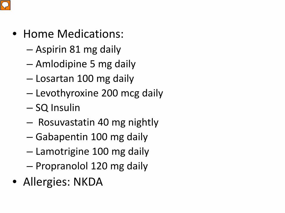
These are her home medications. It is important to note she is only on a baby aspirin daily and not on therapeutic anticoagualation.
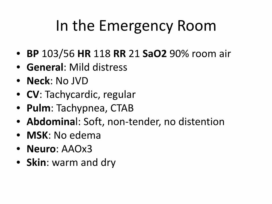
In the Emergency Room
• BP 103/56 HR 118 RR 21 SaO2 90% room air • General: Mild distress • Neck: No JVD • CV: Tachycardic, regular • Pulm: Tachypnea, CTAB • Abdominal: Soft, non-tender, no distention • MSK: No edema • Neuro: AAOx3 • Skin: warm and dry
Presenter
Presentation Notes
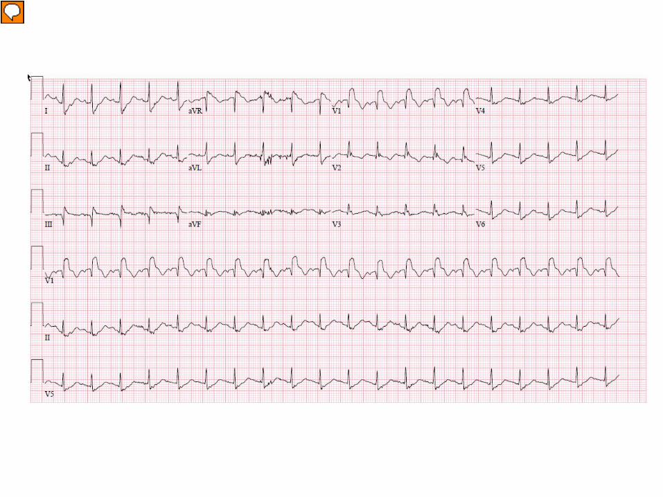
This is her ECG on presentation. As you can see it shows sinus tachycardia with a right bundle branch block. She did have a prior ECG in our system from several years ago and the right bundle is new this admission
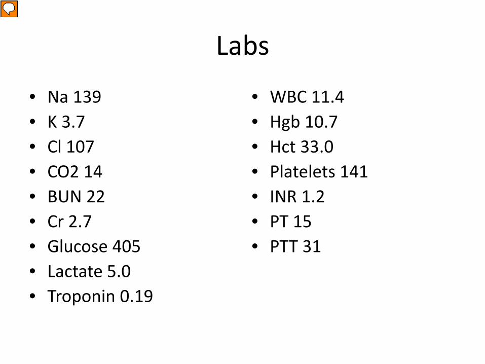
Labs
• Na 139 • K 3.7 • Cl 107 • CO2 14 • BUN 22 • Cr 2.7 • Glucose 405 • Lactate 5.0 • Troponin 0.19
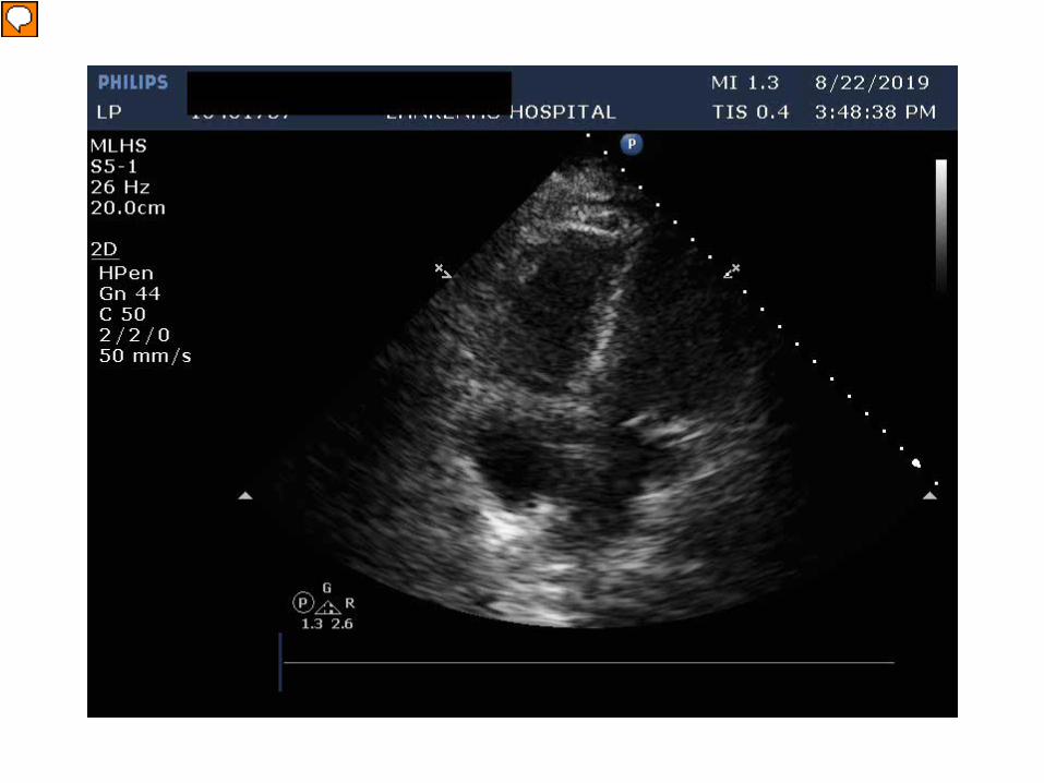
Given her presentation, ECG changes, and inability to get a CT with IV contrast to look for pulmonary embolism due to her acute kidney injury on baseline CKD, a STAT echocardiogram was performed to look for evidence of right heart strain
Presenter
Presentation Notes
This is a transthoracic apical four chamber. You can see the left ventricular cavity is small and hyperdynamic with flattening of the intraventricular septum consistent with right ventricular pressure and volume overload. The RV is severely dilated with severely reduced systolic function, however, the apex is hyperdynamic which is consistent the McConnell’s sign.
Presenter
Presentation Notes
These next two images are parasternal long and short axis views. They again demonstrate the hyperdynamic LV with flattening of the septum consistent with RV pressure and volume overload.
Presenter
Presentation Notes
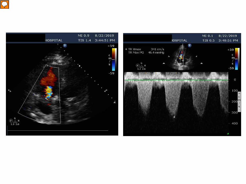
These next images show her tricuspid regurgitation and RVSP of 46 mmHg plus right atrial pressure. At this point given her history, presenting symptoms, echo findings, and inability to get a CTA due to CrCl the decision was made to proceed directly to the cath lab for pulmonary angiography.
Presenter
Presentation Notes
These two images demonstrate significant clot burden in the left and right pulmonary arteries. During the study the patient became progressively unstable and with her RV failure the decision was made to proceed to mechanical thrombectomy using the Flowtriever device.
Presenter
Presentation Notes
Using a femoral approach the Flowtriever device was advanced to the pulmonary arteries and mechanical aspirations of large clot were performed. This is an image status post clot removal.
• During the procedure, a large clot piece was noted on the tip of Flowtriever cannula without being able to pull it out. Maintaining the negative suction, the clot was pulled down to iliac vein, and infra-renal IVC filter was placed
• Due to persistent hypotension and severe RV dysfunction, was made to proceed with RV mechanical support with right ventricular assist device (Proteck Duo)
• Patient also developed atrial fibrillation with rapid ventricular response during procedure and emergent cardioversion was performed
• The patient did have minor hemoptysis during the procedure thought to be due to reperfusion injury
• Also started on inhaled Epoprostenol • Patient transferred to CICU
Presenter
Presentation Notes
This is a transthoracic echocardiogram performed the following day. You can see the RVAD in the right sided structures. You can also see the RV size and function has significantly improved.
Hospital Stay
• Day 2 bedside hemodynamic wean successful. RV size and function improved on echocardiogram. Decision was made to decannulate right ventricular assist device.
• Continued to be hemodynamically stable • Transitioned to direct oral anticoagulant • Repeat echocardiogram as outpatient
Presenter
Presentation Notes
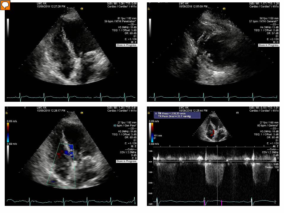
This is her echocardiogram from 1 month later. You can see the LV is no longer small or hyperdynamic. The RV size and function has normalized. The severity of the tricuspid regurgitation has been reduced to trace and her RVSP now is 23 mmHg plus right atrial pressure.
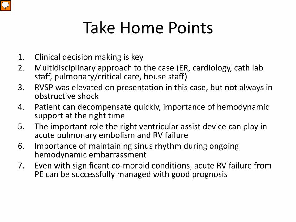
Take Home Points 1. Clinical decision making is key 2. Multidisciplinary approach to the case (ER, cardiology, cath lab
staff, pulmonary/critical care, house staff) 3. RVSP was elevated on presentation in this case, but not always in
obstructive shock 4. Patient can decompensate quickly, importance of hemodynamic
support at the right time 5. The important role the right ventricular assist device can play in
acute pulmonary embolism and RV failure 6. Importance of maintaining sinus rhythm during ongoing
hemodynamic embarrassment 7. Even with significant co-morbid conditions, acute RV failure from
PE can be successfully managed with good prognosis
Presenter
Presentation Notes
This case illustrates may of the topics we have discussed this morning. Some take home points from this case include: Clinical decision making is key. In this case we had a high suspicion for pulmonary embolism to the decision was made to proceed directly to the cath lab for a pulmonary angiogram Multidisciplinary approach is key ( In this case the ER, cardiology, cath lab staff, pulmonary/critical care, house staff were all involved and contributed to her successful outcome) RVSP was elevated on presentation in this case, but not always in obstructive shock and that is something to keep in the back of your mind when evaluating acute RV failure This case shows how quickly a Patient can decompensate, and the importance of hemodynamic support at the right time Going on that point this case shows the important role the right ventricular assist device can play in acute pulmonary embolism and RV failure It also shows the Importance of maintaining sinus rhythm during ongoing hemodynamic embarrassment Lastly, Even with significant co-morbid conditions, acute RV failure from PE can be successfully managed with good prognosis