Embed Size (px)
Citation preview
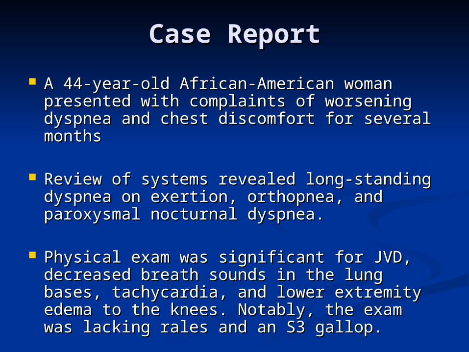
Case ReportCase Report
A 44-year-old African-American woman A 44-year-old African-American woman presented with complaints of worsening presented with complaints of worsening dyspnea and chest discomfort for several dyspnea and chest discomfort for several monthsmonths
Review of systems revealed long-standing Review of systems revealed long-standing dyspnea on exertion, orthopnea, and dyspnea on exertion, orthopnea, and paroxysmal nocturnal dyspnea. paroxysmal nocturnal dyspnea.
Physical exam was significant for JVD, Physical exam was significant for JVD, decreased breath sounds in the lung bases, decreased breath sounds in the lung bases, tachycardia, and lower extremity edema to tachycardia, and lower extremity edema to the knees. Notably, the exam was lacking the knees. Notably, the exam was lacking rales and an S3 gallop. rales and an S3 gallop.
Differential Diagnosis?Differential Diagnosis?
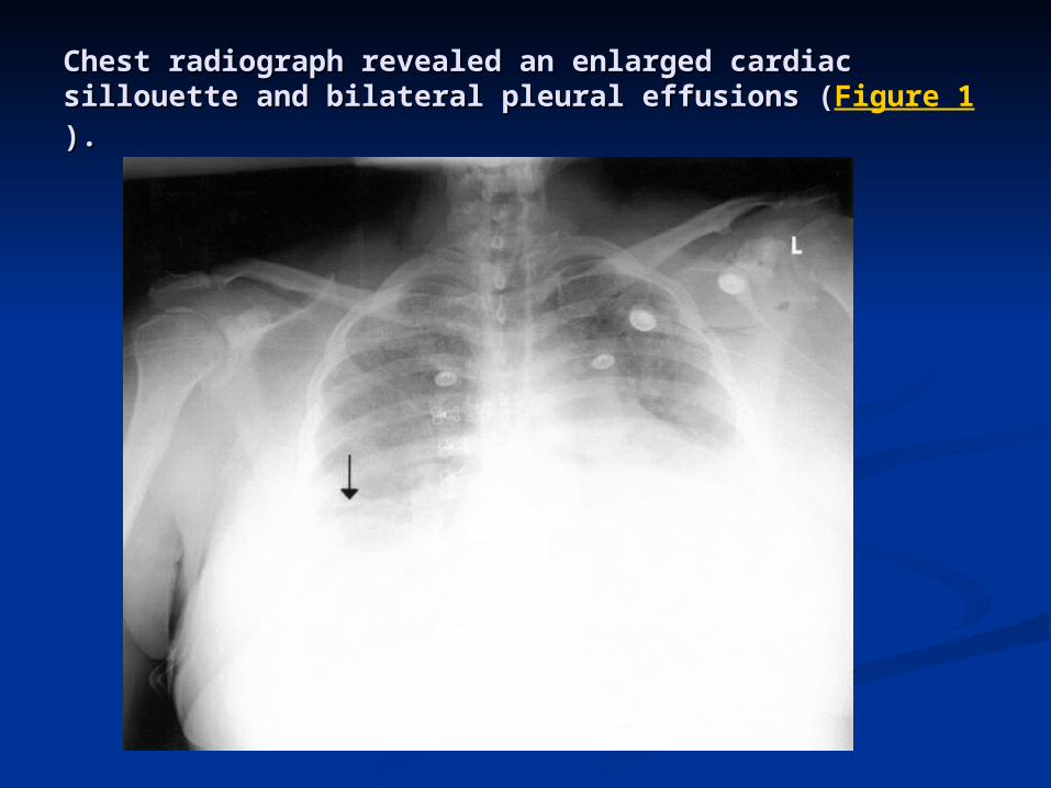
Chest radiograph revealed an enlarged cardiac Chest radiograph revealed an enlarged cardiac sillouette and bilateral pleural effusions (sillouette and bilateral pleural effusions (Figure 1).).
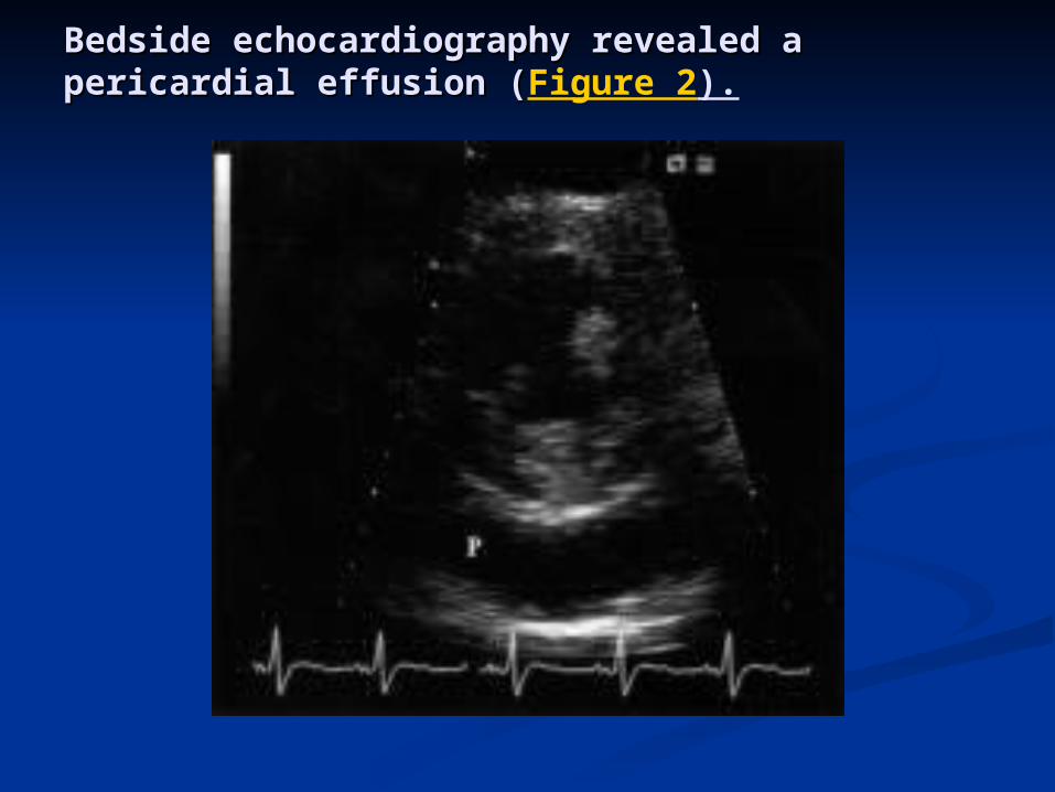
Bedside echocardiography revealed a Bedside echocardiography revealed a pericardial effusion (pericardial effusion (Figure 2).
DataData Laboratory findings included a macrocytic Laboratory findings included a macrocytic
anemia (MCV 114.6 fL, HCT 22.2%) with a anemia (MCV 114.6 fL, HCT 22.2%) with a high corrected reticulocyte count (14.5) and high corrected reticulocyte count (14.5) and normal B12 and folate levels. normal B12 and folate levels.
Auto-immune hemolytic anemia was Auto-immune hemolytic anemia was confirmed by a decreased haptoglobin (<14 confirmed by a decreased haptoglobin (<14 mg/dL), an elevated LDH (1411 U/L), and the mg/dL), an elevated LDH (1411 U/L), and the detection of a warm IgG auto-antibody. detection of a warm IgG auto-antibody.
ANA testing revealed a 160 titer and an anti-ANA testing revealed a 160 titer and an anti-DNA DS level of 101 IU. Right heart DNA DS level of 101 IU. Right heart catheterization revealed tamponade catheterization revealed tamponade physiology.physiology.
DiscussionDiscussion
This case represents a rare presentation in a patient This case represents a rare presentation in a patient with SLE. with SLE. The patient's symptoms were secondary to both tamponade The patient's symptoms were secondary to both tamponade
and hemolytic anemia, perhaps two of the most morbid of the and hemolytic anemia, perhaps two of the most morbid of the diagnostic criteria. diagnostic criteria.
The eventual diagnosis may have been delayed were it The eventual diagnosis may have been delayed were it not for careful attention to the physical exam and not for careful attention to the physical exam and prompt diagnostic testing to validate those findings. prompt diagnostic testing to validate those findings.
Standard treatment for congestive heart failure based Standard treatment for congestive heart failure based solely on reported symptoms would have greatly solely on reported symptoms would have greatly increased her morbidity, and could have been fatal. increased her morbidity, and could have been fatal.
This case emphasizes the importance that our history This case emphasizes the importance that our history AND physical examinations guide our diagnostic and AND physical examinations guide our diagnostic and therapeutic measures.therapeutic measures.
Systemic Lupus Systemic Lupus ErythematosusErythematosusJuly 13, 2009July 13, 2009
Julie Schwartzman, MDJulie Schwartzman, MD
Associate Program Director, Associate Program Director, RheumatologyRheumatology
Director of Arthritis and Lupus ClinicsDirector of Arthritis and Lupus Clinics
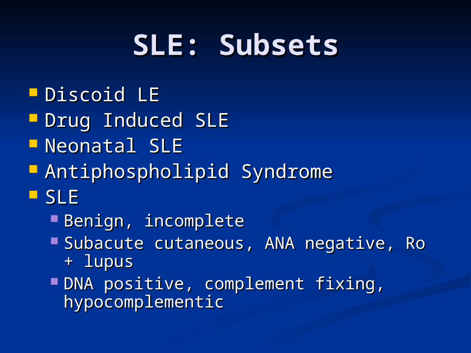
SLE: SubsetsSLE: Subsets
Discoid LEDiscoid LE Drug Induced SLEDrug Induced SLE Neonatal SLE Neonatal SLE Antiphospholipid SyndromeAntiphospholipid Syndrome SLESLE
Benign, incompleteBenign, incomplete Subacute cutaneous, ANA negative, Ro + Subacute cutaneous, ANA negative, Ro +
lupuslupus DNA positive, complement fixing, DNA positive, complement fixing,
hypocomplementichypocomplementic
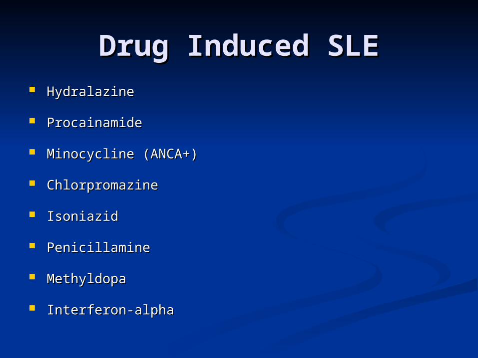
Drug Induced SLEDrug Induced SLE HydralazineHydralazine
ProcainamideProcainamide
Minocycline (ANCA+)Minocycline (ANCA+)
ChlorpromazineChlorpromazine
IsoniazidIsoniazid
PenicillaminePenicillamine
MethyldopaMethyldopa
Interferon-alphaInterferon-alpha
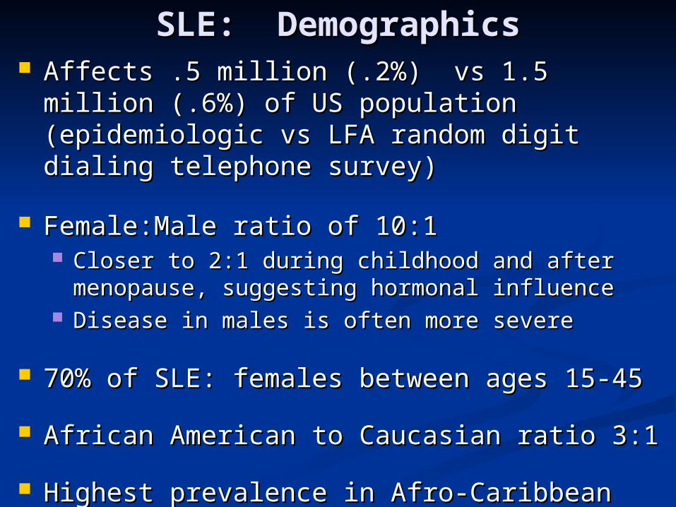
SLE: DemographicsSLE: Demographics Affects .5 million (.2%) vs 1.5 million (.6%) of Affects .5 million (.2%) vs 1.5 million (.6%) of
US population (epidemiologic vs LFA random US population (epidemiologic vs LFA random digit dialing telephone survey)digit dialing telephone survey)
Female:Male ratio of 10:1Female:Male ratio of 10:1 Closer to 2:1 during childhood and after Closer to 2:1 during childhood and after
menopause, suggesting hormonal influencemenopause, suggesting hormonal influence Disease in males is often more severe Disease in males is often more severe
70% of SLE: females between ages 15-4570% of SLE: females between ages 15-45
African American to Caucasian ratio 3:1African American to Caucasian ratio 3:1
Highest prevalence in Afro-Caribbean females Highest prevalence in Afro-Caribbean females 1:2501:250
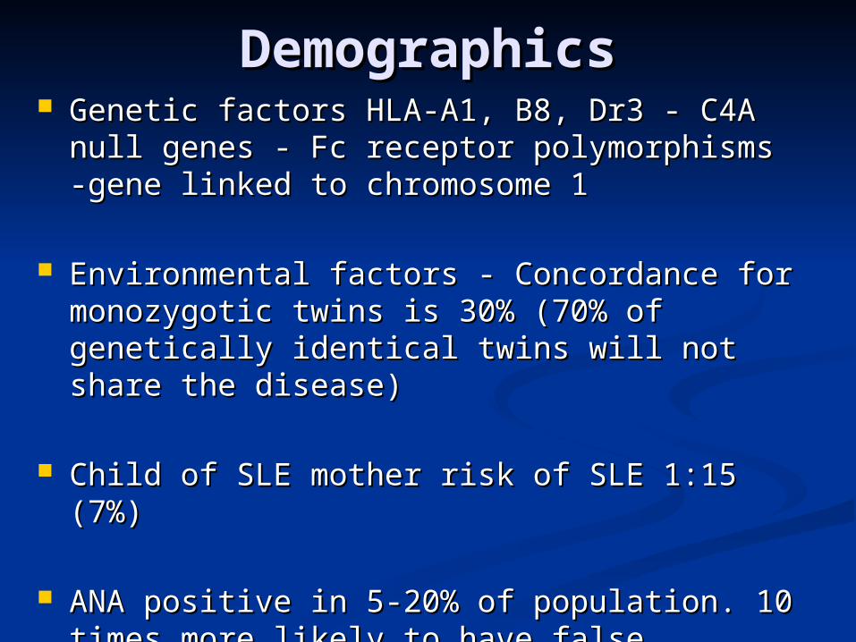
DemographicsDemographics Genetic factors HLA-A1, B8, Dr3 - C4A null Genetic factors HLA-A1, B8, Dr3 - C4A null
genes - Fc receptor polymorphisms -gene genes - Fc receptor polymorphisms -gene linked to chromosome 1linked to chromosome 1
Environmental factors - Concordance for Environmental factors - Concordance for monozygotic twins is 30% (70% of genetically monozygotic twins is 30% (70% of genetically identical twins will not share the disease)identical twins will not share the disease)
Child of SLE mother risk of SLE 1:15 (7%)Child of SLE mother risk of SLE 1:15 (7%)
ANA positive in 5-20% of population. 10 times ANA positive in 5-20% of population. 10 times more likely to have false positive ANA than more likely to have false positive ANA than diseasedisease
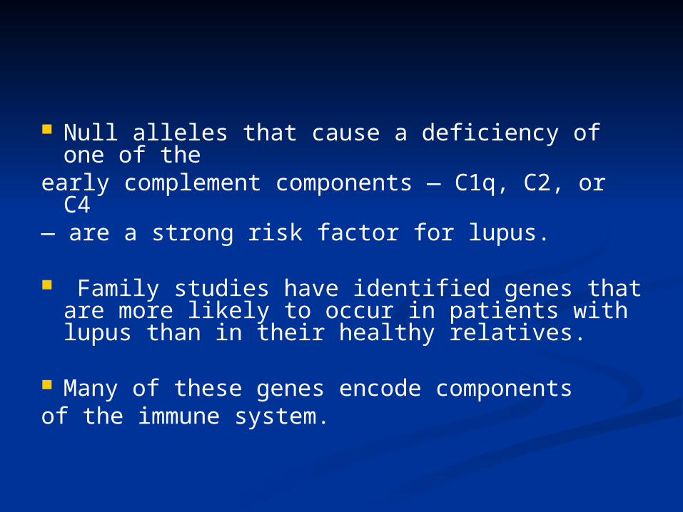
Null alleles that cause a deficiency of one of the
early complement components — C1q, C2, or C4
— are a strong risk factor for lupus.
Family studies have identified genes that are more likely to occur in patients with lupus than in their healthy relatives.
Many of these genes encode componentsof the immune system.

SLE: ETIOLOGYSLE: ETIOLOGY
AUTOANTIBODY PRODUCTIONAUTOANTIBODY PRODUCTION
GENERATION OF CIRCULATING GENERATION OF CIRCULATING IMMUNE COMPLEXESIMMUNE COMPLEXES
EPISODIC COMPLEMENT EPISODIC COMPLEMENT ACTIVATIONACTIVATION
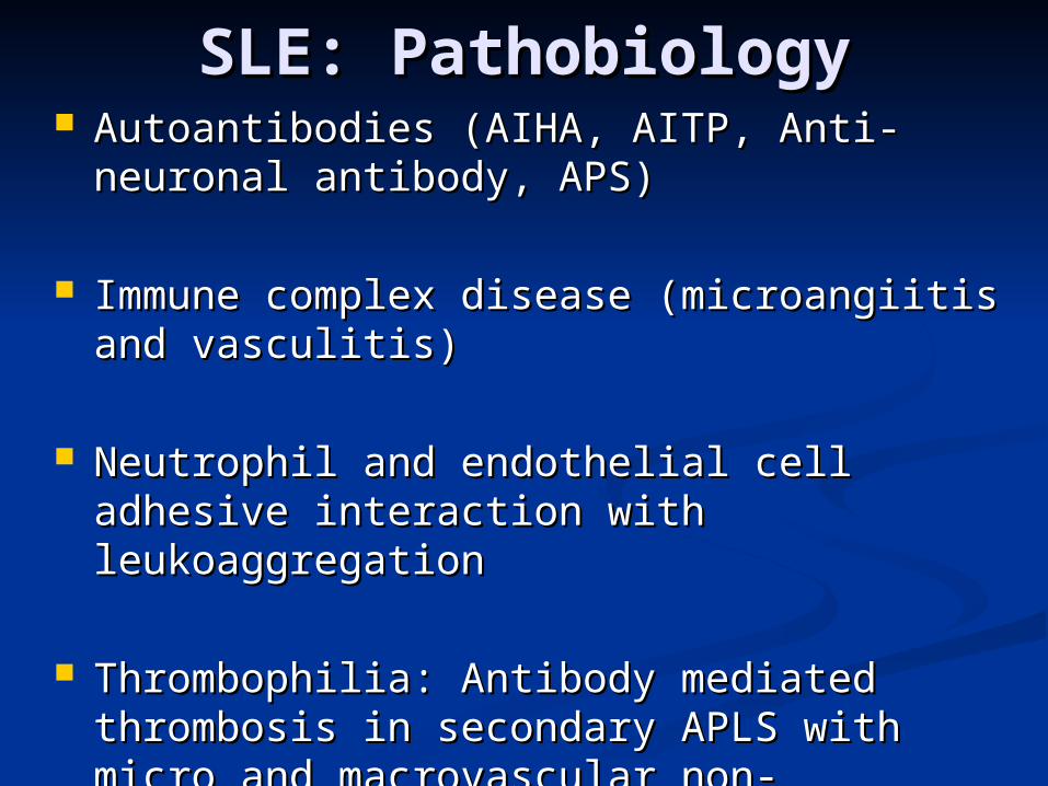
SLE: PathobiologySLE: Pathobiology Autoantibodies (AIHA, AITP, Anti-neuronal Autoantibodies (AIHA, AITP, Anti-neuronal
antibody, APS)antibody, APS)
Immune complex disease (microangiitis and Immune complex disease (microangiitis and vasculitis)vasculitis)
Neutrophil and endothelial cell adhesive Neutrophil and endothelial cell adhesive interaction with leukoaggregationinteraction with leukoaggregation
Thrombophilia: Antibody mediated Thrombophilia: Antibody mediated thrombosis in secondary APLS with micro thrombosis in secondary APLS with micro and macrovascular non-inflammatory and macrovascular non-inflammatory occlusionocclusion
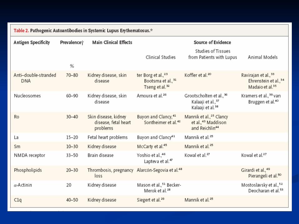
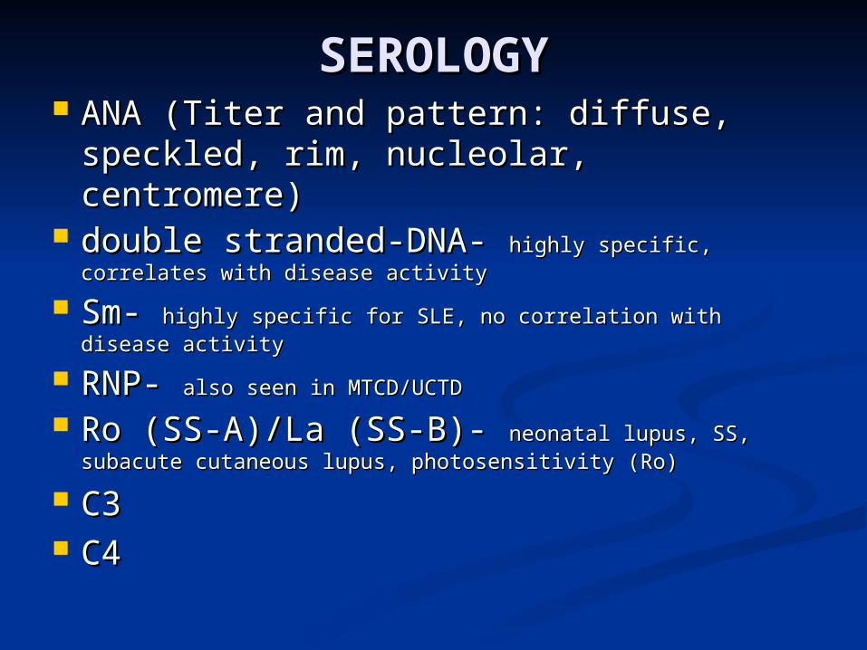
SEROLOGYSEROLOGY ANA (Titer and pattern: diffuse, ANA (Titer and pattern: diffuse,
speckled, rim, nucleolar, centromere)speckled, rim, nucleolar, centromere) double stranded-DNA- double stranded-DNA- highly specific, highly specific,
correlates with disease activitycorrelates with disease activity
Sm- Sm- highly specific for SLE, no correlation with disease highly specific for SLE, no correlation with disease activityactivity
RNP- RNP- also seen in MTCD/UCTDalso seen in MTCD/UCTD
Ro (SS-A)/La (SS-B)- Ro (SS-A)/La (SS-B)- neonatal lupus, SS, neonatal lupus, SS, subacute cutaneous lupus, photosensitivity (Ro)subacute cutaneous lupus, photosensitivity (Ro)
C3C3 C4C4
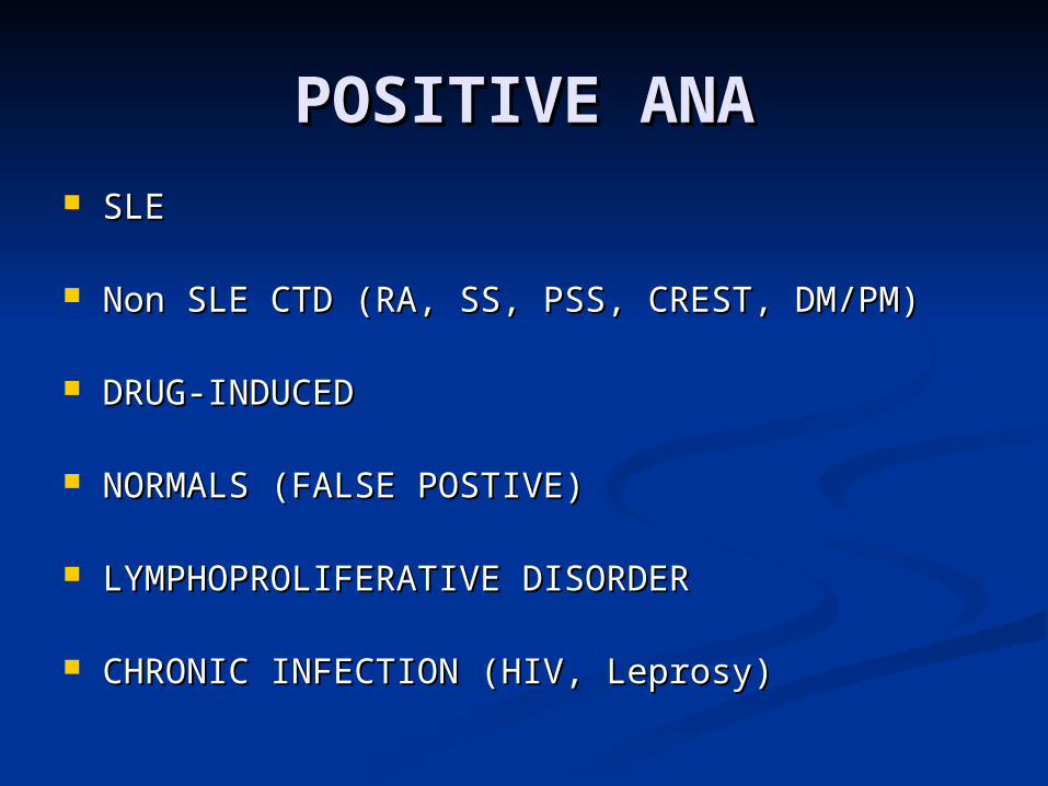
POSITIVE ANAPOSITIVE ANA SLESLE
Non SLE CTD (RA, SS, PSS, CREST, DM/PM)Non SLE CTD (RA, SS, PSS, CREST, DM/PM)
DRUG-INDUCEDDRUG-INDUCED
NORMALS (FALSE POSTIVE)NORMALS (FALSE POSTIVE)
LYMPHOPROLIFERATIVE DISORDERLYMPHOPROLIFERATIVE DISORDER
CHRONIC INFECTION (HIV, Leprosy)CHRONIC INFECTION (HIV, Leprosy)
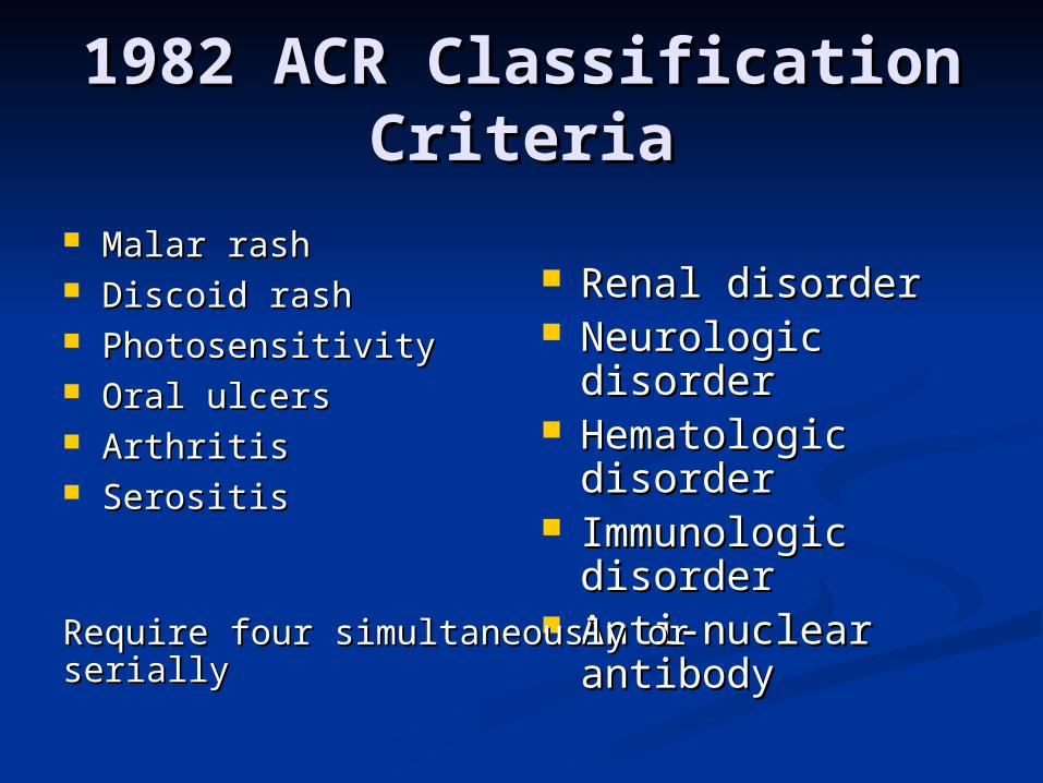
1982 ACR Classification 1982 ACR Classification CriteriaCriteria
Malar rashMalar rash Discoid rashDiscoid rash PhotosensitivityPhotosensitivity Oral ulcersOral ulcers ArthritisArthritis SerositisSerositis
Renal disorderRenal disorder Neurologic Neurologic
disorderdisorder Hematologic Hematologic
disorderdisorder Immunologic Immunologic
disorderdisorder Anti-nuclear Anti-nuclear
antibodyantibodyRequire four simultaneously or serially Require four simultaneously or serially
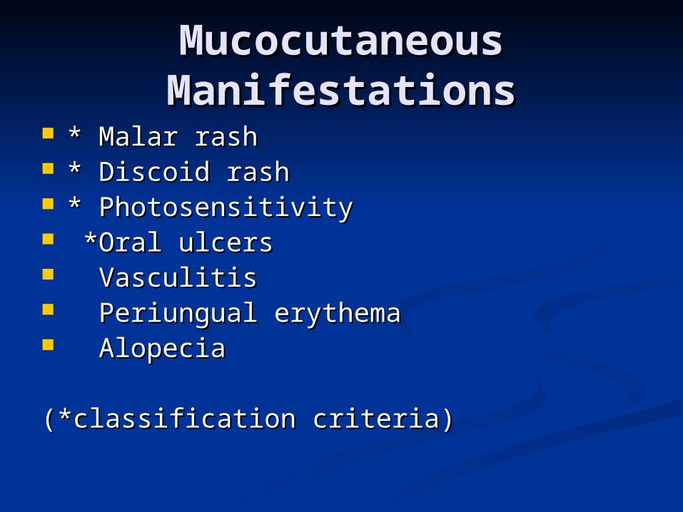
Mucocutaneous Mucocutaneous ManifestationsManifestations
* Malar rash* Malar rash * Discoid rash* Discoid rash * Photosensitivity* Photosensitivity *Oral ulcers *Oral ulcers VasculitisVasculitis Periungual erythemaPeriungual erythema AlopeciaAlopecia
(*classification criteria)(*classification criteria)
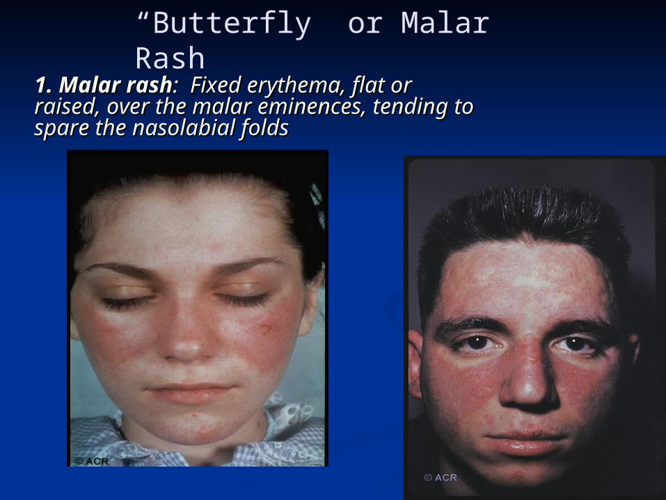
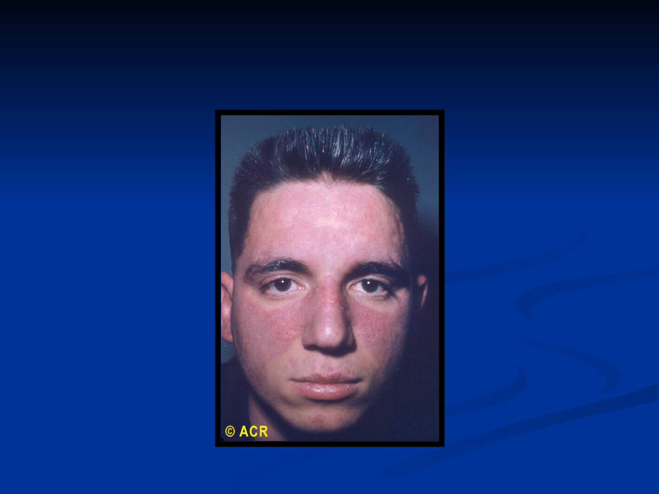
“Butterfly” or Malar Rash
1. Malar rash1. Malar rash: Fixed erythema, flat or : Fixed erythema, flat or raised, over the malar eminences, tending to raised, over the malar eminences, tending to spare the nasolabial foldsspare the nasolabial folds
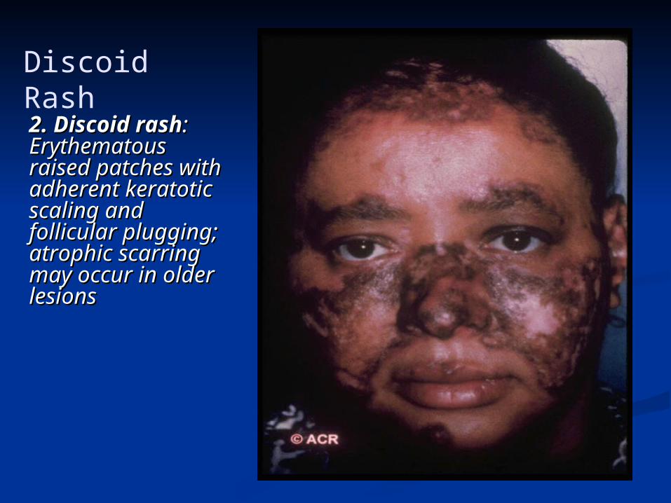
Discoid Rash
2. Discoid rash2. Discoid rash: : Erythematous Erythematous raised patches with raised patches with adherent keratotic adherent keratotic scaling and scaling and follicular plugging; follicular plugging; atrophic scarring atrophic scarring may occur in older may occur in older lesionslesions
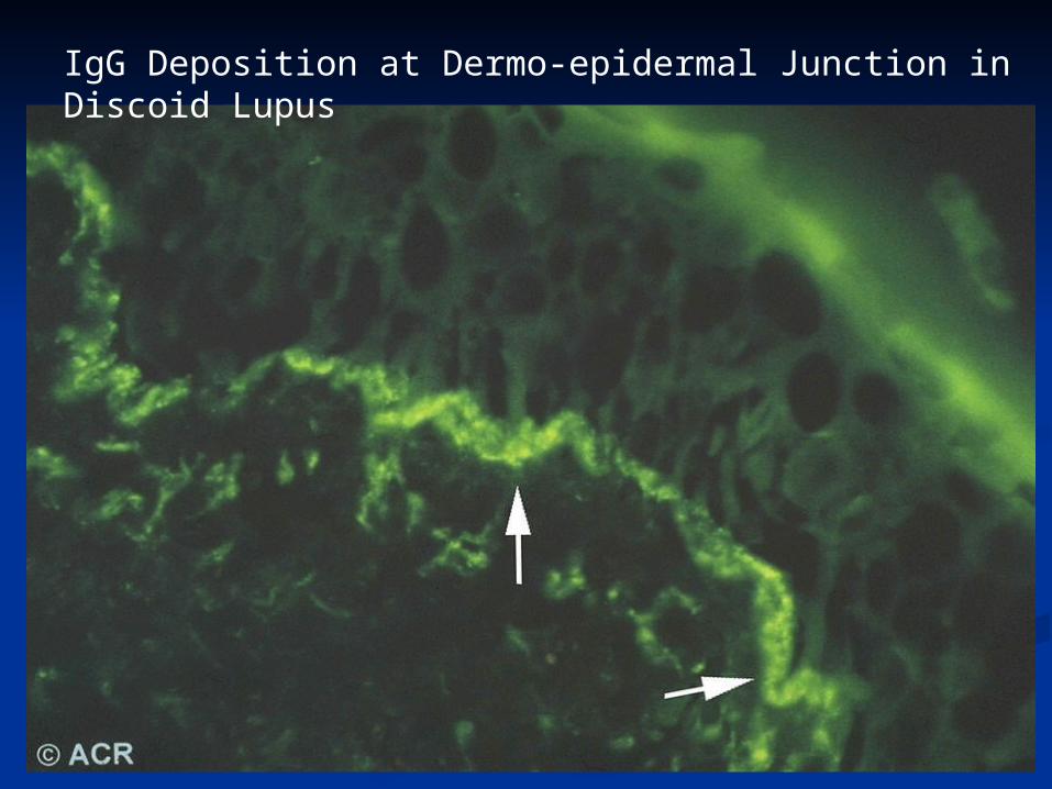
IgG Deposition at Dermo-epidermal Junction in Discoid Lupus
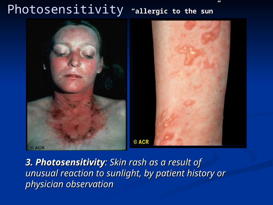
Photosensitivity “allergic to the sun”“allergic to the sun”
3. Photosensitivity3. Photosensitivity: Skin rash as a result of : Skin rash as a result of unusual reaction to sunlight, by patient history or unusual reaction to sunlight, by patient history or physician observation physician observation
Oral Ulcers
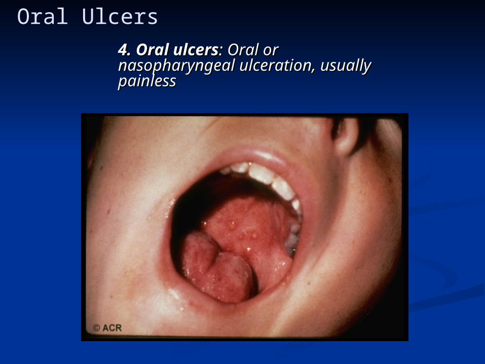
4. Oral ulcers4. Oral ulcers: Oral or : Oral or nasopharyngeal ulceration, usually nasopharyngeal ulceration, usually painlesspainless
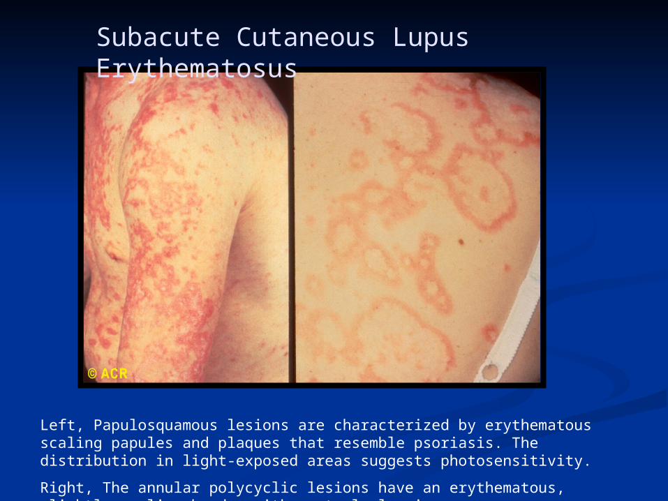
Subacute Cutaneous Lupus Erythematosus
Left, Papulosquamous lesions are characterized by erythematous scaling papules and plaques that resemble psoriasis. The distribution in light-exposed areas suggests photosensitivity.
Right, The annular polycyclic lesions have an erythematous, slightly scaling border with central clearing.
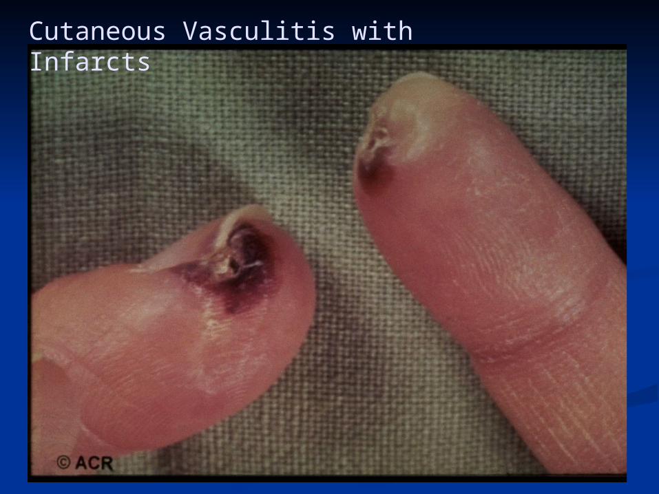
Cutaneous Vasculitis with Infarcts

Alopecia
ArthritisArthritis
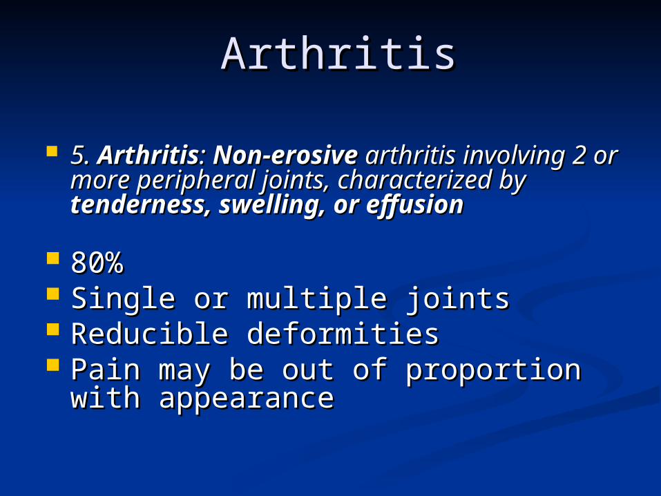
5. 5. ArthritisArthritis: : Non-erosiveNon-erosive arthritis arthritis involving 2 or more peripheral joints, involving 2 or more peripheral joints, characterized by characterized by tenderness, swelling, tenderness, swelling, or effusionor effusion
80%80% Single or multiple jointsSingle or multiple joints Reducible deformitiesReducible deformities Pain may be out of proportion with Pain may be out of proportion with
appearanceappearance
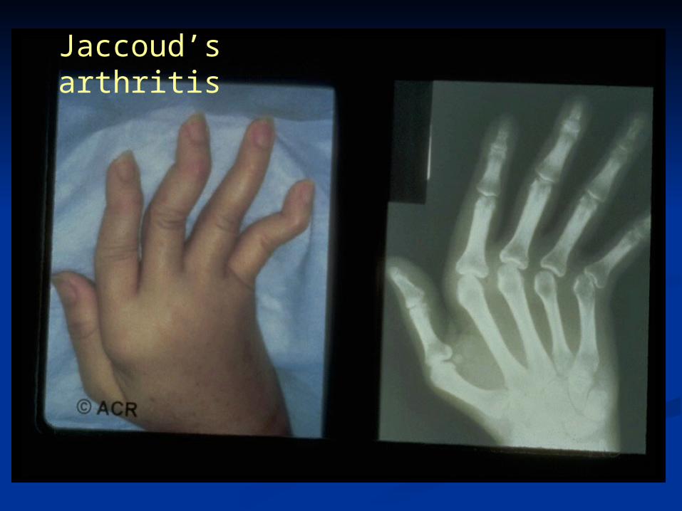
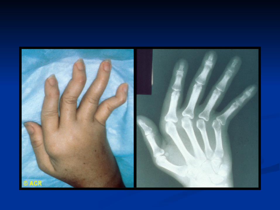
Jaccoud’s arthritis
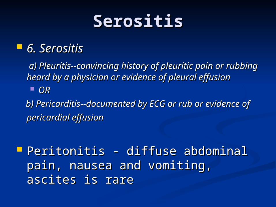
SerositisSerositis 6. Serositis6. Serositis a) Pleuritis--convincing history of pleuritic pain a) Pleuritis--convincing history of pleuritic pain
or rubbing heard by a physician or evidence of or rubbing heard by a physician or evidence of pleural effusion pleural effusion OR OR
b) Pericarditis--documented by ECG or rub or b) Pericarditis--documented by ECG or rub or
evidence of pericardial effusionevidence of pericardial effusion
Peritonitis - diffuse abdominal pain, Peritonitis - diffuse abdominal pain, nausea and vomiting, ascites is rarenausea and vomiting, ascites is rare
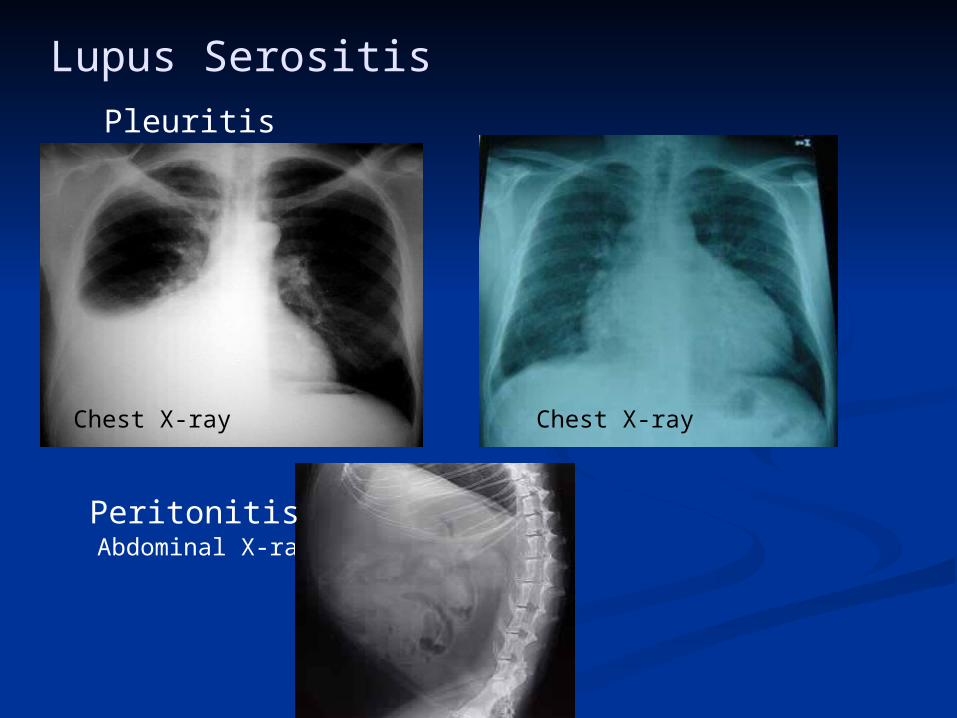
Pleuritis Pericarditis
Chest X-ray
Abdominal X-ray
Lupus Serositis
Peritonitis
Chest X-ray
RenalRenal
7. 7. Renal disorderRenal disorder
a) Persistent proteinuria greater a) Persistent proteinuria greater than 0.5 grams per day than 0.5 grams per day
OROR
b) Cellular casts--may be red cell, b) Cellular casts--may be red cell, hemoglobin, granular, tubular, or hemoglobin, granular, tubular, or mixed mixed
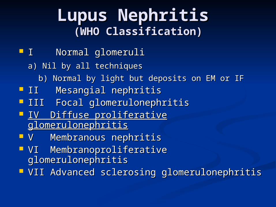
Lupus Nephritis Lupus Nephritis (WHO Classification)(WHO Classification)
I Normal glomeruliI Normal glomeruli
a) Nil by all techniquesa) Nil by all techniques
b) Normal by light but deposits on EM or b) Normal by light but deposits on EM or IFIF
II Mesangial nephritisII Mesangial nephritis III Focal glomerulonephritisIII Focal glomerulonephritis IV Diffuse proliferative glomerulonephritisIV Diffuse proliferative glomerulonephritis V Membranous nephritisV Membranous nephritis VI Membranoproliferative VI Membranoproliferative
glomerulonephritis glomerulonephritis VII Advanced sclerosing glomerulonephritisVII Advanced sclerosing glomerulonephritis
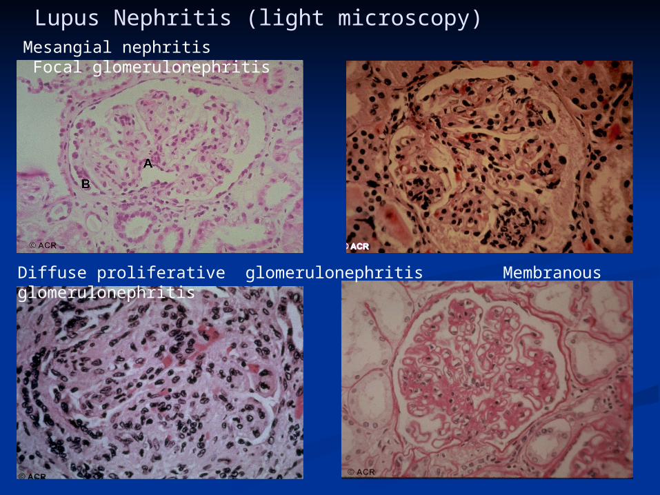
Lupus Nephritis (light microscopy)
Diffuse proliferative glomerulonephritis Membranous glomerulonephritis
Mesangial nephritis Focal glomerulonephritis
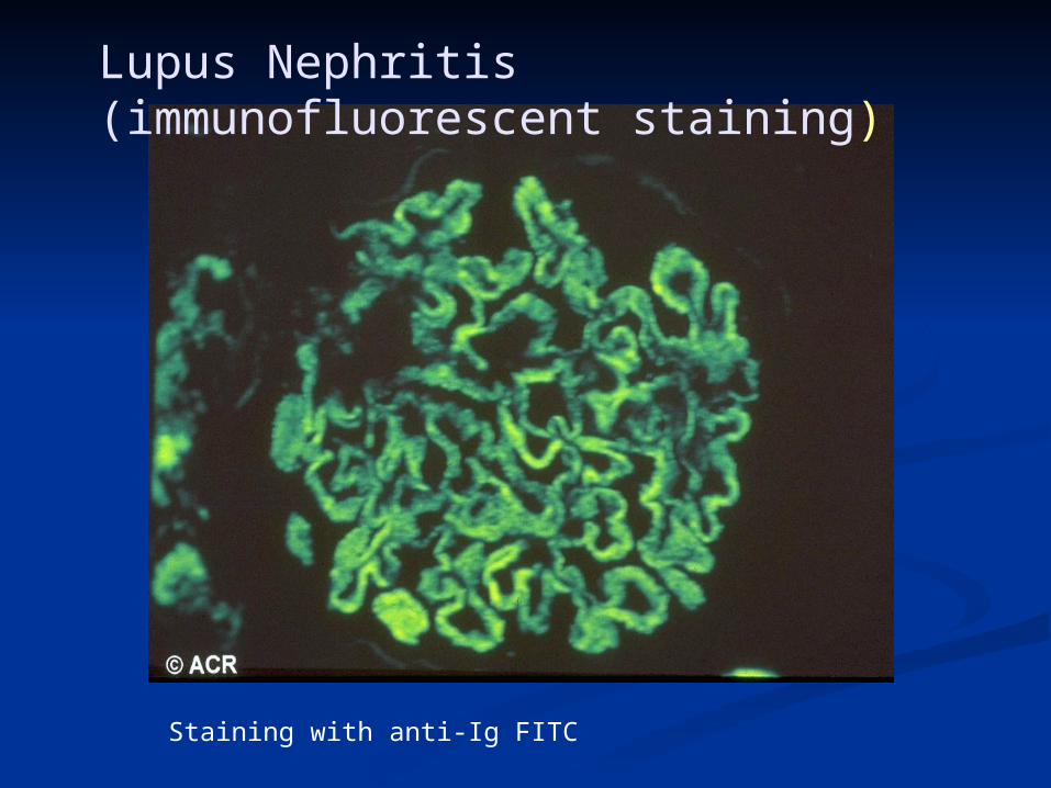
Lupus Nephritis (immunofluorescent staining)
Staining with anti-Ig FITC
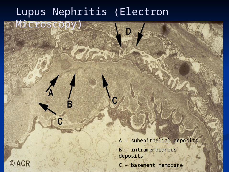
Lupus Nephritis (Electron Microscopy)
A – subepithelial deposits
B – intramembranous deposits
C – basement membrane
D – epithelial foot processes
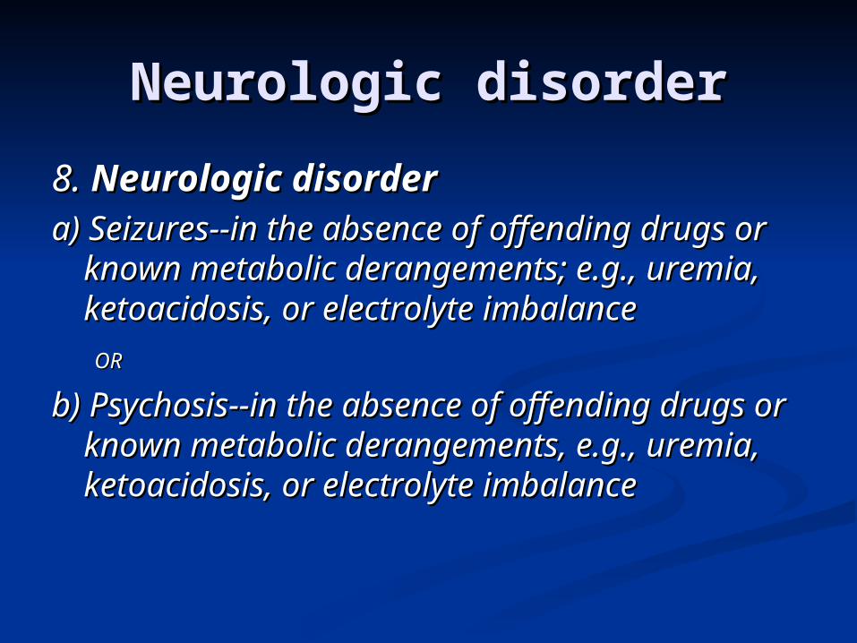
Neurologic disorderNeurologic disorder
8. 8. Neurologic disorderNeurologic disordera) Seizures--in the absence of offending a) Seizures--in the absence of offending
drugs or known metabolic derangements; drugs or known metabolic derangements; e.g., uremia, ketoacidosis, or electrolyte e.g., uremia, ketoacidosis, or electrolyte imbalance imbalance
OROR
b) Psychosis--in the absence of offending b) Psychosis--in the absence of offending drugs or known metabolic derangements, drugs or known metabolic derangements, e.g., uremia, ketoacidosis, or electrolyte e.g., uremia, ketoacidosis, or electrolyte imbalance imbalance
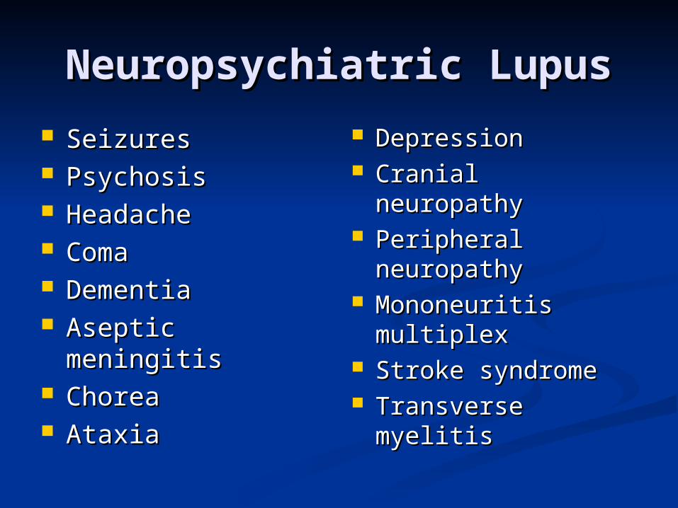
Neuropsychiatric LupusNeuropsychiatric Lupus
SeizuresSeizures PsychosisPsychosis HeadacheHeadache ComaComa DementiaDementia Aseptic meningitisAseptic meningitis ChoreaChorea AtaxiaAtaxia
DepressionDepression Cranial neuropathyCranial neuropathy Peripheral Peripheral
neuropathyneuropathy Mononeuritis Mononeuritis
multiplexmultiplex Stroke syndromeStroke syndrome Transverse Transverse
myelitismyelitis
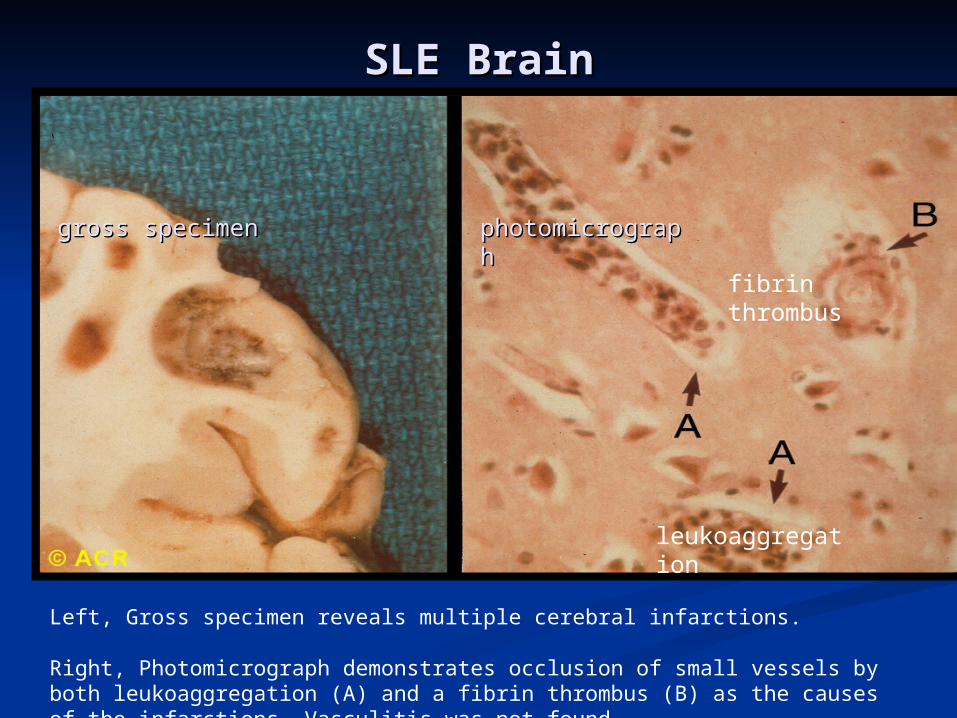
SLE BrainSLE Brain
gross specimengross specimen photomicrographphotomicrograph
leukoaggregation
fibrin thrombus
Left, Gross specimen reveals multiple cerebral infarctions.
Right, Photomicrograph demonstrates occlusion of small vessels by both leukoaggregation (A) and a fibrin thrombus (B) as the causes of the infarctions. Vasculitis was not found
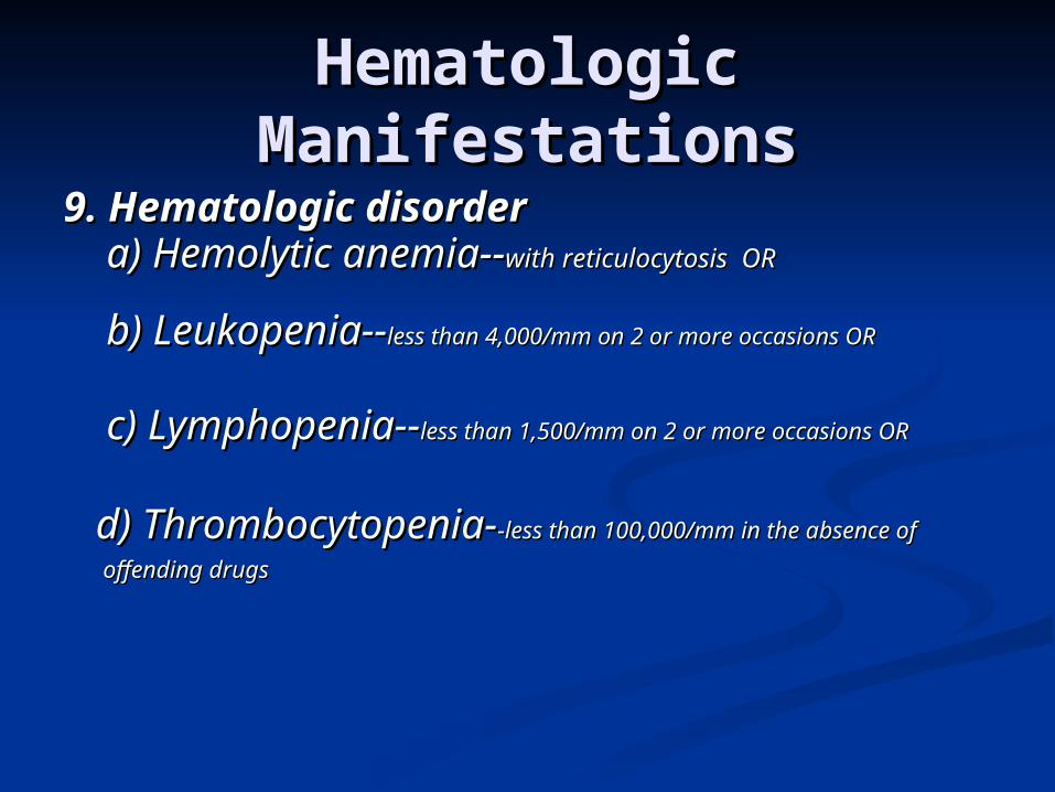
Hematologic Hematologic ManifestationsManifestations
9. 9. Hematologic disorderHematologic disorder a) Hemolytic anemia--a) Hemolytic anemia--with reticulocytosis ORwith reticulocytosis OR
b) Leukopenia--b) Leukopenia--less than 4,000/mm on 2 or more occasions less than 4,000/mm on 2 or more occasions OROR
c) Lymphopenia--c) Lymphopenia--less than 1,500/mm on 2 or more occasions less than 1,500/mm on 2 or more occasions OROR
d) Thrombocytopenia-d) Thrombocytopenia--less than 100,000/mm in the -less than 100,000/mm in the
absence of offending drugsabsence of offending drugs
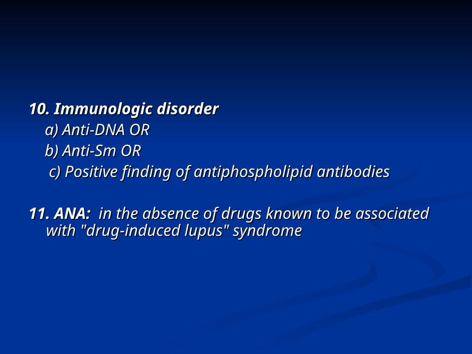
10. Immunologic disorder10. Immunologic disorder a) Anti-DNA ORa) Anti-DNA OR b) Anti-Sm ORb) Anti-Sm OR c) Positive finding of antiphospholipid c) Positive finding of antiphospholipid
antibodies antibodies
11. ANA:11. ANA: in the absence of drugs known to be in the absence of drugs known to be associated with "drug-induced lupus" syndrome associated with "drug-induced lupus" syndrome
Classification CriteriaClassification Criteria
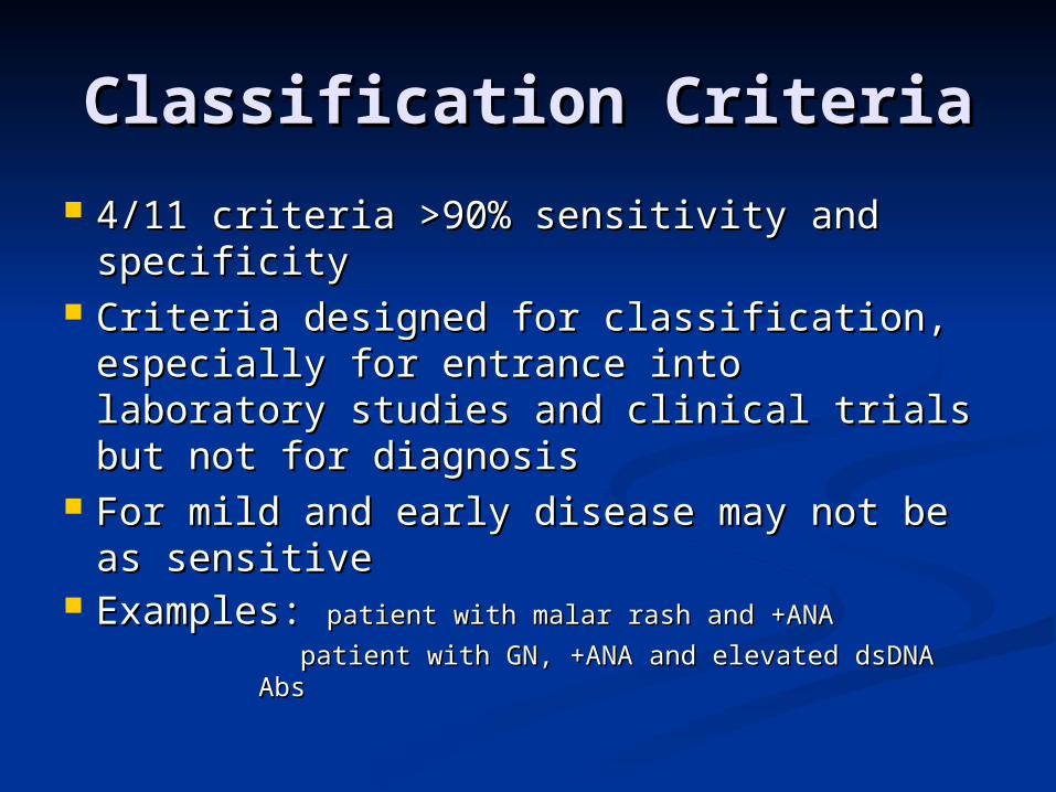
4/11 criteria >90% sensitivity and 4/11 criteria >90% sensitivity and specificity specificity
Criteria designed for classification, Criteria designed for classification, especially for entrance into laboratory especially for entrance into laboratory studies and clinical trials but not for studies and clinical trials but not for diagnosisdiagnosis
For mild and early disease may not be as For mild and early disease may not be as sensitivesensitive
Examples: Examples: patient with malar rash and +ANApatient with malar rash and +ANA
patient with GN, +ANA and elevated dsDNA Abspatient with GN, +ANA and elevated dsDNA Abs
MortalityMortality
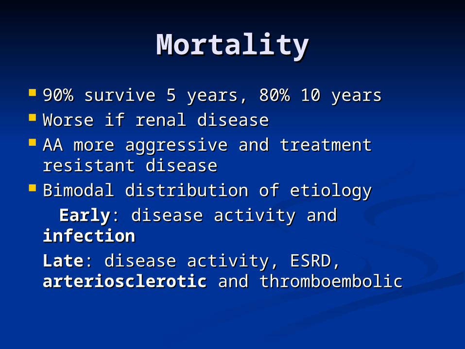
90% survive 5 years, 80% 10 years90% survive 5 years, 80% 10 years Worse if renal diseaseWorse if renal disease AA more aggressive and treatment AA more aggressive and treatment
resistant diseaseresistant disease Bimodal distribution of etiologyBimodal distribution of etiology
EarlyEarly: disease activity and : disease activity and infectioninfection
LateLate: disease activity, ESRD, : disease activity, ESRD, arterioscleroticarteriosclerotic and thromboembolic and thromboembolic
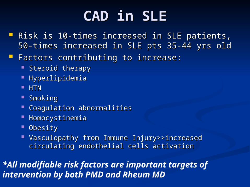
CAD in SLECAD in SLE Risk is 10-times increased in SLE patients, 50-times Risk is 10-times increased in SLE patients, 50-times
increased in SLE pts 35-44 yrs oldincreased in SLE pts 35-44 yrs old Factors contributing to increase:Factors contributing to increase:
Steroid therapySteroid therapy HyperlipidemiaHyperlipidemia HTNHTN SmokingSmoking Coagulation abnormalitiesCoagulation abnormalities HomocystinemiaHomocystinemia ObesityObesity Vasculopathy from Immune Injury>>increased circulating Vasculopathy from Immune Injury>>increased circulating
endothelial cells activationendothelial cells activation
*All modifiable risk factors are important targets of intervention by both PMD and Rheum MD
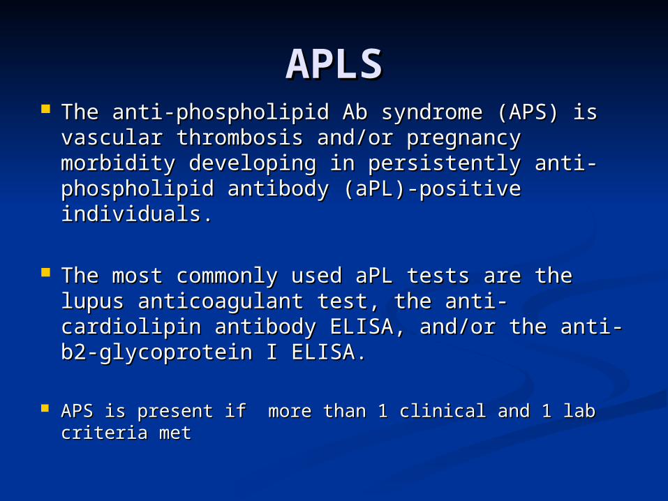
The anti-phospholipid Ab syndrome (APS) is The anti-phospholipid Ab syndrome (APS) is vascular thrombosis and/or pregnancy morbidity vascular thrombosis and/or pregnancy morbidity developing in persistently anti-phospholipid developing in persistently anti-phospholipid antibody (aPL)-positive individuals. antibody (aPL)-positive individuals.
The most commonly used aPL tests are the lupus The most commonly used aPL tests are the lupus anticoagulant test, the anti-cardiolipin antibody anticoagulant test, the anti-cardiolipin antibody ELISA, and/or the anti- b2-glycoprotein I ELISA.ELISA, and/or the anti- b2-glycoprotein I ELISA.
APS is present if more than 1 clinical and 1 lab criteria APS is present if more than 1 clinical and 1 lab criteria metmet
APLSAPLS
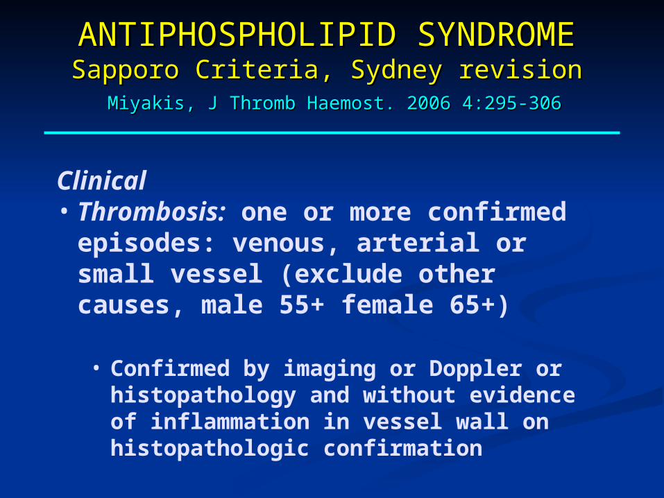
Clinical• Thrombosis: one or more confirmed episodes:
venous, arterial or small vessel (exclude other causes, male 55+ female 65+)
• Confirmed by imaging or Doppler or histopathology and without evidence of inflammation in vessel wall on histopathologic confirmation
ANTIPHOSPHOLIPID ANTIPHOSPHOLIPID SYNDROMESYNDROME
Sapporo Criteria, Sydney revisionSapporo Criteria, Sydney revision Miyakis, J Thromb Haemost. 2006 4:295-306Miyakis, J Thromb Haemost. 2006 4:295-306
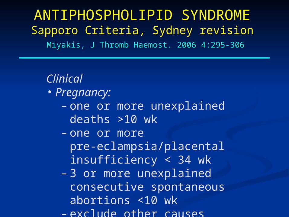
Clinical• Pregnancy:
– one or more unexplained deaths >10 wk– one or more pre-eclampsia/placental
insufficiency < 34 wk– 3 or more unexplained consecutive
spontaneous abortions <10 wk– exclude other causes
ANTIPHOSPHOLIPID ANTIPHOSPHOLIPID SYNDROMESYNDROME
Sapporo Criteria, Sydney revisionSapporo Criteria, Sydney revision Miyakis, J Thromb Haemost. 2006 4:295-306Miyakis, J Thromb Haemost. 2006 4:295-306
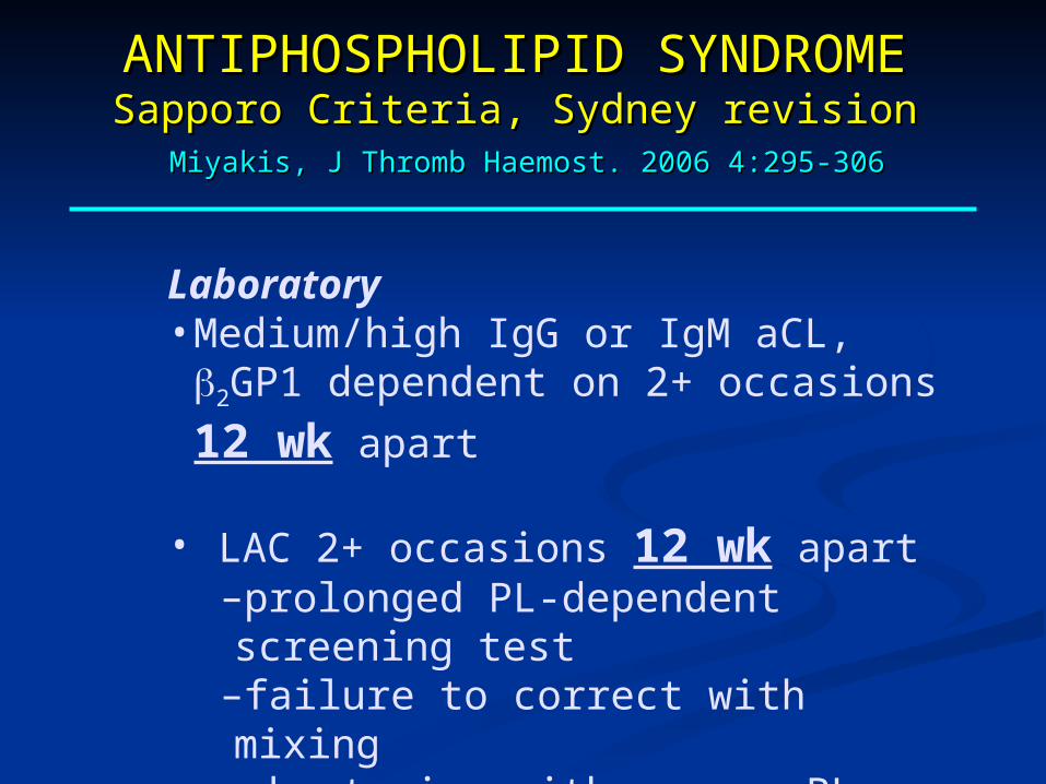
Laboratory• Medium/high IgG or IgM aCL, 2GP1
dependent on 2+ occasions 12 wk apart
• LAC 2+ occasions 12 wk apart–prolonged PL-dependent screening test–failure to correct with mixing–shortening with excess PL –exclusion of other coagulopathies
ANTIPHOSPHOLIPID ANTIPHOSPHOLIPID SYNDROMESYNDROME
Sapporo Criteria, Sydney revisionSapporo Criteria, Sydney revision Miyakis, J Thromb Haemost. 2006 4:295-306Miyakis, J Thromb Haemost. 2006 4:295-306
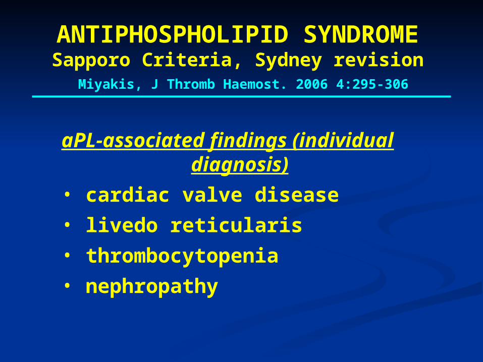
aPL-associated findings (individual diagnosis)
• cardiac valve disease
• livedo reticularis
• thrombocytopenia
• nephropathy
ANTIPHOSPHOLIPID SYNDROMESapporo Criteria, Sydney revision Miyakis, J Thromb Haemost. 2006 4:295-306
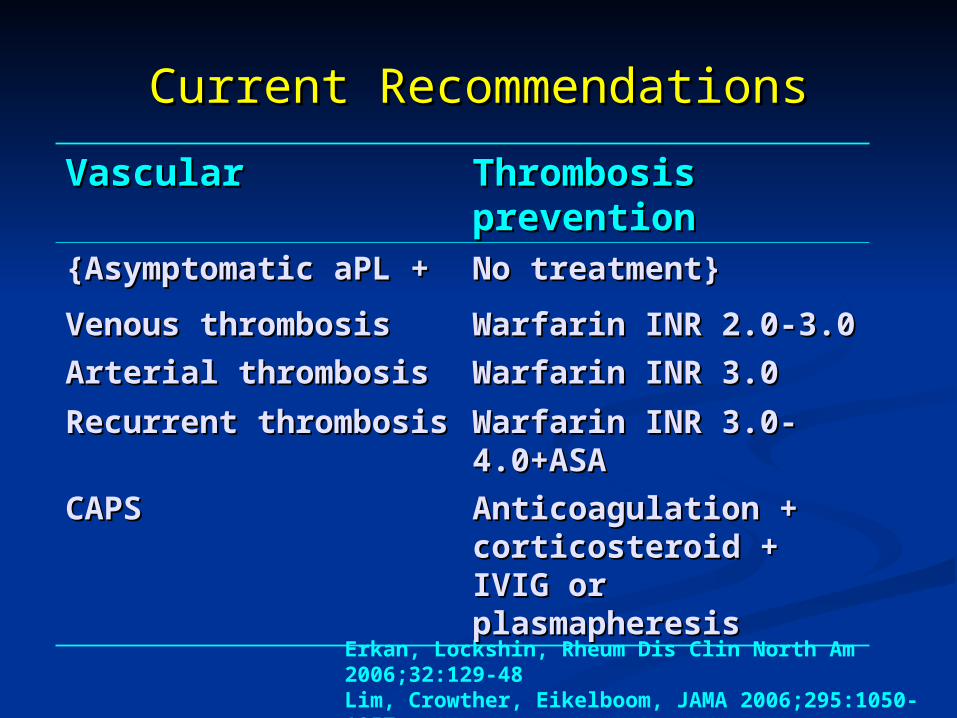
Erkan, Lockshin, Rheum Dis Clin North Am 2006;32:129-48Lim, Crowther, Eikelboom, JAMA 2006;295:1050-1057
VascularVascular Thrombosis Thrombosis preventionprevention
{Asymptomatic aPL {Asymptomatic aPL ++
No treatment}No treatment}
Venous thrombosisVenous thrombosis Warfarin INR 2.0-3.0Warfarin INR 2.0-3.0
Arterial thrombosisArterial thrombosis Warfarin INR 3.0Warfarin INR 3.0
Recurrent Recurrent thrombosisthrombosis
Warfarin INR 3.0-Warfarin INR 3.0-4.0+ASA4.0+ASA
CAPSCAPS Anticoagulation + Anticoagulation + corticosteroid + IVIG corticosteroid + IVIG or plasmapheresisor plasmapheresis
Current RecommendationsCurrent Recommendations
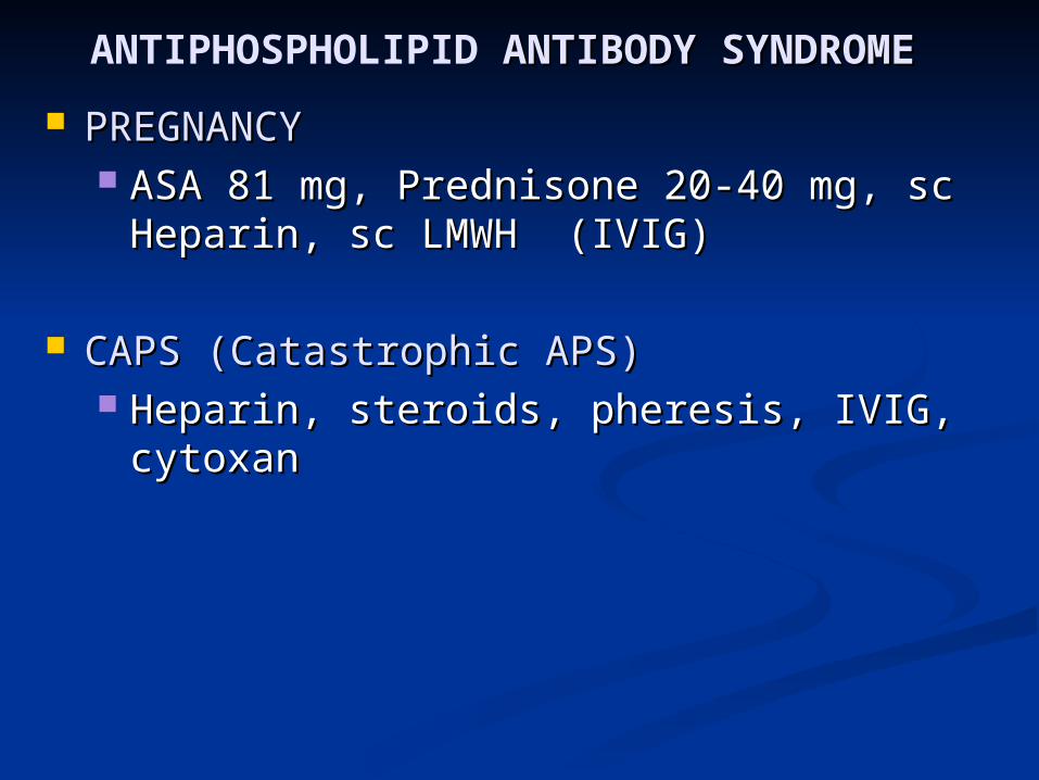
ANTIPHOSPHOLIPID ANTIBODY SYNDROME ANTIBODY SYNDROME
PREGNANCYPREGNANCY ASA 81 mg, Prednisone 20-40 mg, sc ASA 81 mg, Prednisone 20-40 mg, sc
Heparin, sc LMWH (IVIG)Heparin, sc LMWH (IVIG)
CAPS (Catastrophic APS)CAPS (Catastrophic APS) Heparin, steroids, pheresis, IVIG, Heparin, steroids, pheresis, IVIG,
cytoxancytoxan
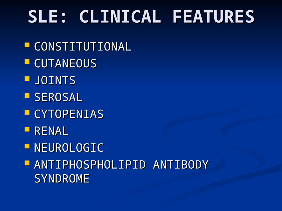
SLE: CLINICAL SLE: CLINICAL FEATURESFEATURES
CONSTITUTIONALCONSTITUTIONAL CUTANEOUSCUTANEOUS JOINTSJOINTS SEROSALSEROSAL CYTOPENIASCYTOPENIAS RENAL RENAL NEUROLOGICNEUROLOGIC ANTIPHOSPHOLIPID ANTIBODY ANTIPHOSPHOLIPID ANTIBODY
SYNDROME SYNDROME
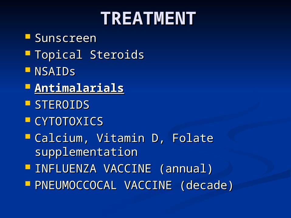
TREATMENTTREATMENT SunscreenSunscreen Topical SteroidsTopical Steroids NSAIDsNSAIDs AntimalarialsAntimalarials STEROIDSSTEROIDS CYTOTOXICSCYTOTOXICS Calcium, Vitamin D, Folate Calcium, Vitamin D, Folate
supplementation supplementation INFLUENZA VACCINE (annual)INFLUENZA VACCINE (annual) PNEUMOCCOCAL VACCINE (decade)PNEUMOCCOCAL VACCINE (decade)
TreatmentTreatment
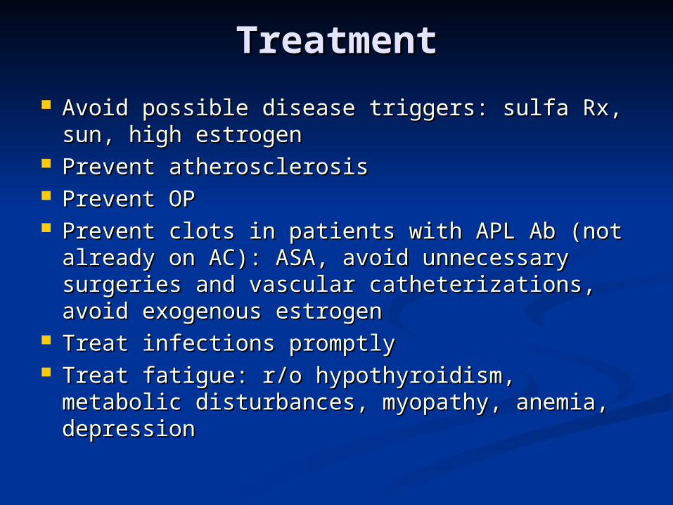
Avoid possible disease triggers: sulfa Rx, Avoid possible disease triggers: sulfa Rx, sun, high estrogensun, high estrogen
Prevent atherosclerosisPrevent atherosclerosis Prevent OPPrevent OP Prevent clots in patients with APL Ab (not Prevent clots in patients with APL Ab (not
already on AC): ASA, avoid unnecessary already on AC): ASA, avoid unnecessary surgeries and vascular catheterizations, surgeries and vascular catheterizations, avoid exogenous estrogenavoid exogenous estrogen
Treat infections promptlyTreat infections promptly Treat fatigue: r/o hypothyroidism, metabolic Treat fatigue: r/o hypothyroidism, metabolic
disturbances, myopathy, anemia, depressiondisturbances, myopathy, anemia, depression
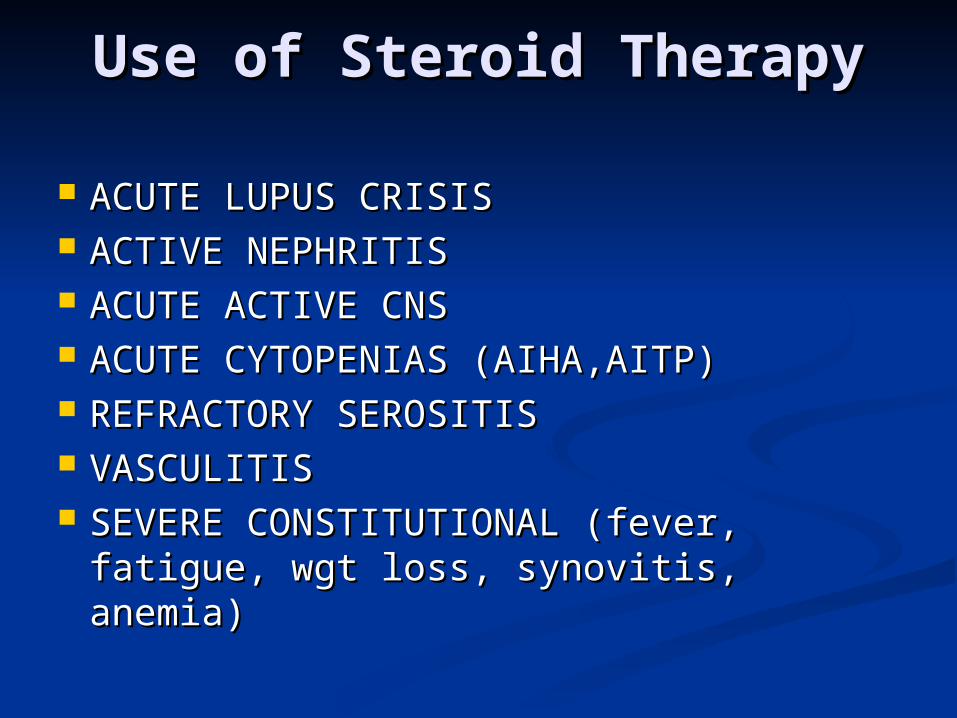
Use of Steroid TherapyUse of Steroid Therapy
ACUTE LUPUS CRISISACUTE LUPUS CRISIS ACTIVE NEPHRITISACTIVE NEPHRITIS ACUTE ACTIVE CNSACUTE ACTIVE CNS ACUTE CYTOPENIAS (AIHA,AITP)ACUTE CYTOPENIAS (AIHA,AITP) REFRACTORY SEROSITISREFRACTORY SEROSITIS VASCULITISVASCULITIS SEVERE CONSTITUTIONAL (fever, SEVERE CONSTITUTIONAL (fever,
fatigue, wgt loss, synovitis, anemia)fatigue, wgt loss, synovitis, anemia)
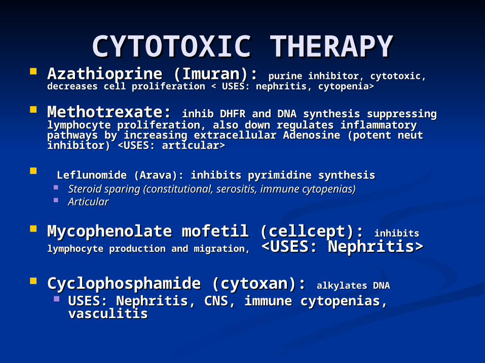
CYTOTOXIC THERAPYCYTOTOXIC THERAPY Azathioprine (Imuran): Azathioprine (Imuran): purine inhibitor, cytotoxic, purine inhibitor, cytotoxic,
decreases cell proliferation < USES: nephritis, cytopenia>decreases cell proliferation < USES: nephritis, cytopenia>
Methotrexate: Methotrexate: inhib DHFR and DNA synthesis suppressing inhib DHFR and DNA synthesis suppressing lymphocyte proliferation, also down regulates inflammatory lymphocyte proliferation, also down regulates inflammatory pathways by increasing extracellular Adenosine (potent neut pathways by increasing extracellular Adenosine (potent neut inhibitor) <USES: articular>inhibitor) <USES: articular>
Leflunomide (Arava): inhibits pyrimidine synthesisLeflunomide (Arava): inhibits pyrimidine synthesis Steroid sparing (constitutional, serositis, immune cytopenias) Steroid sparing (constitutional, serositis, immune cytopenias) ArticularArticular
Mycophenolate mofetil (cellcept): Mycophenolate mofetil (cellcept): inhibits inhibits
lymphocyte production and migration,lymphocyte production and migration, <USES: Nephritis> <USES: Nephritis>
Cyclophosphamide (cytoxan): Cyclophosphamide (cytoxan): alkylates DNAalkylates DNA USES: Nephritis, CNS, immune cytopenias, USES: Nephritis, CNS, immune cytopenias,
vasculitis vasculitis
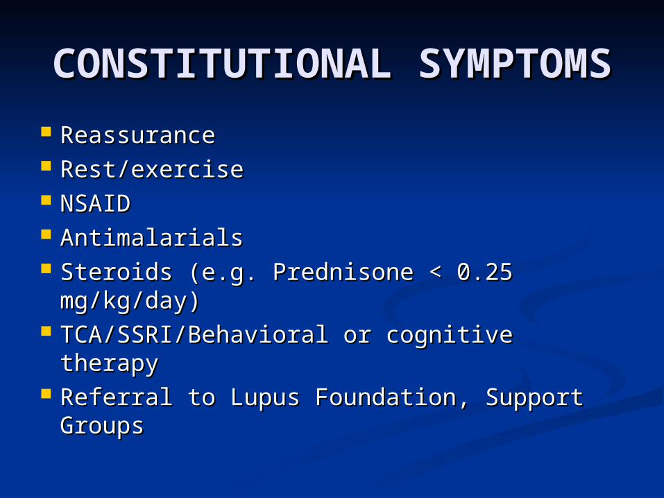
CONSTITUTIONAL CONSTITUTIONAL SYMPTOMSSYMPTOMS
ReassuranceReassurance Rest/exerciseRest/exercise NSAIDNSAID AntimalarialsAntimalarials Steroids (e.g. Prednisone < 0.25 Steroids (e.g. Prednisone < 0.25
mg/kg/day)mg/kg/day) TCA/SSRI/Behavioral or cognitive therapyTCA/SSRI/Behavioral or cognitive therapy Referral to Lupus Foundation, Support Referral to Lupus Foundation, Support
GroupsGroups
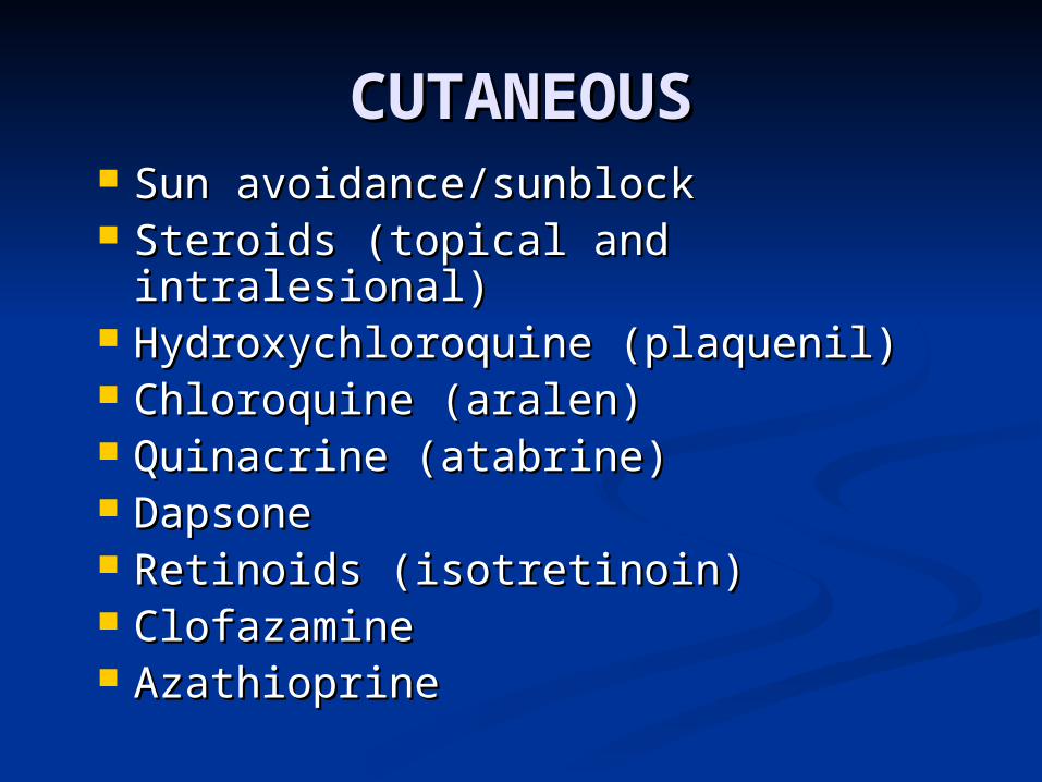
CUTANEOUSCUTANEOUS Sun avoidance/sunblockSun avoidance/sunblock Steroids (topical and intralesional)Steroids (topical and intralesional) Hydroxychloroquine (plaquenil)Hydroxychloroquine (plaquenil) Chloroquine (aralen)Chloroquine (aralen) Quinacrine (atabrine)Quinacrine (atabrine) DapsoneDapsone Retinoids (isotretinoin)Retinoids (isotretinoin) ClofazamineClofazamine AzathioprineAzathioprine
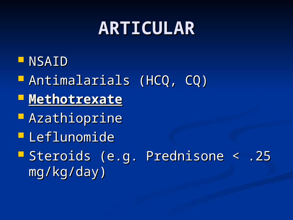
ARTICULARARTICULAR
NSAIDNSAID Antimalarials (HCQ, CQ)Antimalarials (HCQ, CQ) MethotrexateMethotrexate AzathioprineAzathioprine LeflunomideLeflunomide Steroids (e.g. Prednisone < .25 Steroids (e.g. Prednisone < .25
mg/kg/day)mg/kg/day)
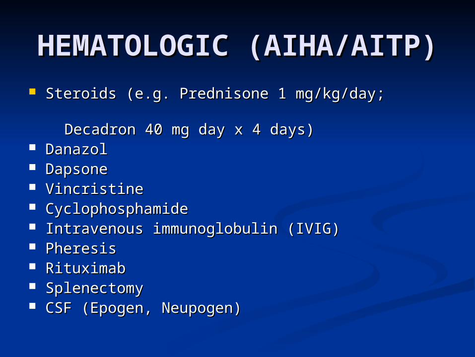
HEMATOLOGIC HEMATOLOGIC (AIHA/AITP)(AIHA/AITP)
Steroids (e.g. Prednisone 1 mg/kg/day; Steroids (e.g. Prednisone 1 mg/kg/day;
Decadron 40 mg day x 4 days)Decadron 40 mg day x 4 days) Danazol Danazol DapsoneDapsone VincristineVincristine CyclophosphamideCyclophosphamide Intravenous immunoglobulin (IVIG)Intravenous immunoglobulin (IVIG) PheresisPheresis RituximabRituximab SplenectomySplenectomy CSF (Epogen, Neupogen)CSF (Epogen, Neupogen)
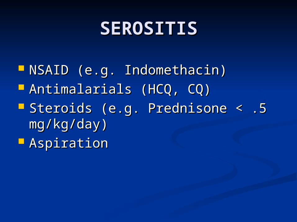
SEROSITISSEROSITIS
NSAID (e.g. Indomethacin)NSAID (e.g. Indomethacin) Antimalarials (HCQ, CQ)Antimalarials (HCQ, CQ) Steroids (e.g. Prednisone < .5 Steroids (e.g. Prednisone < .5
mg/kg/day)mg/kg/day) AspirationAspiration
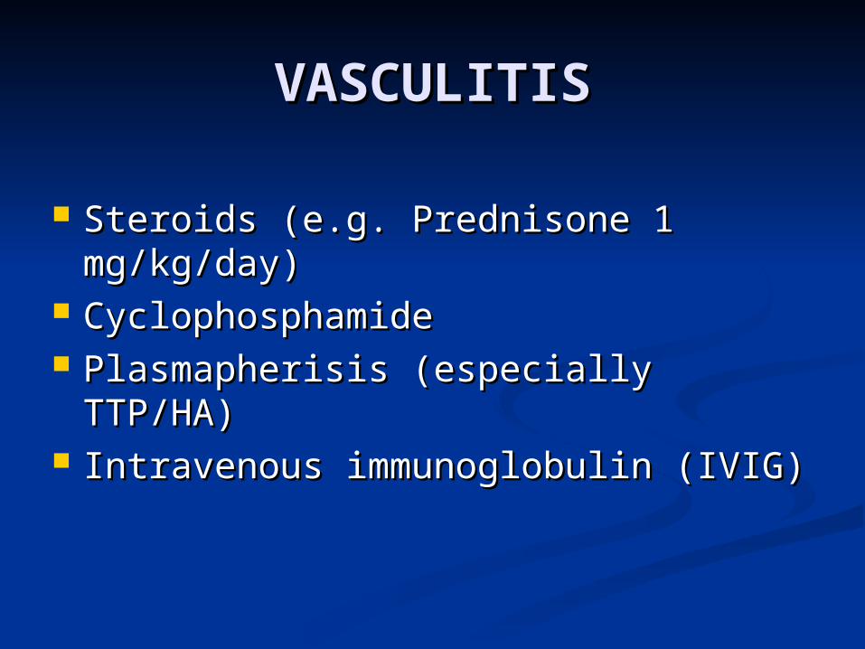
VASCULITISVASCULITIS
Steroids (e.g. Prednisone 1 Steroids (e.g. Prednisone 1 mg/kg/day)mg/kg/day)
CyclophosphamideCyclophosphamide Plasmapherisis (especially TTP/HA)Plasmapherisis (especially TTP/HA) Intravenous immunoglobulin (IVIG)Intravenous immunoglobulin (IVIG)
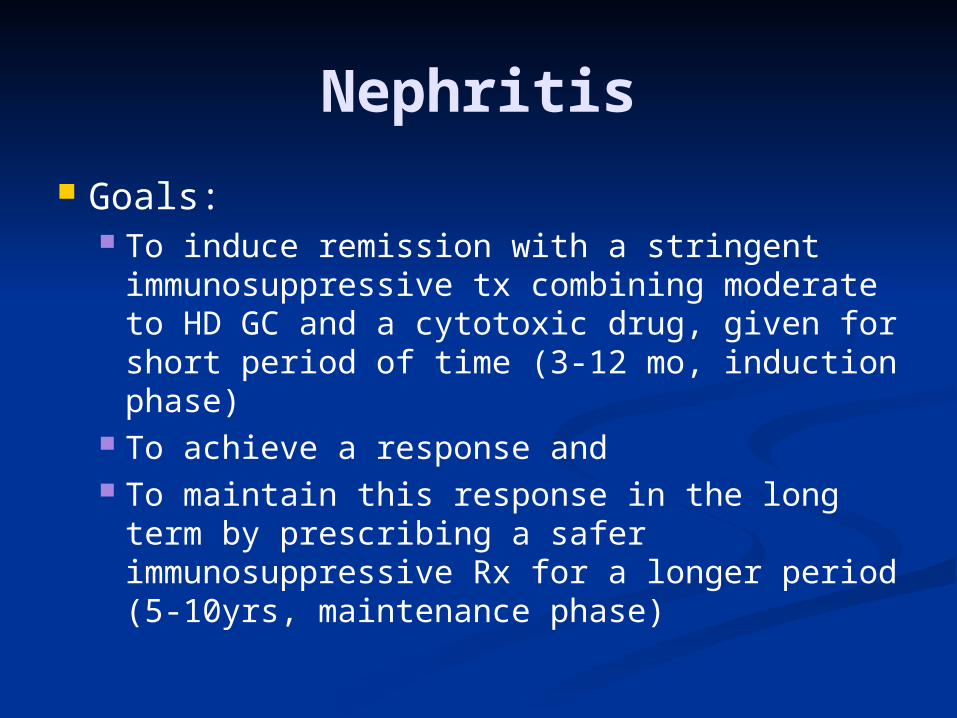
Nephritis
Goals: To induce remission with a stringent
immunosuppressive tx combining moderate to HD GC and a cytotoxic drug, given for short period of time (3-12 mo, induction phase)
To achieve a response and To maintain this response in the long term
by prescribing a safer immunosuppressive Rx for a longer period (5-10yrs, maintenance phase)
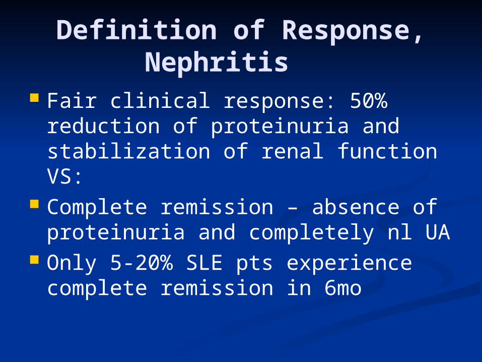
Definition of Response, Nephritis
Fair clinical response: 50% reduction of proteinuria and stabilization of renal function VS:
Complete remission – absence of proteinuria and completely nl UA
Only 5-20% SLE pts experience complete remission in 6mo
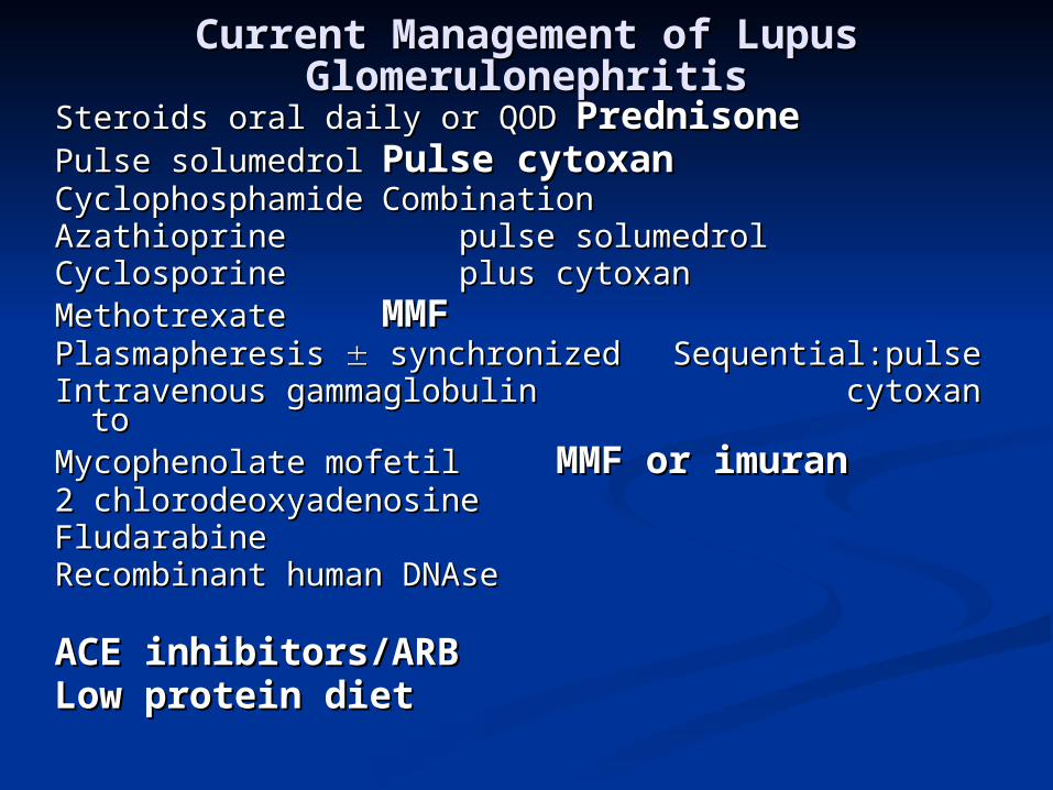
Current Management of Lupus Current Management of Lupus GlomerulonephritisGlomerulonephritis
Steroids oral daily or QODSteroids oral daily or QOD PrednisonePrednisonePulse solumedrolPulse solumedrol Pulse cytoxanPulse cytoxanCyclophosphamideCyclophosphamide Combination Combination AzathioprineAzathioprine pulse solumedrol pulse solumedrol CyclosporineCyclosporine plus cytoxan plus cytoxanMethotrexateMethotrexate MMFMMFPlasmapheresis Plasmapheresis synchronized synchronized
Sequential:pulseSequential:pulseIntravenous gammaglobulinIntravenous gammaglobulin cytoxan to cytoxan toMycophenolate mofetilMycophenolate mofetil MMF or MMF or
imuranimuran2 chlorodeoxyadenosine2 chlorodeoxyadenosineFludarabineFludarabineRecombinant human DNAseRecombinant human DNAse
ACE inhibitors/ARBACE inhibitors/ARBLow protein dietLow protein diet
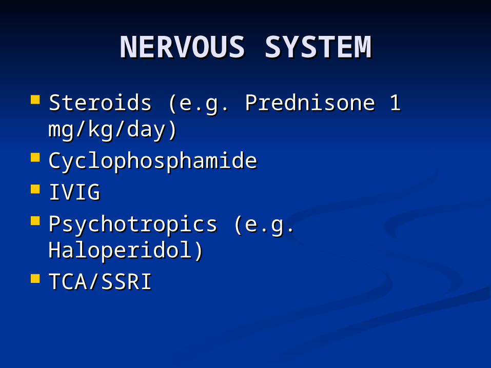
NERVOUS SYSTEMNERVOUS SYSTEM
Steroids (e.g. Prednisone 1 Steroids (e.g. Prednisone 1 mg/kg/day)mg/kg/day)
CyclophosphamideCyclophosphamide IVIGIVIG Psychotropics (e.g. Haloperidol)Psychotropics (e.g. Haloperidol) TCA/SSRITCA/SSRI
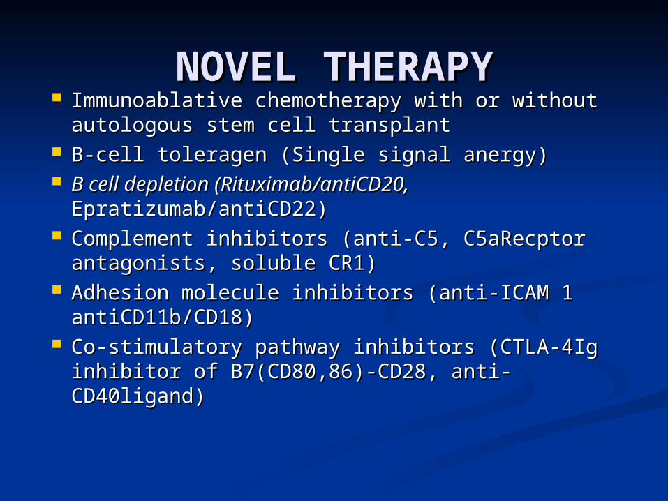
NOVEL THERAPYNOVEL THERAPY Immunoablative chemotherapy with or without Immunoablative chemotherapy with or without
autologous stem cell transplantautologous stem cell transplant B-cell toleragen (Single signal anergy)B-cell toleragen (Single signal anergy) B cell depletion (Rituximab/antiCD20,B cell depletion (Rituximab/antiCD20,
Epratizumab/antiCD22)Epratizumab/antiCD22) Complement inhibitors (anti-C5, C5aRecptor Complement inhibitors (anti-C5, C5aRecptor
antagonists, soluble CR1) antagonists, soluble CR1) Adhesion molecule inhibitors (anti-ICAM 1 Adhesion molecule inhibitors (anti-ICAM 1
antiCD11b/CD18) antiCD11b/CD18) Co-stimulatory pathway inhibitors (CTLA-4Ig Co-stimulatory pathway inhibitors (CTLA-4Ig
inhibitor of B7(CD80,86)-CD28, anti-inhibitor of B7(CD80,86)-CD28, anti-CD40ligand)CD40ligand)
PITFALLSPITFALLS
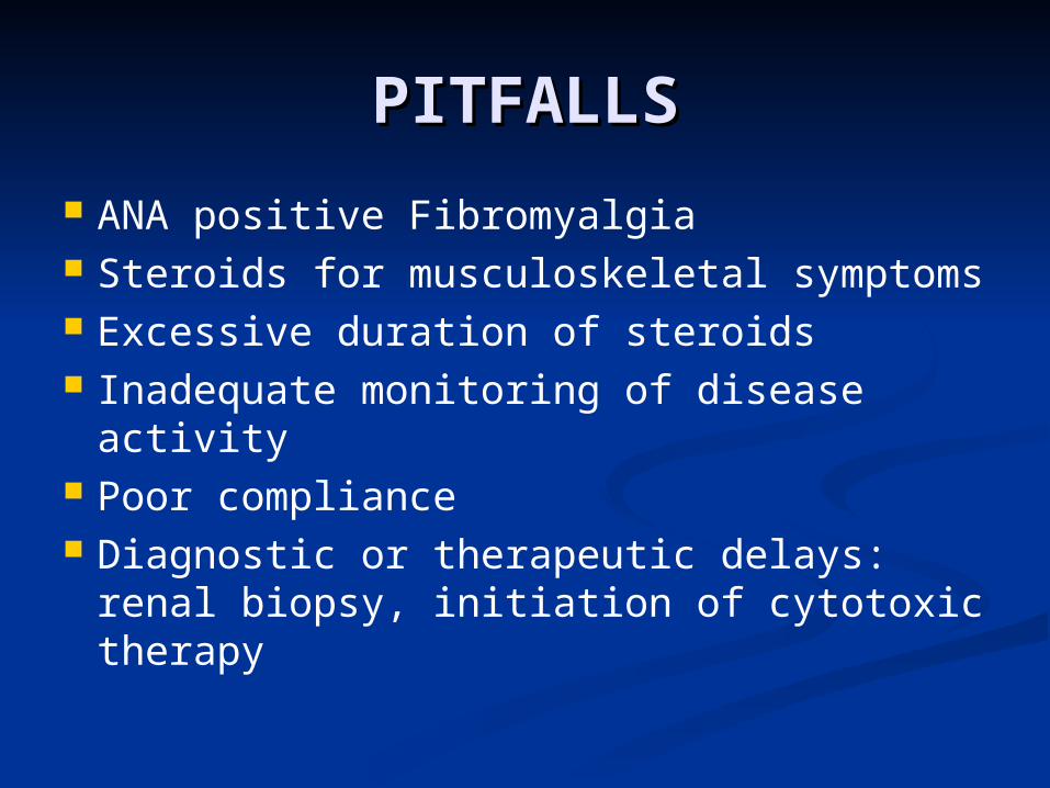
ANA positive Fibromyalgia Steroids for musculoskeletal symptoms Excessive duration of steroids Inadequate monitoring of disease
activity Poor compliance Diagnostic or therapeutic delays: renal
biopsy, initiation of cytotoxic therapy
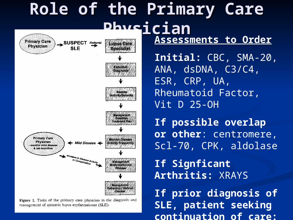
Role of the Primary Care Role of the Primary Care PhysicianPhysician
Assessments to Order
Initial: CBC, SMA-20, ANA, dsDNA, C3/C4, ESR, CRP, UA, Rheumatoid Factor, Vit D 25-OH
If possible overlap or other: centromere, Scl-70, CPK, aldolase
If Signficant Arthritis: XRAYS
If prior diagnosis of SLE, patient seeking continuation of care: Subserologies: Ro, La, Sm, RNP, LAC, Cardiolipin Ab
Reasons for Referral to Reasons for Referral to RheumatologyRheumatology
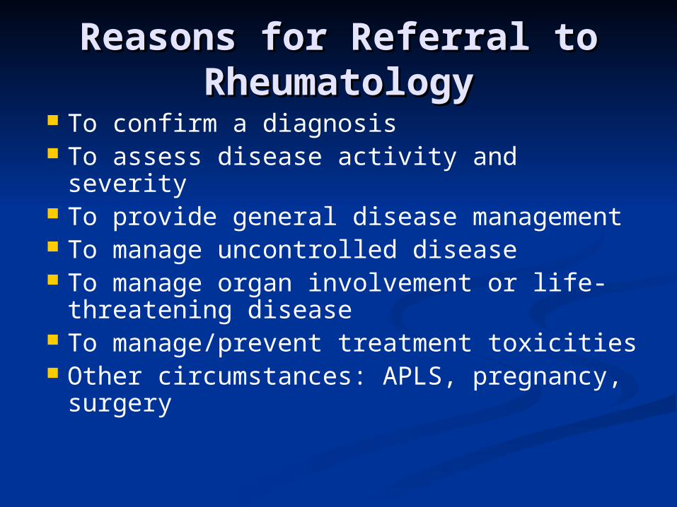
To confirm a diagnosis To assess disease activity and severity To provide general disease
management To manage uncontrolled disease To manage organ involvement or life-
threatening disease To manage/prevent treatment toxicities Other circumstances: APLS, pregnancy,
surgery

Follow Up VisitsFollow Up Visits Frequency depends on activity, Frequency depends on activity,
severity, and extent of SLE, response severity, and extent of SLE, response to treatment, type of treatment, need to treatment, type of treatment, need for toxicity monitoringfor toxicity monitoring
At routine visits, CBC, SMA, UA should At routine visits, CBC, SMA, UA should be checked, even in patients with be checked, even in patients with previously normal valuespreviously normal values
Patients with known renal disease Patients with known renal disease should also have either 24 hour urine should also have either 24 hour urine or spot protein/creatinine checked or spot protein/creatinine checked every 6-8 weeksevery 6-8 weeks
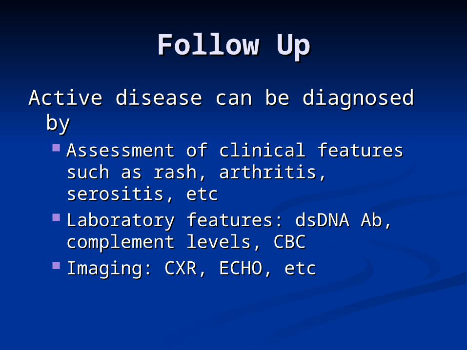
Follow UpFollow Up
Active disease can be diagnosed by Active disease can be diagnosed by Assessment of clinical features such as Assessment of clinical features such as
rash, arthritis, serositis, etcrash, arthritis, serositis, etc Laboratory features: dsDNA Ab, Laboratory features: dsDNA Ab,
complement levels, CBCcomplement levels, CBC Imaging: CXR, ECHO, etcImaging: CXR, ECHO, etc
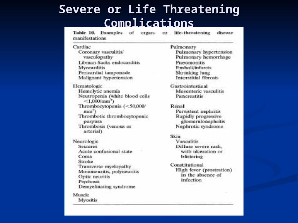
Severe or Life Threatening ComplicationsSevere or Life Threatening Complications
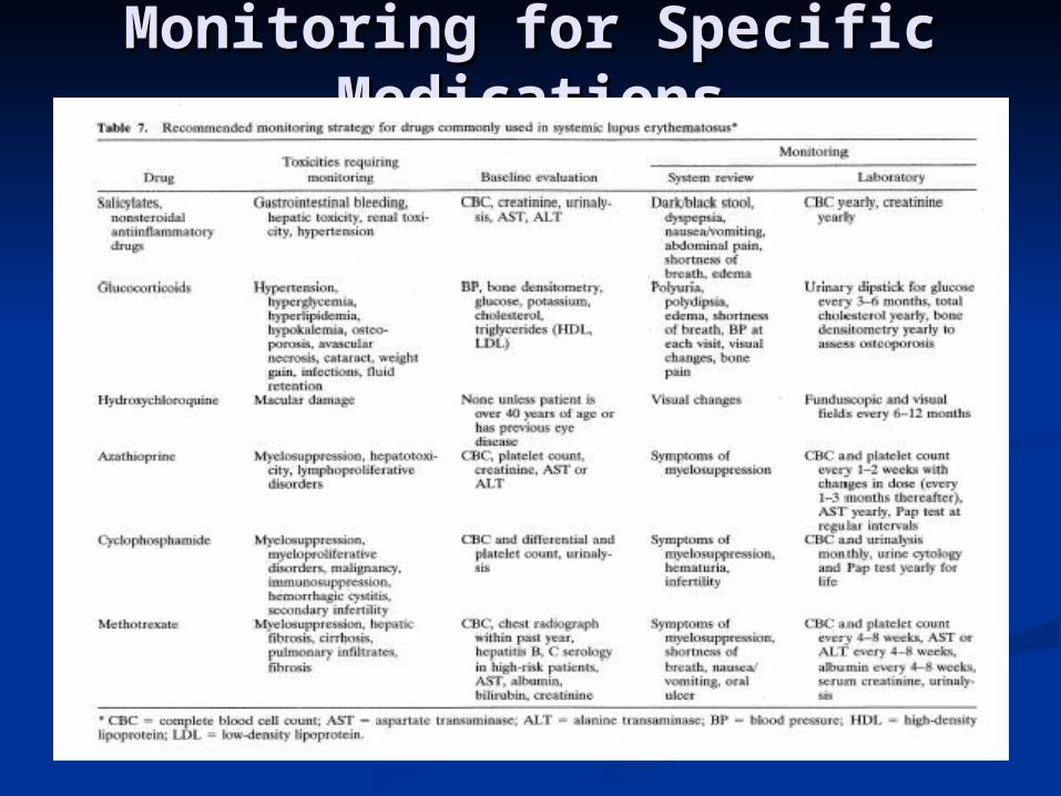
Monitoring for Specific Monitoring for Specific MedicationsMedications
Patients on chronic steroid therapy Patients on chronic steroid therapy must also be on Calcium/Vitamin Dmust also be on Calcium/Vitamin D
Anticipate the need for possible Anticipate the need for possible bisphosphonate therapy, check bisphosphonate therapy, check DEXADEXA
Role of the Primary Care Role of the Primary Care PhysicianPhysician
Patients may have more frequent access Patients may have more frequent access to their primary care physician as to their primary care physician as compared to Rheum or Renal Services compared to Rheum or Renal Services
Understand the importance of disease Understand the importance of disease severity: lupus and it’s treatments are severity: lupus and it’s treatments are highly toxic and clinical status can highly toxic and clinical status can decline rapidlydecline rapidly
Do not hesitate to call the Do not hesitate to call the Rheumatology Consult Service when Rheumatology Consult Service when initiating work-up or to discuss initiating work-up or to discuss continued carecontinued care
Case ReportCase Report
19 yo HF no PMH p/w 3 weeks of 19 yo HF no PMH p/w 3 weeks of intermittent fevers (Tmax 103.4), intermittent fevers (Tmax 103.4), weakness, retrosternal discomfort, weakness, retrosternal discomfort, proximal muscle weakness.proximal muscle weakness.
Denies weight loss, chills, HA, CP, Denies weight loss, chills, HA, CP, SOB, GI/GU sx.SOB, GI/GU sx.
DDX?DDX?
Case PresentationCase Presentation
Initial PE: HR 92, RR 22, O2 sat 99% Initial PE: HR 92, RR 22, O2 sat 99% on RAon RA
Positives: trace alopecia, UE/LE 2/5Positives: trace alopecia, UE/LE 2/5 Initial labs: pancytopenia, SMA-10 Initial labs: pancytopenia, SMA-10
WNL, AST/ALT 560/340, bilis nl, alb WNL, AST/ALT 560/340, bilis nl, alb 2.3, UA: 3+prot and blood, EKG 2.3, UA: 3+prot and blood, EKG tachy, CXR NLtachy, CXR NL
CPK: 3750, LDH 1221, ECHO: small CPK: 3750, LDH 1221, ECHO: small effusioneffusion
Case ReportCase Report
Subsequent Data: ANA 1:1280, Subsequent Data: ANA 1:1280, +dsDNA, C3 28, C4 6 +Sm/RNP+dsDNA, C3 28, C4 6 +Sm/RNP
Patient with clinical picture and lab Patient with clinical picture and lab data consistent with acute data consistent with acute presentation of SLE, with acute presentation of SLE, with acute cytopenia, myositis, serositis, and cytopenia, myositis, serositis, and nephritisnephritis