Embed Size (px)
Citation preview

Case ReportA Rare Presentation of Primary Hyperparathyroidism withConcurrent Aldosterone-Producing Adrenal Carcinoma
Mario Molina-Ayala,1 Claudia Ramírez-Rentería,2 Analleli Manguilar-León,1
Pedro Paúl-Gaytán,1 and Aldo Ferreira-Hermosillo1
1Endocrinology Department, Hospital de Especialidades, Centro Medico Nacional Siglo XXI, IMSS, Cuauhtemoc 330,Colonia Doctores, 06720 Mexico City, DF, Mexico2Experimental Endocrinology Investigation Unit, Hospital de Especialidades, Centro Medico Nacional Siglo XXI, IMSS,Cuauhtemoc 330, Colonia Doctores, 06720 Mexico City, DF, Mexico
Correspondence should be addressed to Aldo Ferreira-Hermosillo; [email protected]
Received 2 March 2015; Revised 5 June 2015; Accepted 9 June 2015
Academic Editor: Carlo Capella
Copyright © 2015 Mario Molina-Ayala et al. This is an open access article distributed under the Creative Commons AttributionLicense, which permits unrestricted use, distribution, and reproduction in any medium, provided the original work is properlycited.
Aldosterone-producing adrenocortical carcinomas are an extremely rare cause of hyperaldosteronism (<1%). Coexistence ofdifferent endocrine tumors warrants additional screening for multiple endocrine neoplasia syndromes, especially in young patientswith large or malignant masses. We present the case of a 40-year-old man with a history of hypertension that presented withan incidental left adrenal tumor during an ultrasound performed for nephrolithiasis. Biochemical assessment showed a mildlyelevated calcium (11.1mg/dL), high parathyroid hormone, and a plasma aldosterone concentration/plasma renin activity ratio of124.5 (normal < 30), compatible with primary hyperparathyroidism with a concomitant primary hyperaldosteronism. A Tc99m-MIBI scintigraphy showed an abnormally increased tracer uptake in the right superior parathyroid and abdominal computedtomography confirmed a left adrenal tumor of 20 cm. The patient underwent parathyroidectomy and adrenalectomy with finalpathology reports of parathyroid hyperplasia and adrenal carcinoma with biochemical remission of both endocrinopathies. Hewas started on chemotherapy, but the patient developed a frontal cortex and an arm metastasis and finally died less than one yearlater.
1. Introduction
Primary hyperparathyroidism due to multiple gland hyper-plasia is the most common presentation of parathyroiddisease in patients with multiple endocrine neoplasia type1 (MEN1), which also includes pituitary tumors (mostlyprolactinomas) and tumors in endocrine pancreas and duo-denum. However, other gastroenteropancreatic neuroen-docrine tumors, adrenocortical adenomas, or thyroid nod-ules can occur in MEN1 [1]. Almost 50% of MEN1 patientscould develop adrenal adenomas or hyperplasia, which aremainly nonfunctioning and benign [2]. Adrenocortical car-cinomas (ACC) are rare manifestations of MEN1 with anincidence of 6% according toWaldmann et al. [2]. In fact, lessthan 1% of ACC produce aldosterone, cortisol, or androgens.Aldosterone-producing adrenocortical carcinomas (APAC)
are infrequent but are highly aggressive and recurrent [3] andcould evolve from initially small and clinically nonfunctionaladenomas.
The purpose of this paper is to report an unusualassociation of primary hyperparathyroidism with an APACthat supports the need for extensive endocrinological assess-ments in patients with adrenal carcinomas and any atypicalpresentation of neuroendocrine disease.
2. Case Report
A 40-year-old man presented with a 4-year history of hyper-tension treated with aldosterone receptor antagonist (ARA2)and in 2008 a left lithotripsy. He had a family history oftype 2 diabetes and cancer (type not specified). He presented
Hindawi Publishing CorporationCase Reports in EndocrinologyVolume 2015, Article ID 910984, 5 pageshttp://dx.doi.org/10.1155/2015/910984
2 Case Reports in Endocrinology
Figure 1: Parathyroid Tc99m-MIB scintigraphy. Images showed an increased uptake of the right superior parathyroid gland on the 150mindelayed image compared with the early 15min image (white arrow).
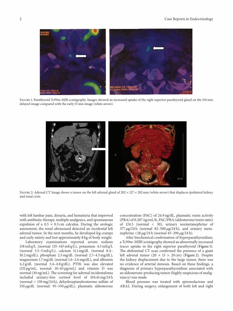
Figure 2: Adrenal CT Image shows a tumor on the left adrenal gland of 202 × 127 × 202mm (white arrow) that displaces ipsilateral kidneyand renal cysts.
with left lumbar pain, dysuria, and hematuria that improvedwith antibiotic therapy, multiple analgesics, and spontaneousexpulsion of a 0.5 × 0.5 cm calculus. During the urologicassessment, the renal ultrasound detected an incidental leftadrenal tumor. In the next months, he developed leg crampsand early satiety and lost approximately 8 kg of body weight.
Laboratory examinations reported serum sodium138mEq/L (normal 135–145mEq/L), potassium 4.5mEq/L(normal 3.5–5mEq/L), calcium 11.1mg/dL (normal 8.4–10.2mg/dL), phosphate 2.5mg/dL (normal 2.7–4.5mg/dL),magnesium 1.7mg/dL (normal 1.6–2.6mg/dL), and albumin4.2 g/dL (normal 3.4–4.8 g/dL). PTHi was also elevated(151 pg/mL, normal: 10–65 pg/mL) and vitamin D wasnormal (30 ng/mL).The screening for adrenal incidentalomaincluded urinary-free cortisol level of 104.61mg/24 h(normal < 130mg/24 h), dehydroepiandrosterone sulfate of150 𝜇g/dL (normal: 95–530𝜇g/dL), plasmatic aldosterone
concentration (PAC) of 24.9 ng/dL, plasmatic renin activity(PRA) of 0.207 ng/mL/h, PAC/PRA (aldosterone/renin ratio)of 124.5 (normal < 30), urinary normetanephrine of377 𝜇g/24 h (normal 82–500 𝜇g/24 h), and urinary meta-nephrine <28𝜇g/24 h (normal 45–290 𝜇g/24 h).
After biochemical confirmation of hyperparathyroidism,a Tc99m-MIBI scintigraphy showed an abnormally increasedtracer uptake in the right superior parathyroid (Figure 1).The abdominal CT scan confirmed the presence of a giantleft adrenal tumor (20 × 13 × 20 cm) (Figure 2). Despitethe kidney displacement due to the large tumor, there wasno evidence of arterial stenosis. Based on these findings, adiagnosis of primary hyperparathyroidism associated withan aldosterone-producing tumor (highly suspicious of malig-nancy) was made.
Blood pressure was treated with spironolactone andARA2. During surgery, enlargement of both left and right
Case Reports in Endocrinology 3
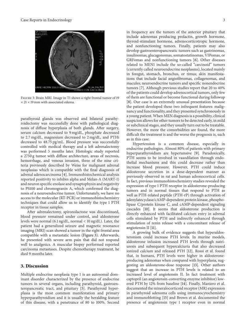
Figure 3: Brain MRI. Image in T1 shows a right frontal tumor of 19× 21 × 19mm with associated edema.
parathyroid glands was observed and bilateral parathy-roidectomy was successfully done with pathological diag-nosis of diffuse hyperplasia of both glands. After surgery,serum calcium decreased to 9mg/dL, phosphate decreasedto 2.7mg/dL, magnesium decreased to 2mg/dL, and PTHidecreased to 48.75 pg/mL. Blood pressure was successfullycontrolled with medical therapy and a left adrenalectomywas performed 5 months later. Histologic study reporteda 2750 g tumor with diffuse architecture, areas of necrosis,hemorrhage, and venous invasion, three of the nine cri-teria previously described by Weiss for malignant adrenalneoplasias which is compatible with the final diagnosis ofadrenal adenocarcinoma [4]. Immunohistochemical analysisreported positivity to inhibin alpha and Melan-A (MART-1)andneuron specific enolase and synaptophysin andnegativityto PS100 and chromogranin A, which confirmed the diag-nosis of a neuroendocrine tumor. Unfortunately, we have noaccess to the molecular (RT-PCR) or immunohistochemistrytechniques that could allow us to identify the type 1 PTHreceptor in tissue samples.
After adrenalectomy, spironolactone was discontinued,blood pressure remained under control, and aldosteronelevels were normal (6.1 ng/dL, normal < 10 ng/dL). Later, thepatient had a generalized seizure and magnetic resonanceimaging (MRI) scan showed a tumor in the right frontal areacompatible with a metastatic lesion (Figure 3). Afterwards,he presented with severe arm pain that did not respondwell to analgesics. A muscular biopsy performed reportedcarcinoma metastases. Despite chemotherapy treatment, hedied 9 months later.
3. Discussion
Multiple endocrine neoplasia type 1 is an autosomal dom-inant disorder characterized by the presence of endocrinetumors in several organs, including parathyroid, gastroen-teropancreatic tract, and pituitary [5]. Parathyroid hyper-plasia is the most common manifestation and primaryhyperparathyroidism and it is usually the heralding featureof this disease, with a penetrance of 80 to 100%. Second
in frequency are the tumors of the anterior pituitary thatinclude adenomas producing prolactin, growth hormone,thyroid-stimulant hormone, adrenocorticotropic hormone,and nonfunctioning tumors. Finally, patients may alsodevelop gastroenteropancreatic tumors such as gastrinomas,insulinomas, glucagonomas, somatostatinomas, VIPomas, orGRFomas and nonfunctioning tumors [6]. Other diseasesrelated to MEN1 include the so-called “carcinoid” tumors(currently called neuroendocrine neoplasms), locatedmainlyin foregut, stomach, bronchus, or timus; skin manifesta-tions that include facial angiofibromas, collagenomas, andmacules; neuroendocrine tumors and specific nonendocrinetumors [7]. Although previous studies report that 20 to 40%of the patients could develop adrenocortical tumors, only fewof them are functional or become functional during followup[8]. Our case is an extremely unusual presentation becausethe patient developed these two infrequent features: malig-nancy and functionality, and they presented synchronously ina young patient.WhenMENdiagnosis is a possibility, clinicalsuspicion allows for other tumors to be detected early, inmildor subclinical stages, and they usually turn out to be treatable.However, the more the comorbidities are found, the moredifficult the treatment is and the worse the prognosis is, suchas in this case.
Hypertension is a common disease, especially inendocrine pathologies. Almost 80% of patients with primaryhyperparathyroidism are hypertensive [9]. Paradoxically,PTH seems to be involved in vasodilation through endo-thelial mechanisms and this could decrease rather thanincrease blood pressure. However, PTH could inducealdosterone secretion in a dose-dependent manner aspreviously observed in rat and human adrenocortical cells.In fact, previous immunohistochemistry studies revealed theexpression of type 1 PTH receptor in aldosterone-producingtumors and in normal tissues that respond to PTH aswell as PTH-related peptide (PTH-rP) through activation ofadenylatecyclase/cAMP-dependent protein kinase, phospho-lipase C/protein kinase C, and cAMP-dependent signalingcascades [10]. It seems that aldosterone production isdirectly enhanced with facilitated calcium entry in adrenalcells stimulated by PTH and indirectly enhanced throughstimulation of renin release with a concomitant release ofangiotensin II [11].
A growing bulk of evidence suggests that hyperaldos-teronism could increase PTH levels. In murine models,aldosterone infusion increased PTH levels through natri-uresis and subsequent hypercalciuria that also decreasedionized calcium and released PTH [12]. Rossi et al. foundthat, in humans, PTH levels were higher in aldosterone-producing adenomas when compared with hyperplasia, sug-gesting an aldosterone-dose response [13]. Other authorssuggest that an increase in PTH levels is related to anincreased level of angiotensin II. In fact treatment withcaptopril (an angiotensin-converting enzyme inhibitor) low-ered PTH by 12% from baseline [14]. Finally, Maniero et al.documented themineralocorticoid receptor (MR) expressionin parathyroid adenoma cells using immunocytochemistryand immunoblotting [15] and Brown et al. documented thepresence of angiotensin type 1 receptor even in normal

4 Case Reports in Endocrinology
parathyroid tissue [14]. These findings suggest that aldos-terone and PTH have a reciprocal but not completely under-stood interaction.
In our institution,we lack a standardizedmethod to assessvitamin D level, which limits the interpretation of results(normal level observed in our case). This must be taken intoaccount because low vitamin D levels are associated withhigher plasma renin activity and angiotensin II concentra-tions that could also increase aldosterone andPTH levels [16].This relationship has been explored as a possible therapeutictarget with conflicting results [17].
Honda et al. reported the case of a 44-year-old womanwith confirmed primary hyperparathyroidism, aldosterone-producing adenoma, and breast cancer [18]. They detected amutation at codon 541 in exon 10 with a loss of heterozygosity(LOH) in each contralateral allele in parathyroid adenomaand breast cancer tissue but not in adrenocortical adenomatissue. However, other reports found LOH of the 11q13 inaldosterone-producing adrenocortical adenomas, suggestingthat patients with primary hyperaldosteronism should bescreened for other components of MEN [19]. Unfortunately,in this case, we could not perform an immunoblottinganalysis on isolated tissues in search for a possible mutationin MEN gene. Despite the report of concomitant hyperal-dosteronism and PHPT, adrenal carcinoma has not beenassociated with this presentation. In general terms, adrenalcarcinomas are rare and aggressive malignancies with anannual incidence of 0.7 to 2 cases per 1million population peryear [20].They are more common in women with a female tomale ratio of 2 : 1 and have a bimodal age distribution witha peak in childhood and a second peak in the fourth andfifth decades [21]. Functional carcinomas are rare and lessthan 1% of the cases are associated with Conn’s syndrome[22]. In fact, Seccia et al. in 2005 reported that since 1955there were only 60 cases of APAC [3]. They observed thatpatients with APAC had a peak of incidence of 40 to 49years of age, were predominantly women, and had a tumor onthe right adrenal. Additionally, they described recurrence in48% of the patients and metastases that involved liver, lungs,abdomen, abdominal lymph nodes, and ipsilateral adrenalsite. This case is also particularly atypical because it involvesa male with a right adrenal tumor with metastases to the armand brain.
Surgery is still the first-line treatment [21] whenever itis possible. It is indicated even in patients with advanceddisease. Additionally, the most common systemic drugsused include mitotane, cisplatin, and etoposide alone orin combination with other agents. Adjuvant treatment asradiation therapy could be indicated to treat symptomaticmetastatic lesions as well as chemoembolization or radiofre-quency ablation [21]. In this case, chemotherapy treatmentwas initiated, but patient was in such an advanced stage ofthe disease that it was finally unsuccessful.
Conflict of Interests
The authors declare that there is no conflict of interestsregarding the publication of this paper.
References
[1] R. V. Thakker, “Multiple endocrine neoplasia type 1 (MEN1),”Best Practice and Research: Clinical Endocrinology and Meta-bolism, vol. 24, no. 3, pp. 355–370, 2010.
[2] J. Waldmann, D. K. Bartsch, P. H. Kann, V. Fendrich, M.Rothmund, and P. Langer, “Adrenal involvement in multipleendocrine neoplasia type 1: results of 7 years prospectivescreening,” Langenbeck’s Archives of Surgery, vol. 392, no. 4, pp.437–443, 2007.
[3] T. M. Seccia, A. Fassina, G. G. Nussdorfer, A. C. Pessina, andG. P. Rossi, “Aldosterone-producing adrenocortical carcinoma:an unusual cause of Conn’s syndrome with an ominous clinicalcourse,” Endocrine-Related Cancer, vol. 12, no. 1, pp. 149–159,2005.
[4] L. M. Weiss, “Comparative histologic study of 43 metastasizingand nonmetastasizing adrenocortical tumors,” The AmericanJournal of Surgical Pathology, vol. 8, no. 3, pp. 163–169, 1984.
[5] S. K. Agarwal, “Multiple endocrine neoplasia type 1,” Frontiersof Hormone Research, vol. 41, pp. 1–15, 2013.
[6] W. S. Rubinstein, “Endocrine cancer predisposition syndromes:hereditary paraganglioma, multiple endocrine neoplasia type1, multiple endocrine neoplasia type 2, and hereditary thyroidcancer,”Hematology/Oncology Clinics of North America, vol. 24,no. 5, pp. 907–937, 2010.
[7] R. V.Thakker, “Multiple endocrine neoplasia type 1 (MEN1) andtype 4 (MEN4),”Molecular and Cellular Endocrinology, vol. 386,no. 1-2, pp. 2–15, 2014.
[8] P. Langer, K. Cupisti, D. K. Bartsch et al., “Adrenal involvementin multiple endocrine neoplasia type 1,” World Journal ofSurgery, vol. 26, no. 8, pp. 891–896, 2002.
[9] G.M. Hedback and A. S. Oden, “Cardiovascular disease, hyper-tension and renal function in primary hyperparathyroidism,”Journal of Internal Medicine, vol. 251, no. 6, pp. 476–483, 2002.
[10] J. Rosenberg, M. Pines, and S. Hurwitz, “Response ofadrenal cells to parathyroid hormone stimulation,” Journal ofEndocrinology, vol. 112, no. 3, pp. 431–437, 1987.
[11] A. Tomaschitz, E. Ritz, B. Pieske et al., “Aldosterone andparathyroid hormone: a precarious couple for cardiovasculardisease,” Cardiovascular Research, vol. 94, no. 1, pp. 10–19, 2012.
[12] V. S. Chhokar, Y. Sun, S. K. Bhattacharya et al., “Hyperparathy-roidism and the calciumparadox of aldosteronism,”Circulation,vol. 111, no. 7, pp. 871–878, 2005.
[13] G. P. Rossi, F. Ragazzo, T.M. Seccia et al., “Hyperparathyroidismcan be useful in the identification of primary aldosteronism dueto aldosterone-producing adenoma,” Hypertension, vol. 60, no.2, pp. 431–436, 2012.
[14] J. M. Brown, J. S. Williams, J. M. Luther et al., “Human inter-ventions to characterize novel relationships between the renin-angiotensin-aldosterone system and parathyroid hormone,”Hypertension, vol. 63, no. 2, pp. 273–280, 2014.
[15] C. Maniero, A. Fassina, V. Guzzardo et al., “Primary hyper-parathyroidism with concurrent primary aldosteronism,”Hypertension, vol. 58, no. 3, pp. 341–346, 2011.
[16] J. P. Forman, J. S. Williams, and N. D. L. Fisher, “Plasma25-hydroxyvitamin D and regulation of the renin-angiotensinsystem in humans,” Hypertension, vol. 55, no. 5, pp. 1283–1288,2010.
[17] A. Vaidya, J. M. Brown, and J. S. Williams, “The renin–angiotensin–aldosterone system and calcium-regulatory hor-mones,” Journal of Human Hypertension, 2015.

Case Reports in Endocrinology 5
[18] M. Honda, T. Tsukada, T. Horiuchi et al., “Primary hyper-parathyroidism associatiated with aldosterone-producingadrenocortical adenoma and breast cancer: relation to MEN1gene,” Internal Medicine, vol. 43, no. 4, pp. 310–314, 2004.
[19] Y. L. Kim, Y. W. Jang, J. T. Kim et al., “A rare case of primaryhyperparathyroidism associated with primary aldosteronism,hurthle cell thyroid cancer andmeningioma,” Journal of KoreanMedical Science, vol. 27, no. 5, pp. 560–564, 2012.
[20] S. H. Golden, K. A. Robinson, I. Saldanha, B. Anton, andP. W. Ladenson, “Clinical review: prevalence and incidenceof endocrine and metabolic disorders in the united states: acomprehensive review,”The Journal of Clinical Endocrinology &Metabolism, vol. 94, no. 6, pp. 1853–1878, 2009.
[21] A. P. Fay, A. Elfiky, G. H. Telo et al., “Adrenocortical carcinoma:the management of metastatic disease,” Critical Reviews inOncology/Hematology, vol. 92, no. 2, pp. 123–132, 2014.
[22] A. Ganguly, “Primary aldosteronism,”TheNew England Journalof Medicine, vol. 339, no. 25, pp. 1828–1834, 1998.

Submit your manuscripts athttp://www.hindawi.com
Stem CellsInternational
Hindawi Publishing Corporationhttp://www.hindawi.com Volume 2014
Hindawi Publishing Corporationhttp://www.hindawi.com Volume 2014
MEDIATORSINFLAMMATION
of
Hindawi Publishing Corporationhttp://www.hindawi.com Volume 2014
Behavioural Neurology
EndocrinologyInternational Journal of
Hindawi Publishing Corporationhttp://www.hindawi.com Volume 2014
Hindawi Publishing Corporationhttp://www.hindawi.com Volume 2014
Disease Markers
Hindawi Publishing Corporationhttp://www.hindawi.com Volume 2014
BioMed Research International
OncologyJournal of
Hindawi Publishing Corporationhttp://www.hindawi.com Volume 2014
Hindawi Publishing Corporationhttp://www.hindawi.com Volume 2014
Oxidative Medicine and Cellular Longevity
Hindawi Publishing Corporationhttp://www.hindawi.com Volume 2014
PPAR Research
The Scientific World JournalHindawi Publishing Corporation http://www.hindawi.com Volume 2014
Immunology ResearchHindawi Publishing Corporationhttp://www.hindawi.com Volume 2014
Journal of
ObesityJournal of
Hindawi Publishing Corporationhttp://www.hindawi.com Volume 2014
Hindawi Publishing Corporationhttp://www.hindawi.com Volume 2014
Computational and Mathematical Methods in Medicine
OphthalmologyJournal of
Hindawi Publishing Corporationhttp://www.hindawi.com Volume 2014
Diabetes ResearchJournal of
Hindawi Publishing Corporationhttp://www.hindawi.com Volume 2014
Hindawi Publishing Corporationhttp://www.hindawi.com Volume 2014
Research and TreatmentAIDS
Hindawi Publishing Corporationhttp://www.hindawi.com Volume 2014
Gastroenterology Research and Practice
Hindawi Publishing Corporationhttp://www.hindawi.com Volume 2014
Parkinson’s Disease
Evidence-Based Complementary and Alternative Medicine
Volume 2014Hindawi Publishing Corporationhttp://www.hindawi.com