Embed Size (px)
Citation preview
CASE REPORT Open Access
Case report and literature review: patientwith gastroduodenal intussusception dueto the gastrointestinal stromal tumor of thelesser curvature of the gastric bodyMihajlo Đokić1, Jerica Novak2* , Miha Petrič1, Branislava Ranković3, Miha Štabuc4 and Blaž Trotovšek1
Abstract
Background: Intussusception in adult patient is rare. Gastroduodenal intussusception due to the gastrointestinalstromal tumors is infrequently described in the literature. Authors present a case of gastroduodenal intussusceptiondue to the low-risk gastrointestinal stromal tumor of the lesser curvature of the gastric body with literature review.
Case presentation: Sixty-two-year-old male was admitted to our hospital with symptoms of acute gastric outletobstruction. Imaging studies confirmed a lesion of the gastric wall producing gastroduodenal intussusception withpylorus obstruction. Upon laparotomy a tumor mass of the lesser curvature of the gastric body that invaginatedthrough the pylorus into the duodenum was found. Desinvagination and resection of the tumor with the adequateresection margins were performed. Histology reveled a low-risk gastrointestinal stromal tumor. Due to favorableoutcome only observation was suggested by the multidisciplinary team.
Conclusions: Gastroduodenal intussusception due to the gastrointestinal stromal tumor of the gastric wall is a rareevent. Surgical resection is the treatment of choice. In selected cases laparosopic resection of the tumor can beperformed.
Keywords: Gastroduodenal intussusception, Gastric gastrointestinal stromal tumor, Gastric outlet obstruction
BackgroundIntussusception rarely occurs among the adult patients.It accounts for 5% of all intussusception cases and inonly 1% causes intestinal obstruction [1]. Gastroduode-nal intussusception is the most infrequent form of intus-susception in adults, it occurs in less than 10% [2].Clinical and radiological findings in a patient with gas-tric outlet obstruction, gastroduodenal intussusceptionand gastrointestinal stromal tumor (GIST) of the lessercurvature of the gastric body is presented.
Case presentation62-year-old Caucasian male presented to the emergencyroom with acutely worsening epigastric pain lasting for
several days and black stool lasting for a week. Symp-toms of vomiting, inappetence and weight loss that havebeen lasting for the past six months without doctor ap-pointment was also reported in medical history. Patienthad a history of diabetes mellitus on insulin therapy.Upon clinical examination abdomen was not distended,there was no signs of guarding or rebound tenderness.Laboratory data showed anemia (hemoglobin 119 g/L,normal range 130–170 g/L; hematocrit 0.343, normalrange 0.4–0.5), leukocytosis (13.5 109/L, normal range4.0–10.0) and normal value of C-reactive protein (below5mg/L, normal range 0–5 mg/L). Tumor markers CEAand Ca 19–9 were within normal range.Due to melena lasting for a week, patient underwent
esophagogastroduodenoscopy (EGD) and ultrasound ofthe abdomen on the outpatient bases only few days priorto admission to the hospital. EGD was technically de-manding due to the poor passage of the endoscope
© The Author(s). 2019 Open Access This article is distributed under the terms of the Creative Commons Attribution 4.0International License (http://creativecommons.org/licenses/by/4.0/), which permits unrestricted use, distribution, andreproduction in any medium, provided you give appropriate credit to the original author(s) and the source, provide a link tothe Creative Commons license, and indicate if changes were made. The Creative Commons Public Domain Dedication waiver(http://creativecommons.org/publicdomain/zero/1.0/) applies to the data made available in this article, unless otherwise stated.
* Correspondence: [email protected] of Surgical Oncology, Ljubljana Institute of OncologyActa ChirBelg, Zaloška cesta 2, 1000 Ljubljana, SloveniaFull list of author information is available at the end of the article
Đokić et al. BMC Surgery (2019) 19:158 https://doi.org/10.1186/s12893-019-0608-3
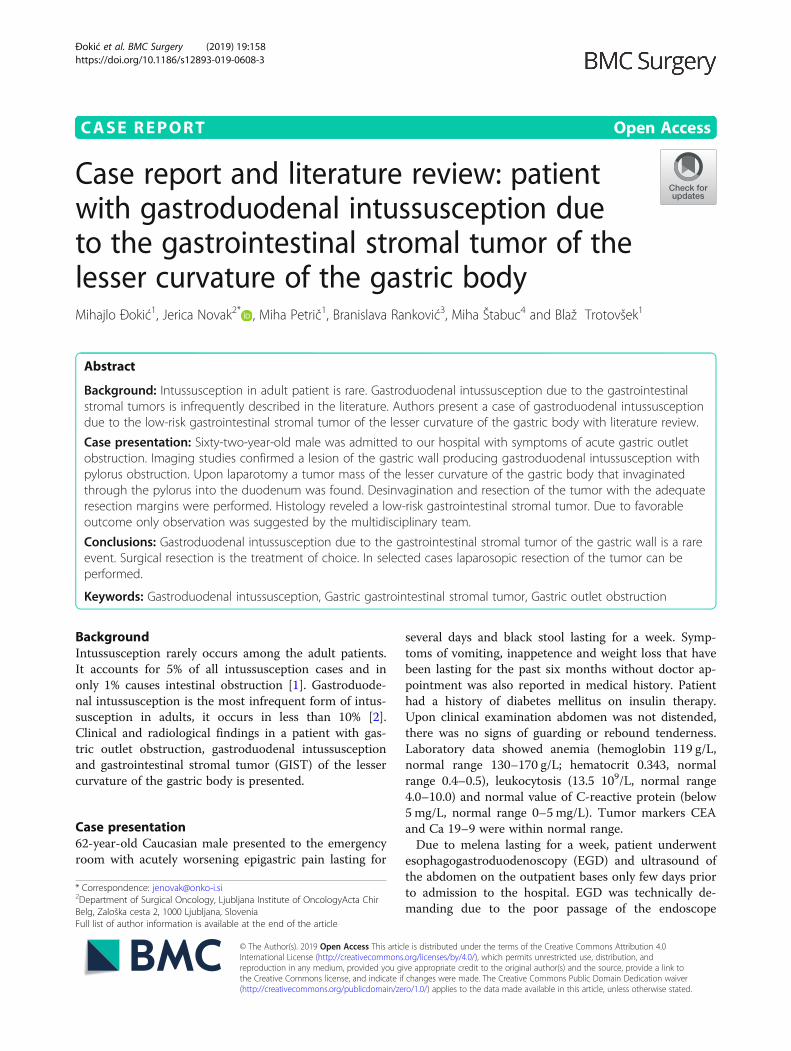
through the stomach, duodenal bulbous and upper partof the duodenum. Inflation of the gastric body was notpossible, therefore the visualization of the gastric wallwas poor with no obvious intraluminal mass orhemorrhage detected. Additionally gastric peristalsis wasdescribed to be absent. Abdominal ultrasound showedtumor formation of the gastric body, measuring 7 × 5cm, but no intussusception was described. CT scan re-vealed a 5.4 × 5.6 × 6.2 cm intraluminal tumor formationof the lesser curvature of the gastric body with well de-fined borders was described. Tumor mass caused inva-gination of the gastric cardia through the antrum andpylorus into the D2 part of the duodenum producinggastric outlet obstruction (Figs. 1, 2). No disseminationto the parenchymal organs was described.Explorative laparotomy was performed. Palpable gas-
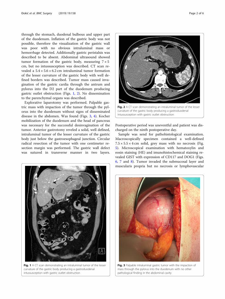
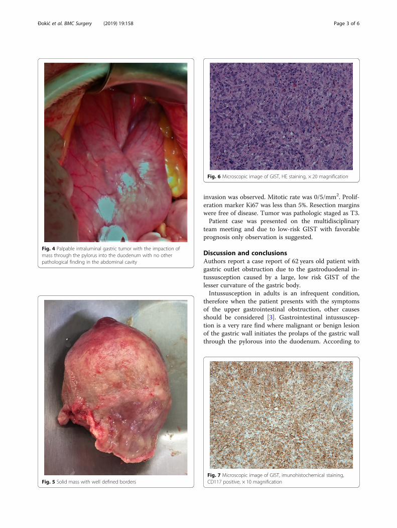
tric mass with impaction of the tumor through the pyl-orus into the duodenum without signs of disseminateddisease in the abdomen. Was found (Figs. 3, 4). Kochermobilization of the duodenum and the head of pancreaswas necessary for the successful desinvagination of thetumor. Anterior gastrotomy reveled a solid, well defined,intraluminal tumor of the lesser curvature of the gastricbody just below the gastroesophageal junction. Circularradical resection of the tumor with one centimeter re-section margin was performed. The gastric wall defectwas sutured in transverse manner in two layers.
Postoperative period was uneventful and patient was dis-charged on the ninth postoperative day.Sample was send for pathohistological examination.
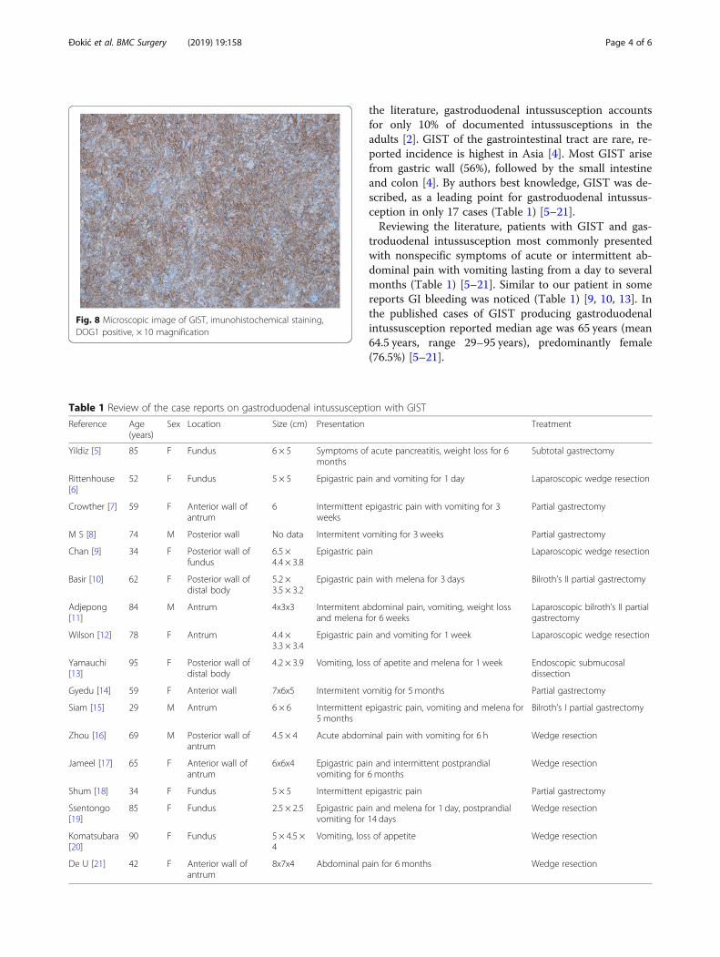
Macroscopically specimen contained a well-defined7.5 × 5.5 × 4 cm solid, grey mass with no necrosis (Fig.5). Microscopical examination with hematoxylin andeosin staining (HE) and imunohistochemical staining re-vealed GIST with expression of CD117 and DOG1 (Figs.6, 7 and 8). Tumor invaded the submucosal layer andmuscularis propria but no necrosis or lymphovascular
Fig. 1 A CT scan demonstrating an intraluminal tumor of the lessercurvature of the gastric body producing a gastroduodenalintussusception with gastric outlet obstruction
Fig. 2 A CT scan demonstrating an intraluminal tumor of the lessercurvature of the gastric body producing a gastroduodenalintussusception with gastric outlet obstruction
Fig. 3 Palpable intraluminal gastric tumor with the impaction ofmass through the pylorus into the duodenum with no otherpathological finding in the abdominal cavity
Đokić et al. BMC Surgery (2019) 19:158 Page 2 of 6
invasion was observed. Mitotic rate was 0/5/mm2. Prolif-eration marker Ki67 was less than 5%. Resection marginswere free of disease. Tumor was pathologic staged as T3.Patient case was presented on the multidisciplinary
team meeting and due to low-risk GIST with favorableprognosis only observation is suggested.
Discussion and conclusionsAuthors report a case report of 62 years old patient withgastric outlet obstruction due to the gastroduodenal in-tussusception caused by a large, low risk GIST of thelesser curvature of the gastric body.Intussusception in adults is an infrequent condition,
therefore when the patient presents with the symptomsof the upper gastrointestinal obstruction, other causesshould be considered [3]. Gastrointestinal intussuscep-tion is a very rare find where malignant or benign lesionof the gastric wall initiates the prolaps of the gastric wallthrough the pylorous into the duodenum. According to
Fig. 4 Palpable intraluminal gastric tumor with the impaction ofmass through the pylorus into the duodenum with no otherpathological finding in the abdominal cavity
Fig. 5 Solid mass with well defined borders
Fig. 6 Microscopic image of GIST, HE staining, × 20 magnification
Fig. 7 Microscopic image of GIST, imunohistochemical staining,CD117 positive, × 10 magnification
Đokić et al. BMC Surgery (2019) 19:158 Page 3 of 6
the literature, gastroduodenal intussusception accountsfor only 10% of documented intussusceptions in theadults [2]. GIST of the gastrointestinal tract are rare, re-ported incidence is highest in Asia [4]. Most GIST arisefrom gastric wall (56%), followed by the small intestineand colon [4]. By authors best knowledge, GIST was de-scribed, as a leading point for gastroduodenal intussus-ception in only 17 cases (Table 1) [5–21].Reviewing the literature, patients with GIST and gas-
troduodenal intussusception most commonly presentedwith nonspecific symptoms of acute or intermittent ab-dominal pain with vomiting lasting from a day to severalmonths (Table 1) [5–21]. Similar to our patient in somereports GI bleeding was noticed (Table 1) [9, 10, 13]. Inthe published cases of GIST producing gastroduodenalintussusception reported median age was 65 years (mean64.5 years, range 29–95 years), predominantly female(76.5%) [5–21].
Fig. 8 Microscopic image of GIST, imunohistochemical staining,DOG1 positive, × 10 magnification
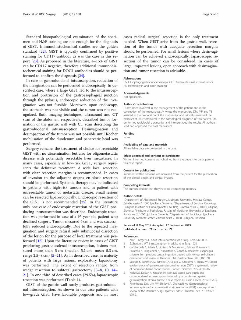
Table 1 Review of the case reports on gastroduodenal intussusception with GIST
Reference Age(years)
Sex Location Size (cm) Presentation Treatment
Yildiz [5] 85 F Fundus 6 × 5 Symptoms of acute pancreatitis, weight loss for 6months
Subtotal gastrectomy
Rittenhouse[6]
52 F Fundus 5 × 5 Epigastric pain and vomiting for 1 day Laparoscopic wedge resection
Crowther [7] 59 F Anterior wall ofantrum
6 Intermittent epigastric pain with vomiting for 3weeks
Partial gastrectomy
M S [8] 74 M Posterior wall No data Intermitent vomiting for 3 weeks Partial gastrectomy
Chan [9] 34 F Posterior wall offundus
6.5 ×4.4 × 3.8
Epigastric pain Laparoscopic wedge resection
Basir [10] 62 F Posterior wall ofdistal body
5.2 ×3.5 × 3.2
Epigastric pain with melena for 3 days Bilroth’s II partial gastrectomy
Adjepong[11]
84 M Antrum 4x3x3 Intermitent abdominal pain, vomiting, weight lossand melena for 6 weeks
Laparoscopic bilroth’s II partialgastrectomy
Wilson [12] 78 F Antrum 4.4 ×3.3 × 3.4
Epigastric pain and vomiting for 1 week Laparoscopic wedge resection
Yamauchi[13]
95 F Posterior wall ofdistal body
4.2 × 3.9 Vomiting, loss of apetite and melena for 1 week Endoscopic submucosaldissection
Gyedu [14] 59 F Anterior wall 7x6x5 Intermitent vomitig for 5 months Partial gastrectomy
Siam [15] 29 M Antrum 6 × 6 Intermittent epigastric pain, vomiting and melena for5 months
Bilroth’s I partial gastrectomy
Zhou [16] 69 M Posterior wall ofantrum
4.5 × 4 Acute abdominal pain with vomiting for 6 h Wedge resection
Jameel [17] 65 F Anterior wall ofantrum
6x6x4 Epigastric pain and intermittent postprandialvomiting for 6 months
Wedge resection
Shum [18] 34 F Fundus 5 × 5 Intermittent epigastric pain Partial gastrectomy
Ssentongo[19]
85 F Fundus 2.5 × 2.5 Epigastric pain and melena for 1 day, postprandialvomiting for 14 days
Wedge resection
Komatsubara[20]
90 F Fundus 5 × 4.5 ×4
Vomiting, loss of appetite Wedge resection
De U [21] 42 F Anterior wall ofantrum
8x7x4 Abdominal pain for 6 months Wedge resection
Đokić et al. BMC Surgery (2019) 19:158 Page 4 of 6
Standard histopathological examination of the speci-men and H&E staining are not enough for the diagnosisof GIST. Immunohistochemical studies are the goldenstandard [22]. GIST is typically confirmed by positivestaining for CD117 antibody as was the case in this re-port [23]. As proposed in the literature, 4–15% of GISTcan be CD117 negative, therefore additional immunohis-tochemical staining for DOG1 antibodies should be per-formed to confirm the diagnosis [24].In case of gastroduodenal intususception, reduction of
the invagination can be performed endoscopically. In de-scribed case, where a large GIST led to the intussuscep-tion and protrusion of the gastroesophageal junctionthrough the pylorus, endoscopic reduction of the inva-gination was not feasible. Moreover, upon endoscopy,the stomach was not visible and the tumor was not rec-ognized. Both imaging techniques, ultrasound and CTscan of the abdomen, respectively, described tumor for-mation of the gastric wall with CT scan describing thegastroduodenal intusussception. Desinvagination anddesimpaction of the tumor was not possible until Kochermobilisation of the duodenum and pancreatic head wasperformed.Surgery remains the treatment of choice for resectable
GIST with no dissemination but also for oligometastaticdisease with potentially resectable liver metastases. Inmany cases, especially in low-risk GIST, surgery repre-sents the definitive treatment. A wide local resectionwith clear resection margins is recommended. In casesof invasion to the adjacent organs en-block resectionshould be performed. Systemic therapy may be indicatedin patients with high-risk tumors and in patient withunresectable tumor or metastatic disease. Small lesionscan be resected laparoscopically. Endoscopic resection ofthe GIST is not recommended [25]. In the literatureonly one case of endoscopic resection of the GIST pro-ducing intussusception was described. Endoscopic resec-tion was performed in case of a 95-year-old patient whodeclined surgery. Tumor mesured 4 cm and was success-fully reduced endoscopically. Due to the repeated inva-gination and surgery refusal only submucosal dissectionof the lesion for the purpose of local treatment was per-formed [13]. Upon the literature review in cases of GISTproducing gastroduodenal intussusception, lesions mea-sured more than 5 cm (median 5.1 cm, mean 5.3 cm,range 2.5–8 cm) [5–21]. As in described case, in majorityof patients with large lesions, exploratory laparotomywas performed. The extent of resection ranged fromwedge resection to subtotal gastrectomy [5–8, 10, 14–21]. In one third of described cases (29.5%), laparoscopicresection was performed (Table 1).GIST of the gastric wall rarely produces gastroduode-
nal intussusception. As shown in our case patients withlow-grade GIST have favorable prognosis and in most
cases radical surgical resection is the only treatmentneeded. When GIST arise from the gastric wall, resec-tion of the tumor with adequate resection marginsshould be performed. For small lesions where desinvagi-nation can be achieved endoscopically, laparoscopic re-section of the tumor can be considered. In cases oflarge, impacted lesions, open approach with desinvagina-tion and tumor resection is advisable.
AbbrevationsEGD: Esophagogastroduodenoscopy; GIST: Gastrointestinal stromal tumor;HE: Hematoxylin and eosin staining
AcknowledgementsNot applicable.
Authors’ contributionsTB has been involved in the management of the patient and in theconception of the manuscript. JN wrote the manuscript. DM, MP and TBassisted in the preparation of the manuscript and critically reviewed themanuscript. RB contributed to the pathological diagnosis of this patient. SMperformed radiologial diagnostics and interpretaded the results. All authorsread and approved the final manuscript.
FundingNone.
Availability of data and materialsAll available data are presented in the case.
Ethics approval and consent to participateWritten informed consent was obtained from the patient to participate tothis case report.
Consent for publicationInformed written consent was obtained from the patient for the publicationof the clinical details and clinical images.
Competing interestsThe authors declare that they have no competing interests.
Author details1Department of Abdominal Surgery, Ljubljana University Medical Center,Zaloška cesta 7, 1000 Ljubljana, Slovenia. 2Department of Surgical Oncology,Ljubljana Institute of OncologyActa Chir Belg, Zaloška cesta 2, 1000 Ljubljana,Slovenia. 3Institute of Pathology, Faculty of Medicine, University of Ljubljana,Korytkova 2, 1000 Ljubljana, Slovenia. 4Department of Radiology, LjubljanaUniversity Medical Center, Zaloška cesta 7, 1000 Ljubljana, Slovenia.
Received: 8 May 2019 Accepted: 17 September 2019
References1. Azar T, Berger DL. Adult intussusception. Ann Surg. 1997;226:134–8.2. Stubenbord WT. Intussusception in adults. Ann Surg. 1970.3. Gambardella C, Allaria A, Siciliano G, Mauriello C, Patrone R, Avenia N,
Polistena A, Sanguinetti A, Napolitano S, Conzo G. Recurrent esophagealstricture from previous caustic ingestion treated with 40-year self-dilation:case report and review of literature. BMC Gastroenterol. 2018;18(1):68.
4. Søreide K, Sandvik OM, Søreide JA, Giljaca V, Jureckova A, Bulusu VR. Globalepidemiology of gastrointestinalstromal tumours (GIST): a systematic reviewof population-based cohort studies. Cancer Epidemiol. 2016;40:39–46.
5. Yildiz MS, Doğan A, Koparan IH, Adin ME. Acute pancreatitis andgastroduodenal intussusception induced by an underlying gastricgastrointestinal stromal tumor: a case report. K Gastric Cancer. 2016;16:54–7.
6. Rittenhouse DW, Lim PW, Shirley LA, Chojnacki KA. Gastroduodenalintussusception of a gastrointestinal stromal tumor (GIST): case report andreview of the literature. Surg Laparosc Endosc Percutan Tech. 2013;23(2):e70–3.
Đokić et al. BMC Surgery (2019) 19:158 Page 5 of 6
7. Crowther KS, Wyld L, Yamani Q, JAcob G. Case report: gastroduodenalintussusception of a gastrointestinal stromal tumour. Br J Radiol. 2002;75:987–9.
8. M S PB, Reddy CK, Augustine AJ, Sagari SG. Gastroduodenal intussusceptiondue to pedunculated polypoid gastrointestinal stromal tumour (GIST): a rarecase. J Clin Diagn Res 2015; 9 (1): PD05–06.
9. Chan CT, Wong SK, Ping Tai Y, Li MK. Endo-laparoscopic reduction andresection of gastroduodenal intussusception of gastrointestinal stromaltumor (GIST): a synchronous endoscopic and laparoscopic treatment. SurgLaparosc Endosc Percutan Tech. 2009;19(3):e100–3.
10. Basir N, Yaakub AB, Kafeel G, Telisinghe PU, Tan KK, Sharif F, Chong VH.Gastroduodenal intussusception as a first manifestation of gastricgastrointestinal stromal tumor. Turk J Gastroenterol. 2012;23(2):185–6.
11. Adjepong SE, Parameswaran R, Perry A, Mathews R, Jones R, Butterworth JR,Sigurdsson A. Gastroduodenal intussusception due to gastrointestinalstromal tumor (GIST) treated by laparoscopic billroth II distal gastrectomy.Surg Laparosc Endosc Percutan Tech. 2006;16(4):245–7.
12. Wilson MH, Ayoub F, McGreal P, Collins C. Gastrointestinal stromal tumourpresenting as gastroduodenal intussusception. BMJ Case Rep 2012.bcr2012006787.
13. Yamauchi K, Iwamuro M, Ishii E, Narita M, Hirata N, Okada H.Gastroduodenal intussusception with a gastric gastrointestinal stromaltumor treated by endoscopic submucocal dissection. Intern Med. 2017;56(12):1515–9.
14. Gyedu A, Reich SB, Hoyte-Williams PE. Gastrointestinal stromal tumourpresenting acutely as gastroduodenal intussusception. Acta Chir Belg. 2011;111(5):327–8.
15. Siam FA, Siow SL. Stomach gastrointestinal stromal tumours (GIST)intussuscepted into duodenum: a case report. Malays J Med Sci. 2008;15(4):68–70.
16. Zhou Y, Wu XD, Shi Q, Xu CH. Jia J. Gastroduodenal intussusception andpylorus obstruction induced by a c-KIT-negative gastric gastrointestinalstromal tumor: case report and review of the literature.
17. Jameel ARA, Segamalai D, Murugaiyan G, Shanmugasundaram R, Obla NB.Gastroduodenal intussusception due to gastrointestinal stromal tumour(GIST). J Clin Diagn Res. 2017;11(8):PD09–10.
18. Shum JS, Lo SS, Ka SY, Yeung CW, Ho JT. Gastroduodenal intussusception.Abdom Imaging. 2007;32(6):698–700.
19. Ssentongo P, Egan M, Arkorful TE, Dorvlo T, Scott O, Oh JS, Amponsah-Manu F. Adult intussusception due to gastrointestinal stromal tumor: a rarecase report, comprehensive literature review, and diagnostic challenges inlow-resource countries. Case Rep Surg. 2018;1395230.
20. Komatsubara T, Zuiki T, Lefor AK, Hirota N, Oki J. Unusual gastroduodenalintussusception secondary to a gastrointestinal stromal tumor of the gastricfundus. Int J Surg Open. 2016;5:33–6.
21. De U, Basu S. Gastroduodenal intussusception due to gastrointestinalstromal tumor. Clin Case Rep. 2018;6(11):2276–8.
22. Blay JY, von Mehren M, Blackstein ME. Perspective on updated treatmentguidelines for patients with gastrointestinal stromal tumors. Cancer. 2010Nov 15;116(22):5126–37.
23. Miettinen M, Lasota J. Gastrointestinal stromal tumors: review onmorphology, molecular pathology, prognosis, and differential diagnosis.Arch Pathol Lab Med. 2006;130(10):1466–78.
24. Espinosa I, Lee CH, Kim MK, Rouse BT, Subramanian S, Montgomery K,Varma S, Corless CL, Heinrich MC, Smith KS, Wang Z, Rubin B, Nielsen TO,Seitz RS, Ross DT, West RB, Cleary ML, van de Rijn M. A novel monoclonalantibody against DOG1 is a sensitive and specific marker for gastrointestinalstromal tumors. Am J Surg Pathol. 2008;32(2):210–8.
25. Reid R, Bulusu R, Carroll N, Eatock M, Geh I, Judson I, O’Dwyer P, Warren B,Seddon B, Hill G. Guidelines for the Management of Gastrointestinal StromalTumours (GIST). Association of Upper Gastrointestinal Surgeons of GreatBritain and Ireland, 2009.
Publisher’s NoteSpringer Nature remains neutral with regard to jurisdictional claims inpublished maps and institutional affiliations.
Đokić et al. BMC Surgery (2019) 19:158 Page 6 of 6


![Case Report Journal of Orthopaedic Case Reports 2020 ...€¦ · [8], hip fusion [9], and pelvic supportive osteotomy [7]. Reviewing the literature and with patient approval, femoral](https://img.pdfslide.net/doc/110x75/60fb49b2c2c1962a9273aa57/case-report-journal-of-orthopaedic-case-reports-2020-8-hip-fusion-9-and.jpg)