Embed Size (px)
Citation preview

Introduction Metastatic renal cell carcinoma (RCC) of the skin is very rare. Patients with renal cell carci-noma (RCC) develop metastasis in approxi-mately 30% of cases [1]. Common sites of me-tastasis of RCC are lungs, liver, bone, brain, and adrenal glands, but RCC can metastasize to any organs [1]. Metastatic RCC of the skin was very rare, and approximately 80 cases have been reported in the English literature [2]. Compre-hensive studies of case series of cutaneous metastatic RCC have not been performed. On the other hand, cutaneous metastasis of vis-ceral organs is a late event. In a large western study, only 77 cases were found to have cutane-ous metastasis among 100,453 cases [3]. The primary sites of cutaneous metastasis of 72 cases were as follows; lung (29%), melanoma (18%), gastrointestinal tract (14%), RCC (6%),
genitourinary tracts other than RCC (4%), head and neck (9%), hematologic (5%), breast (5%), and others (2%) [3]. In contrast, in a large orien-tal study, no cutaneous metastasis of RCC was recognized [4]. Herein reported were two cases of RCC with cutaneous metastasis. Case reports Case 1: A 75-year-old man presented with right lumbago, and consulted to a hospital. Imaging modalities including CT and MRI revealed a right renal tumor. Nephrectomy of the right kidney was performed. Pathological diagnosis of the renal tumor was RCC of clear cell type (Fuhrman’s grade II). He denied follow-up. Nine years later, he (at the age of 84 years) was ad-mitted to our hospital because of brain infarc-tion. A physical examination revealed a neck skin tumor. Clinical diagnosis was hemangioma.
Int J Clin Exp Pathol 2012;5(2):175-178 www.ijcep.com /ISSN: 1936-2625/IJCEP1201006
Case Report
Cutaneous metastasis of renal cell carcinoma: a report of two cases Tadashi Terada Departments of Pathology, Shizuoka City Shimizu Hospital, Shizuoka, Japan Received January 17, 2012; accepted January 27, 2012; Epub February 12; Published February 28, 2012 Abstract: Cutaneous metastasis of renal cell carcinoma (RCC) is very rare. The author herein report two cases of RCC with cutaneous metastasis. Case 1: is a 75-year-old man with right lumbago. Imaging modalities including CT and MRI revealed a right renal tumor. Nephrectomy was performed. Pathological diagnosis of the renal tumor was RCC of clear cell type (Fuhrman’s grade II). He denied follow-up. Nine years later, he (at the age of 84 years), a neck skin tumor emerged. Clinical diagnosis was hemangioma. Imaging modalities including CT and MRI showed several tu-mors in both lungs. The resection of the neck tumor was performed. The tumor was composed of clear cell type ar-ranged in a trabecular pattern. Immunohistochemically, the tumor cells were positive for pancytokeratins, cytokeratin 18, CD10, Ki-67 (labeling=13%), but negative for CD34, factor-VIII-related antigen, CEA, EMA, melanosome (HMB45), S100 protein, p53, and HepPar-1. Metastatic RCC was diagnosed. Despite interferon therapy, he died of 6 months after the second admission. Case 2 is a 66-year-old man with gross hematuria. Imaging modalities revealed left renal tumor. A nephrectomy was performed. The pathological diagnosis was RCC of clear cell type (grade II). The tumor was invasive into the renal pelvis. He was treated by chemoradiation, but metastases of lungs, skin (thigh), and lib emerged, and died of cachexia 9 months after the admission. Necropsy of the skin tumor was performed. The skin tumor was composed of clear cells arranged in a trabecular pattern. Immunohistochemically, the tumor cells were positive for pancytokeratins (AE1/3, CAM5.2), CD10, p53, and Ki-67 (labeling=20%), but negative for CD34, factor-VIII-related antigen, CEA, melanosome (HMB45), S100 protein, and HepPar-1. A diagnosis of RCC (grade II) was diag-nosed. Keywords: Skin, metastasis, renal cell carcinoma, immunohistochemistry
Metastatic RCC in skin
176 Int J Clin Exp Pathol 2012;5(2):175-178
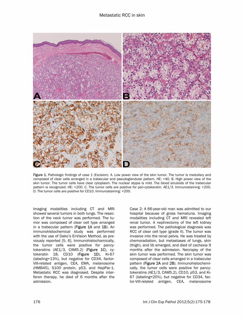
Imaging modalities including CT and MRI showed several tumors in both lungs. The resec-tion of the neck tumor was performed. The tu-mor was composed of clear cell type arranged in a trabecular pattern (Figure 1A and 1B). An immunohistochemical study was performed with the use of Dako’s EnVision Method, as pre-viously reported [5, 6]. Immunohistochamically, the tumor cells were positive for pancy-tokeratins (AE1/3, CAM5.2) (Figure 1C), cy-tokeratin 18, CD10 (Figure 1D), Ki-67 (labeling=13%), but negative for CD34, factor-VIII-related antigen, CEA, EMA, melanosome (HMB45), S100 protein, p53, and HepPar-1. Metastatic RCC was diagnosed. Despite inter-feron therapy, he died of 6 months after the admission.
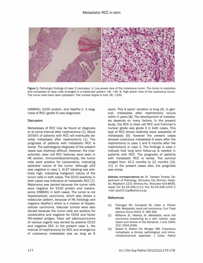
Case 2: A 66-year-old man was admitted to our hospital because of gross hematuria. Imaging modalities including CT and MRI revealed left renal tumor. A nephrectomy of the left kidney was performed. The pathological diagnosis was RCC of clear cell type (grade II). The tumor was invasive into the renal pelvis. He was treated by chemoradiation, but metastases of lungs, skin (thigh), and lib emerged, and died of cachexia 9 months after the admission. Necropsy of the skin tumor was performed. The skin tumor was composed of clear cells arranged in a trabecular pattern (Figure 2A and 2B). Immunohistochemi-cally, the tumor cells were positive for pancy-tokeratins (AE1/3, CAM5.2), CD10, p53, and Ki-67 (labeling=20%), but negative for CD34, fac-tor-VIII-related antigen, CEA, melanosome
Figure 1. Pathologic findings of case 1 (Excision). A. Low power view of the skin tumor. The tumor is medullary and composed of clear cells arranged in a trabecular and pseudoglandular pattern. HE: ×40. B. High power view of the skin tumor. The tumor cells have clear cytoplasm. The nuclear atypia is mild. The blood sinuloids of the trabecular pattern is recognized. HE: ×200. C. The tumor cells are positive for pan-cytokeratin. AE1/3. Immunostaining: ×200. D. The tumor cells are positive for CD10. Immunostaining: ×200.
Metastatic RCC in skin
177 Int J Clin Exp Pathol 2012;5(2):175-178
(HMB45), S100 protein, and HepPar-1. A diag-nosis of RCC (grade II) was diagnosed. Discussion Metastases of RCC may be found at diagnosis or at some interval after nephrectomy [1]. About 20-50% of patients with RCC will eventually de-velop metastasis after nephrectomy [1]. The prognosis of patients with metastatic RCC is worse. The pathological diagnosis of the present cases was relatively difficult. However, the char-acteristic clear cell RCC features were seen in HE section. Immunohistochemically, the tumor cells were positive for cytokeratins, indicating epithelial nature of the tumor. Although p53 was negative in case 1, Ki-67 labeling was rela-tively high, indicating malignant nature of the tumor cells in both cases. The CD10 positivity in both cases was indicative of metastatic RCC [7]. Melanoma was denied because the tumor cells were negative for S100 protein and melano-some (HMB45) in both cases. The tumor is not hepatocellular carcinoma, which also shows a trabecular pattern, because of HE histology and negative HepPar1 which is a marker of hepato-cellular carcinoma. Vascular tumors were also denied because the tumor cells are positive for cytokeratins and negative for CD34 and factor VIII-related antigen. Clear cell adenocarcinoma of various organs was denied by positive CD10 and negative CEA. In the present case 1, the interval of nephrectomy for RCC and emergence of cutaneous metastasis was as long as 9
years. This 9 years’ duration is long [4]. In gen-eral, metastasis after nephrectomy occurs within 5 years [8]. The development of metasta-sis depends on many factors. In the present study, the RCC is clear cell RCC and Fuhrman’s nuclear grade was grade II in both cases. This type of RCC shows relatively lower possibility of metastasis [9]; however the present cases showed cutaneous metastasis 9 years after the nephrectomy in case 1 and 9 months after the nephrectomy in case 2. The findings in case 1 indicate that long term follow-up is needed in patients with RCC. The prognosis of patients with metastatic RCC is worse. The survival ranged from 10.2 months to 22 months [10, 11]. In the present cases also, the prognosis was worse. Address correspondence to: Dr. Tadashi Terada, De-partment of Pathology, Shizuoka City Shimizu Hospi-tal, Miyakami 1231 Shimizu-Ku, Shizuoka 424-8636, Japan Tel: 81-54-336-1111; Fax: 81-54-336-1315; E-mail: [email protected] References [1] Flamigan RC, Campbell SC, Clark JI, Picken
MM. Metastatic renal cell carcinoma. Curr Treat Options Oncol 2003; 4: 385-390.
[2] Williams JC, Heaney JA. Metastatic renal cell carcinoma presenting as a skin nodule: case report and review of the literature. J Urol 1994; 152: 2094-2095.
[3] Saeed S, Keehn CA, Morgan MB. Cutaneous metastasis: a clinical, pathological, and immu-nohistochemical appraisal. J Cutan Pathol
Figure 2. Pathologic findings of case 2 (necropsy). A. Low power view of the cutaneous tumor. The tumor is medullary and composed of clear cells arranged in a trabecular pattern. HE: ×40. B. High power view of the cutaneous tumor. The tumor cells have clear cytoplasm. The nuclear atypia is mild. HE, ×200.
Metastatic RCC in skin
178 Int J Clin Exp Pathol 2012;5(2):175-178
2004; 31: 419-430. [4] Hu SC, Chen GS, Wu CS, Chai CY, Chen WT, Lan
CE. Rates of cutaneous metastasis from differ-ent internal malignancies: experience from a Taiwanese medical center. J Am Acad Dermatol 2009; 60: 379-387.
[5] Terada T, Kawaguchi M, Furukawa K, Sekido Y, Osamura Y. Minute mixed ductal-endocrine carcinoma of the pancreas with predominant intraductal growth. Pathol Int 2002; 52: 740-746.
[6] Terada T, Kawaguchi M. Primary clear cell ade-nocarcinoma of the peritoneum. Tohoku J Exp Med 2005: 206: 271-275.
[7] Leroy X, Farine MO, Buob D, Wacrenier A, Copin MC. Diagnostic value of cytokeratin 7, CD10 and mesothelin in distinguishing ovarian clear cell carcinoma from metastasis of renal clear cell carcinoma. Histopathology 2007; 51: 874-876.
[8] Klatte T, Seligson DB, LaRochelle J, Shuch B, Said JW, Riggs SB, Zomorodian N, Kabbinavar FF, Pantuck AJ, Belldegrun AS. Molecular signa-tures of localized clear renal cell carcinoma to predict disease-free survival after nephrectomy. Cancer Epidermol Biomarkers Prev 2009; 18: 894-900.
[9] Amin MB, Tamboli P, Javidan J, Stricker H, de-Peralta Venturina M, Deshpande A, Menon M. Prognostic impact of histologic subtyping of adult renal epithelial neoplasms: an experience of 405 cases. Am J Surg Pathol 2002; 26: 281-291.
[10] Motzer RJ, Bacik J, Schwarz LH, Reuter V, Russo P, Marison S, Mazumdar M. Prognostic factors for survival in previously treated pa-tients with metastatic renal cell carcinoma. J Clin Oncol 2004; 22: 453-463.
[11] Heng DY, Xie W, Regan MM, Warren MA, Gol-shayan AR, Sahi C, Eigl BJ, Ruether JD, Cheng T, North S, Venner P, Knox JJ, Chi KN, Koll-mannsberger C, McDermott DF, Oh WK, Atkins MB, Bukowski RM, Rini BI, Choueiri TK. Prog-nostic factors for overall survival in patients with metastatic renal cell carcinoma treated with vascular endothelial growth factor-targeted agents: results from a large, multicenter study. J Clin Oncol 2009; 27: 5694-5799.