Embed Size (px)
Citation preview
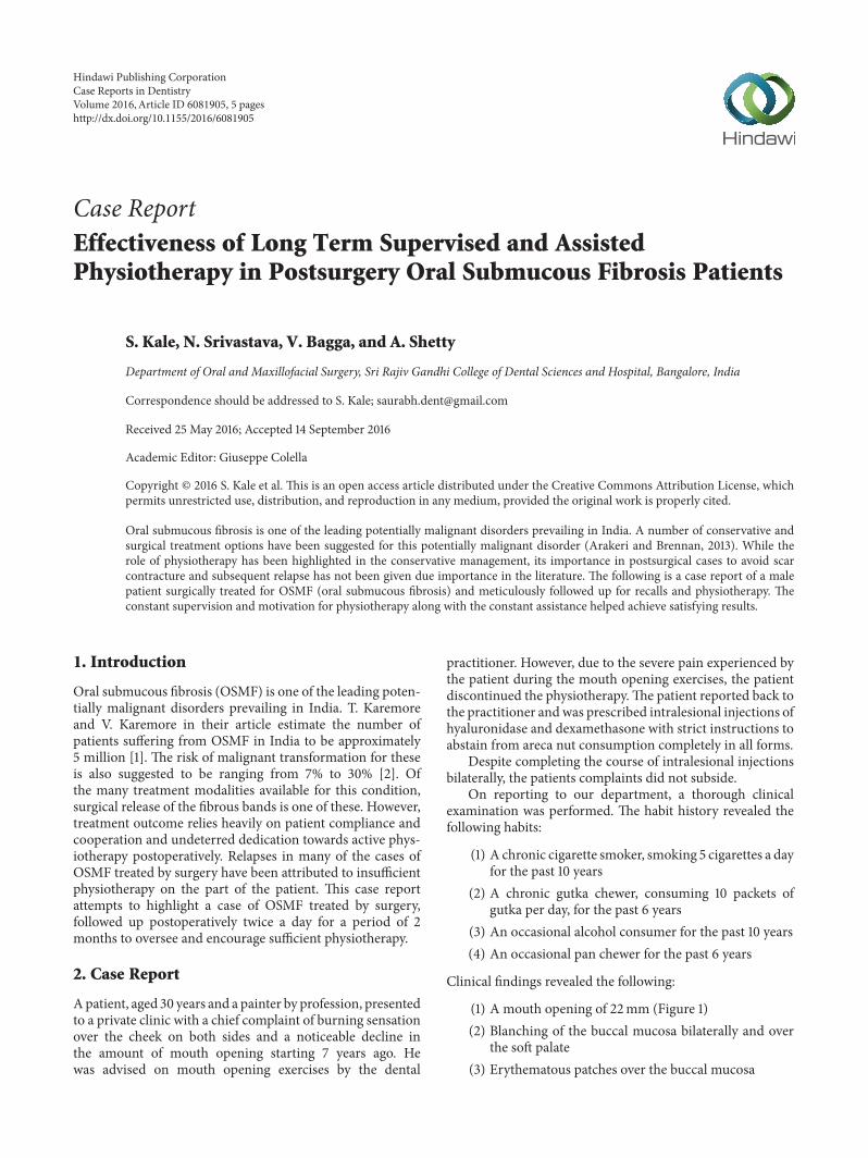
Case ReportEffectiveness of Long Term Supervised and AssistedPhysiotherapy in Postsurgery Oral Submucous Fibrosis Patients
S. Kale, N. Srivastava, V. Bagga, and A. Shetty
Department of Oral and Maxillofacial Surgery, Sri Rajiv Gandhi College of Dental Sciences and Hospital, Bangalore, India
Correspondence should be addressed to S. Kale; [email protected]
Received 25 May 2016; Accepted 14 September 2016
Academic Editor: Giuseppe Colella
Copyright © 2016 S. Kale et al. This is an open access article distributed under the Creative Commons Attribution License, whichpermits unrestricted use, distribution, and reproduction in any medium, provided the original work is properly cited.
Oral submucous fibrosis is one of the leading potentially malignant disorders prevailing in India. A number of conservative andsurgical treatment options have been suggested for this potentially malignant disorder (Arakeri and Brennan, 2013). While therole of physiotherapy has been highlighted in the conservative management, its importance in postsurgical cases to avoid scarcontracture and subsequent relapse has not been given due importance in the literature. The following is a case report of a malepatient surgically treated for OSMF (oral submucous fibrosis) and meticulously followed up for recalls and physiotherapy. Theconstant supervision and motivation for physiotherapy along with the constant assistance helped achieve satisfying results.
1. Introduction
Oral submucous fibrosis (OSMF) is one of the leading poten-tially malignant disorders prevailing in India. T. Karemoreand V. Karemore in their article estimate the number ofpatients suffering from OSMF in India to be approximately5 million [1]. The risk of malignant transformation for theseis also suggested to be ranging from 7% to 30% [2]. Ofthe many treatment modalities available for this condition,surgical release of the fibrous bands is one of these. However,treatment outcome relies heavily on patient compliance andcooperation and undeterred dedication towards active phys-iotherapy postoperatively. Relapses in many of the cases ofOSMF treated by surgery have been attributed to insufficientphysiotherapy on the part of the patient. This case reportattempts to highlight a case of OSMF treated by surgery,followed up postoperatively twice a day for a period of 2months to oversee and encourage sufficient physiotherapy.
2. Case Report
Apatient, aged 30 years and a painter by profession, presentedto a private clinic with a chief complaint of burning sensationover the cheek on both sides and a noticeable decline inthe amount of mouth opening starting 7 years ago. Hewas advised on mouth opening exercises by the dental
practitioner. However, due to the severe pain experienced bythe patient during the mouth opening exercises, the patientdiscontinued the physiotherapy.The patient reported back tothe practitioner andwas prescribed intralesional injections ofhyaluronidase and dexamethasone with strict instructions toabstain from areca nut consumption completely in all forms.
Despite completing the course of intralesional injectionsbilaterally, the patients complaints did not subside.
On reporting to our department, a thorough clinicalexamination was performed. The habit history revealed thefollowing habits:
(1) A chronic cigarette smoker, smoking 5 cigarettes a dayfor the past 10 years
(2) A chronic gutka chewer, consuming 10 packets ofgutka per day, for the past 6 years
(3) An occasional alcohol consumer for the past 10 years(4) An occasional pan chewer for the past 6 years
Clinical findings revealed the following:
(1) A mouth opening of 22mm (Figure 1)(2) Blanching of the buccal mucosa bilaterally and over
the soft palate(3) Erythematous patches over the buccal mucosa
Hindawi Publishing CorporationCase Reports in DentistryVolume 2016, Article ID 6081905, 5 pageshttp://dx.doi.org/10.1155/2016/6081905

2 Case Reports in Dentistry
Figure 1: Preoperative MO—22mm.
Figure 2: Surgical resection of fibrous bands.
(4) Hockey stick shaped uvula(5) Restricted mobility of the tongue, with the tongue on
protrusion, slightly overlapping the lower incisors(6) Stains and calculus(7) Palpation that confirmed the inspectory findings and
exhibited presence of vertical fibrous bands over thebuccal mucosa and horizontal bands circumorally
The previous history of intralesional injections and theminimal benefit from them drove the treatment plan infavour of surgical resection of the fibrous bands bilaterally inthe region of the buccal mucosa.
The preoperative investigations were done and foundto be within normal limits. Informed consent for surgerywas obtained from the patient. Surgical resection of thefibrous bands was carried out under general anaesthesia (Fig-ure 2). Postresection mouth opening achieved was 32mm.To further enhance the mouth opening, a plan for bilat-eral coronoidectomy was decided on the operating tablewhich further improved the mouth opening to 48mm. Theresultant defect over the buccal mucosa was covered with acollagen membrane impregnated with placental extract andhyaluronidase (Figure 3) [3]. The collagen membrane wassutured over the defect (Figure 4) and a bolster gauze wasplaced over it to stabilize the membrane. A Ryles tube wasinserted postoperatively to aid in feeding.
Active physiotherapy was started from the 1st postopera-tive day under the cover of strong analgesics (intramusculardiclofenac sodium 3mL BD). A Hester’s jaw opener wasused actively to open the mouth. The patient was dischargedon the second postoperative day after a session of assistedphysiotherapy. From the third postoperative day onwards,
Figure 3: Collagen membrane impregnated with placental extractand hyaluronidase.
Figure 4: Collagenmembrane sutured on the defect over the buccalmucosa.
the patient was advised to report to our department everymorning for a period of 2months. Active physiotherapy usingHester’s jaw opener was performed in the department. Thepatient was again attended to everyday in the evenings for asession of active physiotherapy and was also simultaneouslyencouraged. Targets were set for the patient to attain specificmouth openings till certain days to encourage him. Incases where a complaint of pain incapacitated the patientfrom doing physiotherapy, the attending surgeons personallyhelped the patient use the Hester’s gradually. The patientwas advised to perform physiotherapy himself using the jawopener between these 2 assisted sessions of physiotherapy.Mouth opening was gradually increased and the woundwas evaluated regularly. Wound was evaluated on the 10thpostoperative day and certain loose sutures were removed.Betadine irrigation was also done. The characteristic hockeystick shaped uvula and the blanching of the palate associatedwith OSMF could also be observed clearly after surgery(Figure 5).
The improvement in mouth opening observed was asfollows:

Case Reports in Dentistry 3
Figure 5:Hockey stick shaped uvula and the blanching of the palate.
Figure 6: 3rd postoperative day immediately after assisted mouthopening—21mm.
(1) 3rd postoperative day assisted mouth opening—21mm (Figure 6)
(2) 7th postoperative day assisted mouth opening—33mm (Figure 7)
(3) 1-month postoperative assisted mouth opening—40mm (Figure 8)
(4) 35-day postoperative passivemouth opening—35mm(Figure 9)
(5) 6-month postoperative passive mouth opening—43mm (Figure 10)
3. Discussion
The association of OSMF with India dates back to the timesof Sushruta, who recognized OSMF as a mouth and throatmalady and termed it “Vidhari” in around 3000 BC [4].Schwartz in 1952 first found the existence of this conditionin five Indian women from Kenya [5]. Named initially as“atrophia idiopathica (tropica) mucosae oris” by him, it waslater renamed “submucous fibrosis” by Joshi in 1953 [5].Of the many reasons cited for the recurrence of OSMFafter surgery or after other treatment modalities, insufficient
Figure 7: 7th postoperative day assisted mouth opening—33mm.
Figure 8: 30th postoperative day ice cream sticks assisted mouthopening—40mm.
physiotherapy is one of the major causes. While nonstoppageof the habit can be attributed to the addictive nature ofareca nut, insufficient physiotherapy mainly results from thefollowing causes:
(1) Negligence and underestimation on part of thepatient towards the importance of performing phys-iotherapy in the right way and for the right number oftimes
(2) Pain during the physiotherapy incapacitating thepatient from doing active physiotherapy on his own
A number of studies have been performed to assess theeffectiveness of physiotherapy as a conservative treatmentmodality for mild to moderate cases of OSMF. Thakur etal. [6] in their study on 64 patients observed physiotherapyto be a helpful adjunct to micronutrients for conservativemanagement of patients with mild to moderate OSMF. Theresults were statistically significant when physiotherapy andmicronutrient therapy were used in combination comparedto physiotherapy being used alone.
Vijayakumar and Priya [7] in their study on 64 patientswith grade 2 and 3 OSMF evaluated the role of physiotherapyand ultrasound therapy in conservative management of suchpatients.Themean improvement in mouth opening obtainedwas 6mm suggesting that heating a muscle and subsequentphysiotherapy can help achieve improved mouth opening.
Alam et al. [8] through their study on patients withOSMF advocated physiotherapy postsubmucosal injectionsof chemicals for treating OSMF. The main objective of thephysiotherapy according to the author was to counteract thetendency of fibrosis, trismus, and dysphagia occurring after

4 Case Reports in Dentistry
Figure 9: 35th postoperative day passive mouth opening—35mm.
Figure 10: 6-month postoperative passive mouth opening—43mm.
the trauma due to the injections and the irritative nature ofthe chemicals injected.
The literature however is scarce on the importance ofphysiotherapy after surgery to reduce chances of scar con-tracture and relapse. A study done by Cox and Zoellner on54 OSMF patients highlighted the importance of physio-therapy in improving mouth opening [9]. Physiotherapy wasemployed as the sole modality of conservative treatment ascompared to the use of local injections of hyaluronidase. Theresults of the study pointed towards a significant improve-ment in mouth opening in the physiotherapy group. Manystudies invariably mention the need for physiotherapy aftersurgery to improve outcomes and prevent scar contractureand recurrence [10–12].However, the initiation of physiother-apy, its importance, and the desirable duration to continuephysiotherapy after surgery have not been discussed much.A common observation is the patients aversion towardsimmediate postoperative physiotherapy due to the associatedsevere pain. However, we promote starting active physiother-apy immediately 2 days postoperatively to minimize chancesof scar contracture setting in. To overcome the pain, weadvocate keeping the patient under a strong analgesic cover.Also, another reason for the disappointment noted withpostsurgery outcomes is the poor compliance on part ofthe patient to perform physiotherapy to the desired extent,for the desired frequency and in the correct way. Homeprogrammes of physiotherapy can be successful only if thepatient is motivated and made to do the exercises once everyday under supervision. Motivating the patient and a constantsupervision and assistance towards physiotherapywere henceour main objectives. Without supervision and motivation,patients tend to fall short of the desired goal of mouthopening. Repeated over days, this results in a gradual decline
in the potential increase in mouth opening which could havebeen achieved.
The success towards achieving a satisfactorymouth open-ing in this patient can be attributed to the active supervisedphysiotherapy. The patient visited the hospital regularly fora period of 2 months for assisted physiotherapy and wasin turn attended to for the same duration by an attendingsurgeon. This is not feasible for every patient as the routineand compliance will vary for every individual.
The following case report is an attempt to highlightthe need for our intervention into the patients’ home pro-grammes of physiotherapy to maximize the benefits obtainedfrom these. The results can be more definitively seen incontrolled trials as are going on in our institution.
Competing Interests
The authors declare that they have no competing interests.
References
[1] T. Karemore and V. Karemore, “Etiopathogenesis and treat-ment strategies of oral submucous fibrosis,” Journal of IndianAcademy of OralMedicine and Radiology, vol. 23, no. 4, pp. 598–602, 2011.
[2] G. Arakeri and P. A. Brennan, “Oral submucous fibrosis: anoverview of the aetiology, pathogenesis, classification, and prin-ciples of management,” British Journal of Oral andMaxillofacialSurgery, vol. 51, no. 7, pp. 587–593, 2013.
[3] Y. Raghavendra Reddy, N. Srinath, H. Nandakumar, and M.Rajini Kanth, “Role of collagen impregnated with dexametha-sone and placentrix in patients with oral submucous fibrosis,”Journal of Maxillofacial and Oral Surgery, vol. 11, no. 2, pp. 166–170, 2012.
[4] S. Samdariya, D. Kumar, A. Kumar, P. Porwal, and P. Pareek,“Oral submucous fibrosis—a short review,” International Jour-nal of Medical Science and Public Health, vol. 3, no. 11, pp. 1308–1312, 2014.
[5] C. B. More, S. Das, H. Patel, C. Adalja, V. Kamatchi, and R.Venkatesh, “Proposed clinical classification for oral submucousfibrosis,” Oral Oncology, vol. 48, no. 3, pp. 200–202, 2012.
[6] N. Thakur, V. Keluskar, A. Bagewadi, and A. Shetti, “Effective-ness ofmicronutrients and physiotherapy in themanagement oforal submucous fibrosis,” International Journal of ContemporaryDentistry, vol. 2, no. 1, pp. 101–105, 2011.
[7] M. Vijayakumar and D. Priya, “Physiotherapy for improvingmouth opening & tongue protrution in patients with Oral Sub-mucous Fibrosis (OSMF)—case series,” International Journal ofPharmaceutical Science and Health Care, vol. 3, no. 2, pp. 52–58,2013.
[8] S. Alam, I. Ali, K. Y. Giri et al., “Efficacy of aloe vera gel as anadjuvant treatment of oral submucous fibrosis,” Oral Surgery,Oral Medicine, Oral Pathology and Oral Radiology, vol. 116, no.6, pp. 717–724, 2013.
[9] S. Cox and H. Zoellner, “Physiotherapeutic treatment improvesoral opening in oral submucous fibrosis,” Journal of OralPathology and Medicine, vol. 38, no. 2, pp. 220–226, 2009.
[10] C. R. Bande, A. Datarkar, and N. Khare, “Extended nasolabialflap compared with the platysma myocutaneous muscle flapfor reconstruction of intraoral defects after release of oral

Case Reports in Dentistry 5
submucous fibrosis: a comparative study,”British Journal of Oraland Maxillofacial Surgery, vol. 51, no. 1, pp. 37–40, 2013.
[11] R.M. Borle, P. V. Nimonkar, and R. Rajan, “Extended nasolabialflaps in the management of oral submucous fibrosis,” BritishJournal of Oral andMaxillofacial Surgery, vol. 47, no. 5, pp. 382–385, 2009.
[12] U. Lokesh, G. Veena, Anubhav Jann,, G. K. Vivek, and M.R. Shilpa, “Application of lasers for oral submucus fibrosis—an experimental study,” Archives of CraniOrofacial Sciences(ACOFS), vol. 1, no. 6, pp. 81–86, 2014.

Submit your manuscripts athttp://www.hindawi.com
Hindawi Publishing Corporationhttp://www.hindawi.com Volume 2014
Oral OncologyJournal of
DentistryInternational Journal of
Hindawi Publishing Corporationhttp://www.hindawi.com Volume 2014
Hindawi Publishing Corporationhttp://www.hindawi.com Volume 2014
International Journal of
Biomaterials
Hindawi Publishing Corporationhttp://www.hindawi.com Volume 2014
BioMed Research International
Hindawi Publishing Corporationhttp://www.hindawi.com Volume 2014
Case Reports in Dentistry
Hindawi Publishing Corporationhttp://www.hindawi.com Volume 2014
Oral ImplantsJournal of
Hindawi Publishing Corporationhttp://www.hindawi.com Volume 2014
Anesthesiology Research and Practice
Hindawi Publishing Corporationhttp://www.hindawi.com Volume 2014
Radiology Research and Practice
Environmental and Public Health
Journal of
Hindawi Publishing Corporationhttp://www.hindawi.com Volume 2014
The Scientific World JournalHindawi Publishing Corporation http://www.hindawi.com Volume 2014
Hindawi Publishing Corporationhttp://www.hindawi.com Volume 2014
Dental SurgeryJournal of
Drug DeliveryJournal of
Hindawi Publishing Corporationhttp://www.hindawi.com Volume 2014
Hindawi Publishing Corporationhttp://www.hindawi.com Volume 2014
Oral DiseasesJournal of
Hindawi Publishing Corporationhttp://www.hindawi.com Volume 2014
Computational and Mathematical Methods in Medicine
ScientificaHindawi Publishing Corporationhttp://www.hindawi.com Volume 2014
PainResearch and TreatmentHindawi Publishing Corporationhttp://www.hindawi.com Volume 2014
Preventive MedicineAdvances in
Hindawi Publishing Corporationhttp://www.hindawi.com Volume 2014
EndocrinologyInternational Journal of
Hindawi Publishing Corporationhttp://www.hindawi.com Volume 2014
Hindawi Publishing Corporationhttp://www.hindawi.com Volume 2014
OrthopedicsAdvances in