Embed Size (px)
Citation preview

110
www.ejvs.selcuk.edu.trwww.eurasianjvetsci.org
CASE REPORT
Congenital meningoencephalocele in a Brown Swiss calf: A case report
Turan Yaman1*, Serkan Erdoğan2, Funda Terzi1, Zafer Özyıldız3
1Department of Pathology, 2Department of Anatomy, Faculty of Veterinary Medicine, Dicle University, 21280, Diyarbakır,3Department of Pathology, Faculty of Veterinary Medicine, Harran University, 63200, Şanlıurfa, Turkey
Received: 27.12.2012, Accepted: 25.01.2013*[email protected]
Özet
Yaman T, Erdoğan S, Terzi F, Özyıldız Z. Bir Montofon buzağıda konjenital meningoencephalocele olgusu. Eurasian J Vet Sci, 2013, 29, 2, 110-113
Dicle Üniversitesi Veteriner Fakültesi Pataloji Anabilim Dalı’na ölü olarak getirilen 2 günlük montofon ırkı erkek buzağıda inspeksiyon ve radyolojik muayeneler sonucu meningocele’den şüphelenildi. Yapılan patolojik-anatomik incelemeler sonunda, squama frontalis üzerinde 17x15 cm çapında, fluktuan kıvamlı, üzeri kıllı deri ile kaplı bir kese gözlendi. Kese içerisinde yaklaşık 200 mL kanla karışık seröz ve akışkan bir sıvı ile karşılaşıldı. Beyin meninkslerle birlikte frontal kemiğin rostrodorsaline doğru fıtıklaşmıştı. Os ethmoidale rudimenter olarak gelişmişti ve bu yüzden etmoid labirentler bu bölgede saptanamadı. Ayrıca, os nasale’nin ve os frontale’nin pars nasalis bölümünün olmadığı, nazal konkalar ile sert damağın rostral yarımının bulunmadığı saptandı. Serebral hipoplazi ve makrogyri belirgindi. Sonuç olarak nadir olarak gözlenen meningoencephalocele olgusu, Diyarbakır ilinde ilk defa bir buzağıda anatomopatolojik olarak tanımlandı.
Anahtar kelimeler: Buzağı, konjenital meningoencephalocele
Abstract
Yaman T, Erdogan S, Terzi F, Ozyildiz Z. Congenital meningoencephalocele in a Brown Swiss calf: A case report. Eurasian J Vet Sci, 2013, 29, 2, 110-113
After inspection and radiological examinations, meningocele was suspected on a 2-day old Brown swiss male calf which was brought dead to the Department of Pathology, Faculty of Veterinary Medicine in Dicle University. As a result of the anatomopathologic analysis, a saclike protrusion that was 17x15 cm sized fluctuant and covered with a hairy skin was observed on frontal squama. In the pouch, approximately 200 mL serous fluid mixed with blood were seen. The brain was also protrused with meninges rostro-dorsal to frontal region. Ethmoid bone was formed rudimentary, so ethmoid labyrinths was not detected in this region. On the other hand, absences of nasal bone, nasal part of frontal bone, nasal conchae and rostral half of hard palate were detected. Cerebral hypoplasia with macrogyria was evident. In conclusion, rarely observed meningoencephalocele case was firstly identified as anatomopathologically in a calf in Diyarbakır province.
Keywords: Calf, congenital meningoencephalocele

111
Yaman et alCongenital meningoencephalocele
Meningocele is the saclike protrusion of the meninges filled with cerebrospinal fluid through a defect in the skull (Urman 1983, Hazıroğlu and Milli 1997). In addition to the skull, meningo-cele may also occur in cervical (Alkan et al 1995), thoracic and lumbar vertebrae (Ertürk and Samsar 1978, Rivas et al 1996). Meningocele forms a hernial cyst that is filled with cerebrospi-nal fluid but does not contain neural tissue. By contrast, menin-goencephalocele is the hernial protrusion of the meninges and brain tissue through a defect (cranium bifidum) in the cranium (Urman 1983, Hazıroğlu and Milli 1997, Hoogmoed et al 1999).
Even though congenital defects are more common in central nervous system, muscular system and in gastrointestinal sys-tem, they can also be encountered less frequently in urogeni-tal system, eye, and skin (Dennis 1993). Environmental factors along with the congenital factors are considered to be critical in the etiology and development of meningocele and meningoen-cephalocele (Aslanbey et al 1989, Kohli 1998, Kaya et al 2011). Malnutrition, contact with teratogenic viruses, and the use of drugs with teratogenic side effects are among the significant en-vironmental factors propagating the development of neurologi-cal defects (Lopez and Wilson 2000). Moreover, trace elements, poisonous plants, radiation, hyperthermia, and the pressure applied during rectal examination are among the reported tera-togenic environmental factors (Johnson et al 1985, Alkan et al 1995). Some environmental factors are shown to provoke the development of various meningocele phenotypes through af-fecting the degree of gene penetration (Rivas et al 1996). Simi-larly, autosomal recessive genes are reported to be responsible for the most of the cranial defects in animals. The formation of meningocele in calf (Kumar and Ramakrishna 1996) and lamb (Özaydın et al 1996) are reported as well.
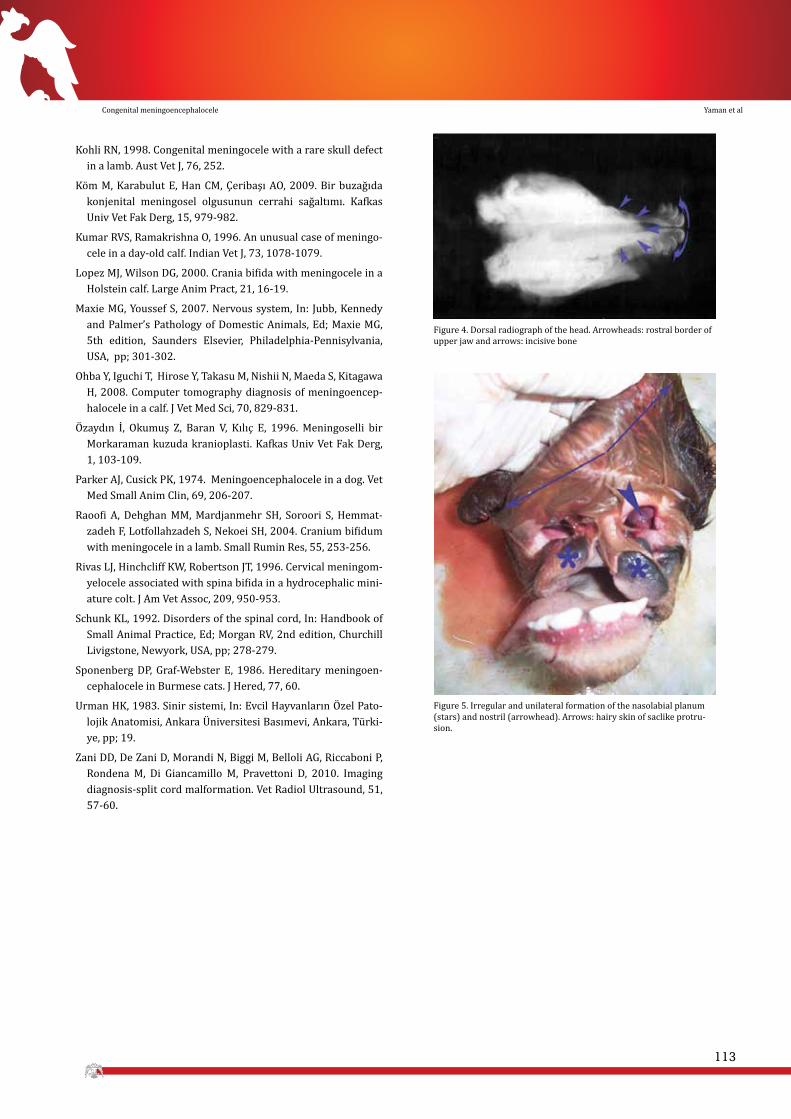
The material of the present study was a calf of Brown Swiss cattle. The calf was two days old, death and weighing 7 kg, and brought the Department of Pathology at the Veterinary Medi-cal School of Dicle University. On the macroscopic examination, there was a saclike protrusion over the frontal squama in the cranium. The protrusion was fluctuant; its size was 17x15 cm and covered by a hairy skin (Figure 1). The sac contained about 200 mL of freely flowing serous liquid mixed with blood (Figure 2). Moreover, there was a patch of hairy skin extending out from the conjunctiva of the lower eyelid in the right eye (dermoid cyst). The upper jaw was shorter than the lower jaw owing to the incomplete development of palatine process of the maxillary bone and the incisive bone. The development of the upper lip was also deficient (Figures 3 and 4). However, the tongue and the teeth were normal otherwise. When the skin of cranium was lifted, we noted that while the frontal squama of the frontal bone was present, its nasal part and the nasal bone were absent in the cranium of the calf. Sphenoid bone was normal; however, the neighboring temporal bone and its petrous portion were con-siderable thicker (Figures 3 and 4). There was an irregular and unilateral formation of the nasolabial planum and nostril (right) only and the continuation of the nasal cavity and the formation of the nasal conchae were not present properly (Figure 5). Fur-
thermore, the ethmoid bone was rudimentary, poorly ossified, and its osseous labyrinths were not formed. The brain was hy-poplastic with prominent macrogyri (Figure 2).
Meningocele and meningoencephalocele are herniation of me-ninges or/and brain substance through defects or malforma-tions of skull during the embryologic development. These her-niations can occur in frontal (Köm et al 2009), parietal (Kaya et al 2011) or occipital (Raoofi et al 2004) regions of the cranium. Frontal herniations are shown to be accompanied with osseous deformities in nasal conchae and ethmoidal bones (Ohba et al 2008). In the present case, meningoencephalocele was originat-ed from the frontal region. Moreover, nasal conchae were aplas-tic and ethmoidal bone was rudimentary. Current observations were consistent with earlier literature reports (Ohba et al 2008, Köm et al 2009). Even though meningoencephalocele is rarely encountered in domestic animals, it can genetically develop in pigs and Burmese cats (Maxie and Youssef 2007) or with the use of griseofulvin during pregnancy. Nevertheless, the formation of meningoencephalocele has been reported in domestic animals, i.e., calves (Raoofi et al 2004), lambs (Back et al 1991), dogs (Sponenberg and Graf-Webster 1986), and foals (Ertürk and Samsar 1978). Although strain and gender have not been report-ed in general to be a predisposing factor for the development of congenital defects in cattle, the ratio of congenital defects among Brown Swiss cattle is reported to be higher (Parker and Cusick 1974, Belge et al 2000).
In meningocele and meningoencephalocele cases, meninges and brain substance can protrude outside of the skull within in a her-niated pouch that is either not covered or covered with a hairy skin (Aslanbey et al 1989, Kumar and Ramakrishna 1996, Kaya et al 2011). In the present case, the herniated pouch was cov-ered by a hairy skin. Reported studies in Turkey indicate that the frequency of the congenital anomalies in ruminants is between 26.8-40.48% (Belge et al 2000). Karabulut et al (2001) demon-strate that the incidence of congenital anomalies among Brown Swiss cattle is higher than other strains of cattle. In addition, Köm et al (2009) reported that the location of the hernial pro-trusion in their case was in the occipital region. In the present case the location of defect was in the frontal region. Moreover, either meningoencephalocele can form separately or can be ac-companied with certain other malformations within the same site or in other part of the body. Potential anomalies that can be accompanied with it are cranioschisis, spina bifida (Ohba 2008, Lopez and Wilson 2000), anal atresia, rectal atresia, umbilical hernia (Kaya et al 2011) and kyphoscoliosis (Zani et al 2010). In the current case, we noticed that the meningocele was accom-panied with agenesis of nasal septum, prognathia, dermoid cyst. While some other studies report that the calves with these mal-formations cannot stand up (Özaydın et al 1996), other report that they can stand up and walk around. In our case, our anam-nesis indicated that the calf was never able to stand up and walk.
Genetic factors, certain medicines used during pregnancy, and abnormal interventions during rectal examinations can play a

112
Yaman et alCongenital meningoencephalocele
role in the development cranial defect and malformations (Alkan et al 1995). In the present case, medical history revealed that the mother had not been exposed to any kind of toxicants, received any kind of medicines, or suffered from high fevered disease and gave to a normal birth without help. Therefore, genetic factors are considered to have played role in the formation of the cur-rent defect in the calf. Autosomal recessive genes are known to control the formation of congenital defects (Hoogmoed et al 1999, Alkan et al 1995). The use of bulls with known healthy pedigree in artificial insemination is critical. Therefore, inspec-tion of mother and inseminating bull for genetic defects will help us to prevent or reduce the incidence of congenital malforma-tions.
In conclusion, determination of the factors causing the congeni-tal anomalies becomes impossible since they are numerous such as malnutrition, exposed diseases, applied medicines, and grow-ing conditions. The present case classified as meningoencepha-locele both pathologically and anatomically and we think that current report will illuminate on the further similar studies on the congenital anomalies.
References
Alkan İ, Bakır B, Dilek FH, Belge A, 1995. İki Akkaraman kuzuda meningoensefalosel olgusu. YYÜ Sağ Bil Derg, 1, 71-75.
Aslanbey D, Öcal MK, Kutsal O, Ünsaldı E, 1989. İki kuzuda rast-lanan meningocele ve meningoencephalocele olgular. AÜ Vet Fak Derg, 36, 379-389.
Back W, van den Belt AJ, Lagerweij E, van Overbeeke JJ, van der Velden MA, 1991. Surgical repair of a cranial meningocele in a calf. Vet Rec, 128, 569-571.
Belge A, Gönenci R, Biricik HS, Ormancı S, 2000. Buzağılarda doğmasal anomali olguları. YYÜ Vet Fak Derg, 11, 23-26.
Dennis SM, 1993. Congenital defects of sheep. Vet Clin North Am Food Anim Pract, 9, 203-217.
Ertürk E, Samsar E, 1978. Bir kuzuda doğmasal spina bifida ve meningocele. AÜ Vet Fak Derg, 28, 261-266.
Hazıroğlu R, Milli ÜH, 1997. Sinir sistemi, In: Veteriner Patoloji, 1. cilt, birinci Baskı, Tamer Matbaacılık, Ankara, Türkiye, pp; 238.
Hoogmoed LV, Yarbrough TB, Lecouteur RA, Hornof WJ, 1999. Surgical repair of a thoracic meningocele in a foal. Vet Surg, 28, 496-500.
Johnson JL, Leipold TT, Hudson DB, 1985. Prominent congenital defects in Nebraska beef cattle. Breed Reprod, 4, 1-8.
Karabulut E, Ünsaldı S, Durgun T, 2001. 1991-2000 yılları ara-sında Fırat Üniversitesi Veteriner Fakültesi cerrahi kliniğine getirilen buzağılardaki doğmasal anomali olguları. FÜ Sağ Bil Derg, 15, 367-374.
Kaya M, Okumuş Z, Doğan E, Çetin EM, Yanmaz LE, 2011. Erzu-rum yöresindeki buzağılarda doğmasal anomalilerin görülme sıklığı ve sağ kalım oranları. FÜ Sağ Bil Vet Derg, 25, 83-93.
Figure 1. Saclike protrusion or pouch (arrows) over the frontal bone.
Figure 2. Protrused brain (in circle) into the pouch (arrows) which contained serous liquid mixed with blood.
Figure 3. Lateral radiograph of the head. Blue star: mandible, red star: maxillary bone, blue arrowheads: rudimentary ethmoidal bone, red arrowheads: parts of saclike protrusion, red arrow: squamous part of temporal bone, blue arrow: petrous part of temporal bone.
113
Yaman et alCongenital meningoencephalocele
Kohli RN, 1998. Congenital meningocele with a rare skull defect in a lamb. Aust Vet J, 76, 252.
Köm M, Karabulut E, Han CM, Çeribaşı AO, 2009. Bir buzağıda konjenital meningosel olgusunun cerrahi sağaltımı. Kafkas Univ Vet Fak Derg, 15, 979-982.
Kumar RVS, Ramakrishna O, 1996. An unusual case of meningo-cele in a day-old calf. Indian Vet J, 73, 1078-1079.
Lopez MJ, Wilson DG, 2000. Crania bifida with meningocele in a Holstein calf. Large Anim Pract, 21, 16-19.
Maxie MG, Youssef S, 2007. Nervous system, In: Jubb, Kennedy and Palmer’s Pathology of Domestic Animals, Ed; Maxie MG, 5th edition, Saunders Elsevier, Philadelphia-Pennisylvania, USA, pp; 301-302.
Ohba Y, Iguchi T, Hirose Y, Takasu M, Nishii N, Maeda S, Kitagawa H, 2008. Computer tomography diagnosis of meningoencep-halocele in a calf. J Vet Med Sci, 70, 829-831.
Özaydın İ, Okumuş Z, Baran V, Kılıç E, 1996. Meningoselli bir Morkaraman kuzuda kranioplasti. Kafkas Univ Vet Fak Derg, 1, 103-109.
Parker AJ, Cusick PK, 1974. Meningoencephalocele in a dog. Vet Med Small Anim Clin, 69, 206-207.
Raoofi A, Dehghan MM, Mardjanmehr SH, Soroori S, Hemmat-zadeh F, Lotfollahzadeh S, Nekoei SH, 2004. Cranium bifidum with meningocele in a lamb. Small Rumin Res, 55, 253-256.
Rivas LJ, Hinchcliff KW, Robertson JT, 1996. Cervical meningom-yelocele associated with spina bifida in a hydrocephalic mini-ature colt. J Am Vet Assoc, 209, 950-953.
Schunk KL, 1992. Disorders of the spinal cord, In: Handbook of Small Animal Practice, Ed; Morgan RV, 2nd edition, Churchill Livigstone, Newyork, USA, pp; 278-279.
Sponenberg DP, Graf-Webster E, 1986. Hereditary meningoen-cephalocele in Burmese cats. J Hered, 77, 60.
Urman HK, 1983. Sinir sistemi, In: Evcil Hayvanların Özel Pato-lojik Anatomisi, Ankara Üniversitesi Basımevi, Ankara, Türki-ye, pp; 19.
Zani DD, De Zani D, Morandi N, Biggi M, Belloli AG, Riccaboni P, Rondena M, Di Giancamillo M, Pravettoni D, 2010. Imaging diagnosis-split cord malformation. Vet Radiol Ultrasound, 51, 57-60.
Figure 4. Dorsal radiograph of the head. Arrowheads: rostral border of upper jaw and arrows: incisive bone
Figure 5. Irregular and unilateral formation of the nasolabial planum (stars) and nostril (arrowhead). Arrows: hairy skin of saclike protru-sion.