Embed Size (px)
Citation preview
Case report
Open Access
Giant scrotal lymphedema of unclear etiology: a case reportGaniyu A Rahman1*, Ismaila A Adigun2, Ibrahim F Yusuf1,Adebiyi B Aderibigbe2 and Amarachukwu C Etonyeaku1
Address: 1Division of General Surgery, Department of Surgery, University of Ilorin Teaching Hospital, Ilorin, Nigeria and 2Division of Plastic andReconstructive Surgery, Department of Surgery, University of Ilorin Teaching Hospital, Ilorin, Nigeria
Email: GAR* - [email protected]; IAA - [email protected]; IFY - [email protected]; ABA - [email protected];ACE - [email protected]
*Corresponding author
Published: 28 May 2009 Received: 7 February 2008Accepted: 23 January 2009
Journal of Medical Case Reports 2009, 3:7295 doi: 10.1186/1752-1947-3-7295
This article is available from: http://jmedicalcasereports.com/jmedicalcasereports/article/view/7295
© 2009 Rahman et al; licensee Cases Network Ltd.This is an Open Access article distributed under the terms of the Creative Commons Attribution License (http://creativecommons.org/licenses/by/3.0),which permits unrestricted use, distribution, and reproduction in any medium, provided the original work is properly cited.
Abstract
Introduction: Scrotal lymphedema is common in the tropics and subtropics. The giant variants cancause a lot of physical disability and psychological disturbances.
Case presentation: We present a 25-year-old Nigerian male with giant scrotal lymphedema withsevere debilitating symptoms, immobility and emotional disturbance. He benefited from a modifiedCharles’ procedure and reconstruction of the penile shaft using a split-thickness skin graft.
Conclusion: Giant scrotal lymphedema related to poverty, ignorance and neglect, is amenable tosurgery. Surgery provides a cosmetically acceptable and functionally satisfying outcome.
IntroductionGiant scrotal lymphedema, also known as elephantiasis,can be caused by obstruction, aplasia or hypoplasia oflymphatic vessels [1]. Though it can be caused by aneoplasm, radiotherapy or lymphadenectomy, most casesare usually caused by infection as a result of lympho-granuloma venereum or filarial infestation with Wucher-eria bancrofti [1-3]. Giant scrotal lymphedema is commonin the tropics and sub-tropics [4,5].
We present our experience of treating an unusuallylarge penoscrotal lymphedema and the result of amodified Charles’ procedure and cutaneous recon-struction of the penile shaft using a split-thickness skingraft.
Case presentationA 25-year-old single Nigerian man was admitted via thesurgical outpatient clinic with a two-year history of scrotalswelling which was initially small in size, non-painful andnot associated with fever. The swelling gradually increasedin size to the extent of impairing free movement of thepatient both because of its weight and size in between thelower limbs. The swelling made sexual intercourse andvoiding in the standing position impossible. The skin wasintact without associated ulcerations, and there was nodifficulty in voiding though the penile shaft was buried inthe swelling. There was no history of chronic coughsuggestive of pulmonary or disseminated tuberculosis. Hehad no problems with his eyesight and no other swellingson his body were known.
Page 1 of 4(page number not for citation purposes)

He had a history of surgery to the two inguinal areas about10 years before presentation; surgical details were notavailable nor was any histology available. He was from apolygamous home and took neither alcohol nor anyother substance of abuse. There was no history of a similarlesion in his relatives or his acquaintances at his place ofabode.
Physical examination showed a healthy looking youngman with a giant scrotal swelling of a size greater than thatof his head. There were hyperpigmented giant ruggae inotherwise intact skin with the penile shaft buried in thescrotal wall skin (Figure 1a and b). The swelling was non-tender, non-pitting and non-reducible with the cord barelydiscernible at the neck. Bilateral inguinal scars were seenwith significant lymphadenopathy.
Hemogram, serum electrolyte, urea and creatinine, urina-lysis, urine microscopy culture and sensitivity were allwithin normal limits. Scrotal ultrasound showed athickened scrotal wall but the scrotal content was normal.
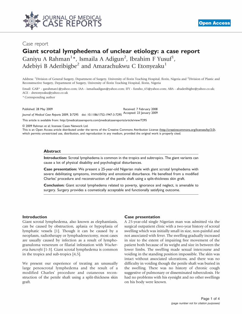
He subsequently underwent a modified Charles’ proce-dure with a primary penile shaft split-thickness skin graft(Figure 2A) performed by a team of general surgeons andplastic surgeons. His immediate postoperative conditionwas satisfactory (Figure 2B). On the postoperative dayseven, the penile skin graft showed good skin take andsome good granulation tissue over the exposed testicles.
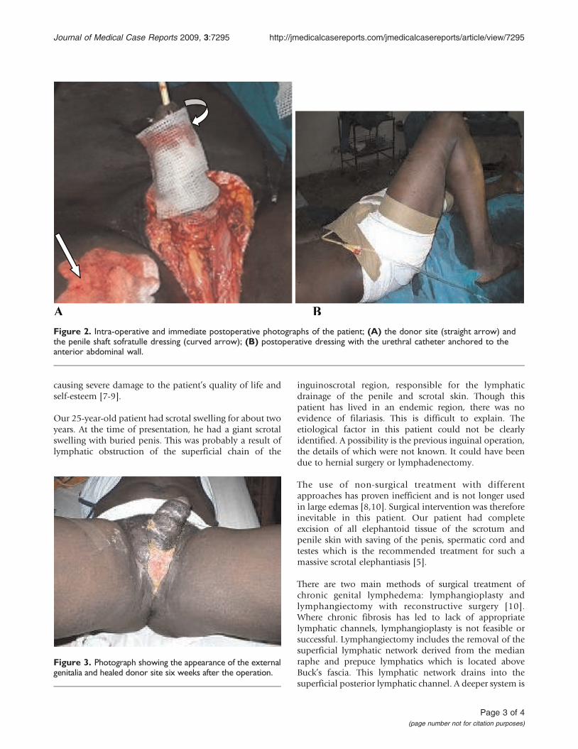
The patient continued to have wound dressing until theperineal wound had healed and contracted appreciablyleaving about a 3 cm raw area of the wound at the sixpostoperative week (Figure 3). He had an uneventfulpostoperative course with the scrotal wound healingcompletely by the eighth postoperative week (Figure 3).He is now about one year post-surgery and doing well. Thepatient is physically and socially satisfied with hisimproved quality of life.
DiscussionLymphedema of the penis and scrotum is due to abnormalaccumulation of lymphatic fluid in subcutaneous tissue ofthe penis and scrotum. Lymphedema has two types:primary and secondary. Primary lymphedema can becongenital-inherited (Milroy’s), praecox or tarda. Second-ary lymphedema has three origins: obstructive (secondaryto neoplasm, radiotherapy, surgical intervention, mechan-ical trauma, bacterial and fungal infections), phlebitial orangioneurotic [6]. Penoscrotal lymphedemamostly occursfollowing an infection or as a reaction to trauma. Scrotallymphedema can be emotionally distressing and physi-cally disabling. It is a condition leading to progressiveenlargement of the scrotum and penis. In addition tothe grotesque aspect, the progression of the conditionimpairs ambulation, makes sexual intercourse impossible,and impairs both voiding in the standing position andproper hygiene of the perineal region, with subsequentmalodor and recurrent episodes of skin infection, all
Figure 1. Photograph of a 25-year-old man showing giant scrotal lymphedema; (A) scrotal lymphedema (arrowed) and(B) the buried penis with external meatus as a dimple (arrowed).
Page 2 of 4(page number not for citation purposes)
Journal of Medical Case Reports 2009, 3:7295 http://jmedicalcasereports.com/jmedicalcasereports/article/view/7295
causing severe damage to the patient’s quality of life andself-esteem [7-9].
Our 25-year-old patient had scrotal swelling for about twoyears. At the time of presentation, he had a giant scrotalswelling with buried penis. This was probably a result oflymphatic obstruction of the superficial chain of the
inguinoscrotal region, responsible for the lymphaticdrainage of the penile and scrotal skin. Though thispatient has lived in an endemic region, there was noevidence of filariasis. This is difficult to explain. Theetiological factor in this patient could not be clearlyidentified. A possibility is the previous inguinal operation,the details of which were not known. It could have beendue to hernial surgery or lymphadenectomy.
The use of non-surgical treatment with differentapproaches has proven inefficient and is not longer usedin large edemas [8,10]. Surgical intervention was thereforeinevitable in this patient. Our patient had completeexcision of all elephantoid tissue of the scrotum andpenile skin with saving of the penis, spermatic cord andtestes which is the recommended treatment for such amassive scrotal elephantiasis [5].
There are two main methods of surgical treatment ofchronic genital lymphedema: lymphangioplasty andlymphangiectomy with reconstructive surgery [10].Where chronic fibrosis has led to lack of appropriatelymphatic channels, lymphangioplasty is not feasible orsuccessful. Lymphangiectomy includes the removal of thesuperficial lymphatic network derived from the medianraphe and prepuce lymphatics which is located aboveBuck’s fascia. This lymphatic network drains into thesuperficial posterior lymphatic channel. A deeper system is
Figure 2. Intra-operative and immediate postoperative photographs of the patient; (A) the donor site (straight arrow) andthe penile shaft sofratulle dressing (curved arrow); (B) postoperative dressing with the urethral catheter anchored to theanterior abdominal wall.
Figure 3. Photograph showing the appearance of the externalgenitalia and healed donor site six weeks after the operation.
Page 3 of 4(page number not for citation purposes)
Journal of Medical Case Reports 2009, 3:7295 http://jmedicalcasereports.com/jmedicalcasereports/article/view/7295

located beneath Buck’s fascia and drains into the deepinguinal lymph nodes [6]. It is important to removeinvolved skin and subcutaneous tissue completely toprevent recurrence.
The major challenge in this procedure is the reconstructionof the penile skin. There are various procedures in thesurgical literature to achieve this [7,8]. We opted for theuse of a split-thickness skin graft. This seems to producebetter results than the use of flaps, even local flaps whichnotably affect tactile sensitivity and erection [9]. Theoutcome of this procedure has been very satisfactory.There is a good cosmetic result for the external genitaliawith easier ambulation, effective hygiene, voiding andsubjectively satisfactory sexual intercourse. The histo-pathological section showed skin tissue with papilloma-tosis. The dermis showed dense lymphoplasmocyticinfiltration and dense fibrocollagenous tissue with dilatedlymphatic channels. These features were consistent withelephantiasis. One year after the procedure, the patient hasdone very well with improvement to his quality of life.
ConclusionMassive penoscrotal lymphedema can cause physicaldisability, social embarrassment and can be psychologi-cally disturbing. The uniqueness of this case is the unclearetiology and achieving complete surgical cure without arotational flap and vacuum assisted devices recommendedin such a large scrotal lymphedema. The patient benefitedfrom a modified Charles’ procedure with a primary penileshaft split-thickness skin graft and achieved a satisfactoryoutcome.
ConsentWritten informed consent was obtained from the patientfor publication of this case report and any accompanyingimages. A copy of the written consent is available forreview by the Editor-in-Chief of this Journal.
Competing interestThe authors declare that they have no competing interests.
Authors’ contributionAll authors were actively involved in the management ofthis patient. They also participated in the design andwriting of the manuscript and all have read and approvedthe final version.
References1. Denzinger S, Watzlawek, Burger M, Wieland WF, Otto W: Giant
scrotal elephantiasis of inflammatory etiology: a case report.J Med Case Rep 2007, 1:23.
2. McDougal WS: Lymphedema of the external genitalia. J Urol2003, 170:711-716.
3. Tammer ME, Plogmeier K, Schneider W: Surgical therapy ofscrotal edema in elephantiasis congenital hereditaria (Melgetype). Urologe A 2002, 41:493-495.
4. Nelson RA, Alberts GL, King LE Jr: Penile and scrotal elephan-tiasis caused by indolent Chlamydia trachomatis infection.Urology 2003, 61:224.
5. Kuepper D: Giant scrotal elephantiasis. Urology 2005, 65:389.6. Brown WL, Wood JE: Lymphedema of the penis. Plast Reconstr
Surg 1977, 59:68-71.7. Bulkley GJ: Scrotal and lymphedema. J Urol 1962, 87:422-429.8. Farina R, Farina G: Elefantiase peno-escrotal (osqueofaloplas-
tia). Rev Bra Cir 1995, 85:205-212.9. Gonella HA: Linfedema peniano. Rev Bra Cir 1967, 72:23-26.10. Apesos J, Anigian G: Reconstruction of penile and scrotal
lymphedema. Ann Plast Surg 1991, 27:570-573.
Page 4 of 4(page number not for citation purposes)
Journal of Medical Case Reports 2009, 3:7295 http://jmedicalcasereports.com/jmedicalcasereports/article/view/7295
Do you have a case to share?
Submit your case report today• Rapid peer review• Fast publication• PubMed indexing• Inclusion in Cases Database
Any patient, any case, can teach ussomething
www.casesnetwork.com