Embed Size (px)
Citation preview

© 2014 Mansour et al. This work is published by Dove Medical Press Limited, and licensed under Creative Commons Attribution – Non Commercial (unported, v3.0) License. The full terms of the License are available at http://creativecommons.org/licenses/by-nc/3.0/. Non-commercial uses of the work are permitted without any further
permission from Dove Medical Press Limited, provided the work is properly attributed. Permissions beyond the scope of the License are administered by Dove Medical Press Limited. Information on how to request permission may be found at: http://www.dovepress.com/permissions.php
Clinical Ophthalmology 2014:8 2403–2407
Clinical Ophthalmology Dovepress
submit your manuscript | www.dovepress.com
Dovepress 2403
C a s e r e p O rt
open access to scientific and medical research
Open access Full text article
http://dx.doi.org/10.2147/OPTH.S73990
Isolated posterior capsular rupture following blunt head trauma
ahmad M MansourMahmoud O Jaroudirola N HamamFadi C Maalouf
Department of Ophthalmology, american University of Beirut, Beirut, Lebanon
Abstract: Closed-globe traumatic cataract is not uncommon in males in the pediatric age
group. However, there is a relative paucity of literature on isolated posterior lens capsule rupture
associated with closed-globe traumatic cataract. We report a case of a 6-year-old boy who
presented with white cataract 1 day after blunt trauma to the forehead associated with posterior
capsular rupture that was detected by B-scan ultrasonography preoperatively. No stigmata of
trauma outside the posterior capsule could be detected by slit-lamp exam, funduscopy, and optical
coherence tomography. Phacoemulsification with posterior chamber intraocular lens implant
was performed 24 hours after trauma, with the patient achieving 6/6 visual acuity 1 week and
6 months after surgery. Our case is unique, being the youngest (amblyogenic age) to be reported,
with prompt surgical intervention, and with no signs of trauma outside the posterior capsule.
Keywords: posterior capsule rupture, forehead trauma, traumatic cataract, pediatric cataract
IntroductionBlunt ocular trauma frequently leads to damage of the cornea, lens, and retina. However,
there is scarce literature1–15 on isolated posterior lens capsule rupture (PCR) following
blunt ocular trauma. We present the case of a child who developed an isolated oval
defect in the posterior lens capsule after blunt trauma with rapid cataract formation.
Case reportOur case is a 6-year-old boy who sustained direct minor trauma to the left forehead. He
was playing at home with his sister and swirling in a circular fashion when he slipped
and hit his forehead against the ground. The following day, he reported blurring of
vision in the left eye and his parents noticed a white left pupil. Twenty-four hours fol-
lowing the incident, examination of the right eye was normal and the left eye had hand
motion vision. Intumescent cataract was evident by slit-lamp examination (Figure 1).
The cornea was clear, the anterior chamber was deep and quiet, the pupil was round
and reactive, and the anterior lens capsule was intact. Intraocular pressure measured
14 mmHg in the right eye and 16 mmHg in the left eye. Gonioscopy revealed a normal
angle without angle recession. The media opacity precluded exam of the posterior
segment. B-scan revealed a flat retina, quiet vitreous, and a breached posterior capsule
centrally (Figure 2). Computerized tomography scan of brain was negative for skull
fracture or intracranial bleed. Since the patient was in the amblyogenic age, cataract
surgery was performed within hours of presentation.
Surgery was started with trypan blue staining of anterior capsule, anterior circular cap-
sulorhexis, and avoiding hydrodissection to prevent expansion of PCR. Slow aspiration
of lens matter with the phacoemulsification probe was performed under low settings of
bottle height, irrigation, and aspiration. After aspiration of two-thirds of the lens content,
Correspondence: ahmad M MansourDepartment of Ophthalmology, american University of Beirut, pO Box 1136044, Beirut, Lebanonemail [email protected]
Journal name: Clinical OphthalmologyArticle Designation: Case reportYear: 2014Volume: 8Running head verso: Mansour et alRunning head recto: Posterior capsular ruptureDOI: http://dx.doi.org/10.2147/OPTH.S73990

Clinical Ophthalmology 2014:8submit your manuscript | www.dovepress.com
Dovepress
Dovepress
2404
Mansour et al
Figure 1 Mature white cataract of the left eye 1 day after blunt trauma to the forehead.
Figure 2 posterior capsular defect (arrow) is evident by B-scan of the left eye.
Figure 3 Large oval defect of the posterior lens capsule of the left eye as noted after aspiration of most of the lens content.Note: arrows delineate the edge of the ruptured posterior capsule.
Figure 4 Well-centered acrylic intraocular lens placed in the sulcus at the end of surgery.
a very large, oval-shaped central posterior capsular rent was
exposed (Figure 3). Anterior vitrectomy was performed, fol-
lowed by cortex aspiration with the vitrector. Although one-
piece foldable intraocular lens implantation in the capsular
bag was initially thought feasible, the intraocular lens had a
tendency to dislodge into the vitreous cavity, necessitating
subsequent fixation in the sulcus without further optic capture
(Figure 4). Intracameral carbachol was applied at the end of
the surgery. One week postoperatively, uncorrected visual
acuity was 6/6 (20/20). The patient had normal funduscopy and
spectral-domain optical coherence tomography of the posterior
pole. Visual acuity stabilized at 6/6 (20/20) over 14 months of
follow-up with well-centered intraocular implant.
DiscussionIsolated PCR in the event of blunt trauma appears rarely
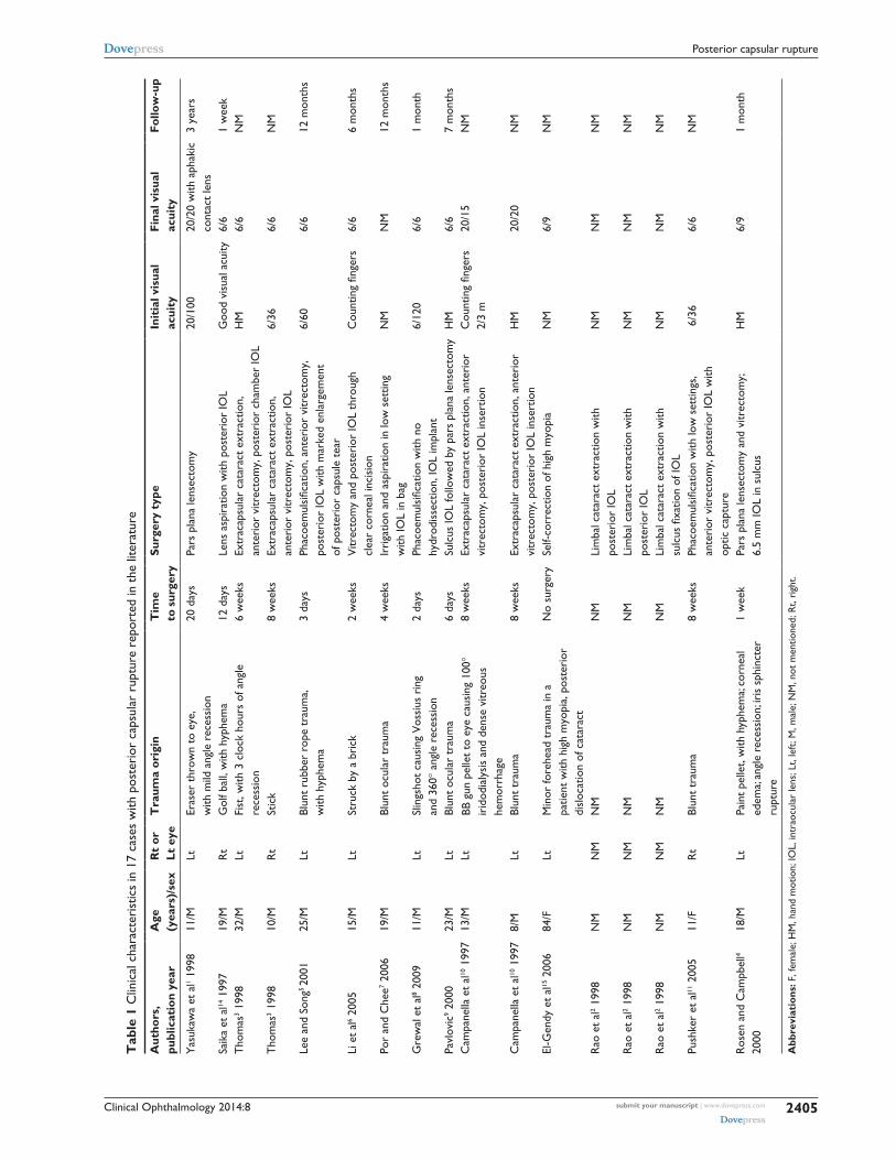
(Table 1).1–15 Our patient presented unique features that dif-
fer from those reported in the literature (Table 1) in several
respects: he is the youngest patient in the literature (6 years)
with PCR; his visual loss was rapid in onset and very severe
to the level of hand motion (unlike a gradual decrease in other
cases); he underwent “immediate” (24 hours after trauma)
surgery to prevent amblyopia (unlike the several days to
weeks in other cases); and his ocular findings were uniquely
restricted to PCR with absence of iritis, hyphema, Vossius
ring, iris sphincter tear, angle recession, or posterior pole
trauma (funduscopic and tomographic).
Clinical Ophthalmology 2014:8 submit your manuscript | www.dovepress.com
Dovepress
Dovepress
2405
posterior capsular rupture
Tab
le 1
Clin
ical
cha
ract
eris
tics
in 1
7 ca
ses
with
pos
teri
or c
apsu
lar
rupt
ure
repo
rted
in t
he li
tera
ture
Aut
hors
, pu
blic
atio
n ye
arA
ge
(yea
rs)/
sex
Rt
or
Lt e
yeT
raum
a or
igin
Tim
e
to s
urge
rySu
rger
y ty
pe
Init
ial v
isua
l ac
uity
Fina
l vis
ual
acui
tyFo
llow
-up
Yas
ukaw
a et
al1 1
998
11/M
Lter
aser
thr
own
to e
ye,
with
mild
ang
le r
eces
sion
20 d
ays
pars
pla
na le
nsec
tom
y20
/100
20/2
0 w
ith a
phak
ic
cont
act
lens
3 ye
ars
saik
a et
al14
199
719
/Mr
tG
olf b
all,
with
hyp
hem
a12
day
sLe
ns a
spir
atio
n w
ith p
oste
rior
IOL
Goo
d vi
sual
acu
ity6/
61
wee
kt
hom
as3 1
998
32/M
LtFi
st, w
ith 3
clo
ck h
ours
of a
ngle
re
cess
ion
6 w
eeks
extr
acap
sula
r ca
tara
ct e
xtra
ctio
n,
ante
rior
vitr
ecto
my,
pos
teri
or c
ham
ber
IOL
HM
6/6
NM
tho
mas
3 199
810
/Mr
tst
ick
8 w
eeks
extr
acap
sula
r ca
tara
ct e
xtra
ctio
n,
ante
rior
vitr
ecto
my,
pos
teri
or IO
L 6/
366/
6N
M
Lee
and
song
5 20
0125
/MLt
Blun
t ru
bber
rop
e tr
aum
a,
with
hyp
hem
a3
days
Phac
oem
ulsi
ficat
ion,
ant
erio
r vi
trec
tom
y,
post
erio
r IO
L w
ith m
arke
d en
larg
emen
t
of p
oste
rior
cap
sule
tea
r
6/60
6/6
12 m
onth
s
Li e
t al
6 200
515
/MLt
stru
ck b
y a
bric
k2
wee
ksV
itrec
tom
y an
d po
ster
ior
IOL
thro
ugh
cl
ear
corn
eal i
ncis
ion
Cou
ntin
g fin
gers
6/6
6 m
onth
s
por
and
Che
e7 200
619
/MBl
unt
ocul
ar t
raum
a4
wee
ksIr
riga
tion
and
aspi
ratio
n in
low
set
ting
w
ith IO
L in
bag
NM
NM
12 m
onth
s
Gre
wal
et
al8 2
009
11/M
Ltsl
ings
hot
caus
ing
Vos
sius
rin
g
and
360°
ang
le r
eces
sion
2 da
ysPh
acoe
mul
sific
atio
n w
ith n
o
hydr
odis
sect
ion,
IOL
impl
ant
6/12
06/
61
mon
th
pavl
ovic
9 200
023
/MLt
Blun
t oc
ular
tra
uma
6 da
yssu
lcus
IOL
follo
wed
by
pars
pla
na le
nsec
tom
yH
M6/
67
mon
ths
Cam
pane
lla e
t al
10 1
997
13/M
LtBB
gun
pel
let
to e
ye c
ausi
ng 1
00°
irid
odia
lysi
s an
d de
nse
vitr
eous
he
mor
rhag
e
8 w
eeks
extr
acap
sula
r ca
tara
ct e
xtra
ctio
n, a
nter
ior
vitr
ecto
my,
pos
teri
or IO
L in
sert
ion
Cou
ntin
g fin
gers
2/
3 m
20/1
5N
M
Cam
pane
lla e
t al
10 1
997
8/M
LtBl
unt
trau
ma
8 w
eeks
extr
acap
sula
r ca
tara
ct e
xtra
ctio
n, a
nter
ior
vitr
ecto
my,
pos
teri
or IO
L in
sert
ion
HM
20/2
0N
M
el-G
endy
et
al15
200
6 84
/FLt
Min
or fo
rehe
ad t
raum
a in
a
patie
nt w
ith h
igh
myo
pia,
pos
teri
or
disl
ocat
ion
of c
atar
act
No
surg
ery
self-
corr
ectio
n of
hig
h m
yopi
aN
M6/
9N
M
rao
et
al2 1
998
NM
NM
NM
NM
Lim
bal c
atar
act
extr
actio
n w
ith
post
erio
r IO
L N
MN
MN
M
rao
et
al2 1
998
NM
NM
NM
NM
Lim
bal c
atar
act
extr
actio
n w
ith
post
erio
r IO
LN
MN
MN
M
rao
et
al2 1
998
NM
NM
NM
NM
Lim
bal c
atar
act
extr
actio
n w
ithsu
lcus
fixa
tion
of IO
LN
MN
MN
M
push
ker
et a
l11 2
005
11/F
rt
Blun
t tr
aum
a8
wee
ksPh
acoe
mul
sific
atio
n w
ith lo
w s
ettin
gs,
ante
rior
vitr
ecto
my,
pos
teri
or IO
L w
ith
optic
cap
ture
6/3
66/
6N
M
ros
en a
nd C
ampb
ell4
2000
18/M
Ltpa
int
pelle
t, w
ith h
yphe
ma;
corn
eal
edem
a; an
gle
rece
ssio
n; ir
is s
phin
cter
ru
ptur
e
1 w
eek
pars
pla
na le
nsec
tom
y an
d vi
trec
tom
y;
6.5
mm
IOL
in s
ulcu
sH
M6/
91
mon
th
Abb
revi
atio
ns: F
, fem
ale;
HM
, han
d m
otio
n; IO
L, in
trao
cula
r le
ns; L
t, le
ft; M
, mal
e; N
M, n
ot m
entio
ned;
rt,
righ
t.
Clinical Ophthalmology 2014:8submit your manuscript | www.dovepress.com
Dovepress
Dovepress
2406
Mansour et al
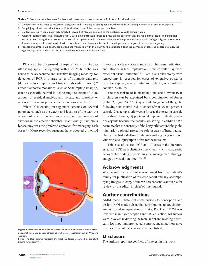
Table 2 proposed mechanisms for isolated posterior capsular rupture following forehead trauma
1. Compression injury leads to equatorial elongation and stretching of strong zonules, which leads to thinning or stretch of posterior capsule.2. Coup injury: direct contusion from rapid focal indentation of the cornea onto the lens.3. Contrecoup injury: rapid anteriorly directed rebound of vitreous can lead to the posterior capsule bursting open.4. Wieger’s ligament acts like a “battering ram”, using the contrecoup forces to press on the posterior capsule: rapid compression and expansion
forces directed along the anteroposterior axis of the eye may avulse the central region of the posterior lens capsule. Wieger’s ligament represents 8 mm in diameter of central lenticulo-vitreous adhesion that is most adherent in the midperipheral region of the lens of the young.
5. Forehead trauma: 1) eye protruded beyond the frontal line with the injury to the forehead hitting the cornea (our case); 2) in deep set eyes, the highly myopic eye renders the cornea at the level of the forehead–cheek line.12
PCR can be diagnosed preoperatively by B-scan
ultrasonography.6 Echography with a 20 MHz probe was
found to be an accurate and sensitive imaging modality for
detection of PCR in a large series of traumatic cataracts
(41 open-globe injuries and two closed-ocular injuries).13
Other diagnostic modalities, such as Scheimpflug imaging,
can be especially helpful in delineating the extent of PCR,
amount of residual nucleus and cortex, and presence or
absence of vitreous prolapse in the anterior chamber.8
When PCR occurs, management depends on several
parameters, such as the extent and location of the tear, the
amount of residual nucleus and cortex, and the presence of
vitreous in the anterior chamber. Traditionally, pars plana
lensectomy was the preferred approach for managing such
cases.1–3 More recently, surgeons have adopted a method
involving a clear corneal incision, phacoemulsification,
and intraocular lens implantation in the capsular bag, with
excellent visual outcome.7,9,11 Pars plana vitrectomy with
lensectomy is reserved for cases of extensive posterior
capsular rupture, marked vitreous prolapse, or significant
zonular instability.
The mechanism of blunt trauma-induced blowout PCR
in children can be explained by a combination of forces
(Table 2; Figure 5):10,16 1) equatorial elongation of the globe
following blunt trauma leads to stretch of zonules and posterior
capsule; 2) anteroposterior vector force to the posterior capsule
from direct trauma; 3) preferential rupture of elastic poste-
rior capsule because the zonules are strong in children.7 We
postulate that the anatomy of the bony orbit around the globe
might play a pivotal protective role in cases of head trauma.
Our patient had a shallow orbital rim, making the globe more
vulnerable to injury upon direct forehead trauma.
This case of isolated PCR and 17 cases in the literature
establish PCR as a distinct clinical entity with diagnostic
echographic findings, special surgical management strategy,
and good visual outcome.1–11,14,15
AcknowledgmentsWritten informed consent was obtained from the patient’s
family for publication of this case report and any accompa-
nying images. A copy of the written consent is available for
review by the editor-in-chief of this journal.
Author contributionsAMM made substantial contributions to conception and
design; MOJ made substantial contributions to acquisition,
analysis, and interpretation of data; RNH and FCM was
involved in initial conception and data collection. All authors
were involved in drafting the manuscript and revising it criti-
cally for important intellectual content, and all authors gave
final approval of the version to be published.
DisclosureThe authors report no conflicts of interest in this work.
Figure 5 artistic rendition of the most probable cause of posterior capsule rupture: equatorial globe and zonular stretch as well as anteroposterior pull by Wieger’s ligament.Note: the black arrows represent the tractional forces generated by the blunt trauma (white arrow).
Clinical Ophthalmology
Publish your work in this journal
Submit your manuscript here: http://www.dovepress.com/clinical-ophthalmology-journal
Clinical Ophthalmology is an international, peer-reviewed journal covering all subspecialties within ophthalmology. Key topics include: Optometry; Visual science; Pharmacology and drug therapy in eye diseases; Basic Sciences; Primary and Secondary eye care; Patient Safety and Quality of Care Improvements. This journal is indexed on
PubMed Central and CAS, and is the official journal of The Society of Clinical Ophthalmology (SCO). The manuscript management system is completely online and includes a very quick and fair peer-review system, which is all easy to use. Visit http://www.dovepress.com/testimonials.php to read real quotes from published authors.
Clinical Ophthalmology 2014:8 submit your manuscript | www.dovepress.com
Dovepress
Dovepress
Dovepress
2407
posterior capsular rupture
References1. Yasukawa T, Kita M, Honda Y. Traumatic cataract with a ruptured posterior
capsule from a nonpenetrating ocular injury. J Cataract Refract Surg. 1998;24:868–869.
2. Rao SK, Parikh S, Padhmanabhan P. Isolated posterior capsule rupture in blunt trauma: pathogenesis and management. Ophthalmic Surg Lasers. 1998;29:338–342.
3. Thomas R. Posterior capsule rupture after blunt trauma. J Cataract Refract Surg. 1998;24:283–284.
4. Rosen WJ, Campbell DG. Posterior capsule rupture after a paint-pellet injury. J Cataract Refract Surg. 2000;26:1422–1423.
5. Lee SI, Song HC. A case of isolated posterior capsule rupture and traumatic cataract caused by blunt ocular trauma. Korean J Ophthalmol. 2001;15:140–144.
6. Li KK, Groenewald C, Wong D. Management of traumatic posterior capsular rupture: corneal approach with high speed vitrector. J Cataract Refract Surg. 2005;31:1666–1668.
7. Por YM, Chee SP. Implantation of foldable intraocular lens with anterior optic capture in isolated posterior capsule rupture. J Cataract Refract Surg. 2006;32:707–708.
8. Grewal DS, Jain R, Brar GS, Grewal SP. Scheimpflug imaging of pediat-ric posterior capsule rupture. Indian J Ophthalmol. 2009;57:236–238.
9. Pavlovic S. Epilenticular intraocular lens implantation in traumatic cataract with a ruptured posterior capsule. Am J Ophthalmol. 2000;130: 352–353.
10. Campanella PC, Aminlari A, DeMaio R. Traumatic cataract and Wieger’s ligament. Ophthalmic Surg Lasers. 1997;28:422–423.
11. Pushker N, Sony P, Khokhar S, Vardhan P. Implantation of foldable intraocular lens with anterior optic capture in isolated posterior capsule rupture. J Cataract Refract Surg. 2005;31:1457–1458.
12. Angra SK, Vajpayee RB, Titiyal JS, Sharma YR, Sandramouli S, Kishore K. Types of posterior capsular breaks and their surgical implications. Ophthalmic Surg. 1991;22:388–391.
13. Tabatabaei A, Kiarudi MY, Ghassemi F, et al. Evaluation of posterior lens capsule by 20 MHz ultrasound probe in traumatic cataract. Am J Ophthalmol. 2012;153:51–54.
14. Saika S, Kin K, Ohmi S, Ohnishi Y. Posterior capsule rupture by blunt ocular trauma. J Cataract Refract Surg. 1997;23:139–140.
15. El-Gendy A, Rahman I, Mahmood U, Nylander A. Traumatic rupture of the posterior capsule resulting in complete posterior prolapse of the lens with subsequent resolution of high myopia. J Cataract Refract Surg. 2006;32:893–894.
16. Wolter JR. Coup-contrecoup mechanism of ocular injuries. Am J Ophthalmol. 1963;56:785–796.