Embed Size (px)
Citation preview
AbstractLeprosy is a chronic granulomatous disease involving theskin and nerves, leading to a debilitating condition.Leprosy has been controlled in most parts of the world;therefore physicians are not very well versed in therecognition, management and assessment of this disease.The protean manifestations of leprosy often lead to delaysin diagnosis and increase the morbidity. We present acase of a 33-year-old male with fever, lymphadenopathy,nodular skin lesions, uveitis and arthritis. Lymphnode,bonemarrow and skin biopsy revealed 3+ AFB smear withnegative AFB cultures, leading to the diagnosis of leprosy.The course of illness was complicated by flare of ErythemaNodosum Leprosum (ENL).
Keywords: Erythema Nodosum Leprosum; Leprosy;chronic fever.
IntroductionLeprosy is one of the most ancient, chronic infectiousdiseases. Although it has been eradicated mostlythroughout the world, it is still endemic in some parts ofSouth East Asia.
Pakistan was able to control leprosy in 1996 with ongoingefforts to eliminate the disease.1 Due to varyingpresentations, crippling complications, andcontagiousness, its timely diagnosis and management areextremely important. This chronic granulomatousinfection can affect skin, nerves, upper respiratory tractmucosa and eyes and causes progressive damage if leftuntreated.
As per World Health Organization classification, leprosycan be classified based on the number of lesions aspaucibacillary (less than or equal to 5 skin lesions with orwithout positive skin smear) and multibacillary (6 or morelesions with or without positive skin smear).2 Usually,presence of erythema nodosum leprosum is seen inpatients with high bacillary load and extensive skinlesions. It occurs due to antigen-antibody complexdeposition and may be associated with systemic
symptoms of uveitis,3 neuritis, lymphadenitis, orchitis,and arthritis.
We present the case of a 33 year old male presenting withlongstanding fever, which was later diagnosed asErythema Nodosum Leprosum as an initial presentationof leprosy.
Ethical approval from the institutional review board of theIndus Hospital, as well as verbal consent from the patientwas taken before submission of this case report.
Case ReportA 33 years old male, resident of Karachi, was seen in May2014 with 1.5 months history of high grade fever withchills and headache at the Family Medicine clinics of The
Vol. 68, No. 4, April 2018
653
CASE REPORT
Leprosy manifesting with type 2 leprae reaction in a patient presenting withchronic fever: A case reportFivzia Farooq Herekar, Hiba Ashraf, Naseem Salahuddin
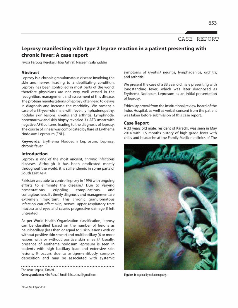
The Indus Hospital, Karachi.Correspondence: Hiba Ashraf. Email: [email protected] Figuire-1: Inguinal Lymphadenopathy.
Indus Hospital, Karachi. He was referred to the InfectiousDisease Clinic as a case of chronic fever. The review ofsystems was significant for dizziness, generalized bodyaches, pain and swelling of both ankles, as well as rednessof the right eye for the last one week. Rest of the systemicreview, past and personal medical history wereunremarkable.
On examination, he was a thin, lean male sittingcomfortably with marked redness of the right eye. He wasfebrile with a temperature of 38°C and normotensive.General physical examination revealed bilateralenlargement of inguinal and femoral lymph nodes,largest measuring 6 cm X3 cm (Figure-1). There wasswelling and redness over the right ankle joint but notenderness. Bilateral shins revealed a fadingerythematous raised, tender lesion, suggestive oferythema nodosum. He was initially worked up for pyrexiaof unknown origin with differentials of disseminatedgonococcal disease, disseminated Tuberculosis (TB),sarcoidosis, Still's disease, and infective endocarditis.
His investigations revealed anaemia. Haemoglobin was
6.4mg/dl with thrombocytosis4 of 772 x 109/L and a TLC of10.2x109/L. His HCV antibody was reactive, and ESR waselevated at 110, C-reactive protein was 46mg/L. Otherinvestigations including blood cultures, urine D/R,autoimmune profile, tests for HIV, VDRL, Malaria andBrucella were negative. His chest X-ray, ultrasoundabdomen and echocardiogram were normal. Bonemarrow trephine and biopsy along with lymph nodeFNAC revealed 3+ AFB, with GeneXpert and AFB culturespending. He was therefore labeled as a case ofDisseminated Extrapulmonary Tuberculosis.
He was started on Anti-tuberculous therapy withIsoniazid, Rifampicin, Ethambutol and Pyrazinamidealong with pyridoxine and showed gradual improvement,where his fever subsided and he reported weight gain.However, no regression of his lymph nodes was noted bythe 3rd month of treatment. Development of new lymphnodes in his submental and submandibular region wasnoted in the follow up visits with an increase in size ofinguinal lymph nodes with abscess formation requiringincision and drainage. Considering it to be TB ImmuneReconstitution Inflammatory Syndrome (IRIS), treatmentwas continued. The AFB culture and sensitivity smear forthe collected pus sample was again 3+AFB. By this time,the initial AFB cultures from the bone marrow and lymphnodes were reported to be negative.
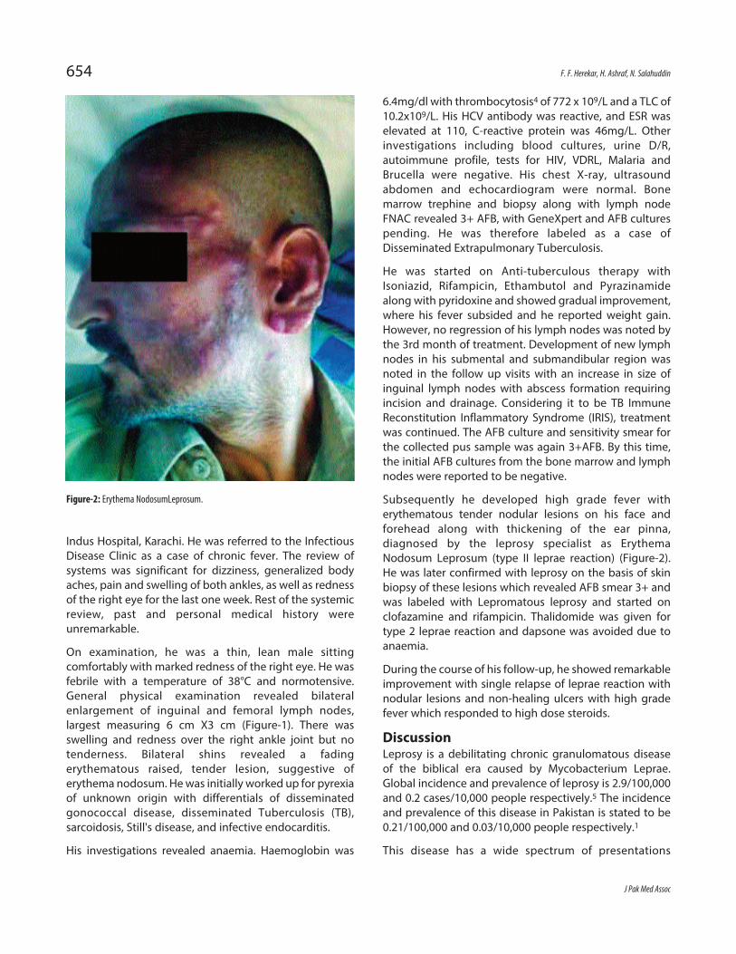
Subsequently he developed high grade fever witherythematous tender nodular lesions on his face andforehead along with thickening of the ear pinna,diagnosed by the leprosy specialist as ErythemaNodosum Leprosum (type II leprae reaction) (Figure-2).He was later confirmed with leprosy on the basis of skinbiopsy of these lesions which revealed AFB smear 3+ andwas labeled with Lepromatous leprosy and started onclofazamine and rifampicin. Thalidomide was given fortype 2 leprae reaction and dapsone was avoided due toanaemia.
During the course of his follow-up, he showed remarkableimprovement with single relapse of leprae reaction withnodular lesions and non-healing ulcers with high gradefever which responded to high dose steroids.
DiscussionLeprosy is a debilitating chronic granulomatous diseaseof the biblical era caused by Mycobacterium Leprae.Global incidence and prevalence of leprosy is 2.9/100,000and 0.2 cases/10,000 people respectively.5 The incidenceand prevalence of this disease in Pakistan is stated to be0.21/100,000 and 0.03/10,000 people respectively.1
This disease has a wide spectrum of presentations
J Pak Med Assoc
654 F. F. Herekar, H. Ashraf, N. Salahuddin
Figure-2: Erythema NodosumLeprosum.
ranging from the most common hypoesthetic skinlesions, to the pathognomonic deformities like saddlenose and Charcot joints. Hypersensitivity reactionsassociated with this disease complicates the course ofillness, hence increasing the morbidity. Type 2 Lepraereaction is associated with lepromatous leprosy with veryhigh burden of the mycobacteria leprae. In 90% of thecases, these reactions occur after starting treatment;however rarely they may occur as a primary presentationor even after completion of therapy.6 The type 2 lepraereaction is manifested by wide spread deposition ofimmune complexes in the skin, joints, lymph nodes, bonemarrow, kidneys, liver, spleen, endothelium and testes.The patient hence presents with the classical nodularlesions, erythema nodosum leprosum, and other systemicsymptoms of high grade fever, uveitis,3 arthralgia etc. Thedifficulty arises when this disease presents only asprolonged fever and lymphadenopathy,7 or initially asone of the leprae reactions in the patients who aretreatment naïve.8
In our patient's initial presentation of prolonged feverwith lymphadenopathy and arthritis without the classicalskin manifestations of leprosy, led to the complexity indiagnosing the disease. Moreover as patients withtuberculosis more frequently present with thesesymptoms in endemic regions, therefore pending AcidFast Bacilli culture report, patient was confidently startedon the standard Anti tuberculous therapy on the basis ofAFB smears. The reason Leprosy could not be confirmedinitially on FNAC is probably because it is a poorer testwhich causes destruction of the tissue hence skin for AFBsmear positive was taken as confirmatory in view of theother clinical findings.9 In a study conducted in India itwas observed that, in patients of leprosy withlymphadenopathy the most frequent site of involvementwas the inguinal region.10
Another manifestation of our patient was arthritis withuveitis3 hence leading us to consider Reiter's syndrome. Ina study conducted by Shiva Prasad and his team theyfound that out of 44 cases of proven leprosy themusculoskeletal manifestations included arthritis, swollenhands and feet, arthralgia and vasculitis, apart from 28cases of lepra reaction presenting with tenosynovitis andarthritis.11 Due to the high bacillary load patient maypresent with anaemia and thrombocytopenia12 or aspancytopenia due to bone marrow infiltration.13
Although our patient had bone marrow infiltration asseen by bone marrow AFB smears being +3 positive, hesuffered from anaemia and thrombocytosis4 rather thanpancytopenia.
ConclusionThe lack of pertinent knowledge regarding leprosyamongst general practitioners and the complicatedcourse and presentation of this disease leads to delayeddiagnosis and hence increasing the morbidity.
Acknowledgement: Dr. Mutahir Zia, Leprosy Specialist,MALC.
Disclaimer: None to declare.
Conflict of Interest: None to declare.
Funding Disclosure: None to declare.
References1. MALC. Leprosy Elimination 2015 [Cited 2016 27th October ].
Available from URL:http://www.malc.org.pk/leprosy_in_pakistan.html.
2. Leprosy Elimination: What is Leprosy [Internet]. 2015 [CitedAugust, 2017]. Available from URL:http://www.who.int/lep/disease/en/.
3. Citirik M, Batman C, Aslan O, Adabag A, Ozalp S, Zilelioglu O.Lepromatous iridocyclitis. Ocul Immunol Inflamm. 2005; 13: 95-9.
4. Rea TH. Elevated platelet counts and thrombocytosis inerythema nodosum leprosum. Int J Lepr Other Mycobact Dis.2002; 70: 167-73.
5. Leprosy [Internet]. February, 2017 [Cited August 2017]. Availablefrom URL: http://www.who.int/mediacentre/factsheets/fs101/en/.
6. Meyerson MS. Erythema nodosum leprosum. Int J Dermatol. 1996;35: 389-92.
7. Vinod K, Chandramohan R, Dutta T, Rajesh N, Basu D. Type 2 LepraReaction as a Cause of Pyrexia of Unknown Origin. J AssocPhysicians India. 2012; 60: 70-2.
8. Bala S, Sen S, Chatterjee G, Gangopadhyay A. Atypical erythemanodosum leprosum as the presenting feature in multibacillaryleprosy: A case report. Indian J Dermatol. 2014; 59: 94-5.
9. Rao IS, Singh MK, Gupta SD, Pandhi RK, Kapila K. Utility of fine-needle aspiration cytology in the classification of leprosy. DiagnCytopathol. 2001; 24: 317-21.
10. Kar H, Mohanty H, Mohanty G, Nayak U. Clinico-pathologicalstudy of lymph node involvement in leprosy. Lepr India. 1983;55: 725-38.
11. Prasad S, Misra R, Aggarwal A, Lawrence A, Haroon N, Wakhlu A, etal. Leprosy revealed in a rheumatology clinic: a case series. Int JRheum Dis. 2013; 16: 129-33.
12. de Oliveira LR, Maltos AL. Mycobacterium leprae in bone marrow.Rev Bras Hematol Hemoter 2014; 36: 300-1.
13. Binitha MP, Saritha S, Riyaz N, Mary V. Pancytopenia due tolepromatous involvement of the bone marrow: Successfultreatment with multidrug therapy. Lepr Rev. 2013; 84: 145-50.
Vol. 68, No. 4, April 2018
Leprosy manifesting with type 2 leprae reaction in a patient presenting with chronic fever: A case report 655