Embed Size (px)
Citation preview
173
Copyright © 2012 by Korean Society of Otorhinolaryngology-Head and Neck Surgery.This is an open-access article distributed under the terms of the Creative Commons Attribution Non-Commercial License (http://creativecommons.org/licenses/by-nc/3.0) which permits unrestricted non-commercial use, distribution, and reproduction in any medium, provided the original work is properly cited.
Nasal Hemangiopericytoma Causing Oncogenic Osteomalacia
Sung Il Cho, MD·Nam Yong Do, MD·Seung Woo Yu, MD·Ji Yun Choi, MD
Department of Otorhinolaryngology, Chosun University College of Medicine, Gwangju, Korea
Case Report
INTRODUCTION
Oncogenic osteomalacia is a very rare cause of osteomalacia. It is treated by surgical tumor resection. This tumor makes parane-oplastic syndrome like hypophosphatemic osteomalacia with hyperphosphaturia, low plasma 1,25-dihydroxyvitamin D and usually a normal serum calcium, parathormone, and 25-hy-droxyvitamin D [1]. Clinical features are bone pain, atrophy of proximal muscles and gait disturbance. Phosphaturia of onco-genic osteomalacia is from phosphaturic factor such as fibroblast growth factor 23 (FGF 23) that is secreted from tumor [2]. Among these tumors that cause osteomalacia, 10 cases have been re-ported worldwide in the ethmoid sinus [3-8]. We report a case of nasal hemangiopericytoma causing oncogenic osteomalacia and review the literature.
CASE REPORT
A 47-year-old woman presented with stuffy nose, postnasal drip-ping from 8 months ago. She had suffered from bone pains since a couple of years ago. Pain in the foot began in 3 years ago and
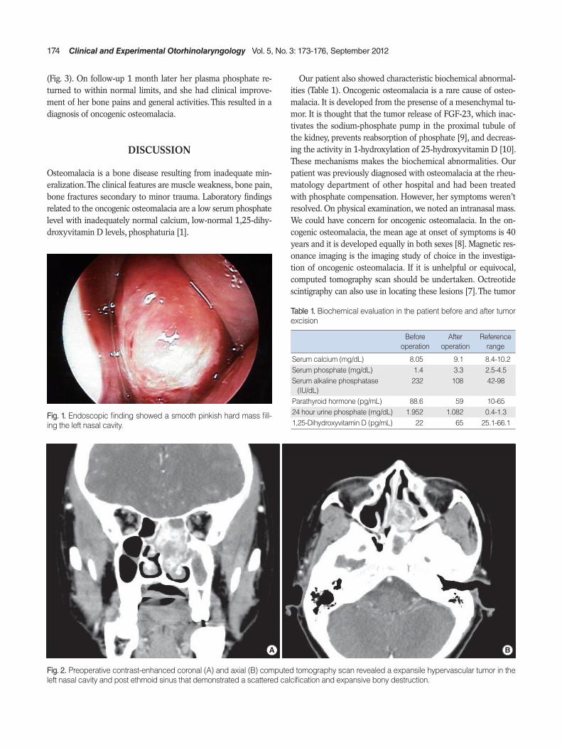
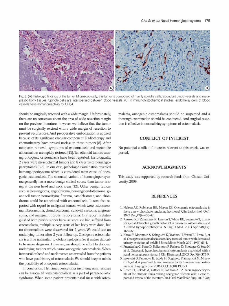
it also developed gradually in her lower leg and back. Fracture of the right femur and osteopenia was detected on general ra-diographs. She underwent conservative treatment and diagnostic workup. She was diagnosed as osteomalacia and took calcium and phosphate. On past medical history, the patient underwent a endoscopic surgery for sinusitis with polyp at 7 years ago. She had no remarkable family history. Nasal endoscopy revealed a smooth pinkish hard mass filling the left nasal cavity. It was ex-tended to superior nasal meatus and made lateral displacement of nasal septum and middle turbinate (Fig. 1). No abnormalities were noted in physical examination of other head and neck area. Nasal computed tomography revealed a expansile hypervascu-lar tumor with dimensions of 3×3×1 cm located in the left na-sal cavity and posterior ethmoid sinus. The mass demonstrated a scattered calcification and expansive bony destruction (Fig. 2). Laboratory examinations before surgery showed hypophospha-temia, normocalcemia, high serum alkaline phophatase, normal parathormone, normal serum 25-hydroxyvitamin D. Subsequent-ly, the patient underwent a biopsy, the histology revealed find-ings that were highly suggestive of fibroma. On the basis of these findings, the patient didn’t undergo selective angiography prior to surgery. We performed wide excision with about 5 mm free margin via endoscopic route under general anesthesia. It was difficult that we dissect the mass because of adhesion to skull base and sphenoid sinus. There was significant bleeding during the excision and the amount of bleeding was around 300 mL, but hemostasis was readily obtained with bipolar cautery and packing. Pathologic examination of the resected mass that was taken from the left nasal cavity showed hemangiopericytoma
Oncogenic osteomalacia is a rare cause that makes abnormalities of bone metabolism. Our case arose in a 47-year-old woman presenting a nasal mass associated with osteomalacia. We excised the mass carefully. After surgery, it was diagnosed as hemangiopericytoma and her symptoms related with osteomalacia were relieved and biochemical abnormalities were restored to normal range. We report and review a rare case of nasal hemangiopericytoma that caused osteomalacia.
Key Words. Nasal hemangiopericytoma, Osteomalacia
• Received October 5, 2009 Revised December 22, 2009 Accepted January 25, 2010
• Corresponding author: Ji Yun Choi, MDDepartment of Otorhinolaryngology, Chosun University Hospital, 365 Pilmun-daero, Dong-gu, Gwangju 501-717, Korea Tel: +82-62-220-3200, Fax: +82-62-225-2702 E-mail: [email protected]
Clinical and Experimental Otorhinolaryngology Vol. 5, No. 3: 173-176, September 2012 http://dx.doi.org/10.3342/ceo.2012.5.3.173pISSN 1976-8710 eISSN 2005-0720
174 Clinical and Experimental Otorhinolaryngology Vol. 5, No. 3: 173-176, September 2012
(Fig. 3). On follow-up 1 month later her plasma phosphate re-turned to within normal limits, and she had clinical improve-ment of her bone pains and general activities. This resulted in a diagnosis of oncogenic osteomalacia.
DISCUSSION
Osteomalacia is a bone disease resulting from inadequate min-eralization. The clinical features are muscle weakness, bone pain, bone fractures secondary to minor trauma. Laboratory findings related to the oncogenic osteomalacia are a low serum phosphate level with inadequately normal calcium, low-normal 1,25-dihy-droxyvitamin D levels, phosphaturia [1].
Our patient also showed characteristic biochemical abnormal-ities (Table 1). Oncogenic osteomalacia is a rare cause of osteo-malacia. It is developed from the presense of a mesenchymal tu-mor. It is thought that the tumor release of FGF-23, which inac-tivates the sodium-phosphate pump in the proximal tubule of the kidney, prevents reabsorption of phosphate [9], and decreas-ing the activity in 1-hydroxylation of 25-hydroxyvitamin D [10]. These mechanisms makes the biochemical abnormalities. Our patient was previously diagnosed with osteomalacia at the rheu-matology department of other hospital and had been treated with phosphate compensation. However, her symptoms weren’t resolved. On physical examination, we noted an intranasal mass. We could have concern for oncogenic osteomalacia. In the on-cogenic osteomalacia, the mean age at onset of symptoms is 40 years and it is developed equally in both sexes [8]. Magnetic res-onance imaging is the imaging study of choice in the investiga-tion of oncogenic osteomalacia. If it is unhelpful or equivocal, computed tomography scan should be undertaken. Octreotide scintigraphy can also use in locating these lesions [7]. The tumor
Fig. 1. Endoscopic finding showed a smooth pinkish hard mass fill-ing the left nasal cavity.
Fig. 2. Preoperative contrast-enhanced coronal (A) and axial (B) computed tomography scan revealed a expansile hypervascular tumor in the left nasal cavity and post ethmoid sinus that demonstrated a scattered calcification and expansive bony destruction.
A B
Table 1. Biochemical evaluation in the patient before and after tumor excision
Before operation
After operation
Reference range
Serum calcium (mg/dL) 8.05 9.1 8.4-10.2Serum phosphate (mg/dL) 1.4 3.3 2.5-4.5Serum alkaline phosphatase
(IU/dL)232 108 42-98
Parathyroid hormone (pg/mL) 88.6 59 10-6524 hour urine phosphate (mg/dL) 1.952 1.082 0.4-1.31,25-Dihydroxyvitamin D (pg/mL) 22 65 25.1-66.1
Cho SI et al.: Nasal Hemangiopericytoma 175
should be surgically resected with a wide margin. Unfortunately, there are no consensus about the area of wide resection margin on the previous literature, however we believe that the tumor must be surgically excised with a wide margin of resection to prevent recurrences. And preoperative embolization is applied because of its significant vascular component. Radiotherapy and chemotherapy have proved useless in these tumors [8]. After neoplasm removal, symptoms of osteomalacia and metabolic abnormalities are rapidly restored [11]. Ten ethmoid tumors caus-ing oncogenic osteomalacia have been reported. Histologically, 2 cases were mesenchymal tumors and 8 cases were hemangio-pericytomas [3-8]. In our case, pathologic examination revealed hemangiopericytoma which is considered main cause of onco-genic osteomalacia. The sinonasal variant of hemangiopericyto-ma generally has a more benign clinical course than tumor aris-ing at the non head and neck areas [12]. Other benign tumors such as hemangioma, angiofibroma, hemangioendothelioma, gi-ant cell tumor, nonossifying fibroma, osteoblastoma, and chon-droma could be associated with osteomalacia. It was also re-ported with regard to malignant tumors which were osteosarco-ma, fibrosarcoma, chondrosarcoma, synovial sarcoma, angiosar-coma, and malignant fibrous histiocytoma. Our report is distin-guished with previous ones because since she had suffered from osteomalacia, multiple survey scans of her body were done, but no abnormalities were discovered for 2 years. We could see an underlying tumor after 2 year follow-up. Oncogenic osteomala-cia is a little unfamiliar to otolaryngologists. So it makes difficul-ty to make diagnosis. However, we should be effort to discover underlying tumors which cause oncogenic osteomalacia. When intranasal or head and neck masses are revealed from the patients who have past history of osteomalacia, We should keep in minds the possibility of oncogenic osteomalacia. In conclusion, Hemangiopericytoma involving nasal sinuses can be associated with osteomalacia as a part of paraneoplastic syndrome. When some patient presents nasal mass with osteo-
Fig. 3. (A) Histologic findings of the tumor. Microscopically, this tumor is composed of mainly spindle cells, abundunt blood vessels and meta-plastic bony tissues. Spindle cells are interspersed between blood vessels. (B) In immunohistochemical studies, endothelial cells of blood vessels have immunoreactivity for CD34.
A B
malacia, oncogenic osteomalacia should be suspected and a thorough examination should be conducted. And surgical resec-tion is effective in normalizing symptoms of osteomalacia.
CONFLICT OF INTEREST
No potential conflict of interests relevant to this article was re-ported.
ACKNOWLEDGMENTS
This study was supported by research funds from Chosun Uni-versity, 2009.
REFERENCES
1. Nelson AE, Robinson BG, Mason RS. Oncogenic osteomalacia: is there a new phosphate regulating hormone? Clin Endocrinol (Oxf). 1997 Dec;47(6):635-42.
2. Jonsson KB, Zahradnik R, Larsson T, White KE, Sugimoto T, Imani-shi Y, et al. Fibroblast growth factor 23 in oncogenic osteomalacia and X-linked hypophosphatemia. N Engl J Med. 2003 Apr;348(17): 1656-63.
3. Kawai Y, Morimoto S, Sakaguchi K, Yoshino H, Yotsui T, Hirota S, et al. Oncogenic osteomalacia secondary to nasal tumor with decreased urinary excretion of cAMP. J Bone Miner Metab. 2001;19(1):61-4.
4. Fuentealba C, Pinto D, Ballesteros F, Pacheco D, Boettiger O, Soto N, et al. Oncogenic hypophosphatemic osteomalacia associated with a nasal hemangiopericytoma. J Clin Rheumatol. 2003 Dec;9(6):373-9.
5. Inokuchi G, Tanimoto H, Ishida H, Sugimoto T, Yamauchi M, Miyau-chi A, et al. A paranasal tumor associated with tumor-induced osteo-malacia. Laryngoscope. 2006 Oct;116(10):1930-3.
6. Beech TJ, Rokade A, Gittoes N, Johnson AP. A haemangiopericyto-ma of the ethmoid sinus causing oncogenic osteomalacia: a case re-port and review of the literature. Int J Oral Maxillofac Surg. 2007 Oct;

176 Clinical and Experimental Otorhinolaryngology Vol. 5, No. 3: 173-176, September 2012
36(10):956-8.7. Kenealy H, Holdaway I, Grey A. Occult nasal sinus tumours causing
oncogenic osteomalacia. Eur J Intern Med. 2008 Nov;19(7):516-9.8. Gonzalez-Compta X, Manos-Pujol M, Foglia-Fernandez M, Peral E,
Condom E, Claveguera T, et al. Oncogenic osteomalacia: case report and review of head and neck associated tumours. J Laryngol Otol. 1998 Apr;112(4):389-92.
9. Shimada T, Mizutani S, Muto T, Yoneya T, Hino R, Takeda S, et al. Cloning and characterization of FGF23 as a causative factor of tu-mor-induced osteomalacia. Proc Natl Acad Sci U S A. 2001 May;98 (11):6500-5.
10. Wilkins GE, Granleese S, Hegele RG, Holden J, Anderson DW, Bon-dy GP. Oncogenic osteomalacia: evidence for a humoral phosphatu-ric factor. J Clin Endocrinol Metab. 1995 May;80(5):1628-34.
11. Furco A, Roger M, Mouchet B, Richard O, Martinache X, Fur A. Os-teomalacia cured by surgery. Eur J Intern Med. 2002 Feb;13(1):67-9.
12. Catalano PJ, Brandwein M, Shah DK, Urken ML, Lawson W, Biller HF. Sinonasal hemangiopericytomas: a clinicopathologic and immu-nohistochemical study of seven cases. Head Neck. 1996 Jan-Feb; 18(1):42-53.


![REVIEW Open Access The modulation of apoptosis by oncogenic … · 2017. 8. 25. · transmissible oncogenic pathogen [4], and in 1932, Shope and Hurst demonstrated the oncogenic activity](https://img.pdfslide.net/doc/110x75/60a5adee03abc344316eb0df/review-open-access-the-modulation-of-apoptosis-by-oncogenic-2017-8-25-transmissible.jpg)