Embed Size (px)
Citation preview
Sugino et al. Diagnostic Pathology 2013, 8:134http://www.diagnosticpathology.org/content/8/1/134
CASE REPORT Open Access
Bronchiolitis obliterans associated withStevens-Johnson Syndrome: histopathologicalbronchial reconstruction of the whole lung andimmunohistochemical studyKeishi Sugino1*, Akira Hebisawa2, Toshimasa Uekusa3, Kazuhito Hatanaka4, Hiroshi Abe5 and Sakae Homma1
Abstract
This study presents an extremely rare case of constrictive bronchiolitis obliterans (BO) associated with Stevens-JohnsonSyndrome (SJS) provides the morphological and immunohistochemical features using histopathological bronchialreconstruction technique. A 27-year-old female developed progressive dyspnea after SJS induced by taking amoxicillinat the age of 10. Finally, she died of exacerbation of type II respiratory failure after 17 years from clinically diagnosed ashaving BO. Macroscopic bronchial reconstruction of the whole lungs at autopsy showed the beginning of bronchialobliterations was in the 4th to 5th branches, numbering from each segmental bronchus. Once they were obliterated,the distal and proximal bronchi were dilated. Microscopic bronchial reconstruction demonstrated the localization ofobliteration was mainly from small bronchi to membranous bronchioli with intermittent airway luminal narrowing orobliteration. Moreover, CD3-, CD20-, and CD68-positive cells were found in the BO lesions. CD34- and D2-40-positivecells were mainly distributed in the peribronchiolar lesions and bronchiolar lumens, respectively. SMA- and TGF-β-positive cells were seen in the fibrous tissue of BO lesions.The virtual slides: The virtual slide(s) for this article can be found here: http://www.diagnosticpathology.diagnomx.eu/vs/1071703140102601.
Keywords: Stevens-Johonson syndrome, Bronchiolitis obliterans, Constrictive bronchiolitis obliterans, Bronchialreconstruction, Immunohistochemistry
BackgroundStevens-Johnson syndrome is also called mucocutaneousocular syndrome and causes severe erythema exsudativummultiform [1]. SJS is caused by various drugs includingantimicrobial or antiepileptic drugs and infectious diseasessuch as mycoplasma and viruses [2]. Although pulmonarycomplications are often observed in SJS, bronchiolitisobliterans (BO) is extremely rare and its incidence is notstill understood.We described a patient of constrictive BO associated
with SJS that progressively deteriorated during long-termperiod and demonstrated characteristic histopathological
* Correspondence: [email protected] of Respiratory Medicine, Toho University Omori Medical Center,Omorinishi 6-11-1, Ota-ku, Tokyo 143-8541, JapanFull list of author information is available at the end of the article
© 2013 Sugino et al.; licensee BioMed CentralCommons Attribution License (http://creativecreproduction in any medium, provided the or
features by bronchial reconstruction and immunohisto-chemical stain.
Case presentationA 25-year-old female had a history of SJS after oral ad-ministration of amoxicillin at the age of 10. Two monthsafter the onset of SJS, she began to suffer from dyspneaon exertion and bilateral pneumothorax repeatedly. Thepatient was referred to our hospital due to fever andprogressive dyspnea at the age of 25. On admission, thepulmonary function tests showed mixed ventilatory andsmall airways impairment as follows: vital capacity (VC)of 1.12 L (36.8% of predicted), forced expiratory volumein 1 second (FEV1) of 0.60 L (53.1% of predicted), re-sidual volume/total lung capacity (RV/TLC) of 52.3%.Blood gas analysis showed PaO2 of 79.5 Torr and PaCO2
Ltd. This is an Open Access article distributed under the terms of the Creativeommons.org/licenses/by/2.0), which permits unrestricted use, distribution, andiginal work is properly cited.
Sugino et al. Diagnostic Pathology 2013, 8:134 Page 2 of 6http://www.diagnosticpathology.org/content/8/1/134
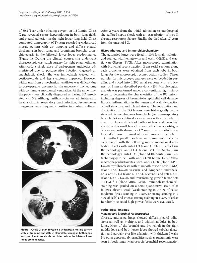
of 60.1 Torr under inhaling oxygen on 1.5 L/min. ChestX-ray revealed severe hyperinflation in both lung fieldsand pleural adhesion in the right lower lung field. Chestcomputed tomography (CT) scan revealed a widespreadmosaic pattern with air trapping and diffuse pleuralthickening in both lungs and prominent broncho-bron-chiolectasis in the bilateral lower lobes predominance(Figure 1). During the clinical course, she underwentthoracoscopic cyst stitch surgery for right pneumothorax.Afterward, a single dose of carbapenem antibiotics ad-ministered due to postoperative infection triggered ananaphylactic shock. She was immediately treated withcorticosteroids and her symptoms improved. However,withdrawal from a mechanical ventilator was difficult dueto postoperative pneumonia, she underwent tracheotomywith continuous mechanical ventilation. At the same time,the patient was clinically diagnosed as having BO associ-ated with SJS. Although azithromycin was administered totreat a chronic respiratory tract infection, Pseudomonasaeruginosa were frequently positive in sputum cultures.
Figure 1 Chest CT scan revealed a widespread mosaic patternwith air trapping and diffuse pleural thickening in both lungsand prominent broncho-bronchiolectasis in the bilateral lowerlobes predominance.
After 2 years from the initial admission to our hospital,she suffered septic shock with an exacerbation of type IIchronic respiratory failure. Finally, she died after 17 yearsfrom the onset of BO.
Histopathology and immunohistochemistryThe autopsied lungs were fixed in 10% formalin solutionand stained with hematoxylin and eosin (H&E) and elas-tic van Gieson (EVG). After macroscopic examinationwith bronchial reconstruction, 2 cm serial sections alongeach bronchus were obtained from each lobe in bothlungs for the microscopic reconstruction studies. Tissuesamples for microscopic analyses were embedded in par-affin, and sliced into 1,200 serial sections with a thick-ness of 4 μm as described previously [3]. Morphologicalanalysis was performed under a conventional light micro-scope to determine the characteristics of the BO lesions,including degrees of bronchiolar epithelial cell damage,fibrosis, inflammation in the lumen and wall, destructionof wall structure, and dilated airway. The localization anddistribution of the BO lesions were histologically recon-structed. A membranous bronchiole (i.e. non-respiratorybronchiole) was defined as an airway with a diameter of2 mm or less and lack of both cartilage and bronchialglands, and a small bronchus was defined as a cartilagin-ous airway with diameter of 2 mm or more, which waslocated in more proximal of membranous bronchiole.4 μm-thick paraffin sections were immunohistochemi-
cally stained with the following mouse monoclonal anti-bodies: T cells with anti-CD3 (clone UCH-T1, Santa CruzBiotechnology), anti-CD4 (clone MT310, Santa CruzBiotechnology), anti-CD8 (clone 5F10, Santa Cruz Bio-technology); B cell with anti-CD20 (clone L26, Dako);macrophages/histiocytes with anti-CD68 (clone KP-1,Dako); myofibroblasts with α-smooth muscle actin (SMA)(clone 1A4, Dako); vascular and lymphatic endothelialcells, anti-CD34 (clone NU-4A1, Nichirei), and anti-D2-40(clone D2-40, Dako), and transforming growth factor-beta1 (TGF-β1) (clone 9016, R&D). Immunohistochemical-staining was graded on a semi-quantitative scale of asfollows: absent, weak (weak staining in < 50% of cells),moderate (weak staining in > 50% or strong staining in <50% of cells) and intense (strong staining in > 50% of cells).Randomly selected high power fields were evaluated.
Pathological findingsMacroscopic bronchial reconstructionGrossly, autopsied lungs showed diffuse pleural adhe-sions as well as multiple, and whitish nodules in bothlungs. Most of the bronchi and bronchioli in the rightmiddle lobe and both lower lobes showed tubular dilata-tion and partially cyst-like dilatation with thickened walls.No other apparent abnormalities such as pneumonia wereseen in both lungs. Macroscopic bronchial reconstruction
Sugino et al. Diagnostic Pathology 2013, 8:134 Page 3 of 6http://www.diagnosticpathology.org/content/8/1/134
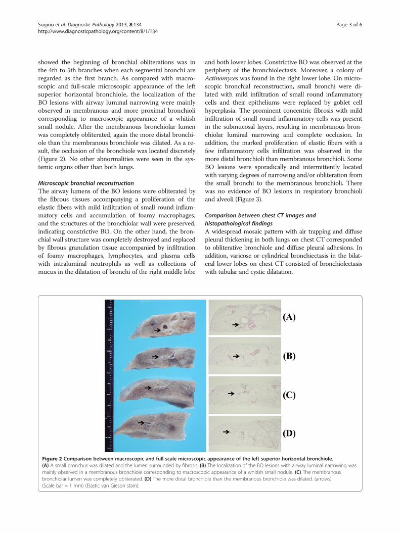
showed the beginning of bronchial obliterations was inthe 4th to 5th branches when each segmental bronchi areregarded as the first branch. As compared with macro-scopic and full-scale microscopic appearance of the leftsuperior horizontal bronchiole, the localization of theBO lesions with airway luminal narrowing were mainlyobserved in membranous and more proximal bronchiolicorresponding to macroscopic appearance of a whitishsmall nodule. After the membranous bronchiolar lumenwas completely obliterated, again the more distal bronchi-ole than the membranous bronchiole was dilated. As a re-sult, the occlusion of the bronchiole was located discretely(Figure 2). No other abnormalities were seen in the sys-temic organs other than both lungs.
Microscopic bronchial reconstructionThe airway lumens of the BO lesions were obliterated bythe fibrous tissues accompanying a proliferation of theelastic fibers with mild infiltration of small round inflam-matory cells and accumulation of foamy macrophages,and the structures of the bronchiolar wall were preserved,indicating constrictive BO. On the other hand, the bron-chial wall structure was completely destroyed and replacedby fibrous granulation tissue accompanied by infiltrationof foamy macrophages, lymphocytes, and plasma cellswith intraluminal neutrophils as well as collections ofmucus in the dilatation of bronchi of the right middle lobe
Figure 2 Comparison between macroscopic and full-scale microscopi(A) A small bronchus was dilated and the lumen surrounded by fibrosis. (Bmainly observed in a membranous bronchiole corresponding to macroscobronchiolar lumen was completely obliterated. (D) The more distal bronch(Scale bar = 1 mm) (Elastic van Gieson stain).
and both lower lobes. Constrictive BO was observed at theperiphery of the bronchiolectasis. Moreover, a colony ofActinomyces was found in the right lower lobe. On micro-scopic bronchial reconstruction, small bronchi were di-lated with mild infiltration of small round inflammatorycells and their epitheliums were replaced by goblet cellhyperplasia. The prominent concentric fibrosis with mildinfiltration of small round inflammatory cells was presentin the submucosal layers, resulting in membranous bron-chiolar luminal narrowing and complete occlusion. Inaddition, the marked proliferation of elastic fibers with afew inflammatory cells infiltration was observed in themore distal bronchioli than membranous bronchioli. SomeBO lesions were sporadically and intermittently locatedwith varying degrees of narrowing and/or obliteration fromthe small bronchi to the membranous bronchioli. Therewas no evidence of BO lesions in respiratory bronchioliand alveoli (Figure 3).
Comparison between chest CT images andhistopathological findingsA widespread mosaic pattern with air trapping and diffusepleural thickening in both lungs on chest CT correspondedto obliterative bronchiole and diffuse pleural adhesions. Inaddition, varicose or cylindrical bronchiectasis in the bilat-eral lower lobes on chest CT consisted of bronchiolectasiswith tubular and cystic dilatation.
c appearance of the left superior horizontal bronchiole.) The localization of the BO lesions with airway luminal narrowing waspic appearance of a whitish small nodule. (C) The membranousiole than the membranous bronchiole was dilated. (arrows)
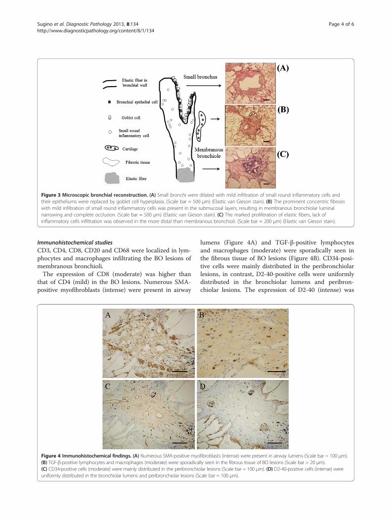
Figure 3 Microscopic bronchial reconstruction. (A) Small bronchi were dilated with mild infiltration of small round inflammatory cells andtheir epitheliums were replaced by goblet cell hyperplasia. (Scale bar = 500 μm) (Elastic van Gieson stain). (B) The prominent concentric fibrosiswith mild infiltration of small round inflammatory cells was present in the submucosal layers, resulting in membranous bronchiolar luminalnarrowing and complete occlusion. (Scale bar = 500 μm) (Elastic van Gieson stain). (C) The marked proliferation of elastic fibers, lack ofinflammatory cells infiltration was observed in the more distal than membranous bronchioli. (Scale bar = 200 μm) (Elastic van Gieson stain).
Sugino et al. Diagnostic Pathology 2013, 8:134 Page 4 of 6http://www.diagnosticpathology.org/content/8/1/134
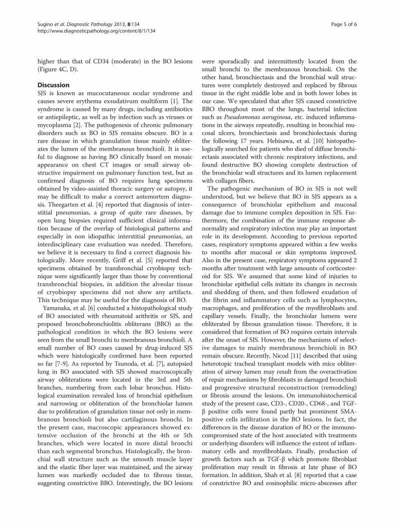
Immunohistochemical studiesCD3, CD4, CD8, CD20 and CD68 were localized in lym-phocytes and macrophages infiltrating the BO lesions ofmembranous bronchioli.The expression of CD8 (moderate) was higher than
that of CD4 (mild) in the BO lesions. Numerous SMA-positive myofibroblasts (intense) were present in airway
Figure 4 Immunohistochemical findings. (A) Numerous SMA-positive myo(B) TGF-β-positive lymphocytes and macrophages (moderate) were sporadica(C) CD34-positive cells (moderate) were mainly distributed in the peribronchiouniformly distributed in the bronchiolar lumens and peribronchiolar lesions (S
lumens (Figure 4A) and TGF-β-positive lymphocytesand macrophages (moderate) were sporadically seen inthe fibrous tissue of BO lesions (Figure 4B). CD34-posi-tive cells were mainly distributed in the peribronchiolarlesions, in contrast, D2-40-positive cells were uniformlydistributed in the bronchiolar lumens and peribron-chiolar lesions. The expression of D2-40 (intense) was
fibroblasts (intense) were present in airway lumens (Scale bar = 100 μm).lly seen in the fibrous tissue of BO lesions (Scale bar = 20 μm).lar lesions (Scale bar = 100 μm). (D) D2-40-positive cells (intense) werecale bar = 100 μm).
Sugino et al. Diagnostic Pathology 2013, 8:134 Page 5 of 6http://www.diagnosticpathology.org/content/8/1/134
higher than that of CD34 (moderate) in the BO lesions(Figure 4C, D).
DiscussionSJS is known as mucocutaneous ocular syndrome andcauses severe erythema exsudativum multiform [1]. Thesyndrome is caused by many drugs, including antibioticsor antiepileptic, as well as by infection such as viruses ormycoplasma [2]. The pathogenesis of chronic pulmonarydisorders such as BO in SJS remains obscure. BO is arare disease in which granulation tissue mainly obliter-ates the lumen of the membranous bronchioli. It is use-ful to diagnose as having BO clinically based on mosaicappearance on chest CT images or small airway ob-structive impairment on pulmonary function test, but asconfirmed diagnosis of BO requires lung specimensobtained by video-assisted thoracic surgery or autopsy, itmay be difficult to make a correct antemortem diagno-sis. Theegarten et al. [4] reported that diagnosis of inter-stitial pneumonias, a group of quite rare diseases, byopen lung biopsies required sufficient clinical informa-tion because of the overlap of histological patterns andespecially in non idiopathic interstitial pneumonias, aninterdisciplinary case evaluation was needed. Therefore,we believe it is necessary to find a correct diagnosis his-tologically. More recently, Griff et al. [5] reported thatspecimens obtained by transbronchial cryobiopsy tech-nique were significantly larger than those by conventionaltransbronchial biopsies, in addition the alveolar tissueof cryobiopsy specimens did not show any artifacts.This technique may be useful for the diagnosis of BO.Yamanaka, et al. [6] conducted a histopathological study
of BO associated with rheumatoid arithritis or SJS, andproposed bronchobronchiolitis obliterans (BBO) as thepathological condition in which the BO lesions wereseen from the small bronchi to membranous bronchioli. Asmall number of BO cases caused by drug-induced SJSwhich were histologically confirmed have been reportedso far [7-9]. As reported by Tsunoda, et al. [7], autopsiedlung in BO associated with SJS showed macroscopicallyairway obliterations were located in the 3rd and 5thbranches, numbering from each lobar bronchus. Histo-logical examination revealed loss of bronchial epitheliumand narrowing or obliteration of the bronchiolar lumendue to proliferation of granulation tissue not only in mem-branous bronchioli but also cartilaginous bronchi. Inthe present case, macroscopic appearances showed ex-tensive occlusion of the bronchi at the 4th or 5thbranches, which were located in more distal bronchithan each segmental bronchus. Histologically, the bron-chial wall structure such as the smooth muscle layerand the elastic fiber layer was maintained, and the airwaylumen was markedly occluded due to fibrous tissue,suggesting constrictive BBO. Interestingly, the BO lesions
were sporadically and intermittently located from thesmall bronchi to the membranous bronchioli. On theother hand, bronchiectasis and the bronchial wall struc-tures were completely destroyed and replaced by fibroustissue in the right middle lobe and in both lower lobes inour case. We speculated that after SJS caused constrictiveBBO throughout most of the lungs, bacterial infectionsuch as Pseudomonas aeruginosa, etc. induced inflamma-tions in the airways repeatedly, resulting in bronchial mu-cosal ulcers, bronchiectasis and bronchiolectasis duringthe following 17 years. Hebisawa, et al. [10] histopatho-logically searched for patients who died of diffuse bronchi-ectasis associated with chronic respiratory infections, andfound destructive BO showing complete destruction ofthe bronchiolar wall structures and its lumen replacementwith collagen fibers.The pathogenic mechanism of BO in SJS is not well
understood, but we believe that BO in SJS appears as aconsequence of bronchiolar epithelium and mucosaldamage due to immune complex deposition in SJS. Fur-thermore, the combination of the immune response ab-normality and respiratory infection may play an importantrole in its development. According to previous reportedcases, respiratory symptoms appeared within a few weeksto months after mucosal or skin symptoms improved.Also in the present case, respiratory symptoms appeared 2months after treatment with large amounts of corticoster-oid for SJS. We assumed that some kind of injuries tobronchiolar epithelial cells initiate its changes in necrosisand shedding of them, and then followed exudation ofthe fibrin and inflammatory cells such as lymphocytes,macrophages, and proliferation of the myofibroblasts andcapillary vessels. Finally, the bronchiolar lumens wereobliterated by fibrous granulation tissue. Therefore, it isconsidered that formation of BO requires certain intervalsafter the onset of SJS. However, the mechanisms of select-ive damages to mainly membranous bronchioli in BOremain obscure. Recently, Nicod [11] described that usingheterotopic tracheal transplant models with mice obliter-ation of airway lumen may result from the overactivationof repair mechanisms by fibroblasts in damaged bronchioliand progressive structural reconstruction (remodeling)or fibrosis around the lesions. On immunohistochemicalstudy of the present case, CD3-, CD20-, CD68-, and TGF-β positive cells were found partly but prominent SMA-positive cells infiltration in the BO lesions. In fact, thedifferences in the disease duration of BO or the immuno-compromised state of the host associated with treatmentsor underlying disorders will influence the extent of inflam-matory cells and myofibroblasts. Finally, production ofgrowth factors such as TGF-β which promote fibroblastproliferation may result in fibrosis at late phase of BOformation. In addition, Shah et al. [8] reported that a caseof constrictive BO and eosinophilic micro-abscesses after
Sugino et al. Diagnostic Pathology 2013, 8:134 Page 6 of 6http://www.diagnosticpathology.org/content/8/1/134
SJS. It is known that eosinophils are important sources ofa variety of pro-fibrogenic mediators such as TGF-α [12],TGF-β [13], vascular endothelial growth factor [14], andinterleukin-13 [15]. Although eosinophils were not foundin the BO lesions at autopsy in the present case, increasedeosinophils could be determined at an earlier stage of BO.
ConclusionThis study provides important information on the mor-phological and immunohistochemical features in an ex-tremely rare constrictive BO associated with SJS usinghistopathological bronchial reconstruction technique.The involvement of epithelial-mesenchymal transition orpro-fibrotic growth factors will need to be investigatedin the future to clarify the mechanism of development offibrotic lesions in BO associated with SJS.
ConsentWritten informed consent was obtained from the patient'snext-of-kin for publication of this manuscript and anyaccompanying images. A copy of the written consent isavailable for review by the Editor-in-Chief of this journal.
AbbreviationsSJS: Stevens-Johnson syndrome; BO: Bronchiolitis obliterans;BBO: Bronchobronchiolitis obliterans; CT: Computed tomography;H&E: Hematoxylin and eosin; EVG: Elastic van Gieson; TGF-β: Transforminggrowth factor-beta; SMA: α-smooth muscle actin.
Competing interestsAll authors declare that they have no competing interest.
Authors’ contributionsK S participated in the design of the study and histopathological evaluation,and drafted the manuscript. S H assisted in drafting the manuscript andrevised the manuscript. A H, T U, and K H made contributions for analyzingthe histopathological characteristics. H A sliced each lung specimen intoserial sections and carried out the H&E, EVG, and immunohistochemicalstains evaluation. All the authors read and approved the final manuscript.
AcknowledgementsThis work was supported by a grant-in-aid for interstitial lung diseases fromthe Japanese Ministry of Health, Labour and Welfare.
Author details1Department of Respiratory Medicine, Toho University Omori Medical Center,Omorinishi 6-11-1, Ota-ku, Tokyo 143-8541, Japan. 2Department of Pathology,Tokyo National Hospital, Tokyo, Japan. 3Department of Pathology, LaborHealth and Welfare Organization Kanto Rosai Hospital, Kanagawa, Japan.4Department of Molecular and Cellular Pathology, Kagoshima GraduateSchool of Medicine and Dental Sciences, Kagoshima, Japan. 5Department ofPathology, Juntendo University School of Medicine, Tokyo, Japan.
Received: 25 June 2013 Accepted: 30 July 2013Published: 6 August 2013
References1. Bastuji-Garin S, Rzany B, Stern RS, Shear NH, Naldi L, Roujeau JC: Clinical
classification of cases of toxic epidermal necrolysis, Stevens-Johnsonsyndrome, and erythema multiform. Arch Dermatol 1993, 129:92–96.
2. Letko E, Papaliodis DN, Papaliodis GN, Daoud YJ, Ahmed AR, Foster CS:Stevens-Johnson syndrome and toxic epidermal necrolysis: a review ofthe literature. Ann Allergy Asthma Immunol 2005, 94:419–436.
3. Sugino K, Hebisawa A, Uekusa T, Hatanaka K, Abe H, Homma S:Histopathological bronchial reconstruction of human bronchiolitisobliterans. Pathol Int 2011, 61:192–201.
4. Theegarten D, Muller HM, Bonella F, Wohlschlaeger J, Costabel U:Diagnostic approach to interstitial pneumonias in a single centre: reporton 88 cases. Diagn Pathol 2012, 7:160. doi:10.1186/1746-1596-7-160.
5. Griff S, Ammenwerth W, Schönfeld N, Bauer TT, Mairinger T, Blum TG,Kollmeier J, Grüning W: Morphometrical analysis of transbronchialcryobiopsies. Diagn Pathol 2011, 6:53. 10.1186/1746-1596-6-53.
6. Yamanaka A, Maeda M, Yamamoto R: Chronic obstractive bronchiolitis -including bronchobronchiolitis obliterans- (in Japanese). Nihon KyobuRinsho 1986, 45:539–554.
7. Tsunoda N, Iwanaga T, Saito T, Kitamura S, Saito K: Rapidly progressivebronchiolitis obliterans associated with Stevens-Johnson syndrome.Chest 1990, 98:243–245.
8. Shah AP, Xu H, Sime PJ, Trawick DR: Severe airflow obstruction andeosinophilic lung disease after Stevens-Johnson syndrome. Eur Respir J2006, 28:1276–1279.
9. Date H, Sano Y, Aoe M, Goto K, Tedoriya T, Sano S, Andou A, Shimizu N:Living-donor lobar lung transplantation for bronchiolitis obliterans afterStevens-Johnson syndrome. J Thoracic Cardiovasc Surg 2002, 123:389–391.
10. Hebisawa A, Baba M, Yasuda K, Tamura A: Pathology of diffusebronchiectasis (in Japanese). Pathol Clin Med 2002, 20:898–903.
11. Nicod LP: Mechanisms of airway obliteration after lung transplantation.Proc Am Thorac Soc 2006, 3:444–449.
12. Wong DT, Weller PF, Galli SJ, Elovic SA, Band TH, Gallagher GT, Chiang T,Chou MY, Matossian K, McBride J, Todd R: Human eosinophils expresstransforming growth factor alpha. J Exp Med 1990, 172:673–681.
13. Zhang K, Flanders KC, Phan SH: Cellular localization of transforminggrowth factor-beta expression in bleomycininduced pulmonary fibrosis.Am J Pathol 1995, 147:352–361.
14. Hoshino M, Takahashi M, Aoike N: Expression of vascular endothelialgrowth factor, basic fibroblast growth factor, and angiogeninimmunoreactivity in asthmatic airways and its relationship toangiogenesis. J Allergy Clin Immunol 2001, 107:295–301.
15. Lukacs NW, Hogaboam C, Chensue SW, Blease K, Kunkel SL: Type 1/type 2cytokine paradigm and the progression of pulmonary fibrosis. Chest2001, 120:5S–8S.
doi:10.1186/1746-1596-8-134Cite this article as: Sugino et al.: Bronchiolitis obliterans associated withStevens-Johnson Syndrome: histopathological bronchial reconstructionof the whole lung and immunohistochemical study. Diagnostic Pathology2013 8:134.
Submit your next manuscript to BioMed Centraland take full advantage of:
• Convenient online submission
• Thorough peer review
• No space constraints or color figure charges
• Immediate publication on acceptance
• Inclusion in PubMed, CAS, Scopus and Google Scholar
• Research which is freely available for redistribution
Submit your manuscript at www.biomedcentral.com/submit