Embed Size (px)
Citation preview

CASE REPORT Open Access
Efficacy of postural techniques assessed byvideofluoroscopy for myasthenia gravis withdysphagia as the presenting symptom:a case reportHui-Chun Juan1, Isabel Tou2, Shu-Chen Lo2, I-Hsien Wu1*
Abstract
Introduction: Oropharyngeal weakness leading to dysphagia is rarely the presenting symptom of myastheniagravis, but it can be a significant source of morbidity and mortality. The earliest possible diagnosis of myastheniagravis should be made for better management of this cause of treatable dysphagia. A detailed evaluation ofswallowing by videofluoroscopy can assist in making an accurate diagnosis and in individualizing appropriate dietcompensatory techniques.
Case presentation: We present the case of a 57-year-old Taiwanese man with dysphagia as the presentingsymptom of myasthenia gravis, and evaluate the pathological findings of swallowing and effectiveness ofcompensatory postural techniques for dysphagia using videofluoroscopy.
Conclusions: Videofluoroscopy is a valuable technique for evaluating myasthenia gravis dysphagia, because itallows swallowing interventions to be precisely individualized in accordance with the results obtained.
IntroductionMyasthenia gravis (MG) is an autoimmune disorder inwhich autoantibodies are directed against acetylcholinereceptors in the neuromuscular junctions [1]. It is char-acterized by painless and fatigable weakness of skeletalmuscles. Although over 60% of patients with MG haveocular symptoms at presentation, dysphagia related toweakness of the oropharyngeal muscles can also be apresenting symptom. Dysphagia with aspiration is a sig-nificant source of morbidity and mortality in MG [2];consequently, a detailed assessment of swallowing isvery important in patients with MG dysphagia.Videofluoroscopy is a useful evaluation tool for obser-
ving the oral, pharyngeal, and esophageal stages of swal-lowing physiology in patients ingesting radiopaque foods[3], and swallowing evaluation by videofluoroscopy inpatients with MG has been widely utilized in previousstudies. However, subsequent videofluoroscopic
monitoring of compensatory postural techniques toguide appropriate management of swallowing in MGhas been less frequently mentioned. Here, we report thecase of a patient with dysphagia as his main presentingsymptom and describe the use of videofluoroscopy toevaluate his swallowing status and the effectiveness ofcompensatory postural techniques.
Case presentationA 57-year-old Taiwanese man presented to our facilitywith a one-month history of progressive difficulty inswallowing, particularly liquids. In addition, a bodyweight loss of 10 kg was noted. His medical historyincluded chronic sinusitis and chronic serous otitismedia. A chest X-ray revealed incidental right middlelobe collapse when he was admitted for surgical treat-ment for chronic paranasal sinusitis. Obstructive pneu-monitis with a right lower lung field mass lesion wassuspected. A physical examination revealed left ptosis,dysarthria, and mild bilateral shoulder girdle weaknesswithout a definite diurnal change. He was subsequentlydiagnosed as having MG based upon the decremental
* Correspondence: [email protected] of Physical Medicine and Rehabilitation, Chi Mei MedicalCenter, Liouying, TaiwanFull list of author information is available at the end of the article
Juan et al. Journal of Medical Case Reports 2010, 4:370http://www.jmedicalcasereports.com/content/4/1/370 JOURNAL OF MEDICAL
CASE REPORTS
© 2010 Juan et al; licensee BioMed Central Ltd. This is an Open Access article distributed under the terms of the Creative CommonsAttribution License (http://creativecommons.org/licenses/by/2.0), which permits unrestricted use, distribution, and reproduction inany medium, provided the original work is properly cited.
response to repetitive stimulation on electrophysiologicaltesting in association with a positive anti-acetylcholinereceptor antibody test. The MG stage was assessed asgrade IIA according to the Osserman classification atthat time.A clinical swallowing evaluation by a speech/language
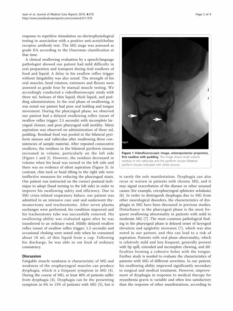
pathologist showed our patient had mild difficulty inoral preparation and transport during trial swallows offood and liquid. A delay in his swallow reflex triggerwithout fatigability was also noted. The strength of hisoral muscles, head rotators, extensors and flexors wereassessed as grade four by manual muscle testing. Weaccordingly conducted a videofluoroscopic study withthree mL boluses of thin liquid, thick liquid, and pud-ding administration. In the oral phase of swallowing, itwas noted our patient had poor oral holding and tonguemovement. During the pharyngeal phase, we observedour patient had a delayed swallowing reflex (onset ofswallow reflex trigger: 2.5 seconds) with incomplete lar-yngeal closure, and poor pharyngeal wall motility. Silentaspiration was observed on administration of three mLpudding. Residual food was pooled in the bilateral pyri-form sinuses and valleculae after swallowing three con-sistencies of sample material. After repeated consecutiveswallows, the residues in the bilateral pyriform sinusesincreased in volume, particularly on the left side(Figure 1 and 2). However, the residues decreased involume when his head was turned to the left side andthere was no evidence of silent aspiration (Figure 3). Incontrast, chin tuck or head tilting to the right side wereineffective measures for reducing the pharyngeal stasis.Our patient was instructed on the correct postural tech-nique to adopt (head turning to the left side) in order toimprove his swallowing safety and efficiency. Due toMG crisis-related respiratory failure, our patient wasadmitted to an intensive care unit and underwent thy-momectomy and tracheostomy. After seven plasmaexchanges were performed, his condition improved andhis tracheostomy tube was successfully removed. Hisswallowing ability was evaluated again after he wastransferred to an ordinary ward. Mildly delayed swallowreflex (onset of swallow reflex trigger: 1.5 seconds) andoccasional choking were noted only when he consumedabout 10 mL of thin liquid from a cup. Followinghis discharge, he was able to eat food of ordinaryconsistency.
DiscussionFatigable muscle weakness is characteristic of MG andweakness of the oropharyngeal muscles can producedysphagia, which is a frequent symptom in MG [4].During the course of MG, at least 40% of patients sufferfrom dysphagia [4]. Dysphagia can be the presentingsymptom in 6% to 15% of patients with MG [5], but it
is rarely the sole manifestation. Dysphagia can alsorecur or worsen in patients with chronic MG, and itmay signal exacerbation of the disease or other unusualcauses (for example, cricopharyngeal sphincter achalasia)[6]. In order to distinguish dysphagia due to MG fromother neurological disorders, the characteristics of dys-phagia in MG have been discussed in previous studies.Disturbance in the pharyngeal phase is the most fre-quent swallowing abnormality in patients with mild tomoderate MG [7]. The most common pathological find-ing in the pharyngeal phase is delayed onset of laryngealelevation and epiglottic inversion [7], which was alsonoted in our patient, and this can lead to a risk ofaspiration. Patients with oral phase abnormality, whichis relatively mild and less frequent, generally presentwith lip spill, extended and incomplete chewing, and dif-ficulties forming a cohesive bolus with the tongue.Further study is needed to evaluate the characteristics ofpatients with MG of different severities. In our patient,his swallowing ability improved significantly secondaryto surgical and medical treatment. However, improve-ment of dysphagia in response to medical therapy formyasthenia gravis is variable and often less satisfactorythan the response of other manifestations, according to
Figure 1 Videofluoroscopic image, anteroposterior projection,first swallow with pudding. The image shows small volumeresidues in the valleculae and the pyriform sinuses (bilateralpyriform sinuses indicated with white arrows).
Juan et al. Journal of Medical Case Reports 2010, 4:370http://www.jmedicalcasereports.com/content/4/1/370
Page 2 of 4

previous studies [8]. Further monitoring of MG-relateddysphagia could be necessary after other symptomsimprove.For an evaluation of dysphagia, a thorough history
and clinical examination provide valuable information.The swallowing ability of patients with myastheniawith dysphagia typically, but not always, shows fatig-ability during meals and as the day progresses. How-ever, clinical examination alone is insufficient to detectand grade dysphagia in MG, and additional instrumen-tal assessment tools may be necessary [9]. Accordingto the study of Colton-Hudson et al. in 2002, theseverity of MG dysphagia as determined by video-fluoroscopic study was worse than that predicted byclinical evaluation. They accordingly suggested routinevideofluoroscopic examination [7]. Videofluoroscopy isregarded as the gold standard in dysphagia diagnosisand management [3]. For patients with MG with dys-phagia as the presenting or sole manifestation, video-fluoroscopy is helpful for early and accurate diagnosisbecause insidious fatigability after consecutive swallow-ing can be detected, as in our patient. Videofluoro-scopy can also be combined with the Tensilon test toassist in diagnosis of bulbar MG. The combination is
particularly valuable for the subgroup of patients withMG who have prominent bulbar symptomatology, andit is more reliable than videofluoroscopy alone [10].After medical management of MG, videofluoroscopy isindicated for following up the course of dysphagia tomodify subsequent treatment strategies, particularly forthose patients with dysphagia that does not improve asquickly as other manifestations. The limitations ofvideofluoroscopy swallow studies are generally relatedto radiation exposure; however, radiation exposure israrely a limiting factor in adults [3]. Fiberoptic endo-scopic evaluation of swallowing (FEES) is another com-mon instrumental swallowing evaluation tool, whichhas greater portability than videofluoroscopy andinvolves no radiation exposure [3]. For individuals withphysical limitations that prevent the use of fluoroscopy(for example, those who cannot be transported to theradiological ward or those who are unable to sit in anupright position), FEES can be beneficial. FEES is alsomore useful than videofluoroscopy for direct visualiza-tion of the anatomy of pharynx, larynx and vocal cord.Although there are some widely applied instrumentaltools for evaluation of dysphagia, we chose video-fluoroscopy in our patient’s case because it can analyzefunctional impairment of swallowing mechanisms and
Figure 2 Videofluoroscopic image, anteroposterior projection,after five swallows with pudding. Residues were increased involume, particularly in the left pyriform sinuses (indicated withwhite arrow), compared with Figure 1.
Figure 3 Videofluoroscopic image, anteroposterior projection,with the head rotated to the left side. Residues decreased involume and no aspiration was detected.
Juan et al. Journal of Medical Case Reports 2010, 4:370http://www.jmedicalcasereports.com/content/4/1/370
Page 3 of 4
test the efficacy of compensatory diet modifications,postures and behavior techniques.In the management of MG, dysphagia is an important
symptom to consider. In addition to medical management,swallowing therapy also plays an important role. Appropri-ate interventions include diet modification, behavioraltechniques, postural techniques, and, if necessary, non-oral routes of feeding. Active exercises to maximize thestrength of the oropharyngeal muscles are generally lim-ited by fatigability and not recommended for dysphagiaassociated with MG [7]. Diet modification, postural tech-niques and behavioral techniques aim to improve swallow-ing safety and efficiency while allowing for oral feeding.Behavioral techniques include some compensatory swal-lowing skills (for example, effortful swallow, Mendelsohnmaneuver, supersupraglottic swallow) to reduce aspirationand/or improve pharyngeal clearance. Postural techniquesinclude compensatory postures, and there are some indi-cations for their use. However, none of these techniquesare effective for all patients. For example, head rotating tothe weak side diverts the bolus to the contralateral stron-ger side and it is appropriate for unilateral pharyngealweakness. Oral and pharyngeal weakness is an indicationfor head tilting toward the stronger side. When reducedoral bolus control with aspiration before or during theswallow is detected, the chin-tuck maneuver maybe help-ful [3]. However, for our patient, only a head-tilting pos-ture was effective according to videofluoroscopicassessment. Therefore, precise clinical and instrumentalevaluations are necessary in selecting the most appropriatetechnique(s) to use. Our patient safely maintained an oraldiet before his MG crisis using compensatory posturaltechniques, and it indeed improved his quality of life.
ConclusionsFor patients with MG with dysphagia as the presentingsymptom, videofluoroscopy is helpful for diagnostic dif-ferentiation and swallowing therapy is an importantintervention. On the basis of our patient’s case, we canconclude that postural techniques are effective forpatients with MG dysphagia. Proper postural techniquescan maintain adequate oral nutrition and hydration ofsuch patients while minimizing the risk of aspiration,and this is significant for improving quality of life. Theefficacy of these techniques should be demonstrated byvideofluoroscopic survey. We accordingly suggest rou-tine videofluoroscopic evaluation for dysphagia relatedto MG to assist diagnosis and aid in preparing an indivi-dualized plan for swallowing therapy.
ConsentWritten informed consent was obtained from the patientfor publication of this case report and any accompanying
images. A copy of the written consent is available forreview by the Editor-in-Chief of this journal.
Author details1Department of Physical Medicine and Rehabilitation, Chi Mei MedicalCenter, Liouying, Taiwan. 2Department of Physical Medicine andRehabilitation, Chi Mei Medical Center, Tainan, Taiwan.
Authors’ contributionsHCJ is the principal author who performed the literature search and draftedthe case report. IT consulted with our patient and had input into thediscussion. SCL performed the videofluoroscopy and swallowing evaluation.IHW reviewed the literature and defined the content of discussion. Allauthors read and approved the final manuscript
Competing interestsThe authors declare that they have no competing interests.
Received: 2 May 2010 Accepted: 19 November 2010Published: 19 November 2010
References1. Engel AG: Myasthenia gravis and myasthenic syndromes. Ann Neurol
1984, 16:519-534.2. Thomas CE, Mayer SA, Gungor Y, Swarup R, Webster EA, Chang I,
Brannagan TH, Fink ME, Rowland LP: Myasthenic crisis: clinical features,mortality, complications, and risk factors for prolonged intubation.Neurology 1997, 48:1253-1260.
3. Palmer JB, Monahan DM, Matsuo K: Rehabilitation of patients withswallowing disorders. In Physical Medicine and Rehabilitation. 3 edition.Edited by: Braddom RL. Philadelphia, PA: Elsevier Inc; 2007:597-616.
4. Huang MH, King KL, Chien KY: Esophageal manometric studies in patientswith myasthenia gravis. J Thorac Cardiovasc Surg 1988, 95:281-285.
5. Grob D, Arsura EL, Brunner NG, Namba T: The course of myasthenia gravisand therapies affecting outcome. Ann N Y Acad Sci 1987, 505:472-499.
6. Rison RA: Reversible oropharyngeal dysphagia secondary tocricopharyngeal sphincter achalasia in a patient with myasthenia gravis:a case report. Cases J 2009, 2:6565.
7. Colton-Hudson A, Koopman WJ, Moosa T, Smith D, Bach D, Nicolle M: Aprospective assessment of the characteristics of dysphagia inmyasthenia gravis. Dysphagia 2002, 17:147-151.
8. Cook IJ, Kahrilas PJ: AGA technical review on management oforopharyngeal dysphagia. Gastroenterology 1999, 116:455-478.
9. Warnecke T, Teismann I, Zimmermann J, Oelenberg S, Ringelstein EB,Dziewas R: Fiberoptic endoscopic evaluation of swallowing withsimultaneous Tensilon application in diagnosis and therapy ofmyasthenia gravis. J Neurol 2008, 255:224-230.
10. Schwartz DC, Waclawik AJ, Ringwala SN, Robbins J: Clinical utility ofvideofluorography with concomitant Tensilon administration in thediagnosis of bulbar myasthenia gravis. Dig Dis Sci 2005, 50:858-861.
doi:10.1186/1752-1947-4-370Cite this article as: Juan et al.: Efficacy of postural techniques assessedby videofluoroscopy for myasthenia gravis with dysphagia as thepresenting symptom: a case report. Journal of Medical Case Reports 20104:370.
Juan et al. Journal of Medical Case Reports 2010, 4:370http://www.jmedicalcasereports.com/content/4/1/370
Page 4 of 4


![Videofluoroscopic Swallowing Studies[1]](https://img.pdfslide.net/doc/110x75/577cc0d51a28aba711914509/videofluoroscopic-swallowing-studies1.jpg)