Embed Size (px)
Citation preview

CASE REPORT Open Access
Follicular bronchiolitis and lymphocyticinterstitial pneumonia in a Japanese manTadashi Terada
Abstract
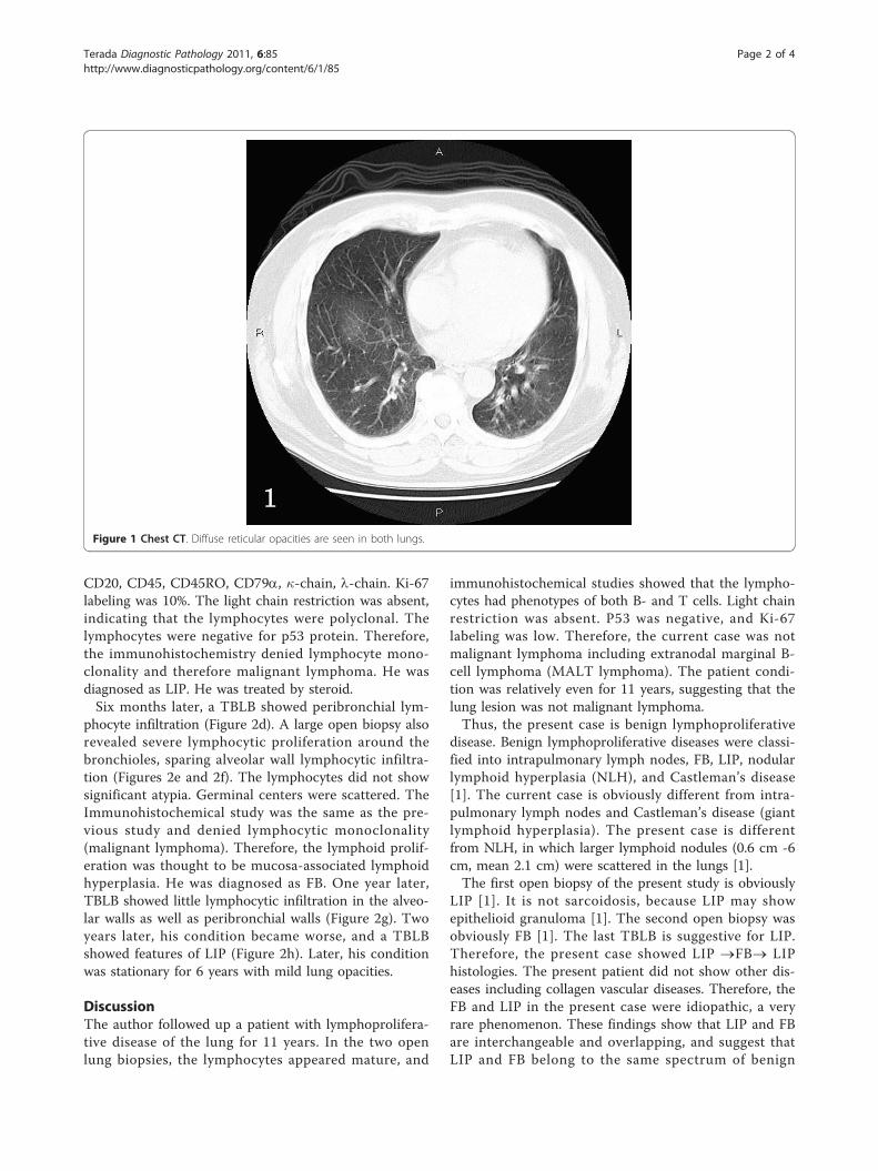
A 44-year-old Japanese man consulted to our hospital because of cough and sputum. Chest-XP and CT revealeddiffuse reticular opacities in both lungs.A transbronchial lung biopsy (TBLB) showed a moderate infiltration of lymphocytes in the alveolar septae. He wasdiagnosed as interstitial pneumonia, and treated by drugs. One year later, his condition deteriolated, and a largeopen biopsy was performed. It showed a diffuse severe infiltration of lymphocytes in the alveolar walls and a fewepithelioid granulomas. No bronchiolitis was seen.Immunohistochemical study denied lymphocyte monoclonality, and he was diagnosed as lymphocytic interstitialpneumonia (LIP). He was treated by steroid.Six months later, TBLB showed peribronchial lymphocyte infiltration. A large open biopsy also revealed a severelymphocytic infiltration around the bronchioles, sparing alveolar wall lymphocytic infiltration. Immunohistochemicalstudy denied malignant lymphoma. He was diagnosed as follicular bronchiolitis (FB). One year later, TBLB showedlittle lymphocytic infiltration in the alveolar walls as well as peribronchial walls. Two years later, his conditionbecame worse, and TBLB showed features of LIP. Later, his condition was stationary for 6 years with mild lungopacities for 6 years. These findings show that LIP and FB are interchangeable and overlapping, and suggest thatLIP and FB belong to the same spectrum of benign lymphoproliferative disorders of the lungs.
Keywords: lung, follicular bronchitis, lymphocytic interstitial pneumonia, histopathology, immunohistochemistry
IntroductionFollicular bronchiolitis (FB) is a benign lymphoprolifera-tive lung disease characterized by hyperplastic mucosa-associated lymphoid tissue present around the peribron-chial spaces [1]. Patients with FB are often associatedwith collagen vasculitis diseases, immunodeficiency state,hyperimmune state, and hereditary factors. Idiopathic FBis rare. Lymphocytic interstitial pneumonia (LIP) is also abenign lymphoproliferative lung disease characterized bysevere lymphocytic infiltration of the alveolar septae [1].Patients with LIP are also often associated with collagenvascular diseases, immunological diseases, immunodefi-ciency diseases, lung infections, and drug induced dis-eases. When FB and LIP are pathologically diagnosed,exclusion of malignant lymphoma is mandatory [1].Herein reported is a case with benign lung lymphoproli-ferative diseases with 11 years follow-up.
Case reportA 44-year-old Japanese man consulted to our hospitalbecause of cough and sputum. Chest-XP and CTrevealed diffuse reticular opacities in both lungs (Figure1). He had no other diseases including collagen vasculardiseases, immunological diseases, immunodeficiency dis-eases, and hypersencitivity disorders. A transbronchiallung biopsy (TBLB) showed a moderate infiltration oflymphocytes in the alveolar septae (Figure 2a). He wasdiagnosed as interstitial pneumonia, and treated bydrugs. One year later, his condition deteriolated, and alarge open biopsy (video-assisted thracostomic biopsy)was performed. It showed a diffuse severe infiltration oflymphocytes in the alveolar walls and a few epithelioidgranulomas (Figures 2b and 2c). The lymphocytes werefree from significant atypia. No bronchiolitis was seen.An immunohistochemical study was performed byDako’s envision method (Dako, Glostrup, Denmark) aspreviously described [2]. The immunohistochemicalstudy showed that the lymphocytes were positive CD3,Correspondence: [email protected]
Department of Pathology, Shizuoka City Shimizu Hospital, Shizuoka, Japan
Terada Diagnostic Pathology 2011, 6:85http://www.diagnosticpathology.org/content/6/1/85
© 2011 Terada; licensee BioMed Central Ltd. This is an Open Access article distributed under the terms of the Creative CommonsAttribution License (http://creativecommons.org/licenses/by/2.0), which permits unrestricted use, distribution, and reproduction inany medium, provided the original work is properly cited.
CD20, CD45, CD45RO, CD79a, �-chain, l-chain. Ki-67labeling was 10%. The light chain restriction was absent,indicating that the lymphocytes were polyclonal. Thelymphocytes were negative for p53 protein. Therefore,the immunohistochemistry denied lymphocyte mono-clonality and therefore malignant lymphoma. He wasdiagnosed as LIP. He was treated by steroid.Six months later, a TBLB showed peribronchial lym-
phocyte infiltration (Figure 2d). A large open biopsy alsorevealed severe lymphocytic proliferation around thebronchioles, sparing alveolar wall lymphocytic infiltra-tion (Figures 2e and 2f). The lymphocytes did not showsignificant atypia. Germinal centers were scattered. TheImmunohistochemical study was the same as the pre-vious study and denied lymphocytic monoclonality(malignant lymphoma). Therefore, the lymphoid prolif-eration was thought to be mucosa-associated lymphoidhyperplasia. He was diagnosed as FB. One year later,TBLB showed little lymphocytic infiltration in the alveo-lar walls as well as peribronchial walls (Figure 2g). Twoyears later, his condition became worse, and a TBLBshowed features of LIP (Figure 2h). Later, his conditionwas stationary for 6 years with mild lung opacities.
DiscussionThe author followed up a patient with lymphoprolifera-tive disease of the lung for 11 years. In the two openlung biopsies, the lymphocytes appeared mature, and
immunohistochemical studies showed that the lympho-cytes had phenotypes of both B- and T cells. Light chainrestriction was absent. P53 was negative, and Ki-67labeling was low. Therefore, the current case was notmalignant lymphoma including extranodal marginal B-cell lymphoma (MALT lymphoma). The patient condi-tion was relatively even for 11 years, suggesting that thelung lesion was not malignant lymphoma.Thus, the present case is benign lymphoproliferative
disease. Benign lymphoproliferative diseases were classi-fied into intrapulmonary lymph nodes, FB, LIP, nodularlymphoid hyperplasia (NLH), and Castleman’s disease[1]. The current case is obviously different from intra-pulmonary lymph nodes and Castleman’s disease (giantlymphoid hyperplasia). The present case is differentfrom NLH, in which larger lymphoid nodules (0.6 cm -6cm, mean 2.1 cm) were scattered in the lungs [1].The first open biopsy of the present study is obviously
LIP [1]. It is not sarcoidosis, because LIP may showepithelioid granuloma [1]. The second open biopsy wasobviously FB [1]. The last TBLB is suggestive for LIP.Therefore, the present case showed LIP ®FB® LIPhistologies. The present patient did not show other dis-eases including collagen vascular diseases. Therefore, theFB and LIP in the present case were idiopathic, a veryrare phenomenon. These findings show that LIP and FBare interchangeable and overlapping, and suggest thatLIP and FB belong to the same spectrum of benign
1Figure 1 Chest CT. Diffuse reticular opacities are seen in both lungs.
Terada Diagnostic Pathology 2011, 6:85http://www.diagnosticpathology.org/content/6/1/85
Page 2 of 4

a b
c d
e f
g h Figure 2 Lung Biopsies. (A)The first transbronchial lung biopsy (TBLB). It shows infiltration of lymphocytes in the alveolar septae. HE, ×200. (B)The first large open biopsy. It shows severe lymphocyte infiltration in the alveolar septae. HE, ×100. (C)The first open biopsy. Higher power view.The alveolar septae with severe lymphocytic infiltration and granuloma formation are seen. HE, ×200. (D)TBLB shows peribronchial lymphocyticinfiltration. HE, ×200. (E)The second large open biopsy. Heavy infiltration of lymphocytes is seen around the bronchiole (MALT hyperplasia).Germinal centers are recognized. The alveolar septae are free from significant changes. HE, ×40. (F) The second open biosy. High power view.The peribronchial lymphoid tissue has germinal center, and consists of mature lymphocytes. HE, ×200. (G)TBLB shows no or minimal lymphocyticinfiltration in the alveolar septae. HE, ×100. (H)TBLB shows lymphocytic infiltration in the alveolar septae. HE, ×40.
Terada Diagnostic Pathology 2011, 6:85http://www.diagnosticpathology.org/content/6/1/85
Page 3 of 4
lymphoproliferative disorders of the lungs. Similar sug-gestion was reported elsewhere [3].
ConsentWritten informed consent was obtained from the patientfor publication of this case report and accompanyingimages.
Competing interestsThe authors declare that they have no competing interests.
Received: 18 May 2011 Accepted: 21 September 2011Published: 21 September 2011
References1. Travis WD, Galvin JR: Non-neoplastic pulmonary lymphoid lesions. Thorax
2001, 56:964-971.2. Terada T, Kawaguchi M, Furukawa K, Sekido Y, Osamura Y: Minute mixed
ductal-endocrine carcinoma of the pancreas with predominantintraductal growth. Pathol Int 2002, 52:740-746.
3. Nocholson AG, Wotherspoon AC, Diss TC, Hansell DM, Du Bois R,Sheppard MN, Isaacson PG, Corrins B: Reactive pulmonary lymphoiddisorders. Histopathology 1995, 26:405-412.
doi:10.1186/1746-1596-6-85Cite this article as: Terada: Follicular bronchiolitis and lymphocyticinterstitial pneumonia in a Japanese man. Diagnostic Pathology 20116:85.
Submit your next manuscript to BioMed Centraland take full advantage of:
• Convenient online submission
• Thorough peer review
• No space constraints or color figure charges
• Immediate publication on acceptance
• Inclusion in PubMed, CAS, Scopus and Google Scholar
• Research which is freely available for redistribution
Submit your manuscript at www.biomedcentral.com/submit
Terada Diagnostic Pathology 2011, 6:85http://www.diagnosticpathology.org/content/6/1/85
Page 4 of 4