Embed Size (px)
Citation preview
CASE REPORT Open Access
Primary acantholytic squamous cell carcinoma ofthe cecum: a case reportZoran Jukić1, Iva Ledinsky2, Monika Ulamec3, Mario Ledinsky2, Božo Krušlin3,4, Davor Tomas3,4*
Abstract
Background: Acantholytic squamous cell carcinoma (ASCC) is an uncommon histopathologic variant of SCC,characterized by marked acantholysis, wherein the tumor cells demonstrate defective cohesion to one another inthe cancer nest leading to a pseudoglandular or pseudovascular appearance. The most common site of ASCC isthe sun-exposed areas of the skin. Sporadic cases of ASCC have also been reported in various mucosal membranesand organs but to our knowledge this is the first case of primary ASCC of the large bowel.
Case presentation: A 59-year-old woman underwent right hemicolectomy due to large tumor in cecum andinitial part of the ascending colon. Microscopically, the tumor consisted of nests of focally keratinizing large,atypical, squamous epithelial cells. Approximately 70% of the tumor showed acantholytic changes and acantholysiswas equally distributed through the entire tumor. Immunohistochemically tumor cells were diffusely positive forcytokeratin (CK) AE1/AE3 and focally positive for epithelial membrane antigen and syndecan 1. All other testedantibodies (CK7, CK 20, CK MNF116, E-cadherin, beta-catenin, p63, p16, CD31, CD34, CEA, estrogen, progesterone)showed negative reaction. Periodic acid Schiff and alcian blue staining showed no intracellular or extracellularmucinous material in the tumor. The diagnosis of acantholytic squamous cell carcinoma of the cecum wassuspected and additional examination was recommended to exclude possibility of metastatic carcinoma. Extensiveclinical examination which also included whole-body PET/CT scan showed no additional tumors. After theexclusion of possible metastatic disease the diagnosis of primary acantholytic squamous cell carcinoma of thececum was confirmed. Six months after surgery the metastasis in small intestine and recurrence in the abdominalcavity at the site of surgery appeared and had the same morphological characteristic as the primary tumor in thececum.
Conclusion: We report a unique case of ASCC arising in cecum and on this way expands the range of tumorsoriginating in colon. Reports of more cases of colonic ASCC would possibly help to elucidate origin, clinicalbehavior and therapy of these tumors.
BackgroundPrimary squamous cell carcinoma (SCC) of the cecum israre entity and only limited numbers of cases aredescribed [1-4]. The first case of SCC was reported bySchmidtman in 1919 [5]. Acantholytic squamous cellcarcinoma (ASCC) is an uncommon histopathologic var-iant of SCC, characterized by marked acantholysis,wherein the tumor cells demonstrate defective cohesionto one another in the cancer nest leading to a pseudo-glandular or pseudovascular appearance [6]. It was first
described in details by Lever [7] in 1947 as adenoa-canthoma of the sweat glands. Synonyms include ade-noid SCC, pseudoglandular SCC, SCC with gland-likefeatures, angiosarcoma-like SCC, and pseudovascularadenoid SCC. The most common site of ASCC is thesun-exposed areas of the skin, particularly on the headand neck of elderly men [6]. Sporadic cases of ASCChave also been reported in various mucosal membranesand organs [8-12] but to our knowledge this is the firstcase of primary ASCC of the large bowel.
Case presentationA 59-year-old woman who presented with a one monthhistory of lower abdominal pain and intermittent watery,
* Correspondence: [email protected] of Pathology, Sestre milosrdnice University Hospital, Zagreb,CroatiaFull list of author information is available at the end of the article
Jukić et al. Diagnostic Pathology 2011, 6:5http://www.diagnosticpathology.org/content/6/1/5
© 2011 Jukićć et al; licensee BioMed Central Ltd. This is an Open Access article distributed under the terms of the Creative CommonsAttribution License (http://creativecommons.org/licenses/by/2.0), which permits unrestricted use, distribution, and reproduction inany medium, provided the original work is properly cited.
bloody stool was admitted to our hospital. On physicalexamination, she was found to have right lower quad-rant abdominal tenderness without guarding. Blood testsrevealed anemia and C reactive protein was slightly ele-vated. Her past medical history included hysterectomydue to multiple myomas 25 years earlier and segmen-tectomy of right breast 3 years ago. Patohistologicaldiagnosis of breast lesion was fibrocystic breast diseasewith usual and atypical intraductal epithelial prolifera-tion and foci of adenosis without sign of intraductal orinvasive carcinoma. Her mother and brother died fromconventional colon cancer in age of 69 and 63 years,respectively. Father died of small cell lung cancer in theage of 67 years. Colonoscopy was performed andshowed large tumor in cecum and initial part of theascending colon. A biopsy confirmed malignant natureof the process and diagnosis of squamous cell carcinomawas suggested. Abdominal CT showed large tumor incecoascending region of the colon and enlarged regionallymph nodes. Signs of distant metastasis are not found.Chest X-ray was unremarkable. A right hemicolectomywith removal of adjacent pericolic lymph nodes was car-ried out.Grossly, in the cecoascending region, an ulcero-prolif-
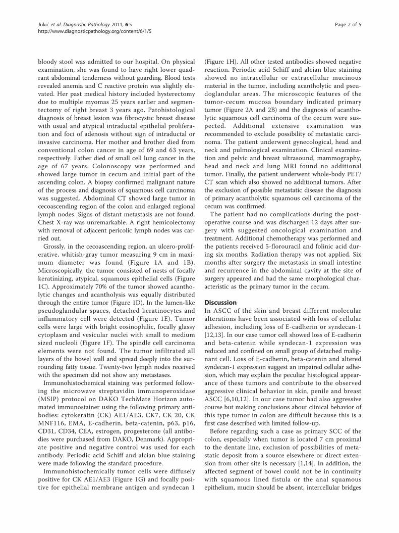
erative, whitish-gray tumor measuring 9 cm in maxi-mum diameter was found (Figure 1A and 1B).Microscopically, the tumor consisted of nests of focallykeratinizing, atypical, squamous epithelial cells (Figure1C). Approximately 70% of the tumor showed acantho-lytic changes and acantholysis was equally distributedthrough the entire tumor (Figure 1D). In the lumen-likepseudoglandular spaces, detached keratinocytes andinflammatory cell were detected (Figure 1E). Tumorcells were large with bright eosinophilic, focally glassycytoplasm and vesicular nuclei with small to mediumsized nucleoli (Figure 1F). The spindle cell carcinomaelements were not found. The tumor infiltrated alllayers of the bowel wall and spread deeply into the sur-rounding fatty tissue. Twenty-two lymph nodes receivedwith the specimen did not show any metastases.Immunohistochemical staining was performed follow-
ing the microwave streptavidin immunoperoxidase(MSIP) protocol on DAKO TechMate Horizon auto-mated immunostainer using the following primary anti-bodies: cytokeratin (CK) AE1/AE3, CK7, CK 20, CKMNF116, EMA, E-cadherin, beta-catenin, p63, p16,CD31, CD34, CEA, estrogen, progesterone (all antibo-dies were purchased from DAKO, Denmark). Appropri-ate positive and negative control was used for eachantibody. Periodic acid Schiff and alcian blue stainingwere made following the standard procedure.Immunohistochemically tumor cells were diffusely
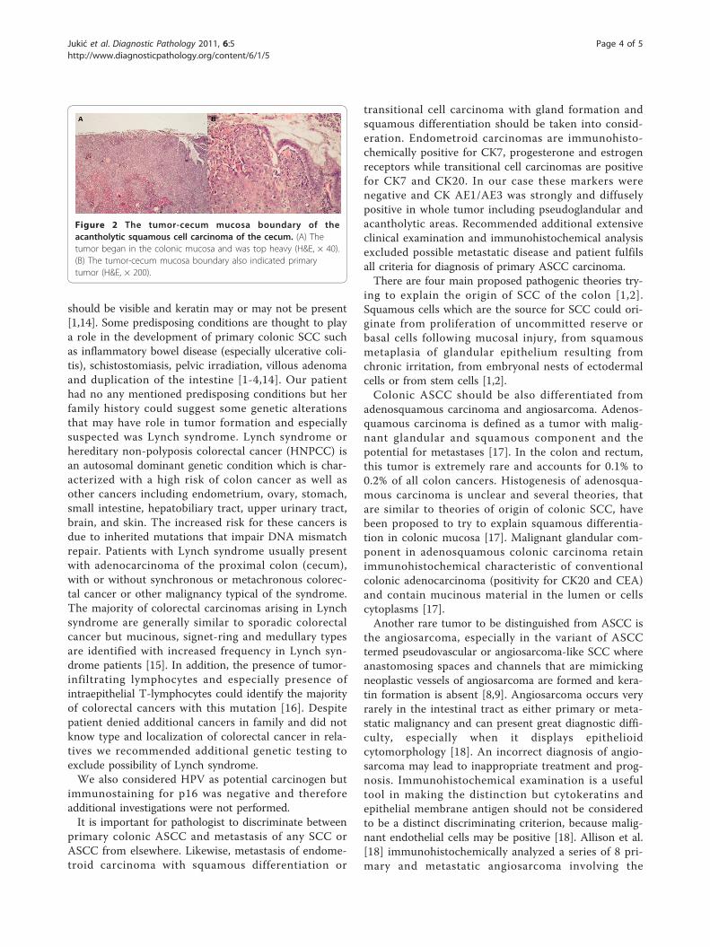
positive for CK AE1/AE3 (Figure 1G) and focally posi-tive for epithelial membrane antigen and syndecan 1
(Figure 1H). All other tested antibodies showed negativereaction. Periodic acid Schiff and alcian blue stainingshowed no intracellular or extracellular mucinousmaterial in the tumor, including acantholytic and pseu-doglandular areas. The microscopic features of thetumor-cecum mucosa boundary indicated primarytumor (Figure 2A and 2B) and the diagnosis of acantho-lytic squamous cell carcinoma of the cecum were sus-pected. Additional extensive examination wasrecommended to exclude possibility of metastatic carci-noma. The patient underwent gynecological, head andneck and pulmological examination. Clinical examina-tion and pelvic and breast ultrasound, mammography,head and neck and lung MRI found no additionaltumor. Finally, the patient underwent whole-body PET/CT scan which also showed no additional tumors. Afterthe exclusion of possible metastatic disease the diagnosisof primary acantholytic squamous cell carcinoma of thececum was confirmed.The patient had no complications during the post-
operative course and was discharged 12 days after sur-gery with suggested oncological examination andtreatment. Additional chemotherapy was performed andthe patients received 5-florouracil and folinic acid dur-ing six months. Radiation therapy was not applied. Sixmonths after surgery the metastasis in small intestineand recurrence in the abdominal cavity at the site ofsurgery appeared and had the same morphological char-acteristic as the primary tumor in the cecum.
DiscussionIn ASCC of the skin and breast different molecularalterations have been associated with loss of cellularadhesion, including loss of E-cadherin or syndecan-1[12,13]. In our case tumor cell showed loss of E-cadherinand beta-catenin while syndecan-1 expression wasreduced and confined on small group of detached malig-nant cell. Loss of E-cadherin, beta-catenin and alteredsyndecan-1 expression suggest an impaired cellular adhe-sion, which may explain the peculiar histological appear-ance of these tumors and contribute to the observedaggressive clinical behavior in skin, penile and breastASCC [6,10,12]. In our case tumor had also aggressivecourse but making conclusions about clinical behavior ofthis type tumor in colon are difficult because this is afirst case described with limited follow-up.Before regarding such a case as primary SCC of the
colon, especially when tumor is located 7 cm proximalto the dentate line, exclusion of possibilities of meta-static deposit from a source elsewhere or direct exten-sion from other site is necessary [1,14]. In addition, theaffected segment of bowel could not be in continuitywith squamous lined fistula or the anal squamousepithelium, mucin should be absent, intercellular bridges
Jukić et al. Diagnostic Pathology 2011, 6:5http://www.diagnosticpathology.org/content/6/1/5
Page 2 of 5
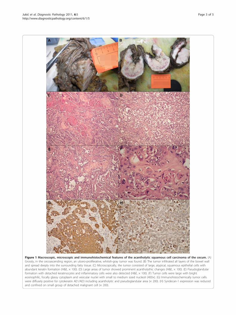
Figure 1 Macroscopic, microscopic and immunohistochemical features of the acantholytic squamous cell carcinoma of the cecum. (A)Grossly, in the cecoascending region, an ulcero-proliferative, whitish-gray tumor was found. (B) The tumor infiltrated all layers of the bowel walland spread deeply into the surrounding fatty tissue. (C) Microscopically, the tumor consisted of large, atypical, squamous epithelial cells withabundant keratin formation (H&E, × 100). (D) Large areas of tumor showed prominent acantholythic changes (H&E, × 100). (E) Pseudoglandularformation with detached keratinocytes and inflammatory cells were also detected (H&E, × 100). (F) Tumor cells were large with brighteosinophilic, focally glassy cytoplasm and vesicular nuclei with small to medium sized nucleoli (400×). (G) Immunohistochemically tumor cellswere diffusely positive for cytokeratin AE1/AE3 including acantholytic and pseudoglandular area (× 200). (H) Syndecan-1 expression was reducedand confined on small group of detached malignant cell (× 200).
Jukić et al. Diagnostic Pathology 2011, 6:5http://www.diagnosticpathology.org/content/6/1/5
Page 3 of 5
should be visible and keratin may or may not be present[1,14]. Some predisposing conditions are thought to playa role in the development of primary colonic SCC suchas inflammatory bowel disease (especially ulcerative coli-tis), schistostomiasis, pelvic irradiation, villous adenomaand duplication of the intestine [1-4,14]. Our patienthad no any mentioned predisposing conditions but herfamily history could suggest some genetic alterationsthat may have role in tumor formation and especiallysuspected was Lynch syndrome. Lynch syndrome orhereditary non-polyposis colorectal cancer (HNPCC) isan autosomal dominant genetic condition which is char-acterized with a high risk of colon cancer as well asother cancers including endometrium, ovary, stomach,small intestine, hepatobiliary tract, upper urinary tract,brain, and skin. The increased risk for these cancers isdue to inherited mutations that impair DNA mismatchrepair. Patients with Lynch syndrome usually presentwith adenocarcinoma of the proximal colon (cecum),with or without synchronous or metachronous colorec-tal cancer or other malignancy typical of the syndrome.The majority of colorectal carcinomas arising in Lynchsyndrome are generally similar to sporadic colorectalcancer but mucinous, signet-ring and medullary typesare identified with increased frequency in Lynch syn-drome patients [15]. In addition, the presence of tumor-infiltrating lymphocytes and especially presence ofintraepithelial T-lymphocytes could identify the majorityof colorectal cancers with this mutation [16]. Despitepatient denied additional cancers in family and did notknow type and localization of colorectal cancer in rela-tives we recommended additional genetic testing toexclude possibility of Lynch syndrome.We also considered HPV as potential carcinogen but
immunostaining for p16 was negative and thereforeadditional investigations were not performed.It is important for pathologist to discriminate between
primary colonic ASCC and metastasis of any SCC orASCC from elsewhere. Likewise, metastasis of endome-troid carcinoma with squamous differentiation or
transitional cell carcinoma with gland formation andsquamous differentiation should be taken into consid-eration. Endometroid carcinomas are immunohisto-chemically positive for CK7, progesterone and estrogenreceptors while transitional cell carcinomas are positivefor CK7 and CK20. In our case these markers werenegative and CK AE1/AE3 was strongly and diffuselypositive in whole tumor including pseudoglandular andacantholytic areas. Recommended additional extensiveclinical examination and immunohistochemical analysisexcluded possible metastatic disease and patient fulfilsall criteria for diagnosis of primary ASCC carcinoma.There are four main proposed pathogenic theories try-
ing to explain the origin of SCC of the colon [1,2].Squamous cells which are the source for SCC could ori-ginate from proliferation of uncommitted reserve orbasal cells following mucosal injury, from squamousmetaplasia of glandular epithelium resulting fromchronic irritation, from embryonal nests of ectodermalcells or from stem cells [1,2].Colonic ASCC should be also differentiated from
adenosquamous carcinoma and angiosarcoma. Adenos-quamous carcinoma is defined as a tumor with malig-nant glandular and squamous component and thepotential for metastases [17]. In the colon and rectum,this tumor is extremely rare and accounts for 0.1% to0.2% of all colon cancers. Histogenesis of adenosqua-mous carcinoma is unclear and several theories, thatare similar to theories of origin of colonic SCC, havebeen proposed to try to explain squamous differentia-tion in colonic mucosa [17]. Malignant glandular com-ponent in adenosquamous colonic carcinoma retainimmunohistochemical characteristic of conventionalcolonic adenocarcinoma (positivity for CK20 and CEA)and contain mucinous material in the lumen or cellscytoplasms [17].Another rare tumor to be distinguished from ASCC is
the angiosarcoma, especially in the variant of ASCCtermed pseudovascular or angiosarcoma-like SCC whereanastomosing spaces and channels that are mimickingneoplastic vessels of angiosarcoma are formed and kera-tin formation is absent [8,9]. Angiosarcoma occurs veryrarely in the intestinal tract as either primary or meta-static malignancy and can present great diagnostic diffi-culty, especially when it displays epithelioidcytomorphology [18]. An incorrect diagnosis of angio-sarcoma may lead to inappropriate treatment and prog-nosis. Immunohistochemical examination is a usefultool in making the distinction but cytokeratins andepithelial membrane antigen should not be consideredto be a distinct discriminating criterion, because malig-nant endothelial cells may be positive [18]. Allison et al.[18] immunohistochemically analyzed a series of 8 pri-mary and metastatic angiosarcoma involving the
Figure 2 The tumor-cecum mucosa boundary of theacantholytic squamous cell carcinoma of the cecum. (A) Thetumor began in the colonic mucosa and was top heavy (H&E, × 40).(B) The tumor-cecum mucosa boundary also indicated primarytumor (H&E, × 200).
Jukić et al. Diagnostic Pathology 2011, 6:5http://www.diagnosticpathology.org/content/6/1/5
Page 4 of 5
gastrointestinal tract and found that tumor cells wereimmunoreactive for cytokeratins AE1/AE3 (7/8), cyto-keratin 7 (2/8), Cam5.2/cytokeratin 8 (5/8), and cytoker-atin 19 (5/8). In one case weakly and focal positivity forepithelial membrane antigen was also observed [18]. Incontrast to an angiosarcoma, the cells of ASCC arenegative for endothelial markers such as CD31, CD34and von Willebrand factor and these markers must beincluded in differentiating ASCC from angiosarcoma[8,9,18].The immunohistochemical and histochemical findings
of the present tumor were compatible with the previousdata of ASCC and exclude possibility of adenosquamouscarcinoma or angiosarcoma [8-13].
ConclusionWe report a unique case of ASCC arising in cecum andon this way expands the range of tumors originating incolon. Reports of more cases of colonic ASCC wouldpossibly help to elucidate origin, clinical behavior andtherapy of these tumors.
ConsentWritten informed consent was obtained from the patientfor publication of this case report and accompanyingimages. A copy of the written consent is available forreview by the Editor-in-Chief of this journal.
Author details1Department of Surgery, Nova Gradiska General Hospital, Nova Gradiska,Croatia. 2Department of Surgery, Sestre milosrdnice University Hospital,Zagreb, Croatia. 3Department of Pathology, Sestre milosrdnice UniversityHospital, Zagreb, Croatia. 4School of medicine, University of Zagreb, Zagreb,Croatia.
Authors’ contributionsZJ supplied relevant clinical information about the patient, drafted themanuscript and was involved in manuscript revision. IL and ML wereinvolved in literature search and preparing the material. MU acquiredphotomicrographs and drafted the manuscript. BK participated in thehistopathological evaluation and drafted the manuscript. DT participated inthe histopathological evaluation, outlined the general concept of themanuscript, and has been involved in drafting and revising it critically. Allauthors have read and approved the final manuscript.
Competing interestsThe authors declare that they have no competing interests.
Received: 6 December 2010 Accepted: 11 January 2011Published: 11 January 2011
References1. Balsano NA: Squamous cell carcinoma of the cecum. Arch Surg 1985,
120:1176-1177.2. Landquest DE, Marcun JN, Thorson AG, Massop D: Primary squamous cell
carcinoma of the colon arising in a villous adenoma. Hum Pathol 1988,19:362-364.
3. Wyatt MG, Clarke TJ, Teasdale C: Primary squamous cell carcinoma of thecaecum. Eur J Surg Oncol 1991, 17:392-394.
4. Mondal SK: Primary squamous cell carcinoma of the cecum. J Cancer ResTher 2009, 5:328-330.
5. Schmidtman M: Zur Kenntnis Seltener Krebs-Formen. Virchows Arch PathAnat 1919, 226:100-118.
6. LeBoit PE, Weedon D, Sarasian A, (Eds): Pathology and genetics of skintumours. World Health Organization classification of tumours Lyon: IARCPress; 2006.
7. Lever WF: Adenoacanthoma of sweat glands. Carcinoma of sweat glandswith glandular and epidermal elements: report of four cases. ArchDermatol 1947, 56:157-171.
8. Papadopoulou E, Tosios KI, Nikitakis N, Papadogeorgakis N, Sklavounou-Andrikopoulou A: Acantholytic squamous cell carcinoma of the gingiva:report of a case and review of the literature. Oral Surg Oral Med OralPathol Oral Radiol Endod 2010, 109:67-71.
9. Horie Y, Kato M: Pseudovascular squamous cell carcinoma of the uterinecerviks: A lesion that may simulate an angiosarcoma. Patol Int 1999,49:170-174.
10. Cunha IW, Guimaraes GC, Soares F, Velazquez E, Torres JJ, Chaux A, Ayala G,Cubilla AL: Pseudoglandular (adenoid, acantholytic) penile squamous cellcarcinoma: a clinicopathologic and outcome study of 7 patients. Am JSurg Pathol 2009, 33:551-555.
11. Kuraoka K, Takehara K, Oshita S, Saito A, Taniyama K: Achantolythicsquamous cell carcinoma of the uterine cervix. Pathol Int 2010,60:245-246.
12. Aulmann S, Schnabel PA, Helmchen B, Dienemann H, Drings P, Otto HF,Sinn HP: Immunohistochemical and cytogenetic characterization ofacantholytic squamous cell carcinoma of the breast. Virchows Arch 2005,446:305-309.
13. Bayer-Garner IB, Smoller BR: The expression of syndecan-1 is preferentiallyreduced compared with that of E-cadherin in acantholytic squamouscell carcinoma. J Cutan Pathol 2001, 28:83-89.
14. Comer TP, Beahrs OH, Dockerty MB: Primary squamous cell carcinomaand adenocarcinoma of the colon. Cancer 1971, 28:1111-1117.
15. Gatalica Z, Torlakovic E: Pathology of the hereditary colorectal carcinoma.Fam Cancer 2008, 7:15-26.
16. Jass JR: HNPCC and sporadic MSI-H colorectal cancer: a review of themorphological similarities and differences. Fam Cancer 2004, 3:93-100.
17. Dong Y, Wang J, Ma H, Zhou H, Lu G, Zhou X: Primary adenosquamouscarcinoma of the colon: report of five cases. Surg Today 2009, 39:619-623.
18. Allison KH, Yoder BJ, Bronner MP, Goldblum JR, Rubin BP: Angiosarcomainvolving the gastrointestinal tract: a series of primary and metastaticcases. Am J Surg Pathol 2004, 28:298-307.
doi:10.1186/1746-1596-6-5Cite this article as: Jukić et al.: Primary acantholytic squamous cellcarcinoma of the cecum: a case report. Diagnostic Pathology 2011 6:5.
Submit your next manuscript to BioMed Centraland take full advantage of:
• Convenient online submission
• Thorough peer review
• No space constraints or color figure charges
• Immediate publication on acceptance
• Inclusion in PubMed, CAS, Scopus and Google Scholar
• Research which is freely available for redistribution
Submit your manuscript at www.biomedcentral.com/submit
Jukić et al. Diagnostic Pathology 2011, 6:5http://www.diagnosticpathology.org/content/6/1/5
Page 5 of 5


![Tgfbr2-deficient Squamous Cell Carcinoma [Abstract] Squamous Cell Carcinoma . ... we report the strategy combining mechanical tumor dissociation, ... Tgfbr2flox/flox mice) (Guasch](https://img.pdfslide.net/doc/110x75/5b4d9eda7f8b9a414e8b707e/tgfbr2-deficient-squamous-cell-carcinoma-abstract-squamous-cell-carcinoma-.jpg)